Characterization of a dual function macrocyclase
enables design and use of ef
fi
cient
macrocyclization substrates
Clarissa M. Czekster
1, Hannes Ludewig
1, Stephen A. McMahon
1& James H. Naismith
1,2,3,4Peptide macrocycles are promising therapeutic molecules because they are protease resis-tant, structurally rigid, membrane permeable, and capable of modulating protein–protein interactions. Here, we report the characterization of the dual function macrocyclase-peptidase enzyme involved in the biosynthesis of the highly toxic amanitin toxin family of macrocycles. The enzyme first removes 10 residues from the N-terminus of a 35-residue substrate. Conformational trapping of the 25 amino-acid peptide forces the enzyme to release this intermediate rather than proceed to macrocyclization. The enzyme rebinds the 25 amino-acid peptide in a different conformation and catalyzes macrocyclization of the N-terminal eight residues. Structures of the enzyme bound to both substrates and biophysical analysis characterize the different binding modes rationalizing the mechanism. Using these insights simpler substrates with only five C-terminal residues were designed, allowing the enzyme to be more effectively exploited in biotechnology.
DOI: 10.1038/s41467-017-00862-4 OPEN
1Biomedical Sciences Research Complex, The University of St Andrews, North Haugh, St Andrews K16 9ST, UK.2Biotherapy Centre, Sichuan University,
C
yclic peptide macrocycles hold promise in pursuing chal-lenging targets involved in protein–protein interactions implicated in diseases as diverse as cancer and anti-microbial infections1. Due to their constrained, pre-organized, and protease-resistant structures, these molecules can modulate key complex macromolecular interactions in a manner that has proven extremely difficult for conventional small molecules1,2. In contrast to most linear peptides, many peptide macrocycles are highly cell permeable3. Ribosomally synthesized and post-translationally modified peptides (RiPPs) are a particularly attractive class of macrocycles because their enzymatic synthesis is driven by enzymes working in cascade to process a genetically encoded and highly variable peptide precursor4. The peptide precursor can be modified by macrocyclization, oxidation, het-erocyclization, hydroxylation, and prenylation in a predictable and scalable manner5. The patellamide pathway is a paradigm in this system, in which catalysis and recognition are physically separated in many of the enzymatic steps leading to a unique combination of specificity and promiscuity6. The macrocyclase in the patellamide biosynthetic pathway (PatGmac) belongs to the subtilisin class of proteases, requiring a substrate with a C-terminal AYD motif, preceded by heterocyclized cysteine or a proline residue7,8. The enzyme is otherwise almost insensitive to the core sequence that becomes the macrocycle and only the thiazoline (or proline) are part of thefinal product, as the AYD is cleaved off during the reaction. This combination of specificity through the use of disposable tags (leader and/or tail sequences) and promiscuity in the core sequence produces a system that is almost infinitely variable. This has made RiPPs appealing for exploitation in biotechnology. In some RiPPs systems, a linker that can also be varied in both length and composition separates the recognition tag and core peptide9,10. Despite the appeal of their promiscuity, the PatG family of macrocyclases face a severe drawback as they are slow11, 12, although in vitro addition of reductant does increase catalytic efficiency13.In addition to PatG, there are four other broad classes of peptide macrocyclases12, 14–16 that operate through an acyl enzyme intermediate. The sortase class of enzymes, which cata-lyze transpeptidation by recognizing a C-terminal LXPTG motif17, the butelase enzyme, which is an asparagine/aspartate (Asx) peptide ligase18, the NRPS thioesterases19and the prolyl oligopeptidase (POP) class of enzymes. A further important class of macrocyclases is that of the ATP-grasp superfamily, which as the name suggests rely on ATP hydrolysis to drive macrocyclia-tion20. The enzymes that catalyze close to traceless peptide bond formation regardless of the peptide sequence—, i.e., only one residue from the precursor peptide recognition tag is carried over to thefinal cyclic product—are PatGmac family members, bute-lase, and POP macrocyclases. The POPB from Basidiomycete fungi such as Amanita bisporigera and Galerina marginata
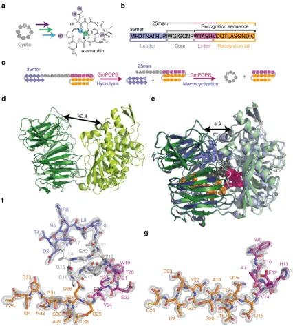
(GmPOPB) species have been reported as having kcat values comparable to butelase, the fastest rate observed for peptide macrocyclisation15,21. GmPOPB is the macrocyclase responsible for macrocyclization of amatoxins, eight amino-acid ribosomal peptides with the core sequence IWGIGC(N/D)P. Amatoxins are cyclic peptides further modified by a characteristic sulfoxide cross-link between tryptophan and a cysteine (Fig. 1a), and hydroxylation (the extent of which vary). The genomes of G. marginataand other amatoxin producingAmanitaspecies pos-sess more than 50 gene sequences annotated as AMA1 (amatoxin precursors) in which there is considerable diversity in the long C-terminal tail that follows the core sequence15,22. Amatoxins are the cause for the toxicity ofAmanitaand Galerinamushrooms. They are readily absorbed through the gut, and a lethal dose in adults is<10 mg23. Amatoxins are stable to inactivation by either the mammalian digestive tract or cooking, thus consumption of
even small numbers of such mushrooms is often fatal. Amatoxin toxicity arises from its accumulation in the liver where it inhibits RNA polymerase II leading to irreparable liver failure23. The highly stable and potent toxicity of amatoxins has led to their exploration as warheads for targeted cancer therapy24, 25. The amatoxin peptide precursor is produced as a 35 amino-acid linear substrate (Fig.1b), which isfirst processed to a 25 residue peptide (25mer) by removal of the highly conserved 10 N-terminal amino-acid leader26 that is discarded. The newly exposed N-terminal eight residues of the 25mer product are then macro-cyclized and the tail, which is necessary for macrocyclization, is discarded (Fig. 1c). Remarkably, both proteolysis and macro-cyclization steps are carried out by the same enzyme, GmPOPB15. We report the functional and structural characterization of GmPOPB and establish the molecular features that determine whether the enzyme catalyzes proteolysis or macrocyclization. Informed by structural and biophysical studies, we have designed a much simpler substrate with fewer C-terminal residues that can be macrocyclized by GmPOPB at synthetically useful rates. The shorter substrate is more cost effective to produce by solid-phase chemical synthesis allowing the generation of more chemically diverse macrocycles, a valuable biotechnological tool.
Results
Structural biology of Apo and substrate-bound GmPOPB. The apo protein crystals belong to space group P212121, with one monomer in the asymmetric unit. The structure was determined at 2.4 Å resolution by molecular replacement using the β -propeller domain of the proline oligopeptidase from porcine brain (residues 82–450, PDB:1h2z) as search model. The refined apo model (PDB:5N4F) includes residues 7–222, 230–695, and 704–726, and the missing regions are presumed to be disordered portions of the protein. The protein contains two domains as observed in other POP enzymes27. The domain containing the putative catalytic residues (Ser577, Asp661, His698) comprises residues 1–81 and 450–728, and the other domain is a seven bladedβ-propeller, comprising residues 82–449 (Fig.1d). In the apo structure, the two domains are in an“open”conformation, in an arrangement reminiscent of a hinged lid on a bottle. This open conformation has been observed in other POPs in crystal form when free of ligand28,29. The catalytic serine sits at the tip of a loop and points toward theβ-propeller domain (Supplementary Fig. 7). The side chain oxygen of Ser577 is 5.6 Å from the side chain carboxylate of Asp661; His698 is on a loop that is dis-ordered. Ser577 and Asp661 of GmPOPB occupy the same position as Ser554 and Asp641 in the porcine proline oligo-peptidase structure30.
density for the N-terminal residues of the 25mer, we were unable to satisfactorily model it. To observe interactions when the catalytic residue Ser577 is present, we also obtained complex structures of the H698A mutant bound to the 35mer peptide (PDB:5N4E) and the D661A mutant bound to the 25mer peptide (PDB:5N4D).
In all 35mer and 25mer complexes, the enzyme adopts the same “closed” conformation in which the lid (the propeller domain) sits on top of the catalytic domain (Fig. 1e). In both
25mer complexes the N-terminal residues of the substrate (IWGIGCN) are disordered; in the S577A–35mer complex the N-terminal two residues (MF) are missing, while in the H698A complex only thefirst N-terminal residue is absent. There are no large differences between the protein backbone positions in the complexes with the 35mer and 25mer substrates (root mean square deviation (rmsd) of 0.48 Å over 720 Cαpositions for the S577A structures). There are also no major differences between the H698A–35mer and S577A–35mer complexes, and between
4 Å
C35 I34 D33
N32 G31
S30 A29
T27
L28 Q26
D25
V24 H23
E22 A21
W19 P18
T20 N17
C16 G15
I14 G13
W12 I11
P10 L9 R8
T7 T4
N5
D3 A6
22 Å
C25 I24
N22
G21
S20 A19
T17
L18 Q16
D15
V14 H13
E12 A11
W9
T10
D23
a
MFDTNATRLPIWGIGCNPWTAEHVDQTLASGNDIC
Leader Core
Recognition sequence 35mer
25mer
Linker Recognition tail
c
f
d e
C
P P
P C
P
P P
P
C
GmPOPB GmPOPB +
Hydrolysis Macrocyclization
35mer 25mer
P
b
Cyclic
P
g + H
H
NH2
O
O O O
O O
O O
O O HO
OH HO
HO
NH
NH N H H N H N
NH HN
N H N
S
α-amanitin
Fig. 1Amanitin biosynthesis and the role of GmPOPB.aBiosynthesis ofα-amanitin from the cyclic peptide produced by GmPOPB involves oxidation (blue
circle), hydroxylations (purple circles), and cross linking (green circle). Amino acids in the cyclic peptide are depicted as geometrical shapes, and the core proline is labeled.b35mer substrate used by GmPOPB to catalyze peptide bond hydrolysis and macrocyclization. Peptide is divided in four regions, leader (blue), core (light gray), linker (magenta), and tail (orange). The color coding for peptide regions forc,e–gfollows this.cScheme of the reactions catalyzed by GmPOPB.dApoGmPOPB in an open state showing 22 Å separation between theβ-propeller (dark green) and the peptidase domain (light green).e
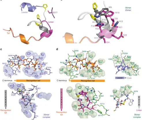
[image:3.595.81.508.48.522.2]the S577A–25mer and D661A–25mer complexes. Table1shows the data collection and refinement statistics for all structures. The recognition tail adopts an identical distorted 310helix conforma-tion inserted into the middle of theβ-propeller domain in both the 25mer and 35mer complexes (Fig.1f, g and2). The carboxyl terminus sits in a pocket where it makes water-mediated hydrogen bonds to the protein. To our surprise, there are only a few hydrogen bonds between the protein and the tail (Fig.2c for the S577A–25mer complex and Fig. 2d for the S577A–35mer complex). Comparison to the apo structure reveals that binding of substrate induces no significant changes in the core of the propeller domain (rmsd of 0.75 Å over 360 Cαpositions for the S577A structures). The changes that occur (relative to the apo structure) are in loops around the catalytic site.
Significant differences between the two substrates are observed for the residues of the linker region, since it occupies distinct conformations on the 25mer and 35mer complexes (Fig.2b). V24 faces Arg79 and V14 is toward Phe506. H23 does not form any hydrogen bonds, while H13 is hydrogen bonding with Glu601. Tyr494 is within hydrogen bonding distance from E22, while on the 25mer complex Tyr494 is interacting with W9. This peptide twisting causes a tryptophan present in the peptides from both complexes (residue 19 in 35mer, 9 in the 25mer), C-terminal to the site of cleavage, to occupy a binding pocket close to the active site. The oxyanion-stabilizing Tyr496 is in close proximity to the core proline (P8) in the 25mer structure (D661A mutant), while Tyr496 hydrogen bonds with P18 in the 35mer complex (Fig.2d). Substrate residues I11–P18 (the core peptide is not seen in the 25mer, apart from weak density from P8 in the D661A–25mer complex) form a twisted loop, which makes contacts with the protein and within itself; atoms that will ultimately form the macrocycle are 7.6 Å apart (Fig.2d). The core peptide interposes between substrate P18 and enzyme Ser577, which is over 8 Å away. Substrate P10, the site of proteolytic cleavage, is positioned
for attack by Ser577 (the Cβof the mutated residue is 3.3 Å from the carbonyl with plausible geometry). In the crystal structure of H698A–35mer complex, the hydroxyl of Ser577 is in hydrogen bonding distance from P10, in a position suited for nucleophilic attack but the structure is less ordered, notably the loop containing the mutation H698A. Tyr496 is on the opposite face of the carbonyl 2.8 Å from the oxygen and positioned to stabilize the tetrahedral intermediate from attack of Ser577 (Fig.2d). Both the interaction and the role of Tyr496 are conserved in other POPs. In the 35mer complex structures, residues 2–9 adopt a helical arrangement that ends up exposed to solvent at the N-terminus and makes few contacts with the protein (Fig. 2d). In none of the GmPOPB structures that we have obtained are the catalytic residues arranged in the traditional manner, the closest approach of the His698 and Ser577 is 13 Å and residues block simple movement (Supplementary Fig. 7). To confirm the importance of the putative catalytic triad for GmPOPB, the mutants S577A, D661A, and H698A were evaluated for activity, and were inactive with both 25mer and 35mer substrates, using 5μM GmPOPB and monitoring the reaction progress after 24 h.
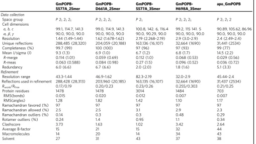
[image:4.595.46.556.73.359.2]Mutations in the histidine loop decrease enzyme activity. To study other residues involved in hydrolysis and macrocyclization, additional mutants H698N, R663A, R663Q, R663K, and W695Δ (deletion) were generated. These mutants were designed based on comparison of sequence alignments between other POP enzymes and the very similar POPA enzyme fromG. marginata, enzymes that solely act as proteases. Arg663 is highly conserved in POPs and thought to play a role in catalysis or substrate binding, since it makes hydrogen bonds with the peptide substrate31. Supple-mentary Fig.9a depicts a sequence alignment comparing POPA, POPB, PCY1 (another macrocyclase from the prolyl oligopepti-dase family), and porcine POP while Supplementary Fig. 9b Table 1 Data collection and refinement statistics
GmPOPB-S577A_25mer
GmPOPB-D661A_25mer
GmPOPB-S577A_35mer
GmPOPB-H698A_35mer
apo_GmPOPB
Data collection
Space group P212121 P212121 P21 P212121 P21212
Cell dimensions
a,b,c 99.1, 114.7, 141.3 99.0, 114.9, 141.3 100.8, 142. 6, 116.4 99.2, 115 141. 5 90.89, 105.62, 86.96
α,β,γ 90.0, 90.0, 90.0 90.0, 90.0, 90.0 90.0, 90.29, 90.0 90.0, 90.0, 90.0 90.0, 90.0, 90.0
Resolution 1.44 (1.49–1.44) 1.62 (1.678–1.62) 2.19 (2.268–2.19) 2.9 (3.0–2.9) 2.4 (2.49–2.4)
Unique reflections 288,485 (28,320) 204,059 (20,188) 163,136 (16,107) 32,664 (1690) 31,441 (2534)
Completeness (%) 99.7 (99) 100 (100) 97 (96) 97 (93) 99 (77)
MeanI/sigma (I) 9.3 (1.3) 6.9 (1.0) 6.7 (1.2) 6.8 (1.7) 14.5 (2.2)
R-merge 0.114 (1.01) 0.059 (0.69) 0.112 (1.0) 0.068 (0.53) 0.029 (0.56)
R-meas 0.063 (0.588) 0.084 (0.98) 0.27 (1.5) 0.096 (0.52) 0.036 (0.72)
Redundancy 6.0 (6.6) 6.7 (6.6) 2.0 (2.0) 1.8 (1.6) 5.1 (3.3)
Refinement
Resolution range 43.3–1.44 46.9–1.62 82.3–2.19 32.0–2.9 45.44–2.4
Reflections used in refinement 288,428 (28,313) 203,960 (20,185) 163,135 (16,107) 32,664 (1690) 31,437 (2534)
Rwork/Rfree 0.17/0.19 0.20/0.23 0.23/0.26 0.255/0.303 0.21/0.25
Protein residues 1478 1478 3014 1484 703
RMS(bonds) 0.015 0.020 0.012 0.007 0.007
RMS(angles) 1.28 1.82 1.42 1.10 1.17
Ramachandran favored (%) 97 97 97 97 97
Ramachandran allowed (%) 2.5 2.5 3.1 2.9 2.3
Ramachandran outliers (%) 0.14 0.3 0.3 0.48 0.29
Rotamer outliers (%) 0.24 1.4 0.95 1.1 0.34
Clashscore 3.73 1.63 1.02 3.42 2.64
Average B-factor 15 21 15 32 44
Macromolecules 14 20 14 34 43
depicts the position of these residues in GmPOPB. H698N was insoluble and not evaluated. The other mutants possessed diminished activities for both peptide bond hydrolysis and macrocyclization. The amount of cyclic peptide present after incubation for 16 h with the 25mer substrate was R663Q> R663A>W695Δ>R663K. When the 35mer was used as sub-strate, the mutants demonstrated diminished activity for peptide bond hydrolysis and almost undetectable activity for macro-cyclization (Supplementary Fig.9c).
Kinetic characterization and substrate scope of GmPOPB. Previous analysis employed GmPOPB isolated from the G. marginata mushroom after transformation with Agrobacterium tumefaciens15. We examined kinetic parameters and performed a substrate specificity study on the enzyme isolated employing a
bacterial overexpression system. Substrates tested are shown in Supplementary Fig. 1. Our results on the native overexpressed enzyme confirm the previous findings15 obtained for protein purified from mushroom that the full-length 35mer substrate is cleaved and the resulting 25mer is released (Supplementary Fig.3). The kinetic data for expressed protein with the 25mer but not 35mer have been previously reported15,21. The 25mer then rebinds (in competition with the 35mer) for macrocyclization. Cleavage and macrocyclization do not occur in a single binding event15. The 25mer accumulates as an intermediate although the proteolysis reaction is slower than cyclic peptide formation (Supplementary Fig.2). Very similar values forKmandkcatwere obtained for all full-length substrates evaluated, with Kmvalues ranging from 8 to 51μM, whilekcatwas between 3.2 and 35 min−1 (Supplementary Fig. 2; Supplementary Table 2). Conservative amino-acid substitutions within the peptide substrate had no
Y494 E601
D504
F507 F506
S508 K714 S502 A503
D504
V524
F506 A503
S508 N707 R79
Y494
W715 A576
E601
K714 L
i n k e r
K83 S421
L420
S151
L90
F84
S85 F98
A408 S406
R410
T88
A86
S107 K149
F150 N409
R79 G427 c
Recognition tail C-terminus
F98 S85
T419 A408
S151
K83
F84 Y96
A86 S406
R410
N409
T88
S107 Recognition tail
H23
E22
V24
W19
T20
A21
V11
H13 E12
T10 A11
W9 35mer
25mer
Linker
Tail
Leader S577
a
d b
F150
C-terminus
L i n k e r
Recognition tail
25mer complex
35mer complex Y496
S577 Y262
E259
S282
I261 T281
T501 V664 F499
R663 F278
Core
Leader G427
L90 K149
R663
A576 T501
Y496
S502
Linker
C o r e H23
V24
E22 A21 T20 W19
R79 W9
T10 E12
V14 H13 A11
Recognition tail
Fig. 2Interactions between GmPOPB and linker regions for 35mer and 25mer substrates are different.aOverview of bound peptides. Colors for distinct
peptide regions are as in Fig.1b, except that the linker in the 25mer complex is indark gray. Position of the catalytic Ser577, and core and leader proline residues (yellow) are indicated insticks.bComparison of the linker region between 35mer (magenta) and 25mer (gray) complexes. Highlighted are the very different position of Glu and His in contrast to tryptophan, which in both structures occupies the same pocket although the conformation is different.c
[image:5.595.51.544.235.651.2]effect either on kinetic parameters or yield of cyclic product. Less conservative substitutions such as mutation to alanine or 9mer core (IWGIGCANP the bold underlined residue represents the insertion) led to reduced macrocyclization and increased linear peptide, the product of peptide hydrolysis instead of macro-cyclization (Supplementary Fig.8).
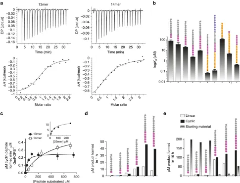
Equilibrium binding of substrates and products. Binding of the inactive mutant S577A to the 25mer, 35mer, a series of truncated substrates (10mer–14mer), as well as the recognition sequence (17mer) WTAEHVDQTLASGNDIC, the truncated recognition sequences VDQTLASGNDIC and TLASGNDIC, and the leader peptide MFDTNATRLP were evaluated by isothermal titration calorimetry (ITC). The results of S577A with both the 25mer and recognition sequence have previously been reported21. Binding of H698A to the 25mer was also measured. The only peptide showing no detectable binding at concentrations up to 1 mM was the 10-residue leader peptide MFDTNATRLP. The full-length substrates and products displayed tight binding
(Kd-25mer–S577A=67±14 nM21, Kd-25mer–H698A=47±11 nM, Kd-35mer=120±30 nM,Kd-recognition=430±10 nM21; binding is dominated by enthalpic contributions (Supplementary Fig. 6b) (Fig.3a shows representative ITC traces for the 13mer and 14mer substrates, Fig. 3b shows Kd values for all peptides evaluated, Supplementary Fig. 10 shows raw data for all binding curves). The inactive mutant H698A has identicalKd-25mer to the S577A mutant suggesting the lack of activity results from catalytic incompetence rather than disruption of substrate binding. Interestingly, despite being longer and having the potential for more interactions with the protein, the 35mer peptide shows slightly weaker binding compared to the 25mer, mostly due to decreased ΔH. A comparison of the complex structures shows that in the 35mer complex there is disorder of side chains in the segment TAEHVD (linker region) but not in the 25mer. To investigate the role of recognition tag peptides corresponding to the entire recognition sequence (linker plus tail, WTAEHVDQ-TLASGNDIC—17 residues), the recognition tail plus the valine from the linker (VDQTLASGNDIC 12 residues) and the highly conserved portion of the tail (TLASGNDIC 9 residues) were
Linear Cyclic Starting material
μ
M product formed
in 16 h
log
Kd
(
μ
M)
μ
M product formed
in 1 h
μ
M cyclin peptide formed min
–1
μ
M
GmPOPB
–1
0.4 0.3 0.2 0.1 0.0
50 40 30 20 10 0
200
150
100
50
0
0 200 400 600 800
[Peptide substrates] μM 13mer
14mer
[25mer] μM 10
5
0
0 100 200
100
P
P
P
P
P
P
P
P
C
C
C
C
C
P
P
P
P
P
P
P
P
P
P
P
P
10
1
0.1
0.01
DP (
μ
cal/s)
DP (
μ
cal/s)
0 –0.02 –0.04 –0.06 –0.08 –0.1 0
–0.02 –0.04 –0.06 –0.08 –0.1 –0.12 –0.14 –0.16
Δ
H
(kcal/mol)
Δ
H
(kcal/mol)
0 –0.1 –0.2 –0.3 –0.4 –0.5 –0.6 –0.7 –0.8 0
–0.1 –0.2 –0.3 –0.4 –0.5 –0.6 –0.7 –0.9 –1 –0.8
Molar ratio Molar ratio
0
0.2 0.4 0.6 0.8 11.2 1.4 1.6 1.8 22.2 0 0.5 1 1.5 2 2.5 3
Time (min)
0 5 10 15 20 25 30
Time (min)
14mer 13mer
0 5 10 15 20 25 30
a b
c d e
Fig. 3Binding and kinetics of substrates and recognition tail truncations.aITC binding curves for the 25mer peptide (obtained with the S577A mutant) and
the 17 amino-acid recognition (linker and tail) sequence.bKdvalues of peptides examined by ITC shows the 35mer and 25mer bind with similar affinity, the
leader does not bind, the tail on its own binds weakly and the contributes to binding energy. The peptides are colored as Fig.1c with N-terminus at top.Error barsare standard error of the mean from the average of at least two independent measurements (Supplementary Table3).cMichaelis–Menten curves show both the 13mer and 14mer are substrates for the enzyme. Fitted values forKd,Km, andkcatare shown in Supplementary Tables2and3.Error barsare
[image:6.595.54.540.50.420.2]tested for binding and gave Kd-17mer–recogSeq=0.43±0.01μM21, Kd-12mer–recogSeq=5±1μM and Kd-9mer–recogSeq=121±19μM (Supplementary Table 3; Supplementary Figs. 6 and 10). Pre-viously, we showed that the recognition sequence dominates binding, with a difference inΔGof only 1.34 kcal mol−1between the 17mer recognition sequence and the 25mer peptide21. To explore how much contribution to the binding energy comes from the linker region, we evaluated the binding of truncated recognition sequences. Our data show that the linker region is important in binding as the loss of the linker (shrinking the recognition sequence from 17 to 12 amino acids) reduces binding affinity 20-fold. On its own, the highly conserved nine-residue tail bound rather weakly, consistent with the few interactions observed with the protein. Following from thisfinding, a series of truncated peptides (core plus parts of the linker) were tested and revealed a trend in which binding affinity increased from 10mer to 13mer (Kd-10mer=83±17μM,Kd-11mer=39±18μM,Kd-12mer=21 ±5μM,Kd-13mer=2.4±0.1μM) peptides, but decreased slightly with the 14mer peptide (Kd-14mer=9.5±1.1μM) (Fig.3b); the 9mer was not sufficiently soluble for analysis. We noted that the difference in affinity between the 35mer and the core plus linker (13mer) was ~20-fold.
Discussion
GmPOPB can form and hydrolyze peptide bonds depending on the substrate length and structure. Both reactions proceed by similar chemical mechanisms, passing through an acyl enzyme intermediate. Typical POPs catalyze peptide bond cleavage fol-lowing a proline, and less efficiently an alanine, showing strong preference for substrates shorter than 30 amino acids32. GmPOPB is unusual in that it processes a 35 amino-acid sub-strate, the longest observed for a POP. POP enzymes possess an aspartate, histidine, and serine catalytic triad. Molecular dynamic simulation studies comparing the porcine and bacterial POPs have proposed a mechanism in which inter-domain“breathing”is required for catalysis33. Although structures of apo and POPs bound to short peptide substrates and inhibitors (ranging from 230to 7 amino acids34) are available, no substrate complex with a long peptide has previously been determined. Interestingly, in GmPOPB complexes the residues of the presumed catalytic triad are not aligned. His698 is 12 Å away (much further than in any other POP, Supplementary Fig.7) from Ser577 but is essential for
catalysis rather than binding. The enzyme–substrate complex shows that, apart from His698, there is no other residue in proximity to the active site capable of acting as general acid/ general base with pKa values near to the 8.0 measured from kinetic analysis21. The domain breathing motion similar to, but larger than, other POPs could correctly position the His698. Mutations of the highly conserved Arg663 and deletion of Trp695 (an insertion relative to other POPs, Supplementary Fig. 9a), which we predicted would affect loop structure and dynamics, were severely compromised in activity with both 25mer and 35mer substrates, consistent with our prediction. We cannot, however, exclude the possibility that His698 may be required for positioning toward a productive conformation of the enzyme–substrate complex rather than act as a base per se. The histidine loop is disordered on the H698A mutant structure and the H698N mutant is insoluble, hinting at a stabilizing role for His698. Analogous to lipid acyl hydrolases35, the enzyme would function with a catalytic dyad in which Ser577 is activated by a water molecule bridged to Asp661.
Comparison of the three-dimensional structures of the enzyme in complex with 25mer and 35mer substrates reveals only minor rearrangements of the protein, mostly in loops that accommodate the longer substrate. Both 35mer (proteolysis) and 25mer (macrocyclization) substrates bind to GmPOPB with high affinity driven mainly by enthalpy (Fig.3b, Supplementary Fig.7; Supplementary Table 3). Consistent with the observation of similar binding affinities of the 25mer and 35mer (Kd 67 and 120 nM, respectively)21, the 10-residue leader (only present in 35mer) does not bind. In both the 25mer and 35mer complex structures, the recognition tail (C-terminal 11 residues) is embedded deeply into the β-propeller domain in an essentially identical arrangement. The linker region, however, adopts very different arrangements in the two complexes, thus its interactions with the protein are quite distinct in the two structures (Fig. 2). ITC measurements show that the linker region, particularly the portion following the core peptide, makes substantial contribution to the binding energy. This is in contrast to the heterocyclase class of RIPP enzymes, where the linker plays no role and can be varied36. Our data show that the structure of the linker is important in binding and determines the orientation of the substrate at the active site (thus its fate).
25mer natural substrate
Shorter designed substrate
5 amino acids
Macrocyclic peptide Macrocyclic
peptide Recog.Seq
Recognition sequence
release
17 amino acids 25mer
binding
Macrocyclization Cyclic
peptide release
25mer
25mer 25mer linker rearrangement
25mer release (unreactive)
Leader release Peptide bond
hydrolysis
Leader
35mer 35mer
binding
b a
Fig. 4GmPOPB catalyzes peptide bond hydrolysis and macrocyclization.aThe sequence of events catalyzed by GmPOPB starting with the 35mer peptide
[image:7.595.58.540.53.215.2]Previous kinetic assays and ours reveal that after removal of the leader, the remaining 25mer is released from GmPOPB, then it rebinds and undergoes the macrocyclization reaction15. In the 35mer complex, the core and linker adopt a tightly packed arrangement that is wedged between the active site loops. We conclude that the linker and/or core are unable to refold to the conformational arrangement seen in the 25mer complex (required for macrocyclization) in situ on a timescale comparable to dissociation. We propose the arrangement of and interactions between C-terminal 25 residues and protein that are seen in the 35mer complex act as a kinetic trap, which can only be escaped by dissociation (Fig.4a). Conformationally trapped peptide reaction intermediates have been identified in other systems. For example, inhibition of proteases by serpins is accomplished by a suicide substrate mechanism, in which the complex is trapped in an inactive arrangement37.
Having identified the key role of the linker, we predicted that it should be possible to design simpler macrocyclization substrates that lacked the recognition tail. This would be valuable since the use of 25mer substrates to make eight residue macrocycles is not economic. ITC shows a 10-fold reduction in binding from 35mer to 13mer, kinetic analysis reveals the 13mer substrate has a Km (25μM) within error to the 25mer substrate (50μM), while the 14mer possesses higher Km (380μM). Similar kcat values were observed with both shorter substrates (0.49 and 0.58 min−1 for the 13mer and 14mer, respectively, Fig. 3c; Supplementary Table2) but these are smaller than the 25mer (18 min−1). Linear peptide (product of hydrolysis instead of macrocyclization) was observed when shorter peptides were utilized as substrates (Fig.3d) consistent with the linker playing a key role in substrate positioning. After 16 h of reaction both 13mer and 14mer sub-strates produce similar amounts of macrocycle, but the 14mer generates less linear product. Linear peptide produced this may not be a significant drawback, as purification of macrocycles from liner peptides is straightforward12, 38. Compared to PatG this represents a significant improvement, since biocatalytic reactions with PatG in vitro can require over 7 days and utilize up to stoichiometric amounts enzyme12,38.
GmPOPB is an unusual enzyme catalyzing, depending on substrate length, proteolysis, or macrocyclization using the same catalytic machinery (Fig.4a). Further complexity comes from the fact that GmPOPB itself generates its 25 residue macrocyclization substrate. The internal structure of the substrate is critical to how the enzyme binds the substrate and to which reaction is catalyzed. As a consequence of this requirement for a specific substrate structure, the enzyme must release the 25mer peptide, allowing it to refold rather than simply moving to the second reaction in a processive manner. Previous work had identified the crucial nature of the recognition sequence in the substrate, but suggested its full length was a requirement for macrocyclization. Our structural work supported by calorimetry and kinetics reveals that shorter peptides are suitable substrates, if their design preserves important interactions with the protein and maintains the peptide structural recognition. GmPOPB recognizes residues within the linker connecting the core and the recognition tail, and this recognition is critical to position the substrate for macrocycliza-tion. A substrate withfive or six C-terminal residues (as opposed to 17) chosen to mimic the linker can be efficiently macrocyclized at synthetically useful rates (Fig. 4b). This work highlights the power of structural and mechanistic studies to redesign substrates or enzymes for use in biotechnology.
Note added in proof:Since the submission of this manuscript two papers were published studying POPs, further demonstrating the importance of this class of macrocyclase enzymes. One reports the structure of the related PCY1 enzyme55and the other discusses broadening of the substrate profile of GmPOPB56.
Methods
Materials. Peptides were purchased from Biosynthesis, as free amine and free
carboxylic acids, at a purity>90%. Buffers and chemicals, unless specified, were from Sigma.
Expression of recombinant proteins. The plasmid pJExpress414 encoding the
codon optimizedG. marginata POPBgene was purchased from DNA 2.0. Plasmids were transformed into BL21(DE3) cells (Agilent). Cultures (50 mL) were grown overnight at 37 °C in the presence of 100μg/mL ampicillin, then diluted 100-fold into 6 L Terrific Broth (TB) media. These cultures were grown at 37 °C with shaking (200 rpm) until the optical density at 600 nm (OD600) reached 0.6. Cells were cooled down for 1 h to 16 °C, and protein expression was then induced by the addition of 0.5 mM isopropylβ-D-thiogalactopyranoside (IPTG, Generon). Cul-tures were incubated for an additional 16 h and centrifuged at 6000×gfor 15 min. Cell pellets were resuspended in 250 mL Ni–NTA lysis/wash buffer A (50 mM HEPES pH 8.0, 300 mM NaCl, 10 mM imidazole, 10% glycerol, and 2 mM
β-mercaptoethanol) supplemented with complete EDTA-free protease inhibitor tablets (Roche Applied Science). The resulting suspension was lysed by two pas-sages through a cell homogenizer at 30,000 psi, and purified by nickel chromato-graphy. Each desired protein was eluted using a step elution with lysis buffer supplemented with 250 mM imidazole (buffer B). Eluted protein was dialyzed overnight against buffer C (50 mM HEPES (pH 8.0), 50 mM NaCl, 10% glycerol, and 2 mMβ-mercaptoethanol) while simultaneously the His-tag was cleaved by TEV protease (prepared in house39). This dialyzed TEV-cleaved mixture was loaded onto a Histrap column connected in tandem to a Hitrap Q-FF column. Both columns were washed with buffer C, and GmPOPB was eluted during this wash. Fractions were pooled and concentrated to<8 mL (at 10 mg/mL approximately). Protein was loaded onto a Superdex S200 gelfiltration column (GE Healthcare) pre-equilibrated with storage buffer D (50 mM HEPES (pH 8.0), 50 mM NaCl, 10% glycerol, and 2 mMβ-mercaptoethanol). Fractions containing pure protein were combined, concentrated, divided in aliquots,flash frozen, and stored at−80 °C. Protein concentrations were determined by absorbance at 280 nm40.
Site-directed mutagenesis. Mutants S577A, D661A, H698N, R663A, R663Q,
R663K, and W695Δwere generated by a published mutagenesis protocol41. Oli-gonucleotides for mutagenesis were purchased from IDT. Sequences of primers used for mutagenesis and sequencing are given in Supplementary Table1. Sequencing was performed using at least three primers to cover the entire gene sequence (Eurofins).
General procedure for kinetic assays. Comparison between distinct substrates
was performed in 50 mM Tris pH 8.0, 50 mM NaCl, 10 mM DTT with varying concentrations of substrates at room temperature. All reactions were performed in duplicates. Reactions were started by adding GmPOPB (50 nM for GmAMA1_C6S, 1μM for the 13mer and 14mer, and 20 nM for other substrates) to the assay mixture containing buffer and peptide. Reactions were quenched at several time points by adding 50μL reaction mixture to 20μL 6% TFA. Reactants were sepa-rated from products for quantification by injecting 50μL of each quenched time point mix onto a ZORBAX SB-C18, 5µm, 9.4 × 50 mm (Agilent) column con-nected to an Agilent LC-MS (G6130B Single Quad, Agilent Technologies). Reac-tants were separated from products using a gradient from H2O containing 0.1% TFA or 0.1% formic acid and 5% acetonitrile to 50% acetonitrile,
at 1.5 ml/min for 8 min. Peaks with ultra violet (UV) absorbance at 220 and 280 nm were integrated, the area of peaks corresponding to reactant and products was used to calculate the percentage of product formed after a correction for differences in the extinction coefficient of each peptide was applied (ε280–25mer= 11,000 M−1cm−1,ε220
–25mer=46,000 M−1cm−1,ε280–cyclic=5500 M−1cm−1, ε220–cyclic=34,000 M−1cm−1, andε280–tail=5500 M−1cm−1). The sum of product +substrate was assumed equal to the total initial amount of substrate, product converted from % to concentration. This value was divided by concentration of enzyme present to yieldv/Et(min−1). When enzyme mutants and peptides con-taining alanine in the core sequence were tested for activity, higher concentrations 5μM enzyme and 100μM substrate were incubated for 1 and 18 h at room tem-perature. For progress curves with the 35mer substrate, measurements were tri-plicate and quantification relied on ion counts from mass spectrometry. Mass signals corresponding to 35mer (1282.9 Da—M+3H), 25mer (900.7 Da—M+2H), leader peptide (1165.5 Da—M+H), recognition sequence (930.4 Da—M+2H), cyclic peptide (841.3 Da—M+H), linear peptide (859.4 Da—M+H) were mon-itored, the area of each was integrated and quantified using a calibration curve performed with the 25mer, 35mer, cyclic, and linear peptides as standards. Authentic cyclic peptide was quantified by UV absorbance. Data showing products formed after 1 and 16 h with truncated peptides were performed twice. UV and ion count approaches gave similar results for the 25mer. Kinetic data werefitted to a Michaelis–Menten equation using GraphPad Prism, and values reported are average and standard error of the mean.
Isothermal titration calorimetry. All titrations were performed on a MicroCal
Malvern Instruments, Northampton, MA, USA). Peptide ligand solutions were prepared in 20 mM Tris pH 8.0 containing 1 mM DTT, prior to buffer exchange by three cycles of dilution in 50 mM Tris pH 8.0 with 50 mM NaCl, 10 mM DTT followed by concentration using a Microsep Advance centrifugal device equipped with a 1 kDa cut off membrane (Pall Corporation). The same three cycles of dilution in 50 mM Tris pH 8.0 with 50 mM NaCl and 10 mM DTT followed by concentration were performed with the protein to be used in the titration using a Vivaspin protein concentrator spin column with a 30 kDa cut off (GE Healthcare).
Afinal dilution to the concentration to be used for titration was performed using
the buffer that passed through during the protein buffer exchange, both for the protein and peptide to be used to avoid any possible buffer mismatch. The stirred cell contained 300μL of protein (the inactive mutant GmPOPB_S577A at 20μM for 35mer, 36μM for 10mer, 36μM for 11mer, 29μM for 12mer, 42μM for 13mer, 29μM for 14mer, 37μM for 9mer recognition sequence, 21μM for 12mer recog-nition sequence), and the injection syringe contained 75μL of peptide ligand (200
μM for 35mer, 924μM for 10mer, 761μM for 11mer, 484μM for 12mer, 442μM for 13mer, 582μM for 14mer, 1 mM for 9mer recognition sequence, 677μM for 12mer recognition sequence). Titrations of peptide into protein solutions were conducted at 20 °C. For all the titration experiments, a total of 19 injections of 2μL were made at 120 s intervals. The heat released due to thefirst injection (0.4μL) was excluded from data analyses. Binding data with the H698A mutant were performed by titrating enzyme (319μM stock) into 25mer peptide (27μM). Blank runs in which peptide (or H698A) was titrated into buffer were performed to correct for the heats of dilution and mixing, and the dilution isotherm for each peptide ligand was subtracted from the respective binding isotherm prior to curve
fitting. Equilibrium dissociation constants (Kd) as well asΔHandΔSvalues for
binding of each peptide to protein were obtained byfitting the calorimetric data with a single-site model using the stoichiometry parameternfixed at 1.0 using
Malvern PEAQ-ITC data analysis software. The ITC data for S577A with both the 25mer and recognition sequence (17mer) have previously been published21. We performed all ITC binding experiments at least in duplicate, and calculations of average and standard error of the mean were performed with GraphPad Prism.
Structural biology. ApoGmPOPB crystals were obtained by vapor diffusion at
20 °C using the hanging drop method. The initial conditions in the drop were 100 mg/mL GmPOPB, 30% PEG4000, and 100 mM MES buffer, pH 6.5. Several crystal clusters appeared after incubation at 20 °C for 1 week, which were crushed and used for microseeding using a 80 mg/mL GmPOPB solution and the same precipitant. Crystals were cryoprotected by addition of 10% glycerol to precipitant solution, andflash cooled in liquid nitrogen. All complex structures were obtained by vapor diffusion at 20 °C using the sitting drop method. Complexes with both 25mer and 35mer peptides were obtained by co-crystallization of 100 mg/ml protein and two-fold molar excess of peptide, and contained 12.5 mM Hexammine cobalt chloride as additive. For the S577A–25mer complex, crystals were obtained with 28% PEG6000, 100 mM Bicine pH 9.0, 60 mM magnesium formate, and 2.42% DMSO. Crystals were cryoprotected by addition of 12% glycerol to pre-cipitant solution, andflash cooled in liquid nitrogen. For the D661A–25mer complex, crystals were obtained with 28% PEG6000, 100 mM Tris pH 8.3, and 90 mM sodium/potassium phosphate. Crystals were cryoprotected by the addition of 12% glycerol to precipitant solution, andflash cooled in liquid nitrogen. Crystals of S577A–35mer complex were obtained with 28% PEG6000, 100 mM Bicine pH 8.7, 64 mM sodium potassium phosphate. Crystals were cryoprotected by addition of 12% glycerol to precipitant solution, andflash cooled in liquid nitrogen. For the H698A–25mer complex, crystals were obtained with 27% PEGMME2000, 90 mM Bicine pH 8.7, and 100 mM potassium thiocyanate. Crystals were cryoprotected by addition of 13% glycerol to precipitant solution, andflash cooled in liquid nitrogen. Data were collected at 100 K at the European Synchrotron Radiation Facility (ESRF) beamline ID30A-3 (S577A–25mer complex), Diamond Light Source beamlines I02 (apo GmPOPB), I04-1 (S577A–35mer complex), I03
(D661A–25mer), or in house on a Rigaku 007HFM rotating anode X-ray generator with a Saturn 944 CCD detector (H698A–35mer). Data were processed with HKL200042(S577A–25mer and H698A–35mer complexes) or Xia2-DIALS43(apo GmPOPB, S577A–35mer and D661A–25mer complexes). All structures were solved by molecular replacement with PHASER44, followed by density improvement using PARROT45, then automatic building using Buccaneer46and Arp/wARP47. Manual rebuilding was performed with COOT48, and refinement was performed with REFMAC549implemented in the CCP4 program suite50, Phenix51, and PDB_REDO52. Structuralfigures were generated with PyMOL (DeLano Scientific, LLC). In Fig.4the solution structures for 13mer, 25mer, and 35mer free were generated by PEPFOLD53,54and the macrocyclic peptide was adapted fromα-amanitin (PDB: 3CQZ).
Data availability. Sequences and plasmids for all clones used in this study have been deposited on Addgene, with the following IDs: 92234 (GmPOPB-Wild type), 92235 (GmPOPB-S577A). 92236 (GmPOPB D661A), 92237 (GmPOPB-H698A), 92238 (GmPOPB-R663A), 92239 (GmPOPB-R663K), 92240 (GmPOPB-R663Q), 92241 (GmPOPB-W695Δ), and 92242 (GmPOPB-H698N). Coordinates have been deposited in the Protein Data Bank, with accession codes 5N4B (S577A mutant bound to 25mer peptide), 5N4C (S577A mutant bound to 35mer peptide), 5N4D (D661A mutant bound to 25mer peptide), 5N4E (H698A mutant bound to 35mer peptide), and 5N4F (apoGmPOPB). All the other data supporting thefindings of
this study are provided within the article and supplementaryfiles, and available from the corresponding author upon reasonable request.
Received: 21 February 2017 Accepted: 2 August 2017
References
1. Driggers, E. M., Hale, S. P., Lee, J. & Terrett, N. K. The exploration of macrocycles for drug discovery-an underexploited structural class.Nat. Rev. Drug Discov.7, 608–624 (2008).
2. Craik, D. J. Chemistry. Seamless proteins tie up their loose ends.Science311, 1563–1564 (2006).
3. Hewitt, W. M. et al. Cell-permeable cyclic peptides from synthetic libraries inspired by natural products.J. Am. Chem. Soc.137, 715–721 (2015). 4. Arnison, P. G. et al. Ribosomally synthesized and post-translationally modified
peptide natural products: overview and recommendations for a universal nomenclature.Nat. Prod. Rep.30, 108–160 (2013).
5. Ortega, M. A. & van der Donk, W. A. New insights into the biosynthetic logic of ribosomally synthesized and post-translationally modified peptide natural products.Cell Chem. Biol.23, 31–44 (2016).
6. Schmidt, E. W. et al. Patellamide A and C biosynthesis by a microcin-like pathway inProchloron didemni, the cyanobacterial symbiont of Lissoclinum patella.Proc. Natl Acad. Sci. USA102, 7315–7320 (2005).
7. Oueis, E. et al. Derivatisable cyanobactin analogues: a semisynthetic approach.
Chembiochem16, 2646–2650 (2015).
8. Houssen, W. E. et al. An efficient method for the in vitro production of azol(in) e-based cyclic peptides.Angew. Chem. Int. Ed. Engl.53, 14171–14174 (2014). 9. Burkhart, B. J., Hudson, G. A., Dunbar, K. L. & Mitchell, D. A. A prevalent
peptide-binding domain guides ribosomal natural product biosynthesis.Nat. Chem. Biol.11, 564–570 (2015).
10. Koehnke, J. et al. Structural analysis of leader peptide binding enables leader-free cyanobactin processing.Nat. Chem. Biol.11, 558–563 (2015).
11. Oueis, E., Jaspars, M., Westwood, N. J. & Naismith, J. H. Enzymatic macrocyclization of 1,2,3-triazole peptide mimetics.Angew. Chem. Int. Ed. Engl.55, 5842–5845 (2016).
12. Koehnke, J. et al. The mechanism of patellamide macrocyclization revealed by the characterization of the PatG macrocyclase domain.Nat. Struct. Mol. Biol.
19, 767–772 (2012).
13. Tianero, M. D. et al. Metabolic model for diversity-generating biosynthesis.
Proc. Natl Acad. Sci. USA113, 1772–1777 (2016).
14. Barber, C. J. et al. The two-step biosynthesis of cyclic peptides from linear precursors in a member of the plant family Caryophyllaceae involves cyclization by a serine protease-like enzyme.J. Biol. Chem.288, 12500–12510 (2013).
15. Luo, H. et al. Peptide macrocyclization catalyzed by a prolyl oligopeptidase involved in alpha-amanitin biosynthesis.Chem. Biol.21, 1610–1617 (2014). 16. Nguyen, G. K. et al. Butelase 1 is an Asx-specific ligase enabling peptide
macrocyclization and synthesis.Nat. Chem. Biol.10, 732–738 (2014). 17. Antos, J. M., Truttmann, M. C. & Ploegh, H. L. Recent advances in
sortase-catalyzed ligation methodology.Curr. Opin. Struct. Biol.38, 111–118 (2016). 18. Nguyen, G. K. et al. Butelase 1: a versatile ligase for peptide and protein
macrocyclization.J. Am. Chem. Soc.137, 15398–15401 (2015).
19. Kohli, R. M., Trauger, J. W., Schwarzer, D., Marahiel, M. A. & Walsh, C. T. Generality of peptide cyclization catalyzed by isolated thioesterase domains of nonribosomal peptide synthetases.Biochemistry40, 7099–7108 (2001). 20. Li, K., Condurso, H. L., Li, G., Ding, Y. & Bruner, S. D. Structural basis for
precursor protein-directed ribosomal peptide macrocyclization.Nat. Chem. Biol.12, 973–979 (2016).
21. Czekster, C. M. & Naismith, J. H. Kinetic landscape of a peptide bond-forming prolyl oligopeptidase.Biochemistry56, 2086–2095 (2017).
22. Pulman, J. A., Childs, K. L., Sgambelluri, R. M. & Walton, J. D. Expansion and diversification of the MSDIN family of cyclic peptide genes in the poisonous agaricsAmanita phalloidesandA. bisporigera.BMC Genomics17, 1038 (2016). 23. Barbato, M. P. Poisoning from accidental ingestion of mushrooms.Med. J.
Aust.158, 842–847 (1993).
24. Weerakkody, D. et al. Novel pH-sensitive cyclic peptides.Sci. Rep.6, 31322 (2016).
25. Zanotti, G., Petersen, G. & Wieland, T. Structure-toxicity relationships in the amatoxin series. Structural variations of side chain 3 and inhibition of RNA polymerase II.Int. J. Pept. Protein Res.40, 551–558 (1992).
26. Walton, J. D., Hallen-Adams, H. E. & Luo, H. Ribosomal biosynthesis of the cyclic peptide toxins of Amanita mushrooms.Biopolymers94, 659–664 (2010).
28. Canning, P., Rea, D., Morty, R. E. & Fulop, V. Crystal structures ofTrypanosoma bruceioligopeptidase B broaden the paradigm of catalytic regulation in prolyl oligopeptidase family enzymes.PLoS ONE8, e79349 (2013).
29. Shan, L., Mathews, I. I. & Khosla, C. Structural and mechanistic analysis of two prolyl endopeptidases: role of interdomain dynamics in catalysis and specificity.
Proc. Natl Acad. Sci. USA102, 3599–3604 (2005).
30. Szeltner, Z. et al. Substrate-dependent competency of the catalytic triad of prolyl oligopeptidase.J. Biol. Chem.277, 44597–44605 (2002).
31. Harris, M. N., Madura, J. D., Ming, L.-J. & Harwood, V. J. Kinetic and mechanistic studies of prolyl oligopeptidase from the hyperthermophile
Pyrococcus furiosus.J. Biol. Chem.276, 19310–19317 (2001).
32. Lone, A. M., Nolte, W. M., Tinoco, A. D. & Saghatelian, A. Peptidomics of the prolyl peptidases.AAPS J.12, 483–491 (2010).
33. Kaushik, S., Etchebest, C. & Sowdhamini, R. Decoding the structural events in substrate-gating mechanism of eukaryotic prolyl oligopeptidase using normal mode analysis and molecular dynamics simulations.Proteins82, 1428–1443 (2014).
34. Fulop, V., Szeltner, Z., Renner, V. & Polgar, L. Structures of prolyl oligopeptidase substrate/inhibitor complexes. Use of inhibitor binding for titration of the catalytic histidine residue.J. Biol. Chem.276, 1262–1266 (2001). 35. Rydel, T. J. et al. The crystal structure, mutagenesis, and activity studies reveal that patatin is a lipid acyl hydrolase with a Ser-Asp catalytic dyad.Biochemistry
42, 6696–6708 (2003).
36. Koehnke, J. et al. The cyanobactin heterocyclase enzyme: a processive adenylase that operates with a defined order of reaction.Angew. Chem. Int. Ed. Engl.52, 13991–13996 (2013).
37. Gettins, P. G. Serpin structure, mechanism, and function.Chem. Rev.102, 4751–4804 (2002).
38. Oueis, E., Jaspars, M., Westwood, N. J. & Naismith, J. H. Enzymatic macrocyclization of 1,2,3-triazole peptide mimetics.Angew. Chem. Weinheim Bergstr. Ger.128, 5936–5939 (2016).
39. Oke, M. et al. The scottish structural proteomics facility: targets, methods and outputs.J. Struct. Funct. Genomics11, 167–180 (2010).
40. Pace, C. N., Vajdos, F., Fee, L., Grimsley, G. & Gray, T. How to measure and predict the molar absorption coefficient of a protein.Protein Sci.4, 2411–2423 (1995).
41. Liu, H. & Naismith, J. H. An efficient one-step site-directed deletion, insertion, single and multiple-site plasmid mutagenesis protocol.BMC Biotechnol.8, 91 (2008).
42. Otwinowski, Z. et al. Processing of X-ray diffraction data collected in oscillation mode.Methods Enzymol.276, 307–326 (1997).
43. Winter, G., Lobley, C. M. & Prince, S. M. Decision making in xia2.Acta Crystallogr. D Biol. Crystallogr.69, 1260–1273 (2013).
44. Storoni, L. C., McCoy, A. J. & Read, R. J. Likelihood-enhanced fast rotation functions.Acta Crystallogr. D Biol. Crystallogr.60, 432–438 (2004). 45. Zhang, K. Y., Cowtan, K. & Main, P. Combining constraints for
electron-density modification.Methods Enzymol.277, 53–64 (1997).
46. Cowtan, K. The Buccaneer software for automated model building. 1. Tracing protein chains.Acta Crystallogr. D Biol. Crystallogr.62, 1002–1011 (2006). 47. Lamzin, V. S. & Wilson, K. S. Automated refinement of protein models.Acta
Crystallogr. D Biol. Crystallogr.49, 129–147 (1993).
48. Emsley, P. & Cowtan, K. Coot: model-building tools for molecular graphics.
Acta Crystallogr. D Biol. Crystallogr.60, 2126–2132 (2004).
49. Murshudov, G. N. et al. REFMAC5 for the refinement of macromolecular crystal structures.Acta Crystallogr. D Biol. Crystallogr.67, 355–367 (2011). 50. Collaborative Computational Project, N. The CCP4 suite: programs for protein
crystallography.Acta Crystallogr. D Biol. Crystallogr.50, 760–763 (1994).
51. Afonine, P. V. et al. Towards automated crystallographic structure refinement with phenix.refine.Acta Crystallogr. D Biol. Crystallogr.68, 352–367 (2012). 52. Joosten, R. P., Long, F., Murshudov, G. N. & Perrakis, A. The PDB_REDO
server for macromolecular structure model optimization.IUCrJ1, 213–220 (2014).
53. Lamiable, A. et al. PEP-FOLD3: faster de novo structure prediction for linear peptides in solution and in complex.Nucleic Acids Res.44, W449–W454 (2016).
54. Shen, Y., Maupetit, J., Derreumaux, P. & Tuffery, P. Improved PEP-FOLD approach for peptide and miniprotein structure prediction.J. Chem. Theory Comput.10, 4745–4758 (2014).
55 Chekan, J. R., Estrada, P., Covello, P. S. & Nair, S. K. Characterization of the macrocyclase involved in the biosynthesis of RiPP cyclic peptides in plants.
Proc. Natl. Acad. Sci. USA114, 6551-6556 (2017)
56 Sgambelluri, R. M., Smith, M. O. & Walton, J. D. Versatility of prolyl oligopeptidase B in peptide macrocyclization.ACS Synth. Biol.http://dx.doi.org/ 10.1021/acssynbio.7b00264(2017).
Acknowledgements
H.L. is funded by the George & Stella Lee Scholarship and Criticat EPSRC. This project was also funded by the European Research Council project 339367 NCB-TNT and by the BBSRC (K015508/1). J.H.N. is 1000 talent scholar of the Chinese Academy of Sciences at the University of Sichuan.
Author contributions
C.M.C. conceived the project, carried out experiments, interpreted data, and wrote the paper. S.A.M. and H.L. carried out experiments and interpreted data. J.H.N. conceived the project, interpreted data, and wrote the paper.
Additional information
Supplementary Informationaccompanies this paper at doi:10.1038/s41467-017-00862-4.
Competing interests:The authors declare no competingfinancial interests.
Reprints and permissioninformation is available online athttp://npg.nature.com/ reprintsandpermissions/
Publisher's note:Springer Nature remains neutral with regard to jurisdictional claims in published maps and institutional affiliations.
Open Access This article is licensed under a Creative Commons Attribution 4.0 International License, which permits use, sharing, adaptation, distribution and reproduction in any medium or format, as long as you give appropriate credit to the original author(s) and the source, provide a link to the Creative Commons license, and indicate if changes were made. The images or other third party material in this article are included in the article’s Creative Commons license, unless indicated otherwise in a credit line to the material. If material is not included in the article’s Creative Commons license and your intended use is not permitted by statutory regulation or exceeds the permitted use, you will need to obtain permission directly from the copyright holder. To view a copy of this license, visithttp://creativecommons.org/ licenses/by/4.0/.