Lickometry: A novel and sensitive
method for assessing functional
deficits in rats after stroke
Jewel Ahmed
1, Dominic M Dwyer
2, Tracy D Farr
1,
David J Harrison
3, Stephen B Dunnett
3and
Rebecca C Trueman
1Abstract
The need for sensitive, easy to administer assessments of long-term functional deficits is crucial in pre-clinical stroke research. In the present study, we introduce lickometry (lick microstructure analysis) as a precise method to assess sensorimotor deficits up to 40 days after middle cerebral artery occlusion in rats. Impairments in drinking efficiency compared to controls, and a compensatory increase in the number of drinking clusters were observed. This highlights the utility of this easy to administer task in assessing subtle, long-term deficits, which could be likened to oral deficits in patients.
Keywords
Animal models, behaviour (rodent), experimental, focal ischaemia, stroke
Received 16 June 2016; Revised 14 October 2016; Accepted 21 October 2016
Introduction
There is a striking lack of novel stroke therapeutics, which has been partially attributed to experimental design of preclinical and clinical trials.1,2 One factor highlighted in preclinical research is the necessity to assess long-term functional outcome. Two to three weeks is recommended by The Stroke Treatment Academic Industry Roundtable (STAIR) criteria to demonstrate long-term benefits of putative therapies.3 From studies that have assessed long-term sensori-motor deficits,4–8 few tests are robust and sensitive past the first few weeks, particularly in animals with modest lesions. Tests highlighted as sensitive to chronic deficits focus on skilled motor abilities.9–15 While sen-sitive, many require extensive training or analysis, which is not always logistically feasible in a complex therapeutic study.
The lateral striatum, often damaged during middle cerebral artery occlusion (MCAO), is important for control of oral movements.16 Therefore, we present a straightforward test that requires little training and is sensitive to long-term deficits in oral function: lickome-try. Licking is a stereotyped movement with little
variability. Lick volume indicates the amount of fluid consumed per lick and is a measure of drinking effi-ciency.17Drinking can then be broken down into clus-ters of licking, which require central pattern generators in the brain stem18 and are modulated by cerebellar inputs,19as well pyramidal and non-pyramidal systems. Lickometry can assess the number of licks per cluster and the time between licks within a cluster (inter-lick interval (ILI)); increased variability in ILI is indicative of oral-motor dysfunction,20 as are changes in lick volume.17Under standard (and non-pathological) con-ditions however, in order to consume more fluid, rodents do not alter lick volume, they increase the number of licks and reduce pauses between clusters of drinking.21
1School of Life Sciences, University of Nottingham, UK 2School of Psychology, Cardiff University, Wales, UK
3Brain Repair Group, School of Biosciences, Cardiff University, UK
Corresponding author:
Rebecca Trueman, School of Life Sciences, University of Nottingham, Nottingham NG7 2UH, UK.
Email: [email protected]
Journal of Cerebral Blood Flow & Metabolism
0(00) 1–7
!Author(s) 2016 Reprints and permissions:
Lickometry is also highly translational as facial weakness and lingual deficits are present in patients. Nearly 45–55% of stroke patients who suffer from cortical or large subcortical stroke have alterations in tongue function, which is correlated with central facial palsy,22dysarthria23and the potentially life-threatening condition of dysphagia.24,25This is not surprising con-sidering the essential role of the tongue in speech and swallowing. Assessing rodent oral/tongue function in a high throughput, unbiased, quantitative manner, could provide a translational readout for treatment studies, which takes advantage of natural behaviours.
Materials and methods
For full methods, please see the supplemental informa-tion. The present data are an in-depth analysis of licko-metry, which was briefly presented as an outcome in Trueman et al.10 As that study demonstrated that ECA transection confounds behavioural measures and worsens welfare measures, for the current analysis, MCAO animals without ECA transection are com-pared to the combined naı¨ve and sham groups (without ECA transection). For full details, please see Trueman et al.10 Experiments were performed according to the UK Animals (Scientific Procedures) Act, under Home Office authority and reported following the ARRIVE guidelines (please see supplemental methods for full details, including exclusion criteria). Thirty to forty days after surgery 22 male Wistar rats, who had under-gone 60 min of MCAO via intraluminal filament method, 20 shams and 10 naı¨ve rats were tested in auto-mated drinking chambers with standard water bottles (Med Associates Inc., Hampton, UK), for 15 min daily over 4 days. During this period, the rats were water restricted with water available for 2 h per day. No ani-mals lost more than 15% of their free feeding body weight, nor was there a difference in weight loss between the groups during this period of restriction. Several parameters were assessed: volume of water consumed per 1000 licks (lick volume), total water con-sumption, number of licks per drinking cluster, total number of clusters, total number of licks, ILI, and ILI variability. For detailed methods, see supplemental information and literature.26,27 Two weeks after licko-metry, the total amount of rat chow consumed in 1 h was also assessed. Rats were perfused between 90 and 100 days, brain sections stained with NeuN (1:4000, Chemicon, UK). Intact striatal volume and brain atro-phy was assessed using ImageJ.
These data were compared to other behavioural measures taken in the original study at 28 days after MCAO, for details of the statistical analysis, and these behavioural measures, see the supplementary information.
Results
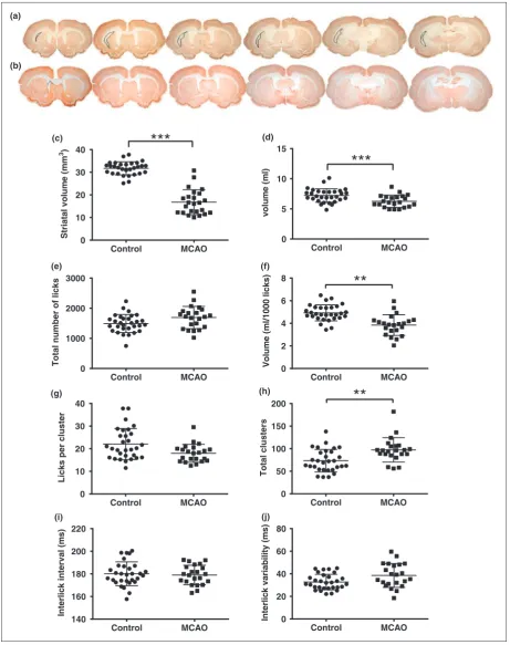
MCAO rats exhibited significant atrophy of the ipsilat-eral hemisphere (Figure 1(a)–(c)) and deficits in 3/7 lickometry parameters. MCAO resulted in a reduction
in water consumption (t50¼3.179, p¼0.0025,
Figure 1(d)). This was not due to performing fewer licks; in fact, there was a trend towards MCAO rats performing more licks (Figure 1(e), t50¼2.143,
p¼0.04, ns due to Bonferroni). However, the volume consumed per 1000 licks was decreased in MCAO ani-mals (Figure 1(f), t50¼4.805, p¼0.0001), indicating a
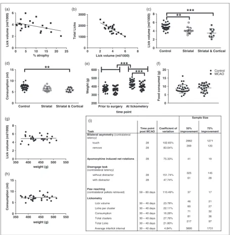
reduction in lick efficiency, which correlated with atro-phy (Figure 2(a), r¼ 0.439, p<0.05). The reduced lick efficiency also correlated with the increase in total licks (Figure 2(b), r¼ 0.649, p<0.001), and likely reflects a compensation mechanism to maintain consumption due to reduced lick efficiency. When examining drinking pat-terns, there was a trend towards the MCAO group making fewer licks per cluster (U¼209, p¼0.02, ns due to Bonferroni) (Figure 1(g)), which was compen-sated by an increase in clusters (U¼169, p¼0.0024, Figure 1(h)). No differences were found in ILI (t50¼0.404, p¼ns, Figure 1(i)), or ILI variability
(t50¼2.422, p¼ns, Figure 1(j)). When Bayes factors
were calculated for all parametric measures, the only measure that supported the null hypothesis was ILI (Bayes factor, K¼4.44). There was strong support for the alternative hypothesis (a difference between
MCAO and control) for consumption (K¼13.97),
volume per lick (K¼1330.59) and ILI variability (K¼2.60) and weak support for total number of licks (K¼1.53).
For the key measures of lick efficiency and consump-tion, we divided MCAO rats into those with striatal only and striatal and cortical lesions. Both groups had signifi-cantly lower lick efficiencies (Figure 2(c), F2,49¼13.61,
p<0.0001, striatal vs control: p¼0.0067, striatal and cortical vs control: p<0.0001). However, only the stri-atal and cortical group reduced the volume of water consumed (Figure 2(d), F2,49¼5.52, p¼0.0069, control
vs cortical and striatal: p<0.0116).
Although the MCAO rats had oral deficits, they maintained food intake. The MCAO rats consumed an equal amount food compared to the control group (Figure 2(f)) and the weight of all rats increased
signifi-cantly from the day of surgery (Figure 2(e),
F1,50¼1004, p<0.0001). However, at the time of
licko-metry, the MCAO animals were lighter than the con-trols (Figure 2(e), F1,50¼45.25, p<0.0001, MCAO vs
Control MCAO 0
10 20 30 40
Striatal
v
o
lu
m
e
(m
m
3)
(c)
***
Control MCAO 0
2 4 6 8
V
o
lu
m
e
(m
l/1000
licks)
**
(f)
Control MCAO 0
50 100 150 200
Tota
l
clu
s
ter
s
(h)
**
Control MCAO 140
160 180 200 220
Interlic
k
in
terval
(m
s
)
(i)
Control MCAO 0
5 10 15
volu
m
e
(m
l)
***
(d)
Control MCAO 0
10 20 30 40
Licks
p
er
cluster
(g)
Control MCAO 0
1000 2000 3000
Total
number
of
lick
s
(e)
Control MCAO 0
20 40 60 80
Interlic
k
variability
(m
s
)
(j) (a)
[image:3.595.74.536.67.651.2](b)
Figure 1. Representative NeuN stained sections from the median animal of the MCAO group, with the lesion outlined (a) and
Control Striatal Striatal & Cortical 0 5 10 15 Consum ption (m l)
**
Control Striatal Striatal & Cortical
0 2 4 6 8 Lick vo lu m e (m l/1000 )
0 2 4 6 8
0 1000 2000 3000
Lick volume (ml/1000)
T o ta l L icks
0 5 10 15 20 25
0 2 4 6 % atrophy Lick v o lume (m l/1000 )
Prior to surgery At lickometery
200 300 400 500 600 time point W e ight (g ) 0 5 10 15 20 Food consumed (g ) Control MCAO
350 400 450 500 550
0 2 4 6 8 weight (g) Lick v o lume (m l/1000 )
350 400 450 500 550
0 5 10 15 weight (g) Consum ptio n (m l)
(a) (b) (c)
(d) (e) (f)
(g) (h)
***
**
***
***
(i) Sample Size Task Time point post MCAO Coefficient of variation 50% improvement 75% improvement Bilateral asymmetry(contralaterallatency)
touch 28 102.65% 2862 1271
remove 28 83.04% 269 120
Apomorphine induced net rotations 28 75.33% 41 19
Disengage task
(contralateral latency)
without distracto 8 151.74% 325 145 with distracto
2 r
2
r 8 47.74% 61 28
Paw reaching
(contralateral pellets retrieved) 59 – 80 days 110.49% 37 17
Lickometry
[image:4.595.63.523.63.529.2]Lick volume 30 – 40 days 23.78% 46 21 Licks per cluster 30 – 40 days 22.11% 60 27 Consumption 30 – 40 days 16.28% 71 32 Total clusters 30 – 40 days 27.76% 81 36 Total Licks 30 – 40 days 21.80% 217 97 Average interlick interval 30 – 40 days 4.84% 3895 1731
Figure 2. Lick volume correlated with brain atrophy (a) and total number of licks (b) for the MCAO animals (n¼22). Lick efficiency
The coefficient of variation for the lickometry meas-ures was lower than other behavioural measure per-formed in the same group of rats (Figure 2(i)), and this resulted in acceptable sample sizes for potential treatment studies (Figure 2(i)).
Discussion
The purpose of the present data was to demonstrate the usefulness of lickometry for assessing long-term post-stroke functional deficits. This test has translational potential, as lack of lingual coordination and deficits in tongue function are common presentations in stroke patients and are associated with dysphagia and dysarth-ria.24,25The results are the first to suggest that MCAO leads to lasting deficits in drinking behaviour.
Peripheral sensory and motor denervation has been reported to produce a reduction in volume per lick, and an increased number of licks without changes in ILI.17 The present results are in agreement, demonstrating that this is most likely a sensorimotor deficit. While lick efficiency decreased in the MCAO group, the total number of licks was slightly increased, and there were more licking clusters without changes to ILI. The increased number of licks and clusters is likely an attempt to compensate for the decreased licking effi-ciency, and indicates MCAO rats do not have reduced motivation to drink despite weighing slightly less than the controls. Although body weight may influence over-all daily consumption, it did not confound this task as it did not influence the amount of water consumed in this short 15 min test and it did not correlate with lick effi-ciency. Additionally, despite having very little variance (4.87% CV), the fact that the ILI was not affected, as supported by the Bayes factor, also indicates that this is a sensorimotor deficit, rather than an alteration in con-trol of the drinking pattern. It should be highlighted that the deficits detected in these animals needed subtle analysis with equipment that could record to the 0.01 s, they did not have such significant impair-ments in oral function that they became malnourished or dehydrated.
Brain atrophy was correlated with lick efficiency, and a sub-analysis of striatal only versus striatal and cortical lesions indicated the decrease in consumption was only evident in the animals with striatal and cor-tical damage. This highlights the ability of this test to discriminate lesion types. However, lick efficiency was reduced in the animals with pure striatal lesions as well, demonstrating the sensitivity of this task to detect def-icits in animals with modest lesions. One limitation of the present study is that the histology was not per-formed at the same timepoint as the lickometry, though we do not expect further lesion evolution beyond the 40-day period.
Previously, only tongue protrusion has been exam-ined after MCAO.28Lickometry provides a more com-prehensive analysis of tongue function and is highly sensitive to long-term deficits. There is very low vari-ation in the data obtained from this task in comparison to the other behavioural measures performed on the same rats. Furthermore, lickometry results in reason-able sample size predictions for treatment studies (n¼21), similar to that required for paw reaching, which is often reported as a sensitive test, and much less than the bilateral asymmetry task (or stick dot, n¼120), which is a commonly used test. Unlike many tests of sensorimotor function,15 drinking is a behav-iour that the rats engage in multiple times a day and thus if spontaneous recovery and/or compensation was to occur, it would happen quickly following MCAO without needing exposure to the test itself. However, as deficits were still seen at 30–40 days, it is unlikely that compensation or significant natural recovery will be seen with this task.
In addition to the high sensitivity, as it is able to detect oral deficits in animals with modest lesions, licko-metry has practical advantages over other behavioural tests. It can be fully automated, and it is quantitative. Drinking is a natural behaviour for animals, so very little training is required. Water restriction is required but only needs to be implemented for a few days (usually 4–5 days) and should not impact the health or welfare of the animals. Nevertheless, there are a few consider-ations with regards to implementation of a lickometry system. For example, the distance between the animal and the drinking tube can influence the licking rate, spe-cifically, the amount of ‘tongue travel’ is negatively related to licking frequency.29 When using lickometry for stroke research, this should be carefully considered, as the animals may have postural difficulties. However, as we saw no alteration in ILI, this did not appear to be a problem with the set-up used in this study. The equip-ment presented here may appear costly, and labs must consider this when choosing to invest in automated sys-tems. However, the reduction in bias and man-power might outweigh the initial cost of the equipment, and less expensive, simpler versions are available than the one presented here.
Overall, lickometry provides a reliable, quantitative measure of long-term oral deficits that is easy to imple-ment in the rodent. Furthermore, this test is highly translational, as oral deficits, while largely unexplored in the rodent, are comparable to symptoms exhibited by the patient population, such as tongue protrusion and the corresponding dysarthria and dysphagia.
Funding
article: The study was funded by grants from the European Union Framework 6 StemStroke (LSHB-CT-2006-037526) and the UK Medical Research Council.
Declaration of conflicting interests
The author(s) declared no potential conflicts of interest with respect to the research, authorship, and/or publication of this article.
Authors’ contributions
RCT, DMD, SBD, DJH and TDF conceived and designed the work. All authors were involved in the analysis and inter-pretation of the data. JA and RCT wrote the manuscript and all authors provided critical revisions.
Supplementary material
Supplementary material for this paper can be found at http:// journals.sagepub.com/doi/suppl/10.1177/0271678X16684141
References
1. Dirnagl U. Bench to bedside: the quest for quality in experimental stroke research. J Cereb Blood Flow
Metab2006; 26: 1465–1478.
2. Gladstone DJ, Black SE and Hakim AM. Toward wisdom from failure: lessons from neuroprotective stroke trials and new therapeutic directions. Stroke 2002; 33: 2123–2136.
3. Fisher M, Feuerstein G, Howells DW, et al. Update of the stroke therapy academic industry roundtable preclin-ical recommendations.Stroke2009; 40: 2244–2250. 4. Encarnacion A, Horie N, Keren-Gill H, et al. Long-term
behavioral assessment of function in an experimental model for ischemic stroke. J Neurosci Methods 2011; 196: 247–257.
5. Lindner MD, Gribkoff VK, Donlan NA, et al. Long-lasting functional disabilities in middle-aged rats with small cerebral infarcts.J Neurosci2003; 23: 10913–10922. 6. Modo M, Stroemer RP, Tang E, et al. Neurological sequelae and long-term behavioural assessment of rats with transient middle cerebral artery occlusion.
J Neurosci Methods2000; 104: 99–109.
7. Freret T, Chazalviel L, Roussel S, et al. Long-term func-tional outcome following transient middle cerebral artery occlusion in the rat: correlation between brain damage and behavioral impairment. Behav Neurosci 2006; 120: 1285–1298.
8. Hudzik TJ, Borrelli A, Bialobok P, et al. Long-term func-tional end points following middle cerebral artery occlu-sion in the rat. Pharmacol Biochem Behav 2000; 65: 553–562.
9. Peeling J, Corbett D, Del Bigio MR, et al. Rat middle cerebral artery occlusion: correlations between histopath-ology, T2-weighted magnetic resonance imaging, and behavioral indices. J Stroke Cerebrovasc Dis 2001; 10: 166–177.
10. Trueman RC, Harrison DJ, Dwyer DM, et al. A critical re-examination of the intraluminal filament MCAO
model: Impact of external carotid artery transection.
Transl Stroke Res2011; 2: 651–661.
11. Klein A, Sacrey L-AR, Whishaw IQ, et al. The use of rodent skilled reaching as a translational model for inves-tigating brain damage and disease.Neurosci Biobehav Rev 2012; 36: 1030–1042.
12. Alaverdashvili M and Whishaw IQ. Motor cortex stroke impairs individual digit movement in skilled reaching by the rat.Eur J Neurosci2008; 28: 311–322.
13. Metz GA and Whishaw IQ. Cortical and subcortical lesions impair skilled walking in the ladder rung walking test: a new task to evaluate fore- and hindlimb stepping, placing, and co-ordination.J Neurosci Methods2002; 115: 169–179. 14. Riek-Burchardt M, Henrich-Noack P, Metz GA, et al.
Detection of chronic sensorimotor impairments in the ladder rung walking task in rats with endothelin-1-induced mild focal ischemia. J Neurosci Methods 2004; 137: 227–233.
15. Trueman RC, Diaz C, Farr TD, et al. Systematic and detailed analysis of behavioural tests in the rat middle cerebral artery occlusion model of stroke: Tests for long-term assessment. J Cereb Blood Flow Metab 2016; Epub ahead of print 17 June 2016. DOI: 10.1177/ 0271678X16654921.
16. Pisa M. Motor functions of the striatum in the rat: critical role of the lateral region in tongue and forelimb reaching.
Neuroscience1988; 24: 453–463.
17. Shires CB, Saputra JM, Stocks RMS, et al. Effects of sensory or motor nerve deafferentation on oromotor function in mice. Otolaryngol Head Neck Surg 2011; 144: 915–920.
18. Travers JB, Dinardo LA and Karimnamazi H. Motor and premotor mechanisms of licking. Neurosci Biobehav Rev1997; 21: 631–647.
19. Bryant JL, Boughter JD, Gong S, et al. Cerebellar cor-tical output encodes temporal aspects of rhythmic licking movements and is necessary for normal licking frequency.
Eur J Neurosci2010; 32: 41–52.
20. Glendinning JI, Gresack J and Spector AC. A high-throughput screening procedure for identifying mice with aberrant taste and oromotor function. Chem
Senses2002; 27: 461–474.
21. Welzl H. Attempt to modify rate and duration of licking in rats by operant conditioning.Behav Processes1976; 1: 319–326.
22. Umapathi T, Venketasubramanian N, Leck KJ, et al. Tongue deviation in acute ischaemic stroke: a study of supranuclear twelfth cranial nerve palsy in 300 stroke patients.Cerebrovasc Dis2000; 10: 462–465.
23. Urban PP, Hopf HC, Fleischer S, et al. Impaired cortico-bulbar tract function in dysarthria due to hemispheric stroke. Functional testing using transcranial magnetic stimulation.Brain1997; 120(Pt 6): 1077–1084.
24. Hirota N, Konaka K, Ono T, et al. Reduced tongue pres-sure against the hard palate on the paralyzed side during swallowing predicts Dysphagia in patients with acute stroke.Stroke2010; 41: 2982–2984.
26. Dwyer DM, Lydall ES and Hayward AJ. Simultaneous contrast: evidence from licking microstructure and cross-solution comparisons.J Exp Psychol Anim Behav Process 2011; 37: 200–210.
27. Wright RL, Gilmour G and Dwyer DM. Microstructural analysis of negative anticipatory contrast: A reconsider-ation of the devalureconsider-ation account.Learn Behav2013; 41: 353–359.
28. Gulyaeva N, Thompson C, Shinohara N, et al. Tongue protrusion: a simple test for neurological recovery in rats following focal cerebral ischemia. J Neurosci Methods 2003; 125: 183–193.
29. Weijnen JA. Licking behavior in the rat: measurement and situational control of licking frequency. Neurosci