Abstract
Primary blast lung injury frequently complicates military conflict and terrorist attacks on civilian populations. The fact that it occurs in areas of conflict, or unpredictable mass casualty events makes clinical study in human casualties implausible. Research in this field is therefore reliant on the use of some form of biological or non-biological surrogate model. We briefly review the modelling work undertaken in this field to date and describe the rationale behind the generation of our in-silico physiological model.
Introduction
First described by Hooker in 1924(1) as a “single gross lesion found post mortem after exposure to air concussion due to high-explosive”, primary blast lung injury (PBLI) is currently defined as “radiological and clinical evidence of acute lung injury occurring within 12 hours of exposure and not due to secondary or tertiary injury”.(2) It is a disease characterized by intra-parenchymal haemorrhage, laceration and pneumothoraces.(3) In the absence of a specific biomarker or radiological hallmark, it can be difficult to distinguish PBLI with confidence from other forms of lung damage in
Modelling based research
Modelling is the use of a surrogate entity to represent a complex system in a readily reproducible manner. Models can be either biological or non-biological. Biological models are further subdivided into in-vitro (cell culture), ex-vivo (live organ) or in-vivo (live animal).(8) Non-biological models are either computational (“in-silico”) or physical (Anthropomorphic) surrogates of the biological system of interest.
As a research technique, the validity of modelling parallels that of clinical trials or laboratory study.(9) Non-biological based research is cheaper than animal modelling, requires less stringent ethical approval and can accommodate scenarios that are unachievable in live animal or human research (such as multiple casualty with multiple injury events). It can do this in an easily repeatable manner so that adequately powered studies which can achieve statistical significance can be undertaken. Modelling also facilitates the Ministry of Defence’s ambition of limiting animal experimentation (10) and the impetus for the scientific community to “Replace, Reduce and Refine” when considering the use of live animals in research.(11, 12)
research continues using both in-vivo and ex-vivo biological models.(14-16) Rodents are commonly used to model lung injury due to a variety of mechanisms including blast. (17) and human cadaveric specimens have been used to examine the effects of under-vehicle explosions on the lower limb.(18)A more recent example of in-vivo blast research is the porcine work undertaken by Garner et al at the Defence Science and Technology Laboritories (DSTL) in Porton Down.(19) This work demonstrated a significant increase in mortality when haemorrhagic shock and blast exposure are combined which subsequently lead to a change in resuscitation protocol within the DMS. The four arms of this study was limited to six to eight subjects for the reasons discussed above and so could only accommodate the study of an immediately life threatening combination of injuries (i.e. course data) and not the intermediate term and more subtle outcomes normally sought in medical intervention research.
One of the earliest examples of anthropomorphic modeling in blast lung research was the Blast Test Device (BTD) developed by the US military. It consists of a chest shaped
Computational modelling has evolved in parallel to advances in computing power. Finite element modeling (FEM) treats the subject of interest as a 3-dimensional mesh of finite blocks each of which has known mechanical properties. These individual components effect change on neighboring units in a predictable manner and thus physical effects on the subject as a whole can be predicted. A FEM model was commissioned by the UK coroner’s office after the suicide bombings on the London transport network in 2005.(22) This quick-running model looked both at primary and secondary (fragmentation) injury resulting from detonation of an explosive device in a crowded area. It was able to generate an abbreviated injury score (AIS) for casualties based on blast injury threshold limits and likely fragmentation injury and so represents a significant step towards arming civil authorities and clinicians with clinically useful information. Whilst much faster than most FEM models, this model still requires 5 hours of run time to recreate 30 minutes of simulated time.(23) A FEM model of PBLI in sheep has recently been developed which can accurately predict the volume of injured lung following a blast but remains unable to inform the medical community regarding the likely level of care such casualties would need and it does not facilitate the study
of potential medical interventions.(24) FEM modelling however normally takes several days per scenario and requires computing power that is not widely available. Despite advances in computer technology, FEM remains predominantly a tool to study structural rather than physiological consequences of injury.
Our current model
series of individual components, each of which are described by a set of independent variables (Fig 2). At the beginning of a modelling study, these variables are initially set so that they represent the patient population to be studied. Once initiated, the model undertakes a series of pre-determined physiological equations for a period of 30 milliseconds which represents one physiological time slice t. The end product of this series of equations then determines the value of the variables used in the next time slice. This iterative process continues for the study run-time T.
The respiratory element of the model consists of the mechanical ventilator and breathing circuit, physiological deadspace (60 mL), anatomical and alveolar shunts and a variable number of ventilated alveoli each of which has its own vascular component. Inhaled gases consist of oxygen, nitrogen, carbon dioxide, water vapour and gas α (anaesthetic or toxic gases). The cardiovascular element is composed of 19 compartments each of which are described by both fixed parameters (unstressed volume and elastance coefficients, resistance and viscosity) and iteratively updated variables (pressure, flow and volume). The systolic/diastolic cycling is modelled through a repeating pulsatile activation function of variable duration.
The numerical simulations of the integrated model provide results that agree with clinical data available in the published literature and the model has also been validated in a number of earlier studies.(27)
Adapting the model to reflect PBLI within the military context
terminal anaesthesia.(30) Our model has produced results closely matching this in-vivo data for both blast and combined blast and haemorrhagic shock.
For the model to be of relevance to the DMS we feel that it needs to meet several criteria. Primarily, it must be validated against the human injury experienced by UK service personnel suffering PBLI in combat. To this end we are creating a clinical database of UK PBLI victims generated in the recent conflict in Afghanistan which will be used to inform the model as to blast-dose related physiological effect and outcome. We need to be able to utilise the model throughout the chain of care from the point of wounding to rehabilitation. It therefor needs to be able to accommodate the study of buddie-buddie care in a pre-hospital environment, potential medical interventions in a Role II/III emergency department and also a variety of ventilatory approaches whilst mechanically ventilated in intensive care. In order to achieve this several adaptions need to be made. It must be able to model spontaneous ventilation in the pre-hospital environment, the effect of possible modulators of pulmonary inflammation and biotrauma that could be administered both in the pre-hospital or emergency department and finally it should be able to replicate the consequences of intensive
care management including ventilator induced lung injury (VILI), oxygen toxicity and a fluctuating fluid volume status. In addition to this we hope to make the software sensitive to the age and gender of the casualty.
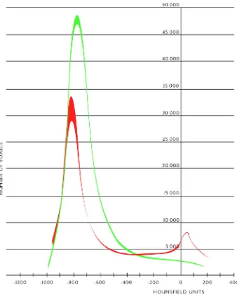
is poorly or non-aerated as a consequence of PBLI (Figure. 4).(31) Early evidence suggests that this method may prove useful in the identification of casualties with PBLI.(32) This work will be used to inform our computerised model of the proportion of non-functioning alveoli in our human casualties in order to increase its fidelity and clinical range.
Future direction.
Despite this extensive modeling activity, it has not kept pace with advances in medicine such as physician lead pre-hospital care, highly orchestrated and effective emergency department management of critically injured casualties, intensive care therapy and computed tomography imaging. It also fails to recognize the fact that improved pre-hospital care will result in increasingly severe cases of PBLI requiring management by the DMS. No model or measurable parameter exists that will either inform clinicians of the degree of injury resulting from shockwave exposure alone, can predict the ongoing physiological compromise surviving casualties will suffer or allow clinicians to model different treatment, mitigating or preventative strategies.(33) It is the ambition of our
group to create a militarily relevant blast lung injury model validated against human combat injury and augmented by specific serological and CT markers of disease severity that will facilitate future research in this field.
Conflict of Interest
References
1.
Hooker DR. Physiological effects of air concussion. American Journal of
Physiology. 1924;67(2):219-74.
2.
I Mackenzie, Tunnicliffe B, J Clasper, P Mahoney, E Kirkman. What the
intensive care doctor needs to know about blast-related lung injury. Journal of
the Intensive Care Society 2013;14(4):303-12.
3.
Wolf SJ, Bebarta VS, Bonnett CJ, Pons PT, Cantrill SV. Blast Injuries. Lancet
2009;374: 405-15.
4.
Smith JE. The epidemiology of blast lung injury during recent military
conflicts: a retrospective database review of cases presenting to deployed
military hospitals, 2003-2009. Philosophical transactions of the Royal Society of
London Series B, Biological sciences 2011;366(1562):291-4.
5
Dearden P. New Blast Weapons. J R Army Med Corps 2001;147: 80-86.
6
Breeze J, Lewis EA, Fryer R, Hepper AE, Mahoney PF, Clasper JC. Defining
the essential anatomical coverage provided by military body armour against high
energy projectiles. J R Army Med Corps 2016;162:284-290.
7
Davis PR, Rickards AC, Ollerton JE. Determining the composition and
benefit of the pre-hospital medical response team in the conflict setting. J R Army
Med Corps 2007;153: 269-273.
8.
Cernak I.. Long-Term Effects of Blast Exposures. For the committee on the
Gulf War and Health. Volume 9. Washington D.C.
9.
Hardman JG, Ross JJ. Modelling: a core technique in anaesthesia and
critical care research. British Journal of Anaesthesia 2006;97(5):589-92.
10.
. wwwgovuk/guidance/research-and-testing-using-animals.
11.
Russell WMS, Burch RL. The Principles of Humane Experimental
Technique. Wheathampsted: Universities Federation for Animal Welfare; 1992.
12.
The Animals Scientific Procedures Act (ASPA), (1986).
13.
Bowen IG, Fletcher E, Richmond D. Estimate of Man's Tolerance to the
Direct Effects of Blast. Technical progress report no. DASA-2113. In: Defence Do,
editor. Washington DC: Defence Atomic Support Agency; 1968.
14.
Breeze JCD, Mabbott A et al. Refrigeration and freezing of porcine tissue
does not affect the retardadtion of fragment simulating projectiles.
15.
Butler BJ, Bo C, Tucker AW et al. Mechanical and histiological
characterisation of trachea tissue subjected to blast-type pressures. Journal of
Physics 2014, Conference series 500.
16.
Chai JK, Cai JH, Deng HP et al. Role of neutrophil elastase in lung injury
induced by burn-blast combined injury in rats. Journal of the International
Society for Burn Injuries 2013;39(4):745-53.
17.
Brown RF, Cooper GJ, Maynard RL. The ultrastructure of rat lung
following acute primary blast injury. International journal of experimental
pathology 1993;74(2):151-62.
19.
Garner J, Watts S, Parry C, Bird J, Cooper G, Kirkman E. Prolonged
permissive hypotensive resuscitation is associated with poor outcome in
primary blast injury with controlled hemorrhage. Ann Surg 2010;251: 1131-39
20.
Yu JH, Vasel EJ, Stuhmiller JH. Modeling of the Non-Auditory Response to
Blast Over-pressure: Design and Field Test of a Blast Overpressure Test Module.
Jaycor Inc; San Diego1990.
21.
Jönsson A, Clemedson C, Arvebo E. An anthropomorpic dummy for blast
research. Proceedings of the International Conference on Protective Clothing
Systems 1981; August 23-27; Stockholm, Sweden.
22.
Pope DJ. The development of a quick-running prediction tool for the
assessment of human injury owing to terrorist attack within crowded
metropolitan environments. Philosophical transactions of the Royal Society of
London Series B, Biological sciences 2011;366:127-43.
23.
Hepper AE, Pope DJ, Bishop M et al. Modelling the blast environment and
relating this to clinical injury: experience from the 7/7 inquest. Journal of the
Royal Army Medical Corps 2014;160:171-4.
24.
Gibbons MM, Dang X, Adkins M, Powell B, Chan P. Finite Element
Modeling of Blast Lung Injury in Sheep. J Biomech Eng 2015;137(4).
25.
Hardman JG, Bedforth NM, Ahmed AB et al. A Physiology simulator:
validation of its respiratory components and its ability to predict the patient's
response to changes in mechanical ventilation. Br J Anaesth 1998;81:327-32.
26.
Hardman JG, Wills JS, Aitkinhead AR. Investigating hypoxaemia during
apnea: validation of a set of physiological models. Anesth Analg 2000;90:614-8.
27.
McCahon RA, Colomb MO, Mahajan RP, Hardman JG. Validation and
application of a high-fidelity, computational model of acute respiratory distress
syndrome to the examination of the indices of oxygenation at constant
lung-state. British Journal of Anaesthesia 2008;101(3):358-65.
28.
Guy RJ, Kirkman K, Watkins PE, Cooper GJ. Physiologic responses to
primary blast. The Journal of trauma 1998;45:983-7.
29.
Spear AM, Davies EM, Taylor C et al. Blast wave exposure to the
extremities causes endothelial activation and damage. Shock 2015;44(5):470-8.
30.
Garner JP, Parry C, Bird J, Kirkman E. Development of a large animal
model for investigating resuscitation after blast exposure. World J Surg
2009;33:2194-202.
31.
Heuer JF, Sauter P, Pelosi P et al. Effects of Pulmonary Acid Aspiration on
the Lungs and Extra-Pulmonary Organs: A Randomised Study in Pigs. Critical
Care 2012;16(2):R35.
32.
Hulse EJ, Vliegenthart ADB, de Potter CMJ et al. Computed tomography
voxel density and micro RNA analysis of blast lung injury. Poster Presentation,
Military Health Services Research Society (MHSRS); 2016.
Legends
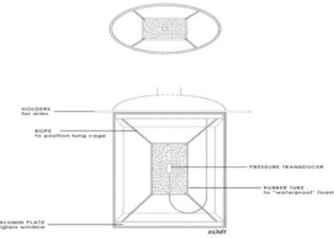
Figure 1. Pictorial representation of the Swedish Dummy Torso.
Figure 2. Pictorial representation of our current in-silico PBLI model.
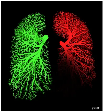
Figure 3. 3-D lung reconstruction. A significant proportion of the left (Red) lower lobe is not aerated in this PBLI casualty.
Figure 4. The histogram data from the 3D CT lung reconstruction denoting the