Accepted Manuscript
Title: Evaluation of extraction and normalisation strategies for the analysis of lipids in placental vessels
Authors: Katrin N. Sander, Daniel P. Hay, Fiona Broughton-Pipkin, Raheela N. Khan, David A. Barrett
PII: S0731-7085(18)31137-3
DOI: https://doi.org/10.1016/j.jpba.2018.08.012
Reference: PBA 12143
To appear in: Journal of Pharmaceutical and Biomedical Analysis
Received date: 14-5-2018 Revised date: 2-8-2018 Accepted date: 6-8-2018
Please cite this article as: Sander KN, Hay DP, Broughton-Pipkin F, Khan RN, Barrett DA, Evaluation of extraction and normalisation strategies for the analysis of lipids in placental vessels, Journal of Pharmaceutical and Biomedical Analysis (2018), https://doi.org/10.1016/j.jpba.2018.08.012
1
Evaluation of extraction and normalisation strategies for the analysis of lipids in
placental vessels
Katrin N. Sandera, b, Daniel P. Haya, Fiona Broughton-Pipkina, Raheela N. Khana, David A. Barrettb.
Affiliations:
aDivision of Medical Science and Graduate Entry Medicine, School of Medicine, University of
Nottingham, The Royal Derby Hospital, Uttoxeter Road , Derby, DE22 3DT, UK
b Advanced Materials and Healthcare Technologies Division, School of Pharmacy, University of
Nottingham, University Park, Nottingham, NG7 2RD, UK
Correspondence to [email protected]; phone: 01159515062
Subject category: Lipids and lipoproteins Mass spectrometry
Evaluation of extraction and normalisation strategies for the analysis of lipids in
placental vessels
Highlights
Rotor-stator disruption was chosen as the most appropriate homogenisation method for the extraction of lipids from tough, fibrous placental tissue
Wet weight determination was evaluated as a valid normalisation procedure as no benefit could be demonstrated in the use of more advanced normalisation factor
2
Abstract
The analysis of lipids in tough or fibrous biological tissues can be challenging due to difficulties in obtaining a representative sample following homogenisation of the tissue. Furthermore, the choice of normalisation method can have a major effect on the quality of quantitative results. Therefore, a range of mechanical homogenisation techniques and normalisation strategies were evaluated for application to human placental vessels. The findings showed that rotor-stator homogenisation in a suitable solvent and wet weight normalisation were the best combination of procedures for quantitative analysis of lipids in placental blood vessels.
3 KeyWords:
Eicosanoids Oxylipins
1.
Introduction
Endogenous bioactive lipids are involved in numerous physiological processes of the human organism and are therefore increasingly subject to quantitative studies in a variety of biofluids and tissues. While the preparation of liquid samples (for example blood plasma) for analysis is relatively simple, extraction of lipids from tissue samples often poses a challenge, particularly when the tissue is made up of fibrous, muscular or similar tough biological material. A prerequisite for a successful analysis is the appropriate choice of a homogenisation and normalisation strategy. An efficient homogenisation method should lead to disruption of the sample resulting in an increased surface area to facilitate quantitative extraction of analytes [1, 2].
Placental vessels, especially chorionic plate vessels, have a high muscular and fibrous content resulting in increased toughness [3] and our initial work with this tissue revealed that it was difficult to obtain homogeneous disruption prior to extraction. Typically, lipid contents are normalised to wet tissue weight but we also noted that a range of different normalisation strategies had been applied to fibrous tissues, including dry weight, protein content, total lipid content, or cross-sectional area. Normalisation to protein content has been recommended by some authors in preference to dry or wet weight, as this factor is less susceptible to changes between sample groups and stable structural proteins account for most proteins in tissue samples [4, 5]. Furthermore, marker proteins for a specific tissue type have also been used, e.g. creatine or myofibrillar protein content for skeletal muscle samples [6]. It has been demonstrated that the measured concentration of analytes can change considerably depending on the selected normalisation method [5-7].Tissue wet weight includes intra- and extracellular water but might also be distorted by rinsing of blood-stained tissue or by residual blood, especially in highly perfused tissues such as placental blood vessels. The variability of the water content can be eliminated by freeze-drying the sample pellets after the extraction of lipids.
Hence, we evaluated four homogenisation methods (Dounce homogenisation, ball mill, cryogenic grinding, rotor-stator homogenisation) and three normalisation methods (wet tissue weight, dry tissue weight, protein content) for the extraction and quantitative analysis of oxylipins from human placental vessels.
2.
Material and methods
2.1. Tissue collection
Placentae were collected after obtaining informed consent from healthy pregnant women delivering at full term gestation. Ethics approval was granted by Derby Research Ethics Committee (REC
Reference No. 09/H0401/90). First branch chorionic plate arteries (CPA) were dissected, briefly rinsed with PBS, stripped of remaining water droplets using forceps and snapfrozen for storage at -80°C within 1h after delivery.
2.2. Comparison of tissue homogenisation methods
CPA from four placentae were minced on dry ice, mixed to form one homogeneous batch of tissue and then transferred into Eppendorf tubes in 0.3 g aliquots. The tissue was subsequently
homogenised using one of the four following methods: For ball milling (n=6), 2 grinding balls
4 (stainless steel, 4 mm ⌀) were added to each of the Eppendorf tubes. The tissue was disrupted (MM301 mixer mill, Retsch, Haan, Germany) by milling 7 times for 5 min at 30 s-1 with intermittent
cooling, until no further visible disruption of the tissue was noted. For the Dounce homogenisation (n=6), a thawing tissue aliquot was homogenised in a cooled Dounce homogeniser for 2 min. For the cryogenic grinding using liquid N2 (n=5), frozen tissue was packed into several layers of cooled
aluminium foil and crushed using a pestle and mortar. For the rotor-stator homogenisation (n=5), frozen tissue was transferred into a glass tube containing 1 mL of ice cold H2O. The tissue was
homogenised for 10 s using a rotor-stator homogeniser (T-25 Ultra-TurraxTM, IKA, Staufen, Germany).
After disruption of the tissue using the described methods, ice cold extraction solvent (0.3% formic acid, 0.07% v/v butylhydroxytoluene in ethanol) was added to each sample and the tissue was extracted for 1 h at 4°C on a shaker. The final solvent composition for samples of all methods before extraction on the shaker was 75% extraction solvent and 25% water. Samples were centrifuged at 15000g for 20 min at 4°C and the supernatant was added to chilled H2O. The final solvent
composition for supernatants of samples of all methods before solid phase extraction (SPE) was 25% extraction solvent and 75% water.
Tissue homogenate supernatants were extracted using SPE columns (Phenomenex Strata-XL 100 µm, polymeric sorbent) according to the instructions of the manufacturer. All samples were disrupted and extracted on the same day and snap frozen before analysis. A previously validated LC-MS/MS method was used for the relative quantification of 10 hydroxyeicosatetraenoic acids (HETEs), 4 epoxyeicosatrienoic acids (EETs), 4 dihydroxyeicosatrienoic acids (DHETs), 2
hydroperoxyeicosatetraenoic acids (HpETEs), 2 octadecadienoic acids (ODEs), 2 leukotrienes, and 6 prostaglandin derivatives [8]. The peak areas were determined and normalised against the initial tissue wet weight. LC-MS/MS-analysis of the samples occurred within one single run.
2.3. Comparison of normalisation methods
CPA from three placentae were minced on dry ice, mixed to form one homogeneous batch of tissue and 12 tissue aliquots were weighed in a range of 300 ± 150 mg. CPA tissues were homogenised using a rotor-stator homogeniser, processed using solid phase extraction (SPE) and analysed via LC-MS as described above. Tissue pellets were kept at -80°C for determination of protein content and dry weight. Pellets were left to thaw on ice and were briefly disrupted using a pipette tip. 1% SDS was added to each tube and pellets were vortexed for 30 min at 20°C with 14000 rpm (vortex at room temperature was necessary to prevent precipitation of sample components). Samples were centrifuged for 20 min at 20°C with 15000 g. The supernatant was diluted 1:10 in 0.9% NaCl and the protein content was determined using a BCA assay. After protein resolubilisation, tissue pellets were freeze-dried using a Thermo Powerdry PL3000 freeze dryer and the dry weight was recorded. The peak areas were determined and normalised against the initial tissue wet weight, dry weight, or protein content.
2.4. Data analysis
LC-MS/MS data analysis was done as described in [8]. Raw data was processed using Analyst (version 1.4, Applied Biosystems, Foster City, USA). Statistical analysis was performed using SPSS software (Version 22, IBM, New York, USA). Graphs were created using Prism (version 6, GraphPad, La Jolla, USA).
5
3.
Results and Discussion
3.1. Comparison of tissue homogenisation methods
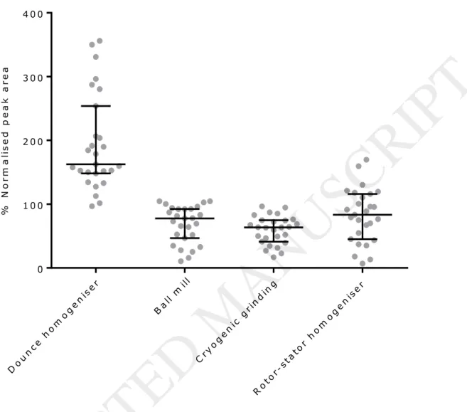
27 oxylipin analytes were detected in the CPA tissue samples and their relative yields using the different homogenisation methods are shown in Figure 1.
The Dounce homogeniser did not visibly disrupt the tissue and produced a highly variable recovery of the lipids. Cryogenic grinding and use of the ball mill resulted in the same problem of a hardly disrupted sample, as assessed by visual inspection, so that the subsequent extraction was essentially performed on unhomogenised tissue samples. Both methods were executed while constantly cooling the tissue using liquid N2. Instead of the anticipated brittle consistency, the tissue samples
became tough and rubbery. The rotor-stator homogenisation was the only method realising a disruption of the tissue into fine particles. In essence, the degree of disintegration of the tissue did not seem to affect the extraction efficiency. Therefore, consideration needs to be given to the fact that tissue disruption does not seem to be necessary to obtain recovery and this suggests that lipids can be extracted by penetration of solvent into the tissue. Homogenisation may slightly enhance recovery but simple partition/diffusion of the lipids into the extraction solvent may have a major contribution to recovery. However, this validation used samples that were aliquoted from one large amount of homogeneously minced tissue. It is likely that in clinical studies, samples are more heterogeneous due to different handling or inter-individual differences between patients. Then varying tissue size, thickness or constitution might affect the extraction efficiency when
homogenisation is only superficial as observed for cryogenic grinding, ball mill or Dounce homogeniser.
All of the tested methods were time consuming, but especially Dounce homogenisation and cryogenic grinding with pestle and mortar are not suitable for high throughput experiments as previously reported by a number of authors [1, 9, 10]. Issues were a time consuming
homogenisation procedure, possibly impairing analyte stability and increased loss of tissue due to transfer operations. In addition to that, the equipment needs to be cleaned between samples. The rotor-stator homogeniser facilitates a faster and cleaner procedure but still every sample needs to be processed individually. The ball mill enabled the highest throughput with a simple procedure that could process up to 20 samples per session. This technique was thoroughly assessed for the
extraction of mouse tissue in high-throughput experiments and shown to deliver good performance [11, 12].
Significance testing by one-way ANOVA and subsequent multiple comparisons using Dunnett T3 showed that the analyte yield significantly changed for most of the compounds based on the used homogenisation method (p<0.05). Multiple comparisons pointed out the Dounce homogeniser as the method for which most of the analytes were significantly different in comparison to other methods (p<0.05). No difference was seen amongst the other methods. Dounce homogenisation gave the highest normalised peak area for the analytes, however, the validity of this result is highly questionable. In our experience tissue residues were not completely removed despite thorough cleaning of the Dounce homogeniser. Tissue blanks (undergoing the same procedure without tissue) showed considerable and very variable peaks for the analytes of interest. The increased yield of analytes by the Dounce homogeniser might therefore partly be caused by these analyte residues. In addition to that, the method is not practical for large sample sets as discussed above and previously noted [12]. The need to process the sample set in batches over several days is likely to introduce variability and susceptibility to errors. As Dounce homogenisation gave widely-varying results, it was considered not to be suited for purpose.
6 Instead, the rotor-stator homogeniser was deemed to be the most appropriate method for future sample processing. This method is quick and uncomplicated, providing an enhanced analytical performance. This is also in line with a publication showing that the rotor-stator homogenisation caused less variability among samples compared to cryogenic grinding using pestle and mortar [10]. It is important to note that the procedure needs to be performed in a small amount of water, which prevents denaturation of the sample and obstruction of the homogeniser. This enables disruption into fine particles within seconds and avoids methodological problems as previously reported [13]. Addition of organic solvent after homogenisation then allows the extraction of lipophilic analytes.
3.2. Comparison of normalisation methods
27 analytes were detected in the samples (n=12). A good correlation of wet weight (ww) and dry weight (dw) was observed per sample, whereas the correlation of protein content (pc) with ww or dw was less pronounced (dw:ww R2=0.95; pc:ww R2=0.69; pc:dw R2=0.76). The relative standard
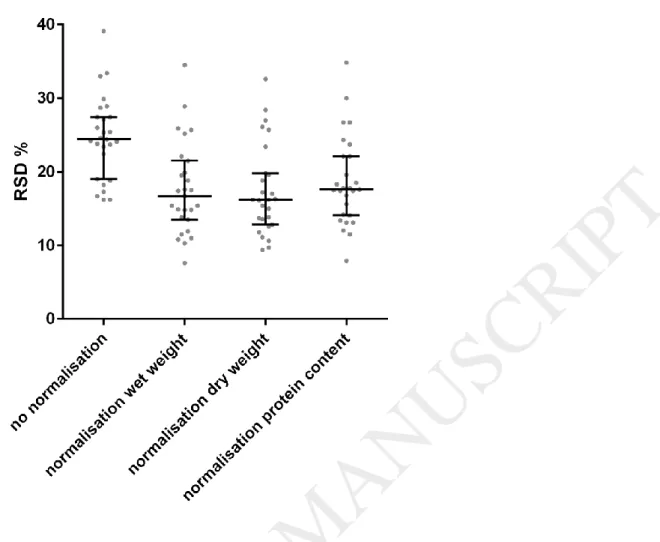
deviation (RSD) across the 12 samples was calculated for each of the 27 analytes. Figure 2 shows that all three normalisation methods significantly improve the RSDs of analyte levels. No significant difference was found between the normalisation strategies.
This result is in line with previous publications, reporting that wet weight, dry weight and protein content normalisation are all effective in improving inter-sample variability [14]. As the additional effort of protein content or dry weight determination did not improve the quality of the data, wet weight normalisation was deemed to be the most suitable normalisation method for this type of tissue sample.
4.
Conclusion
This study aimed to develop a suitable experimental protocol for the quantification of lipids in tough, fibrous placental tissue samples, which is more challenging than the analysis of biofluids. Rotor-stator disruption was chosen as the most appropriate homogenisation method for large studies, as other tested homogenisation methods (ball mill, cryogenic grinding and Dounce homogeniser) hardly disrupted placental vessels or were not suitable for a large number of samples. Furthermore, wet weight determination was identified as a valid normalisation procedure as no benefit could be demonstrated in the use of more advanced normalisation factors such as dry weight or protein content. The applicability of Rotor-stator disruption and wet weight normalisation may be extended to other biological tissues.
5.
Acknowledgments
We thank the patients for participating in this study and the clinical staff of the Department of Obstetrics and Gynaecology at the Royal Derby Hospital for their cooperation.
6.
Sources of funding
This work was supported by the British Heart Foundation [grant number: PG/10/49/28422]; the University of Nottingham (studentship)
7.
Disclosures
None.
8.
References
[1] H. Gao, S. Ho, J. Williams, LC-MS Bioanalysis of Drugs in Tissue Samples, Handbook of LC-MS Bioanalysis, John Wiley & Sons Inc.2013, pp. 297-306.
7 [2] C. Yu, L.H. Cohen, Tissue sample preparation - Not the same old grind, Lc Gc Europe, 17 (2004) 96-+.
[3] M.J. Mulvany, C. Aalkjaer, Structure and function of small arteries, Physiol Rev, 70 (1990) 921-961.
[4] W.W. Christie, X. Han, Lipid Analysis: Isolation, Separation, Identification and Lipidomic Analysis, Elsevier Science2010.
[5] M.A. Marks, Y. Eby, R. Howard, P.E. Gravitt, Comparison of normalization methods for measuring immune markers in cervical secretion specimens, J Immunol Methods, 382 (2012) 211-215.
[6] M.L. Nishio, A.G. Madapallimattam, K.N. Jeejeebhoy, Comparison of six methods for force normalization in muscles from malnourished rats, Med Sci Sports Exerc, 24 (1992) 259-264.
[7] J.J.A. Spath, M.H. Gee, P.A. Gwirtz, Normalization of the measurement of cardiac creatine phosphokinase activity, Cardiology, 64 (1979) 222-230.
[8] J.-H. Zhang, T. Pearson, B. Matharoo-Ball, C.A. Ortori, A.Y. Warren, R. Khan, D.A. Barrett, Quantitative profiling of epoxyeicosatrienoic, hydroxyeicosatetraenoic, and
dihydroxyeicosatetraenoic acids in human intrauterine tissues using liquid
chromatography/electrospray ionization tandem mass spectrometry, Anal Biochem, 365 (2007) 40-51.
[9] B.N. Ametaj, G. Bobe, Y. Lu, J.W. Young, D.C. Beitz, Effect of sample preparation, length of time, and sample size on quantification of total lipids from bovine liver, J Agric Food Chem, 51 (2003) 2105-2110.
[10] C. Lin, H. Wu, R. Tjeerdema, M. Viant, Evaluation of metabolite extraction strategies from tissue samples using NMR metabolomics, Metabolomics, 3 (2007) 55-67.
[11] X. Liang, S. Ubhayakar, B.M. Liederer, B. Dean, A. Ran-Ran Qin, S. Shahidi-Latham, Y. Deng, Evaluation of homogenization techniques for the preparation of mouse tissue samples to support drug discovery, Bioanalysis, 3 (2011) 1923-1933.
[12] W. Roemisch-Margl, C. Prehn, R. Bogumil, C. Roehring, K. Suhre, J. Adamski, Procedure for tissue sample preparation and metabolite extraction for high-throughput targeted metabolomics,
Metabolomics, 8 (2012) 133-142.
[13] F.M. Geier, E.J. Want, A.M. Leroi, J.G. Bundy, Cross-platform comparison of Caenorhabditis elegans tissue extraction strategies for comprehensive metabolome coverage, Anal Chem, 83 (2011) 3730-3736.
[14] W.H. Karasov, R.K. Buddington, J.M. Diamond, Adaptation of Intestinal Sugar and Amino Acid Transport in Vertebrate Evolution, in: R. Gilles, M. Gilles-Baillien (Eds.) Transport Processes, Iono- and Osmoregulation, Springer Berlin Heidelberg1985, pp. 227-239.
8
Figure 1: Median and interquartile range of normalised peak areas of 27 analytes. Each dot represents one analyte, showing the mean of the technical replicates. The mean normalised peak area of each analyte of all technical replicates over a specific method was expressed as percent to the mean normalised peak area of all samples over all methods. Normalisation of the peak area was performed against tissue wet weight (g).
Do u n ce
h om o ge n
ise r
Ba l l m
ill
Cry o ge n
ic g rin d
ing
Ro t o r-s
tato r h o
mo g e nis
e r 0
1 0 0 2 0 0 3 0 0 4 0 0
% N o r m a lis e d p e a k a r e a r e la tiv e
t o a v e r a g e o v e r a ll m e t h o d s
9
Figure 2: Impact of the normalisation strategy on the RSD of 27 analyte levels. RSD was calculated for each analyte across 12 CPA samples. All normalisation methods significantly improved the variability of analyte levels (p<0.01 using 1-way ANOVA with Tukey's multiple comparison). There is no significant difference between the three normalisation methods. Dots represent individual analytes, bars represent median and interquartile ranges.