DEVELOPMENT OF SOLID
MATRIX-ANTIBODY-ANTIGEN (SMAA) COMPLEXES AS MULTIVALENT
SUBUNIT VACCINES
Tomas Hanke
A Thesis Submitted for the Degree of PhD
at the
University of St Andrews
1994
Full metadata for this item is available in
St Andrews Research Repository
at:
http://research-repository.st-andrews.ac.uk/
Please use this identifier to cite or link to this item:
http://hdl.handle.net/10023/13989
Development of Solid Matiix-Antibody-Antigen (SMAA) Complexes as Multivalent Subunit Vaccines
by
Tomas Hanke, B. Sc., M. Sc.
A thesis submitted in partial fulfilment of the requirements for
the degree of Doctor of Philosophy
School of Medical and Biological Sciences University of St. Andrews
All rights reserved
INFORMATION TO ALL USERS
The quality of this reproduction is dependent upon the quality of the copy submitted.
In the unlikely event that the author did not send a com plete manuscript and there are missing pages, these will be noted. Also, if material had to be removed,
a note will indicate the deletion.
uest
ProQuest 10166322
Published by ProQuest LLO (2017). Copyright of the Dissertation is held by the Author.
All rights reserved.
This work is protected against unauthorized copying under Title 17, United States C ode Microform Edition © ProQuest LLO.
ProQuest LLO.
789 East Eisenhower Parkway P.Q. Box 1346
I, Tomas Hanke, hereby certify that this thesis has been composed by myself, that it is a record of my own work and that it has not been accepted in partial or complete fulfilment of any other degree or professional qualification.
k c-
n , m 3
Signed ... Date .Mx.IrT.. j.
I was admitted to the Faculty of Science of the University of St Andrews under Ordinance General No. 12 o n ^ ^ .lf and as a candidate for the degree of Ph. D. on
Signed ... Date
I hereby certify that the candidate has fulfilled the conditions of the Resolution and Regulations app^pfiatq^to the Degree of Ph. D.
Sigi^ed ... Date
In submitting this thesis to the University of St.Andrews, I understand that I am giving permission for it to be made available for use in accordance with the regulations of the University Library for the time being in force, subject to any
copyright vested in the work not being affected thereby. I also understand that the title and the abstract will be published and that a copy of the work may be made and supplied to any bona fide library of research worker.
ACKNOWLEDGEMENTS
I would like to thank my supervisor, Rick Randall, for his patience, advice and help during the work progress, and Greg Winter and Bill Harris for their advice and reagents necessary to carry out the humanizing of SV5-P-k mAb.
I am grateful to Dan Young and Jill Southern for their technical advice and assistance, Kate Spooner for teaching me everything about the Microsoft Word programme and other people in lab 28 for their encouragement and nice company.
Finally, I am indebted to Bill Blythe for his photographic skills.
This work was supported by a grant from the AIDS Directed Programme of the Medical Research Council, U.K.
I
ABSTRACT
In the course of the work presented in this thesis, the construction of solid matrix-antibody-antigen (SMAA) complexes as vaccines was further developed. In particular, it was demonstrated that it is feasible to assemble SMAA complexes using a short oligopeptide tag (Pk) attached to the C-termini of antigens and a Pk tag- specific mAb SV5-P-k. In order to facilitate the purification of recombinant proteins for immunization purposes, a second affinity tag was attached to the antigen
N-termini. Initially, the N-terminal tag was 26-lcDa-large thrombin-removable glutathione 5-transferase (GST), which pemiitted first-step purification on
immobilized glutathione. However, because of problems with protein insolubility and the proteoly tical removal of GST from the hybrid proteins, the GST domain was substituted by an N-terminal 12-amino acid-long tag (His) containing an array of
6 histidines. The His tag was small and thus did not require removal prior to
immunization, and allowed purification of His-linked proteins on a nickel-affinity column. Moreover, it was possible to preform nickel-affinity chromatography under protein denaturing conditions, which allowed purification of insoluble or aggregated proteins. In addition, novel prokaryotic expression vectors were constructed for a single-cloning-step addition of these N- and C-terminal tags to proteins of interest. These vectors were used to individually express all non-glycosylated products encoded by the simian immunodeficiency virus (SIV) in E. coli. The SIV envelope glycoprotein gpl60 with the Pk tag attached to its C-terminus was expressed in insect cells and first-step purified on a lentile lectin column. Following the first purification step on either nickel or lentile columns, all SIV proteins were purified and
successfully incorporated into SMAA complexes using anti-Pk tag mAb SV5-P-k. Thus, efficient purification protocols were developed, which purified recombinant proteins via two different affinity tags attached to their N- and C-termini and isolated predominantly full-size proteins. As a stage in achieving the goal of human
multivalent vaccines, the SV5-P-k mAb was humanized and is currently being expressed in Chinese hamster ovary cells.
iv
1
LIST OF ABBREVIATIONS
i
A A# a-APA ABC ADCC AIDS amp AMV APC APS ATP AZT AZTMP adenine
absorbance at # nm
a- anilino-phenylacetamide ATP-binding cassette
antibody-dependent cell-mediated cytotoxicity acquired immunodeficiency syndrome
ampicillin
avian myoblastoma virus antigen-presenting cells ammonium persulfate adenosine 5'-tris(phosphate)
3'-azidothymidine, zidovudine, Retrovir 3'-azidothymidine monophospate
BHAP bis(heteroaryl)piperazine
BIRG-587 1 l-cyclopropyl-7-methyl-dipyrido-[2,3-b:3',3'-f] 1,4-diazepi-6H-5-one
BSA bovine serum albumin C
CD# cDNA CDR cH
Ch#
CMI ConA cL cpm CTL cytosine cluster designation complementary DNA complenetarity-determining region chimeric cheavy chain of antibody
constant domain of an antibody heavy chain cell-mediated immunity
concanavalin A
chimeric light chain of antibody counts per minute
cytotoxic T lymphocyte dATP dCTP ddC ddl dGTP DMAP DMF DMSO DNA dNTP d4T DTT dTTP 2'-deoxyadenosine 5’-tris(phosphate) 2'-deoxycytidine 5'-tris(phosphate)
3',2'-dideoxycytidine (also DDC, zalcitabine, HTVID) 3',2'-dideoxyinosine (also didanosine, Videx)
2'-deoxyguanosine 5 tris(phosphated)
4-dimethylaminopyridine dimethyl formamide dimethyl sulphoxide deoxyribonucleic acid
2'-deoxynucleotide 5'-tris(phosphates) didehydrothymidine (also Stavudine) dithiothreitol 2'-deoxythymidine 5’-tris(phosphate) EDTA EGTA ELISA env ER ethylenediaminetetracetic acid ethyleneglycol-bis(p-aminoethylether)N,V,7/',V'-tetraacetic acid enzyme-linked immunosorbent assay
envelope
FLT 3'-fluoro-thymi(Hne G
gag guamnegroup-specific antigen H-2
HEL HLA HIV HLA B27 B27 HPLC
designation for the murine MHC locus; chromosome 17 hen egg-white lysozyme
human lymphocyte antigen; designation for the human MHC locus; chromosome 6
human immunodeficiency virus
human lymphocyte antigen (major histocompatibility molecule) high performance liquid cliromatography
ICAM Ig IPTG
intercellular cell-adhesion molecule immunoglobulin
isopropyl-p-D-tliiogalactopyranoside kb
kbp kilobases kilobase pairs LB LCMV LFA LMP LPS LTR Luria broth
lymphocytic choriomeningitis virus lymphocyte function-associated antigen low molecular weight polypeptide lipopolysaccharide
long terminal repeat Mab MB MHC MMTV Mr mRNA monoclonal antibody multiple-banding antigen
major histocompatibility complex murine mammary tumor virur relative molecular mass messenger RNA NaAc NP NP40 nuc sodium acetate nucleoprotein nonidet P-40 endonuclease OD#
ORF optical density at # nm open reading frame PAGE PBS PCR PEG pfu pH
polyacrylamide gel electrophoresis phosphate-buffered saline
polymerase chain reaction polyethylene glycol plaque-forming units
pondus hydrogen (-logio[H+])
PMEA PMSF prod
9-(2-phosphonomethoxyethyl)-adenin phenylmethylsulphonylfluoride
inactivated protease
I
rH rL RNA rpm RRE rRNA rt
reshaped heavy chain of antibody reshaped light chain of antibody ribonucleic acid
revolution per minute rev-responsive element ribosomal RNA reverse transcriptase SDS SIV SMAA SSC ssDNA
sodium dodecyl sulphate simian immunodeficiency virus
solid matrix-antibody-antigen complexes salt-sodium citrate buffer
single-stranded DNA strep streptomycin T TAP TAR TBE 3TC TCR TE TEMED tet THF TIBO TP5 Tris tRNA thymine
transporter associated with antigen presentation
sequence at the 5' end of RNA recognized by tat and cellular proteins
Tris-borate-EDTA buffer
(-)enantiomer of 2'-deoxy-3'-thiacitidine (also Lamivadine) T cell receptor
Tris-EDTA buffer
V,V,V',V'-tetramethylethylenediamine tetracycline
thymic humoral factor
tetrahydro-imidazo[4,5-jk][l,4]-benzodiazepin-2(lH)-one (also R82913) thymopentin 2-amino-2-(hydroxymethyl)propane-1,3-diol transfer RNA UV ultra-violet Vh Vl v/v
variable region of an antibody heavy chain vaiiable region of an antibody light chain volume per volume ratio
w/v weight per volume ratio
X-gal 5-bromo-4-chloro-3-indolyl-b-D-galactoside
Alanine ala A
Arginine arg R
Asparagine asn N
Aspartic acid asp D
Cystein cys C
Glutamine gin Q
Glutamic acid glu E
Glycine giy G
Histidine his H
Isoleucine ile I
Leucine leu L
Lysine lys K
Methionine met M
Phenylalanine phe F
Proline pro P
Serine ser S
Threonine thr T
Tryptophan trp W
Tyrosine tyr Y
Valine val V
GENETIC CODE
TTT phe F TOT ser S TAT tyr Y TGT cys
c
TTC phe F TOC ser S TAG tyr Y TGC cys
c
TTA leu L TCA ser 8 TAA OCH Z TGA OPA
z
TTG leu L TCG ser S TAG AMB Z TGG trp
w
CTT leu L CCT pro P CAT his H CGT arg R CTC leu L CGC pro P CAC his H CGC arg R
OTA leu L CCA pro P CAA gin
Q
CGA arg RCTG leu L CCG pro P GAG gin
Q
CGG arg RATT ile I ACT thr T AAT asn N AGT ser S
ATC ile I ACC thr T AAC asn N AGC ser
s
ATA ile I ACA thr T AAA lys K AGA arg R ATG met M ACG thr T AAG lys K AGG arg R GTT val V GCT ala A GAT asp D GGT gly G
GTC val V GCC ala A GAG asp D GGC
giy
GGTA val V GCA ala A GAA glu E GGA gly G GTG val V GCG ala A GAG glu E GGG gly G
UNITS
degrees Celsius(temperature) g gram (mass)
m metre (length) mol mole (quantity) s second (time)
Ci Curie [radioactivity; 3.7x10^® s"^ (disintegrations per second)] Da Dalton (relative molecular mass)
F Faraday (capacitance)
g gravitational acceleration (9.81 m.s’2)
1 litre (volume; 10“^ m^) M molar concentration (mol.l’^) min. minute (time)
S Svedberg (segmentation) U unit of enzymatic activity
ORDER PREFIXES
d dec! 10-1 k kilo 103
c centi 10-2 M mega 106
m milli 10-3 G giga 109
It micro 10-6 T tera 1012
n nano 10-9
P pico 10-12
f femto 10-15
a atto 10-18
Declaration... ... ii
Acknowledgement... ... iii
Abstract... iv
List of Abbreviations... ...v
Table of Contents... ... x
List of Figures... xiv
List of Tables... ... xv
INTRODUCTION... 1
A. Immune Responses to Viral Infections... 2
A.I. General description of processing and presentation of viral antigens to T cells...2
A.1.1. T cell subpopulations ...2
A. 1.2. Cellular handling of antigens for presentation to T cells...3
A. 1.3. MHC polymorphism...4
A. 1.4. Antigen-presenting cells. ... 7
A.2. Antigen presentation by MHC class I molecules ...9
A.2.1. Calnexin, an accessory molecule for MHC class I biosynthesis...9
A.2.2. Interaction of peptides with MHC class I molecules and analysis of naturally processed MHC class I-associated peptides... 10
A.2.3. MHC class I molecules present self-peptides...11
A.2.4. Prediction of MHC class IT cell sites...12
A.2.5. MHC locus as a cassette for antigen processing and presentation... 13
A.2.6. Proteolytic degradation of antigens for MHC class I presentation... 13
A.2.7. Transport of peptides from the cytosol to the lumen of the ER and association of peptides with MHC class I molecules...14
A.2.8. Non-classical MHC class I molecules...16
A.3. Antigen presentation by MHC class II molecules... 17
A.3.1. Binding of peptides to MHC class II molecules...17
A.3.2, Contact of TCR with MHC-peptide complexes... 19
A.3,3. Prediction of MHC class IIT cell sites... ...20
A.3.4. Proteolytic degradation of antigens for MHC class H presentation and its subcellular localization...20
A.3.5. The role of MHC class Il-associated invariant chain... 21
A.3.6. Presentation of endogenous antigens by MHC class II molecules...22
A.4. Viral mechanisms interfering with antigen processing and presentation...22
A.5. Superantigens... ;... 24
A.5.1. Classification and manifestation of superantigens ...25
A.5.2. Retroviral superantigens ... 26
A.5.3. What is the biological function of superantigens?... 26
B. Control of Viral Diseases... .28
B.l. Traditional approaches to virus vaccines... 29
B.2. New approaches to viral vaccines...30
B.2.1. Subunit vaccines...30
B.2.2. Peptide vaccines... 31
B.2.3. DNA immunization... 34
B.2.4. A case for multivalent vaccines... ...34
B.3. Solid matrix-antibody-antigen (SMAA) complexes as multivalent subunit vaccines... 35
B.3.1. Immunogenicity of SMAA complexes ...35
B.3.2. Construction of SMAA complexes... .37
B.3.2.1. Solid matrix...37
a) ’Fixed’ and killed S. aureus cells ... 37
b) Alum gels...37
B.3.2.2. Antibodies... 38
B.3.2.3. Purification of antigens...39
C. HTV Infection and AIDS... 40
C.l. HIV classification...40
C.2. HIV genome organization... 41
C.3. HIV life cycle...41
C.3.1. Virus entry... 41
C.3.2. Synthesis of HIV nucleic acid and provirus integration... 42
C.3.3. Expression of HIV genes... 43
C.3.4. Nucleocapsid assembly and release of infectious particles... 43
C.4. HIV infection and disease... 44
C.5. Strategies for the control of HIV infection...46
C.5.1. An ideal HIV vaccine... .46
C.5.2. Problems with HIV vaccine development...46
C.5.3. Non-vaccine strategies for controlling HIV replication... 47
C.6. Animal models of HIV infection and AIDS. ... 47
C.6.1. Search for an animal model ... 49
C.6.2. Studies on simian immunodeficiency virus vaccines...49
C.6.2.1. Whole inactivated virus vaccines...52
C.6.2.2. Live attenuated vaccines...53
C.6.2.3. Subunit vaccines...53
D. Objectives of tlie Work... 55
MATERIALS AND METHODS... 57
1. Plasmids... 57
2. Recombinant DNA... 57
3. Agarose-gel electrophoresis...58
4. Recovery of DNA fragments from agarose gels... 58
5. Primers and DNA linkers... 59
6. Isolation of polyA+ RNA from SV5-P-k hybridoma and generation of cDNA... 61
7. Polymerase chain reaction ... .62
8. Processing of PCR products for ligation...62
9. DNA ligation. ... 62
10. Preparation of competent bacteria... 63
13. Oligonucleotide-directed mutagenesis of heavy and light chain
variable regions... 64
14. DNA sequencing...65
15. Production of stably transfected cell lines expressing heavy and light chains derived from the SV5-P-k antibody...65
16. ELISA for detection of antibody H and L chain association, and cloned antibody or antibody fragment specificities...66
17. Isolation of recombinant baculovirus expressing SIV gpl60...66
18. Antibodies and cells...67
19. Screening for pQ9SIVPk recombinants by detection of expressed Pk tag on agar plates...68
20. Expression and two-step pmification of GST-SIV-Pk proteins...68
21. Expression and two-step purification of His-SIV-Pk proteins...69
22. Expression and two-step purification of SIV gpl60-Pk... 70
23. Construction of SMAA complexes...70
24. SDS-PAGE and Western blot analysis ...71
25. Dot blot analysis...71
26. Immunization of mice... 72
RESULTS... 73
A. Construction of SMAA Complexes Using a Specific mAb and Tag-Linked SIV p27...: ...73
A.I. Construction of expression vector pGEX27Pk...73
A.2. Expression of SIV p27-Pk and construction of SMAA complexes... 76
A.3. Isolation of p27-specific monoclonal antibodies... ...79
B. Cloning, expression and two-tag purification of non-glycosylated SIV antigens...81
B.l. Construction of universal pGEX-2T-derived vectors for addition of Pktag... ...81
B.2. Cloning of SIV genes into pGEX-2T-derived expression vectors... 82
B.3. Purification of recombinant Pk-linked SIV proteins expressed from pGEX-2T-derived vectors...82
B.4. Cloning and expression of His-SIV-Pk proteins...86
B.5. Optimization of His-SIV-Pk expression... 89
B.6. Purification of His-SIV-Pk proteins... 90
B.7. Analysis of antibodies to non-glycosylated SIV proteins in sera of SIV-infected macaques...94
C. Cloning, Expression and Two-Step Affinity Purification of SIV Envelope Glycoprotein in Insect Cells... 95
C.l. Expression of the SIV env gene in bacteria... ...96
C.2. Construction of a recombinant baculovirus expressing SIV gpl60-Pk...97
C.3. Two-step purification of SIV gpl60-Pk... 98
C.4. Fidelity of insect cell-produced SIV gpl60-Pk... .101
D. Humanizing of Pk Tag-Specific mAb SV5-P-k... 103
D. 1. Cloning of the SV5-P-k variable regions and expression of Pk
tag-specific Fab and scFv fragments of antibody in E. coli...103
D.2, Construction of eukaryotic vectors expressing engineered immunoglobulin chains... 107
D.2.1. Construction of genes for the reshaped V regions... 107
D.2.1.1. Construction of gene for reshaped heavy chain V region... 107
D.2.1.2. Construction of gene for reshaped light chain V region... 108
D.2.2. Construction of chimeric V region genes... 110
D.2.2.1. Construction of chimeric heavy V gene... 110
D.2.2.2. Construction of chimeric light V gene... 110
D.2.3. Assembly of complete immunoglobulin genes in eukaryotic expression vectors...Ill D.3. Generation of stably transfected CHO cell clones expressing recombinant immunoglobulin chains...112
DISCUSSION...115
1. Pk tag for assembly of SMAA vaccines...115
2. Two-tag purification of proteins...116
3. Levels of protein expression in prokaryotic cells... 119
4. Antigenicities and immunogenicities of recombinant SIV proteins... 122
4.1. Bacterially produced non-glycosylated SIV proteins ... 122
4.2. SIV env produced in insect cells... 124
4.3. Protection experiments in macaque monkeys...124
5. Areas of future investigation ...125
5.1. The nature of solid matrix... 125
5.2. The nature of linkage between antigens and solid matrix... 126
5.3. Increasing the immunogenicity of SMAA complexes... 126
6. SMAA complexes as potential human vaccines...127
REFERENCES... 128
Figure 1. Cellular handling of antigens...5
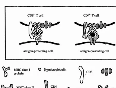
Figure 2. Molecular interactions in antigen-specific T cell recognition of antigen-presenting cells...6
Figure 3. SMAA complexes as multivalent vaccines... 36
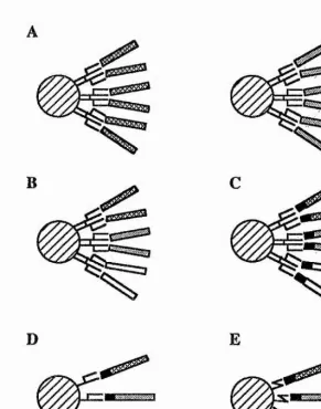
Figure 4. Engineering of antibodies... 39
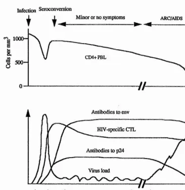
Figure 5. Schematic course of HIV infection and AIDS...44
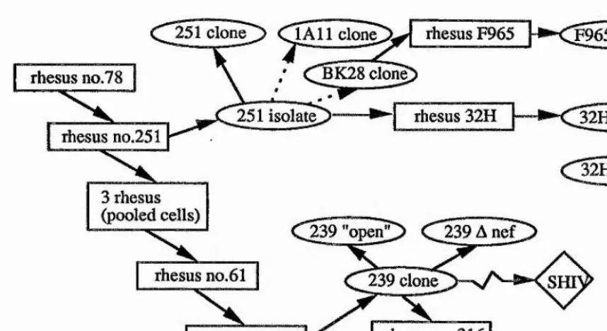
Figure 6. Derivation of SIVmac viruses...1...50
Figure 7. Particle and genome organization of simian immunodeficiency virus ...51
Figure 8. Construction of pGEX27Pk expression vector... 74
Figure 9. DNA linker coding for the Pk tag... .74
Figure 10. Purification of SIV p27-Pk (Coomassie blue-stained SDS-polyacrylamide gel)... 77
Figure 11. Western analysis of SIV p27-Pk-containing SMAA complexes...77
Figure 12. Immunogenicity of the Pk tag...80
Figure 13. Reactivity of mAbs to authentic SIV p27... 80
Figure 14. Western blot of bacterial lysates containing GST-SIV-Pk proteins... ...84
Figure 15. SMAA complexes containing vpr-Pk and vpx-Pk (Coomassie blue-stained SDS-polyacrylamide gel)... 85
Figure 16. Screening of pQ9SIVPk clones by IPTG-induction of Pk tag-linked proteins on agar plate... 87
Figure 17. Western blot analysis of bacterial lysates containing His-SIV-Pk proteins... 90
Figure 18. IPTG dose-response of His-SIV-Pk induction... 91
Figure 19. Time-course of His-SIV-Pk induction... 91
Figure 20. Two-step purification of His-rt-Pk and His-17-Pk (Coomassie blue-stained SDS-polyacrylamide gel). ...93
Figure 21. SMAA complexes containing His-SIV-Pk fusion proteins (Coomassie blue-stained SDS -polyacrylamide gel)...93
Figure 22. Analysis of sera from SIV-infected macaques... 95
Figure 23. Western blot analysis of total cellular lysates of bacteria expressing SIV gpl20Pk gene... ... 96
Figure 24. Time-course of SIV gpl60-Pk expression in 500-ml culture of Sf 21 cells... ... ... ...98
Figure 25. Western blot analysis of antigenicity and immunogenicity of baculovirus-expressed SIV gpl60-Pk... 99
Figure 26. SIV gpl60 -Pk in SMAA complexes (Coomassie blue-stained SDS-polyacrylamide gel)... 100
Figure 27a. Sequences of the light chain PCR product amplified from SV5-P-k hybridoma cDNA... 104
Figure 27b. Sequences of the heavy chain PCR product amplified from SV5-P-k hybridoma cDNA... 105
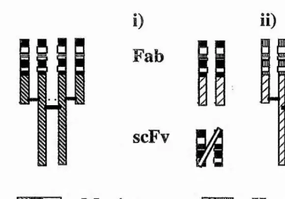
Figure 28. Constructs for expression of chimeric Fab and single chain Fv fragments of antibody in E. coli... 106
Figure 29. Schematic structure of the inserts in M13KOLHuVH and M13VKPCR1 vectors... 108
Figure 30. Sequences of reshaped rH and rL variable regions... 109
Figure 31. Construction and assembly of the rL light chain gene ...112
Figure 32. Principle of the metabolic selection for XGPRT-expressing cells by mycophenolic acid/xanthine... 113
Figure 33. Expression of the rL light chain by CHO-rL cell clones... 114
Figure 34. DNA vectors for expression of tagged proteins...117
Figure 35. Schematics of a two-step purification of SIV 27-Pk expressed from vector pGEX27Pk... 118
LIST OF TABLES
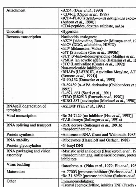
Table 1. Targeted steps in HIV life cycle and some of the corresponding
interfering agents... 48 Table 2. Construction of bacterial vectors expressing GST-SIV-Pk fusion
proteins... ...83 Table 3. Construction of bacterial vectors expressing His-SIV-Pk fusion
proteins...88
Table 4. Expression levels of His-SIV-Pk fusion proteins...92 Table 5. Recombinant heavy and light chains derived from S V5-P-k... Ill
The work presented in this thesis is part of a long term effort to develop a universal strategy for construction of multivalent subunit vaccines, and potentially vaccines against human immunodeficiency virus (HIV). The vaccine, that is being developed in our laboratory, is constructed as solid matrix-antibody-antigen (SMAA) complexes.
Part A of the Introduction presents adaptive immune responses generated to viral infections, i.e. branches of immunity the induction of which is primarily targeted in vaccination. Although antibodies represent important antiviral immune responses, part A mainly focuses on antigen processing and presentation to T cells. This is for several reasons: (i) it is an exciting and currently quickly advancing area of
A. IMMUNE RESPONSES TO VIRAL INFECTIONS.
The nonadaptive defence systems of organisms include external skin and internal circulating cells such as phagocytes or natural killer cells, which serve as a first-line defence against viruses. In vertebrates, there is a second-line protection due to a dual system of humoral and cell-mediated immune responses. These immune responses are generated by a cascade of molecular and cellular associations and interactions. They are adaptive, specific and display a long-term memory. The effectors of humoral responses are antibodies produced by B cells, the binding of which to usually extracellular antigens initiates a variety of elimination responses. Virus-infected cells are more effectively cleared by processes involving
T lymphocytes, which are capable of killing cells displaying virus-derived peptides. A description of the generation of antigen-specific cell-mediated immune responses with particular emphasis on viral proteins and a shorter section on humoral responses follow below.
A.I. General description of processing and presentation of viral antigens to T cells.
A.1.1. T cell subpopulations.
T-cells recognize antigens on the surface of antigen-presenting cells (APC) in association with molecules encoded by genes of either the class I or class II major histocompatibility complex (MHC) (Zinkemagel and Doherty, 1975), a phenomenon known as MHC restriction. Cluster designation markers CD4 and CDS distinguish two major populations of T cells, CD4 CD8+ and CD4+CD8", found in the peripheral
factors termed cytokines, they ’help' to carry out the effector functions of the antigen- specific immune responses, i.e. they ’stimulate' both antigen-activated MHC class I- restricted CTL to kill virus-infected cells and antigen-activated B cells to produce antibodies. They also stimulate cells of the non-specific immune responses such as macrophages, granulocytes or eosinophils. Depending on the local cytokine environment created by cells of the innate immunity early in the response, CD4+ T lymphocytes can differentiate into Thl or Th2 subsets (reviewed by Scott, 1993). Thl cells produce predominantly tumour necrosis factor and interferon-yand are best at activating macrophages. Th2 cells make predominantly interleukins IL-4, IL-5, IL-6 and XL-10 and preferentially activate B cells. Some MHC class H-restricted T cells can function as CTL and as such may have an important role in controlling certain virus infections, for example, those of measles and herpes simplex viruses.
A.1.2. Cellular handling of antigens for presentation to T cells.
Pioneering work on antigen presentation demonsti'ated that internal proteins (e.g. the nucleoprotein), rather than surface glycoproteins, of influenza virus were the major target antigens for influenza virus-specific class I-restricted CTL (Townsend et
fl/., 1984). This was followed by the observation that only part of the gene for influenza virus nucleoprotein (NP) sufficed to sensitize hansfected cells to NP- specific CTL lysis (Townsend et a/., 1985). The finding that T cells recognize peptides rather than whole native antigens, as is the case with antibodies, was finally demonstrated by the ability of synthetic peptides to sensitize target cells to CTL (Townsend et al., 1986).
4 endoplasmic reticulum (ER) where they become associated with newly synthesized MHC class I molecules. These complexes are then transported through the Golgi apparatus to the cell surface for presentation to specific MHC class I-restricted T cells. For the MHC class II presentation, antigens enter acidic vesicles
(endosomes/lysosomes), usually following their capture by specialized cells (see below), where they undergo partial proteolysis. In this compartment, some of the resulting peptides become associated with MHC class II molecules and are transported as MHC-peptide complexes to the cell surface. The intracellular
trafficking of MHC molecules and antigens is described in more detail below and is schematically illustrated in Fig. 1.
The primary interactions between T cell receptors and MHC-peptide complexes (Fig. 2 and below) are essential for the eventual generation of both adaptive immunity and some aspects of innate immunity. The parts of peptides
participating in the T cell recognition of antigen-presenting cells are called T cell sites and consist of an epitope and an agretope (Sette et a/., 1987). Amino acid residues that physically contact a T cell receptor form an epitope and those binding an MHC molecule are called an agretope. However, T cell sites and T cell epitopes are often used interchangeably.
A.1.3. MHC polymorphism.
There is a number of gene clusters encoding MHC class I (e.g. HLA-A, B and C in humans, and H2-K, D and L in mice) and MHC class II (e.g. HLA-DR, DQ and DP in humans; and H-21-A and I-E in mice) molecules. Every individual expresses four to six different class I molecules. Not every peptide generated by proteolysis has affinity for a given MHC molecule (below). As a result, only a selected set of
peptides ('-1%) will be presented to T cells. It has been estimated that each MHC molecule can present approximately 1000 different peptides on a cell, with each
virus budding
virus attachment
antigen uptake
ENDOSOMES
/
virus assemblyMHC class II
genome
a
replication low-pH fusion ^ ^
protein synthesis
— 9 ^
viral proteins
\
LYSOSOMEpeptides # class IMHC
NUCLEUS
GOLGI
MHC class 1 W
p88
RER rough endoplasmic reticulum
MHC class II
invariant chain
putative peptide transporters
[image:23.612.50.510.113.592.2]antigenic peptides
Figure 1. Cellular handling of antigens.
The diagram schematically illustrates the life cycle of a virus (in this case the influenza virus) and how the virus proteins can enter the eiidogenous (MHC class I) and exogenous (MHC class II) antigen processing and presentation pathways in an antigen-presenting cell. Following partial proteolytic degradation, certain virus- derived peptides associate with either MHC class I or II molecules. The MHC-peptide complexes are then transported to the cell surface where they are specifically
CDS*- TceU CI>4^ TceU
antigen-presenting cell antigen-presenting ceU
MHC class I
a chain Q p 2microglobulin CDS CD3complex
MHC class II (X&P chains
CD4
^ antigenic
[image:24.614.101.492.86.384.2]peptides T ceU recepta & P chainsor
Figure 2. Molecular interactions in antigen-specific T cell recognition of antigen- presenting cells.
The left-hand side of the figure depicts the interactions during the recognition of peptides associated with MHC class I molecules by TCR on a CD8+ T cell (usually
a cytotoxic T lymphocyte). The right-hand side of the figure illustrates the recognition of MHC class U-peptide complexes by a CD4+ T cell (a helper or possibly
a cytotoxic T cell). The key determinant of T cell specificity is the T cell receptor (TCR). The TCR is closely associated with the CD3 complex and either a CD8 or CD4 molecule. The CD3 complex is involved in signal transduction across the plasma membrane following TCR engagement and so are most likely the CD8/CD4
molecules.
numbers and letters, e.g. HLA-Aw68 or HLA-B27) and, as a consequence, different individuals in an outbred population will present different sets of peptides to T cells. If 1000 different peptides do indeed bind to one MHC molecules, then every
individual should be able to mount a T cell response to any given virus. Nevertheless, for reasons not clear, it appears that non-responding alleles can be found even for complex viruses (Bennink and Yewdell, 1988).
peptides vs. positive selection of sufficient variety of T cells in the thymus. An excessive MHC polymorphism would not leave, after negative selection of T cells, broad-enough specificity of the T cell population for the organism defense. The same two antagonistic processes have also determined the length of peptides presented by MHC molecules (below).
A.1.4. Antigen-presenting cells.
Cells, which process antigen for presentation to T cells, are termed antigen- presenting cells (APC). Virtually every nucleated cell expresses MHC class I molecules and thus, when infected, can act as a target for activated CTL. MHC class n molecules are constitutively expressed primarily on haematopoietic cells, but a variety of other cells, e.g. epithelial cells in a number of organs, can express MHC class n molecules when activated by cytokines such as interferon 7.
Although any nucleated cell may act as a target cell for MHC class I-restricted T cells, it appears that the initial activation of resting virgin T cells may require additional factors that can only be supplied by specialized antigen-presenting cells. Thus not every antigenic MHC-peptide complex delivered to the cell surface for presentation to T cells is necessarily immunogenic. This has been demonstrated in several studies of diabetes involving transgenic mice (Ohashi et a/., 1991; Oldstone et
a/., 1991; Schonrich et al.y 1991). In one of these studies, mice were made transgenic for lymphocytic choriomeningitis virus (LCMV) glycoprotein (GP) so that they expressed the LCMV GP only in pancreatic isle T cells. The mice were
8
a CTL response to LCMV GP resulting in destruction of pancreatic cells and the development of diabetes.
The number of MHC molecules present on the cell surface depends on the cell type and activation state of the cell, which in turn depends on the presence of
interferons and other cytokines. It has been estimated that an antigen-presenting cell has to display a minimum of 200 antigenic complexes to be recognized by activated
CTL (Christink e t al.y 1991). The overall avidity of cell-to-cell interactions may be
lessened by a negative charge resulting from extensive cell surface glycosylation (Boog et al.y 1989). However, in addition to the formation of the MHC-peptide-T cell
receptor (TCR) complexes, other auxiliary interactions between cell surface molecules contribute to the intercellular signalling between APC and T cells, e.g. interactions of CD8 and MHC class I or CD4 and MHC class II molecules (Fig. 2), LFA-1 and ICAM-1, LFA-3 and CD2 (for review see Springer, 1990), CD28 and B7, and CD5 and CD72 (Linsley et al.y 1991; Vandevelde etal.y 1991). Interactions of
these molecules increase the overall strength of the T cell binding to APC. These interactions also provide necessary additional signals absent in 'non-professional' APC for the priming of resting T cells, i.e. inducing the progression of T cells from Go to G1 phases of the cell cycle, their proliferation, terminal differentiation and
expression of full T cell functions. Because the affinity of TCR for MHC-peptide complexes is very low, the Kd being in the range of 10-4-10-5 M (Matsui et al.y 1991; Weber et al.y 1992), it has been suggested that an antigen-independent adhesion precedes the TCR engagement (Williams and Beyers, 1992).
While any cell infected with virus may act as a target cell for CTL, only specialized antigen-presenting cells may be able to prime CTL. The most potent cell type in this respect seems to be the dendritic cell (Young and Steiman, 1990;
may be due to their low levels of surface glycosylation (Boog et al., 1989) and high levels of expression of surface adhesion molecules (Freudenthal and Steiman, 1990).
A.2. Antigen presentation by MHC class I molecules.
MHC class I molecules are heterodimers consisting of a transmembrane heavy chain (or a chain) and p^-microglobulin (Fig. 2). They can present peptides derived from all proteins synthesized in the cell, including those encoded by mitochondria (Loveland et al., 1990; Shawar et ah, 1991) as well as proteins targeted or delivered to cytosol by in vitro manipulations (Moor et al., 1988). These antigens may include self- and viral proteins or proteins originating from intracellular pathogenic bacteria that enter cytosol (Brunt et al., 1990). Peptides become associated with MHC class I molecules in the ER and/or the Golgi apparatus. The presentation of peptides by MHC class I molecules can be specifically abrogated by treatment of antigen- presenting cells with drug brefeldin A (Misumi et al, 1986). In these cells,
biefeldin A blocks the movement of newly synthesized membrane proteins from the endoplasmic reticulum to the Golgi apparatus, while endocytosis and protein
synthesis are not inhibited.
A.2.1. Calnexin, an accessory molecule for MHC class I biosynthesis.
10
Not all newly synthesized MHC class I heavy chains and Pi-microglobulins assemble into stable complexes and excess free P2-microglobulin is secreted out of the cell (Neefjes and Ploegh, 1988). This suggests that there are limiting amounts of peptides in the ER. Such a situation would ensure that bystanding cells are not sensitized to CTL lysis following capture of secreted antigenic peptides by their unloaded cell surface MHC molecules.
A.2.2. Interaction of peptides with MHC class I molecules and analysis of naturally processed MHC class I-associated peptides.
A landmark in elucidating the way how peptides bind to MHC heterodimers was the determination of crystal structure of human HLA-A2 molecule (Bjorkman et al„ 1987). It revealed a single peptide-binding groove on the MHC heavy chain formed by two a-helices and a p-pleated floor. It was only recently, that the atomic details of peptides bound to MHC class I molecules were determined. Three virus- derived synthetic peptides of different lengths and sequences were bound to the murine H-2K^ molecule in an extended P-structure (Fremont et aL, 1992; Matsamura
is less constrained by the MHC cleft, which can accommodate longer peptides by bulging away from the binding groove.
These principles may not apply to all MHC class I molecules, or, alternatively, just one end of the antigenic peptide may, in some cases, form
sufficiently antigenic complexes. Thus the murine MHC class IH-2L^ molecule was reported to present efficiently two different five-amino acid-long peptides derived from murine cytomegalovirus (Reddehase et al., 1989) and lymphocytic
choriomeningitis virus (Whitton et al., 1989). It was shown independently, that the latter peptide, GVYMG, overlapped the most effective nonapeptide RPQASGVYM, and even a four-residue peptide GVYM still, although inefficiently, sensitized target cells to CTL lysis (Schulz et al., 1991).
In order to analyze directly natural peptides bound to MHC molecules, two groups of workers purified murine class I molecules from virally infected cells and acid eluted the bound peptides. Firstly, a vesicular stomatitis virus-derived peptide was isolated that associated with H -2 K h , but did not bind to H -2 D b molecules (Van
Bleek and Nathenson, 1990). The second study identified different naturally processed influenza peptides presented by either H -2 D h or H -2 K ^ molecules
(Rotzschke et al., 1990). The eluted peptides contained 8 or 9 residues and their fidelities were confirmed by highly efficient recognition of their corresponding synthetic analogues by CTL.
A.2.3. MHC class I molecules present self-peptides.
12 Garrett et al, 1989; Madden et al, 1991). These speculations were proved correct by eluting pools of heterogeneous self-peptides from H-2K^, KP, and HLA-A2.1 molecules (Falk et al, 1991). The majority of these peptides was 8 or 9 amino acids long. The pools of peptides were microsequenced as a mixture and abundances of amino acids at each peptide position revealed agretope motifs characteristic for each MHC molecule. The origins of self-peptides were first identified by separating peptides eluted from HLA-B27 on HPLC and sequencing each peptide individually (Jardetzky etal, 1991). By comparing the peptide sequences with database
sequences, 7 out of 11 peptides was possible to identify as being derived from abundant cytosolic or nuclear proteins such as histones, ribosomal proteins and members of the 90K heat-shock protein family. Obviously, mechanisms inducing self-tolerance must have ensured that MHC molecules presenting self-peptides did not induce deleterious immune responses against 'self. Since then, many allele- specific motifs for MHC class I molecules have been identified (Rammensee et al,
1993).
A.2.4. Prediction of MHC class IT cell sites.
The finding that short synthetic peptides sufficed to sensitize target cells to CTL lysis in vitro and could indeed induce specific CTL in vivo created high expectations for peptide vaccines and initiated a number of studies identifying immunodominant T cell sites. Several computer algorithms were developed which attempted, with varying degrees of success, to predict T cell sites from the protein amino acid sequences based either on the ability of a particular amino acid stretch to form an amphipathic structure (DeLisi and Berzofsky, 1985) or on the best fit to a consensus sequence common to known T cell sites (Rothbard and Taylor, 1988). With increasing amounts of data, it became apparent that neither approach would be entirely satisfactory. However, from the sequences of the peptides eluted from MHC class I, it was possible to identify MHC allele-specific motifs, i.e. agretope amino acids important for binding to an MHC molecule (Jardetzky et al, 1991; Falk etal,
a number of MHC molecules have been determined and summarised by Rammensee and colleagues (1993). The MHC class I binding agretope usually consists of one position in the carboxyl-terminus of peptides requiring a certain amino acid side chain, and one position with strong amino acid preferences elsewhere depending on the MHC allele (Maryanski et aL, 1989 and 1990; Madden et ah, 1991; Fremont et al,, 1992). The remaining positions within the peptide are fairly unrestricted. On the other hand, some amino acids at certain positions in peptide ligands might be ’forbidden’.
Â.2.5. MHC locus as a cassette for antigen processing and presentation.
MHC class I molecules present peptides derived predominantly from
cytoplasmic proteins. Because protein degradation is thought to occur at least initially in the cytoplasm, peptides must cross a membrane to enter the lumen of the ER for association with newly synthesized MHC class I molecules. Genes in the MHC locus code for at least two other classes of proteins that may be involved in antigen
processing. These are subunits of a cytoplasmic proteasome thought to be involved in antigen degradation and putative peptide transporters involved in the transport of peptides from the cytosol to the lumen of the endoplasmic reticulum.
Â.2.6. Proteolytic degradation of antigens for MHC class 1 presentation.
14 a more subtle contribution of the LMP complex to peptide generation. In fact, very little is known about the proteolytic degradation of antigens for class I presentation and alternative mechanisms to proteolysis for generation of peptides have been discussed (Fetten et al., 1991; Boon and Van Pen, 1989).
A.2.7. Transport of peptides from the cytosol to the lumen of the ER and association of peptides with MHC class I molecules.
Studies on mutant cell lines, in which MHC class I heavy chain and p2-microglobulin were efficiently synthesized, but the MHC heterodimers did not seem to reach the cell surface, have suggested a role for peptide transporters in the translocation of peptides from the cytosol to the lumen of the endoplasmic reticulum. In these cells, the surface expression of MHC molecules was significantly increased by addition of virus-derived peptides, even though the same virus previously failed to sensitize the mutant cells to CTL (Townsend et ah, 1989). Further analysis of the phenotypic defect indicated that the presence of peptides was critical for association of the MHC class I heavy chain with P2-ttiicroglobulin (Cerundolo et al., 1990;
Ljunggren et al., 1990; Hosken and Bevan, 1990). Some of the cell lines with presentation defect had been previously characterized as having deletions in the class n region of the MHC locus (Townsend et al., 1990; Ljunggren et al., 1990). Two of the missing genes, TAP^I and TAP-2 (for transporter associated with antigen presentation), belonged to the ’ATP-binding cassette’ (ABC) superfamily (Monaco et al., 1990; Deverson et al., 1990; Trowsdale et al., 1990). All members of this family contain a domain homologous to a putative ATP-binding site and transport molecules ranging from metal ions and small sugars to proteins, some over 100 kDa, across biological membranes. Reconstitution studies of the mutant cell lines revealed that a defect in either the TAP-1 or TAP-2 gene alone resulted in a defect in antigen presentation and a loss of surface expression of MHC class I molecules (Spies and Demars, 1991; Attaya et al., 1992). It has been suggested that the products of the
Direct evidence for the proposed role for the products of TAP genes the peptide transmembrane transport is still awaited. Relatively convincing functional arguments came from an elegant experiment involving a minigene expressing a T cell site from influenza virus matrix protein introduced into an MHC class I-defective cell line T2 (Anderson et al., 1991). Transfected T2 cells were recognized by class I- restricted CTL only if the matrix T cell site was linked to a signal sequence that targeted the hybrid oligopeptide to the lumen of the ER via the signal-recognition particle-dependent transport mechanism. Thus, if peptides reach the lumen of the ER, T2 cells possess all the machinery required for transporting stable MHC-peptide complexes to the cell surface. Additional support for the role of peptide transporters came from studies which demonstrated that the transporters themselves were polymorphic and could influence the selection of peptides finally displayed on the cell surface by MHC class I molecules (Powis et al., 1992).
On the other hand, several observations question the proposed role for the peptide transporter function of the TAP proteins. Spherical vesicles made from membranes of rough ER called 'rough microsomes' are generally impermeable to proteins lacking the signal sequence. However, short peptides of up to ten residues could enter microsomes generated from the ER of T2 cells (Levy et al., 1991). Since these peptides entered the microsomes in the absence of ATP, it was concluded that ATP-dependent transporters were not involved in this translocation. Although the absence of ATP in preparations is always a disputable point, so far there have been no reports of the binding of nucleoside phosphates to TAP proteins (Gaskins et al.,
1992).
16 peptides and found that two, a 9-mer and an 11-mer, were derived from putative signal sequences that would have entered the ER via normal signal-recognition particle-dependent transport. The third peptide of unknown origin was a 13-mer. The presence of correct nonapeptides (in addition to the longer peptides) suggests that some trimming may have occurred in the ER to generate the peptides of the
appropriate length (see also Henderson et al., 1992). Specific antigenic peptides can not be detected in cells unless the MHC molecules to which they bind are also present (Falk et al., 1990; Wallny et al., 1992). Thus, if the right-size peptides are generated in the cytoplasm prior to or during the transport into the ER, peptides in the ER unprotected by binding to MHC molecules must have a very short half-life. By the same token, the peptide short half-life in the ER argues for the presence of a strong proteolytic activity in the lumen of the ER.
A.2.8. Non-classical MHC class I molecules.
All the above work has concerned classical MHC class I, i.e. class la, molecules. The MHC loci in humans and mice also contain clusters of non-classical class I heavy chain genes designated as MHC class Ib molecules (H-2Q, T and M in mice.) Both class la and Ib heavy chains associate with p2-microglobulin. MHC
class Ib molecules display much less polymorphism than class la and class II molecules. The allelic differences in the sequences of class Ib genes are only about
1%, which is typical for most other genes and differs markedly from 15% of the class la genes. The immunological function of MHC class Ib molecules is largely unknown. Originally, it was suggested that they may simply serve as a sequence pool for generating class la polymorphism (Flavell et al, 1986), Evidence for an immune function of MHC class Ib molecules involved several T cell clones bearing yS T cell receptors that were restricted by H-2T molecules (Bluestone et al, 1991).
Subsequently, it was shown that class Ib-containing MHC complexes could present foreign peptides derived from intracellular bacteria and that H-2M3 molecules specialized in presentation of N-formylated peptides (Loveland et al, 1990; Shawar
molecules (Rotzschke et al., 1993). The extrapolation of these studies would be that
other class Ib molecules can also present foreign peptides, however, no function in combating viral infections has yet been reported.
A 3. Antigen presentation by MHC class II molecules.
MHC class n molecules (HLA-DR, DQ and DP in humans and H-21-A and I- E in mice) are heterodimers consisting of transmembrane a and (Î (heavy) chains (Fig. 2) and, in association with antigenic peptides, stimulate CD4+ T lymphocytes. As outlined above, the pathway of antigen processing for presentation to class II- restricted T cells is clearly different from that for MHC class I presentation. A central role in the MHC class II pathway is played by the acidic vesicles
(endosomesAysosomes) of the cell. It is in these vesicles where the class II antigenic peptides are generated and become associated with MHC class II molecules. The two different processing and presentation pathways reflect the fact that antigens
synthesized within the ceil (usually derived from the cytosol of an AFC) are presented by MHC class I molecules, while any antigen, that reaches the endosomal/lysosomal compartment (usually via receptor mediated endocytosis, phagocytosis or pinocytosis), is processed for MHC class II presentation. In this way, antigens for class II presentation never enter the cytosol and remain inside vesicles within the cell. Processing of antigens for class n presentation is sensitive to lysosomotropic agents such as chloroquine, which buffer the acidic environment of the late endosomal/lysosomal compartment and thus prevent the lowering of internal pH necessary for dénaturation and catabolism of internalized antigens.
A 3.1. Binding of peptides to MHC class ÎI molecules.
IS
observed in that the ends of the two side a-helices leave the cleft more 'open'. Peptides did not appear to be buried in the groove as in the case of MHC class I molecules, but rather lay straight in the groove projecting from both ends with no apparent kink in the central region. There was a non-polar pocket near one end of the groove in which an anchoring amino acid side chain resided. Interestingly, the crystal structure suggested a dimerization of HLA-DRl ap heterodimers.
Peptide-binding assays suggest that class II molecules bind peptides with high affinities reaching 10'^ M (Roche et al, 1991) and that strongly binding peptides share allele-specific structural motifs (Jardetzky etal, 1990; O'Sullivan etal, 1991). Site-directed mutagenesis studies of MHC molecules and single-residue substitutions in peptides have provided further information on the importance of individual amino acids in the MHC class II-peptide-TCR interactions (Krieger et al, 1991).
In an analogous series of experiments to the characterisation of naturally occurring peptides bound to MHC class I molecules, peptides have been acid eluted from purified MHC class II molecules and microsequenced. Peptides generated by natural processing and presented by MHC class II complexes were first identified for mouse I-Al) and I-Eb molecules (Rudensky etal, 1991a). They ranged from 13 to at least 17 amino acid residues and this study suggested that the peptides where
precisely defined at their N-termini while their C-termini varied. Since then, data has become available on peptides isolated from several other MHC class II molecules (Hunt et al, 1992; Nelson et al, 1992; Kropshofer et al, 1992; Chicz et al, 1992). The size of naturally processed peptides ranged from 12 to 25 amino acids long, depending on the particular class II molecule. In contrast to earlier work, these studies suggest that there seems to be an allele-specific core sequence involved in binding the peptide to the groove of the MHC class H molecule (Hunt et al, 1992; Chicz et al,
As with class I molecules, antigenic peptides may contribute to the stability of MHC class n ap heterodimers (Sadegh-Nasseri and Germain, 1991; Germain and Hendrix, 1991). Two forms of murine class II molecules have been reported in unboiled spleen immunoprecipitates that differed in stability and potentially in conformation. The more stable form remained associated on SDS-PAGE, while the less stable dissociated into a and p monomers. The relative amounts of the two forms varied for different haplotypes and class II molecules. Pre-incubation of spleen cells with antigen (hen egg-white lysozyme; HEL) significantly increased the levels of the stable form and produced a conformational change detectable by a monoclonal antibody. Other studies showed that the stability of class II aP chain-peptide
complexes is also influenced by pH (Jensen, 1990) and the phospholipid composition of membranes harbouring MHC molecules (Roof et ai, 1990).
A.3.2. Contact of TCR with MHC-peptide complexes.
Similarly to the antibody variable regions, each a and p chain of TCR contains three hypervariable complementarity-determining regions (CDR) (Davis and Bjorkman,
1988). The conformations of the MHC and TCR complexes suggested that the residues of TCR contacting the peptide epitopes predominantly reside in CDR3, that is in the CDR closest to the constant domain (Chothia et al., 1988; Claverie et al.,
1989). The study that supported this model (Jorgensen et al., 1992) involved injecting a series of peptides with a single amino acid substitution in the epitope, i.e. changes affecting the TCR recognition but not the MHC binding, into transgenic mice carrying genes for either the a or P chains of TCR of a T cell line recognizing the original peptide (see Fig. 2). After each immunization of these 'half transgenic mice, hybridomas were prepared using the responding T cells and genes for their
20 A.3.3. Prediction of MHC class H T cell sites.
Since the binding of peptides to class H molecules does not appear to be as selective as the binding of peptides to class I MHC molecules, it may prove difficult to predict agretopes that facilitate the binding of peptides to class H molecules. On the other hand, since the binding of peptides to class II molecules appears to be more permissive than the binding of peptides to class I molecules, the parameters used for predicting class IIT cells sites may not need to be so rigid.
A.3.4. Proteolytic degradation of antigens for MHC class II presentation and its subcellular localization.
Experiments addressing the proteases involved in antigen degradation for MHC class n presentation proved to be largely difficult to interpret, mainly because of the diverse effect of protease inhibitors on APC. The conclusion was that a number of proteases, in particular cathepsin D (Van Noort et al., 1991) and E (Bennett et al.,
1992), residing in the endocytic pathway subtly contributed to the proteolysis depending on the cell type and the particular route by which antigens reached the lysosomal compartment. Indeed, the observations that peptides of different lengths and with poorly defined termini can be isolated from MHC class II molecules
suggests that they might have been generated by different proteases. This in turn may reflect a multiplicity of routes by which the peptides reached the
endosomal/lysosomal compartment. It should also be noted that although MHC class n molecules become loaded with self-peptides (Rudensky et al., 1991a and
1991b; Hunt et al., 1992; Nelson et al., 1992; Kropshofer et al., 1992; Chicz et al.,
1992), it appears that a higher proportion of unloaded MHC class II molecules (1-20%) reaches the cell surface compared to MHC class I molecules (<0.3%; Chen and Parham, 1989).
A.3.5. The role of MHC class H-associated invariant chain.
The trafficking of MHC molecules and antigenic peptides within the cell is the key to the differential presentation of peptides by the two classes of MHC molecules. The intracellular pathways and association of MHC class II heterodimers with peptides is in part regulated by a molecular 'chaperon' termed the invariant chain (Teyton et al., 1990). The invariant chain forms a homotrimer and as such
2 2
(Roche and Cresswell, 1991b). Both signals, the first that retains the invariant chain in the ER until its association with the MHC class H molecules and the second responsible for delivering class E-invariant chain complexes into endosomes, are located in the cytoplasmic tail of the invariant chain (Bakke and Dobberstein, 1990; Lotteau et al, 1991).
A.3.6. Presentation of endogenous antigens by MHC class 11 molecules.
Studies involved in the processing and presentation of the surface antigen of hepatitis B virus (Jin et aL, 1988) and antigens of measles and influenza viruses (Jacobsen et aL, 1989) demonstrated that MHC class E molecules can present endogenous antigens, although less efficiently than the MHC class I molecules. For this presentation to occur, the endogenous antigens still have to follow the
endosomal/lysosomal pathway, for example by internalization of membrane proteins. A series of studies using immunoglobulin X light chain (Weiss and Bogen, 1989 and 1991) and HEL (Brooks et ah, 1991; Moreno et al., 1991) showed that peptides of endogenous origin may also be introduced into the MHC class E pathway by being targeted to particular cellular compartments. Thus peptides derived from mutated
X chains retained in the vacuolar system by preventing the formation of a disulphide bond in the X variable region, or held in the ER by addition of the KDEL sequence were presented by MHC class II molecules. The same T cell site did not associate with MHC class II molecules when retained in the cytosol or nucleus, or when secreted out of the B cells.
Â.4. Viral mechanisms interfering with antigen processing and presentation.
The importance of cell-mediated immunity in controlling virus infections can also be inferred from the different molecular mechanisms viruses have evolved to interfere with the processing and presentation of antigens to T cells. These
23
to contribute to the overall decrease of cell surface expression of MHC complexes, as well as more specific mechanisms. To illustrate this point, a few examples of the ways in which adenoviruses, pox viruses and herpesviruses have evolved to evade the immune response will be briefly described.
Striking examples of evasive strategies have been developed by human adenoviruses, which carry in their genomes a cassette of genes protecting infected cells from the immune response. Most of these genes are clustered in an early
transcriptional region E3. Functions of all the gene products from this region have not been elucidated, but, for example, glycoprotein 19K binds newly synthesised MHC class Î heavy chains and blocks their transport from the ER (Kvist et ah, 1978; Paabo
et ai, 1986; Jefferies and Burgert, 1990), thus preventing the adenovirus-infected cells from displaying MHC class I-peptide complexes (Burgert et al., 1987; Rawle et al., 1989). However, deletion of E3 resulted in disease exacerbation, probably due to increased immunopathology (Ginsberg etal., 1989). Two other proteins from E3 and one from ElB regions protect infected cells against cytolysis induced by tumour necrosis factor. In addition, a protein encoded by El A region has been demonstrated to down-regulate transcription of the MHC class I gene (Schrier et al., 1983; Bernards
etal., 1983).
Different anti-antiviral tactics have been developed by certain poxviruses. Vaccinia virus, for example, possesses genes (B13R and B24R) that code for proteins of the serine-protease inhibitor family (serpins) (Kotwal and Moss, 1989; Smith et al., 1989). A putative function of viral serpins is that they inhibit cellular proteases, some of which are involved in antigen degradation, and thus reduce the presentation of virus-specific peptides. It is known that recombinant vaccinia viruses which express antigens under the control of early promoters can induce CTL responses, while those expressed under the control of late promoters tend only to induce antibody responses (Coupar et al, 1986). Since serpins are expressed late, they may be involved directly in interference with antigen processing and presentation at late times in infection, although direct evidence for their role is still awaited. The
24 produce that interfere with the immune response. For example, vaccinia virus also produces soluble homologues to the interleukin-1 (IL-1) and IL-6 receptors (Smith and Chan, 1991; Alcami and Smith, 1992). Since IL-1 and IL-6 have a broad spectrum of effects and are known to play important roles in the regulation of immune responses, it has been suggested that the vaccinia virus homologues can interfere with the immune response by blocking the effects of these cytokines. It has also been suggested that the serpins may interfere with the conversion of pro-IL-lp to the active form, IL-ip (Ray etal., 1992).
Herpesviruses also appear to have developed a number of specific mechanisms for interfering with antigen processing and presentation. Human cytomegalovirus interferes with the transport of MHC class I molecules from the endoplasmic reticulum to the cell surface and, in addition, encodes a protein of high homology with the MHC class I heavy chain capable of binding the human
P2-microglobulin (Browne et al., 1990). The proposed function of this protein in vivo
is to prevent the assembly of MHC class I complexes by sequestering
P2-microglobulin, and so render the cytomegalovirus-infected cell unrecognizable by CTL.
A.5. Superantigens.
of T cell responses to a peptide antigen ranges usually from 10"^ to 10"^. The fact that superantigens interact with specific regions of the TCR distinguishes them from polyclonal T cell mitogens, such as concanavalin A. Furthermore, superantigens bind the MHC molecule at sites distinct from the peptide-binding groove (Dellabona et al.,
1990) [and different staphylococcal toxins bind to distinct sites on an MHC class II molecule (Scholl et al., 1989)], they do not require intracellular processing (Fraser et al., 1989) and their recognition is only marginally restricted by the MHC allele. Interactions between MHC molecules and superantigens (Dellabona et ah, 1990), MHC molecules and Vp of TCR (Fleischer et ah, 1991), and superantigens and Vp (Pullen et ah, 1990; Gascoigne and Ames, 1991) have been demonstrated and shown to be involved in the generation of T cell responses. The sole binding of some superantigens to MHC class II molecules can induce transcriptional activation and increase in MHC-bearing cell adhesiveness (Trede et ah, 1991), and may synergize in lymphocyte proliferation (Fuleihan etal., 1991).
A.5.1. Classification and manifestation of superantigens.
26
A.5.2. Retroviral superantigens.
Self-superantigens have recently acquired a virological dimension. Although the Mis loci are unlinked to MHC, they co-segregate with integrated genomes of endogenous retroviruses, murine mammary tumour viruses (MMTV; Frankel et al.,
1991; Woodland et al., 1991; Dyson et al., 1991). In addition, an exogenous MMTV encodes a maternally inherited superantigen (Marrack and Kappler, 1991). The superantigen was transmitted from an infected mother to her offspring after birth (presumably in the milk) and caused a deletion of Vpl4+ T cells in the progeny. Further analysis located the superantigen gene to an open reading frame in the 3' long terminal repeat (LTR ORF) of MMTV (Choi et al., 1991). This finding was
confirmed by depletion of Vpl4+T cells in mice transgenic for both the whole MMTV and for just the LTR ORF. LTR ORF comparison of different MMTV
variants located the vp specificity to the C-terminal of the gene product (Acha-Orbea
et al., 1991). Another and potentially more important finding was a demonstration of a Vp5“Specific superantigen expressed by a defective murine leukaemia virus, which induces an AIDS-like disease in mice (Hugin et al., 1991). B cell lymphomas
presenting this retroviral superantigen caused a VP5+ T cell proliferation, which was possible to block by gag p30-specific antibodies. Thus, superantigens may represent yet another mechanism for perturbation of the fine equilibrium of our immune system, potentially contributing to the development human AIDS.
A.5.3. What is the biological function of superantigens?
What is the function of superantigens that preserved them through the
of Mis in positive selection has been also observed (Benoist et al., 1989) as well as their ability to potentiate conventional antigen presentation (Janeway et al., 1983). This led to the suggestion of a co-ligant function for superantigens (Janeway et al.,
1989), whereby Mls-like structures would in special cases stabilize or orient the TCR- MHC interactions. Also, because bacterial superantigen stimulation does not require the CD4 molecule (Fleischer and Schrezenmeier, 1988; Sekaly et al., 1991), these superantigens may substitute for the CD4 function. These properties could be exploited for potentiating immune responses induced by vaccines. In any case, superantigens in mice exert a strong influence on the shaping of the T cell repertoire. Their true biological function and role in autoimmune and immunodeficiency
diseases, however, remains obscure.
A.6. Humoral immune responses.
Humoral immune responses defend an organism primarily against the extracellular phase of viral infections. They are mediated by antibodies, which circulate throughout the body in the lymph and in the blood serum. Antibodies are produced by B lymphocytes, which upon recognition of a foreign antigen differentiate into antibody-secreting plasma or memory B cells. For most antigens, these processes are assisted predominantly by helper Th2 cells (above). Once produced, antibodies can bind to a virtually unlimited number of surface structures on the virus and neutralize it by either preventing attachment to and/or penetration into the host cells, or opsonizing the virus for efficient phagocytosis by macrophages. In some instances, these processes may happen more efficiently in the presence of complement.
Antibodies recognize small antigenic determinants called epitopes, which can be continuous or discontinuous (Barlow et al, 1986). Discontinuous epitopes depend on the tertiary and quaternary protein structures and are always conformationally sensitive, i.e. are destroyed upon antigen dénaturation or proteolytic degradation. Continuous epitopes consist of a linear array of amino acids and are usually
2 8
by antigen-binding sites on antibodies, called paratopes. It is assumed that all epitope- paratope interfaces involve a surface area of approximately 700 Â2, as it was shown in the case of lysozyme (Amit et ah, 1986). From the known folding patterns of many globular proteins, this assumption implies, that no surface epitope region is likely to contain only a single continuous stretch of amino acid residues. Thus, 'continuous epitope', in fact, usually represents only part of a larger discontinuous determinant. It follows that linear peptide fragments are likely to have only a across-reactive
antigenicity with the native protein, from which they were derived. As discussed below, this raises some theoretical questions about peptide vaccines. Several
computer programmes have attempted to predict linear cross-reactive fragments from the primary protein structures (Barlow et al., 1986; Blundell et al., 1987). But, as it is with the prediction of T cell sites, these algorithms predicted well some 'continuous' epitopes, while they failed to identify others.
B. CONTROL OF VIRAL DISEASES.
The spread of many viral diseases has been controlled through primary health care and immunoprophylaxis, which involves either active (vaccines) or passive (antisera) immunization. The advances in molecular biology are also resulting in increasingly effective chemotherapies, which in turn put increasing demands on rapid and specific diagnosis.
The severity of virus infection and disease of an individual depends in part on whether the speed of induction of protective immune responses is faster or slower than the virus replication. Among the factors, that may influence the final outcome of virus infection, are the type of virus and the virulence of a particular virus variant, route of infection, previous exposure to the same or similar viruses/antigens, genotype and immune status of an individual, and presence of other compromising factors.
by increasing the efficiency of protective reaction. While passive immunization supplies virus-specific antibodies, the aim of vaccination is to induce an individual's own natural immunity. This acquired immunity is generated by pre-exposing the immune system to appropriate antigens, which results in a clonal expansion of antigen-specific lymphocytes and generation of circulating antigen-specific memory cells. Most, if not all, of the anti-viral vaccines used to-date prevent viral disease rather than infection. The various types of vaccines and their advantages and disadvantages are discussed below.
B.l. Traditional approaches to virus vaccines.