Azad K. Anand1 . 2 and Benjamin C. P. Lee1
Received December 16, 1981; accepted after revision April 5, 1982.
'Department of Radiology, Section of Neuro -radiology, Cornell Medical Center, New York, NY 10021.
2Present address: Department of Radiology, Section of Neuroradiology. Health Sciences Cen -ter, State University of New York, L-4, Room 147, Stony Brook, NY 11794. Address reprint requests to A. K. Anand.
AJNR 3:567-541, September/October 1982 0195-6108/82/0305-0567 $00.00 © American Roentgen Ray Society
567
Plain and
Metrizamide CT of
Lumbar
Disk
Disease:
Comparison with
Myelography
Computed tomography with and without administration of intrathecal contrast ma-terial was evaluated in 100 patients with suspected lumbar disk disease. Metrizamide computed tomography was performed in 75 patients and plain computed tomography was performed in 25. Metrizamide computed tomography was more accurate than plain computed tomography. It also disclosed many lesions not shown by metrizamide myelography at the lumbosacral level. Very few lesions were revealed by myelography that were not seen by metrizamide computed tomography. The small amount of intrathecal metrizamide needed for scanning has practically no side effects. Experience in 12 patients indicates that the procedure may be performed safely on an outpatient basis. This study suggests that computed tomography should be given serious consid-eration as the primary definitive radiographic examination of suspected lumbar disk disease.
Myelography fails to demonstrate 15% of disk herniations at the lumbosacral level and 5% at L4-L5 [1-3]. Other methods such as diskography, epidurogra-phy, and venography are not used routinely because they cause physical discomfort and are often inconclusive [1, 4]. Computed tomographic (CT) sca n-ning is nonivasive, but should be compared with myelography before its accuracy can be determined. We compared metrizamide myelography and CT, both without and with intrathecal metrizamide.
Materials and Methods
One hundred patients with suspected lumbar disk disease were studied. Plain CT was performed in 25 cases and CT after administration of intrathecal metrizamide in 75 cases. Metrizamide myelography was performed in all 100 palients. Surgical exploration was made in 53 patients.
CT was performed on the GE 8800 scanner using 400 mAs and 120 kV. Lateral
ScoutView images were obtained in all patients for alignment of the sections. Sections of 1.5 and 5 mm were obtained through L3-L4, L4-L5, and L5-S1 disk spaces. Additional 5 mm sections were made in the adjacent vertebral bodies when "reformatting" was needed. Soft pillows were placed below the hips and lower thorax whenever necessary to decrease lumbar lordosis for proper alignment of the sections with a maximum gantry tilt of 15°. In those cases where, despite this maneuver, it was not possible to obtain true axial
sections through the disk spaces, axial sections were "reformatted" using the GEDIS
software program (G.E. experimental software) when this became available during the latter part of the study. Additional sagittal and oblique reformatted images were obtained
whenever better delineation of the lesions was desired.
Metrizamide CT was performed 2-6 hr after myelography. Patients were rolled over
several times before scanning to ensure thorough mixing of the contrast material. In addition, 12 patients were studied apart from myelography by introducing 1 -2 ml of 170 mg Ilml metrizamide into the subarachnoid space immediately before scanning. These 12
patients were followed for 24 hr after the scanning for headaches, nausea, or other
568 ANAND AND LEE AJNR:3, September/October 1982
symptoms and vital signs were recorded.'
Two sets of films were routinely made: one at soft-tissue window settings to visualize the disks and epidural spaces, and another at bone window settings for evaluation of the bony structures. ReView (G.E. software) high-resolution pictures were made for better visu-alization of intrathecal metrizamide, disks, and bony structures. Myelography was performed using 16 ml of 190 mg Ilml concen-tration of metrizamide. The contrast material was not removed but the patients' heads were kept elevated until scanning.
All the examinations were reviewed without clinical data indepen-dently by the two authors. Plain and metrizamide CT were consid-ered positive only when one or more of the following findings were present: (1) Disk margins were visibly protruded behind the normal anterior wall of the spinal canal (fig. 1). The disks were either confined to the level of the intervertebral space or extruded to the adjacent vertebral bodies with or without calcification (figs. 1-3 and 68). (2) There was definite compression of the dural sac or oblit-eration of the epidural fat by the disks (figs. 4-6). (3) There was displacement, compression, or obliteration of the nerve root sleeves (fig. 7). All equivocal findings as well as those where there was incomplete agreement between the authors were considered as
1
2
Fig. 1.-Central disk protrusion. Convex configuration of disk margin (arrowheads).
Fig. 2.- Lateral disk protrusion causes narrowing of exit foramen (arrow-head).
A
8
negative. The myelograms were assessed by the same strict criteria. Surgical findings were compared with the radiologic observations. The primary aim of the study was to compare different diagnositic methods. Therefore, the cases were grouped according to CT and myelographic findings. In group 1, metrizamide or plain CT and myelography were both positive; in group 2, plain or metrizamide CT and myelography were both negative; in group 3, plain or metrizamide CT was positive and myelography was negative; and in group 4, plain or metrizamide CT was negative and myelography was positive. Findings were compared at L3-L4, L4-L5, and L5-S1. Surgical correlation to these findings was possible in 53% of cases.
Results
Our findings are shown in table 1. There was agreement between plain or metrizamide CT and metrizamide myelog-raphy in 75% of cases (groups 1 and 2). Analysis of the other cases showed that discrepancies in the findings were more common at the L5-S1 level than at the L4-L5 level (table 2, groups 3 and 4). At L3-L4, no significant difference was noted. Both plain and metrizamide CT were more ac-curate than myelography in demonstrating protruded disks at L5-S1 than at L4-L5 (group 3). In group 3, where metrizamide or plain CT was superior to myelography at the L4-L5 and L5-S1 levels, metrizamide was used in most cases (13 of 16).
Surgical exploration in 53 cases revealed herniated disks in three patients who had normal plain or metrizamide CT and myelography. There were no negative surgical explo-rations.
Of the 12 patients who had metrizamide CT using a small dose of metrizamide, one complained of mild headaches immediately after scanning. The headache in this patient improved with aspirin in 6-8 hr. No other symptoms or complications were noted in the rest of this group of pa-tients.
Discussion
A definitive diagnosis of disk herniation is made when there is compression of the subarachnoid space and nerve
Fig. 3.- A, Calcified disk protrusion
[image:2.614.54.299.313.492.2] [image:2.614.56.464.554.730.2]AJNR:3, September/October 1982 LUMBAR DISK DISEASE 569
4
5
Fig. 4.-Metrizamide CT. Central compression of subarachnoid space (arrows).
Fig. 5.-Metrizamide CT. Lateral compression of subarachnoid space (arrowheads).
A
B
Fig. 6.-Metrizamide CT. A, almost complete compression of su
barach-noid space by very large disk (arrows). B, 1 cm below. Extruded disk has extended beyond disk level to adjacent vertebral body (arrowheads).
root sleeves on CT and myelography [5-11]. Although some
authors have stated that it is usually possible to distinguish
disk herniations from bulges due to prominent annulus on
myelography [1, 12], it becomes difficult sometimes to make this distinction on CT scans. However, diagnosis of disk
herniation was made in our series only when the strict criteria described above were met. In addition, we cons
id-ered the diagnosis of disk protrusion when the margins of
the disks were clearly visible to be beyond the normal
confines, even though direct compression of the subarach
-noid space was not clearly visible on CT (fig. 1).
In our study, when CT and myelography agreed (groups
1 and 2), there was no problem in diagnosis, and either test
could have been used. Of more concern were those cases
A
B
Fig. 7.- Metrizamide CT. A, Posterior displacement of S1 nerve root
sleeve by herniated disk (arrowheads). B, 0.5 cm below. Incomplete filling of S1 nerve root sleeve due to disk herniation (arrowheads).
TABLE 1: Patients Grouped by Plain or Metrizamide CT and Myelographic Findings
CT Surgically
Group No. Totals
Melrizamide Plain Proven
1, Pas CT and
myelography 46 8 54 38
2, Neg CT and
myelography 11 10 21 3
3, Pas CT and neg
myelography 13 3 16 9
4, Neg CT and pas
myelography 5 4 9 3
Tolals 75 25 100 53
Note.-Pos = positive; neg = negative.
TABLE 2: Comparison of Metrizamide and Plain CT Results at Different Levels
Level
Group No .. Study Totals
L4-L5 L5-S1
3, Pas CT, neg myelography:
Melrizamide CT 4 9 13
Plain CT 1 2 3
(Surgically proven) (2) (7) (9)
4, Neg CT, pas myelography:
Melrizamide CT 3 2 5
Plain CT 2 2 4
(Surgically proven) (1 ) (2) (3)
Tolals 10 15 25
Nole.- Pos = positive: neg = negative.
where there was discrepancy between the studies (groups 3 and 4). Disk herniations were found in a high percentage
of these cases at operation.
[image:3.612.313.553.80.262.2] [image:3.612.50.296.82.277.2] [image:3.612.52.296.332.516.2] [image:3.612.311.558.340.458.2] [image:3.612.316.561.543.673.2]og-570 ANAND AND LEE AJNR:3, September/October 1982
A
8
raphy (group 4). constitute a 9% false-negative rate for CT.
Of these nine cases, four had plain CT studies. No density
difference was appreciated between the sac and disk
mar-gins, however, myelography did show indentation of the
sac. Two of the five cases with metrizamide CT were at the
L5-S1 level, and the epidrual soft-tissue density was
con-sidered to be due to bone averaging because of poor
alignment to the disk plane. These cases were seen during
the early part of the study, when the oblique reconstruction
format was not available. Three other cases had extruded
fragments above or below the disk level where sections
were not made initially and showed no obvious abnormality
at the disk level. However, slices done after the myelogram
at the level of epidural defect behind the vertebral body did
show disk herniation (fig. 8).
Surgical exploration in three patients with negative CT
and myelography (group 2) was done on a clinical basis.
They were labeled positive; however, the description of
herniated disk was vague in the operative notes in these
cases, and surgical findings included the presence of adhe
-sions and fibrosis.
Apart from the imprecise documentation of pathologic
findings at surgery, there were anatomic and technical fa
c-tors that might explain the discrepancy between plain or metrizamide CT and myelographic findings. Compression of the subarachnoid space at the lumbar disk levels down to
L4-L5 was demonstrated on myelography and on CT
be-cause of the usually close proximity of this space to the
back of the vertebral bodies (fig. 38). Such compression is
often more difficult to demonstrate at the lumbosacral level,
where the normal dural sac is frequently separated from the
disk and vertebral bodies by a larger epidural space.
Mye-lography at this lower level may show little or no
compres-sion of the subarachnoid space (fig. 98), but CT may show the margin of the disk protruding into the fat-filled epidural
space (fig. 9A).
The nerve root sleeves pursue an oblique course relative
to the CT plane at the L3-L4 and L4-L5 levels so that subtle compressions may not be apparent. The sleeves become
more vertical in their course at the lumbosacral level and
are at right angles to the CT plane. Therefore, very subtle
c
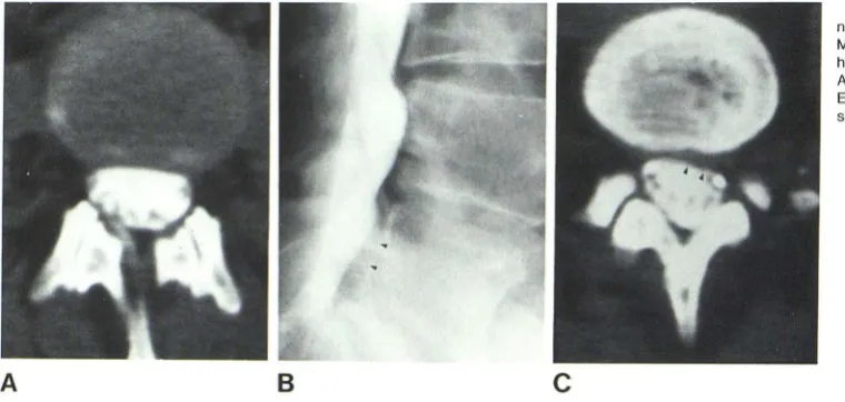
Fig. 8.- A, Metrizamide CT. Promi-nent disk at L4-L5 with no herniation. B,
Myelogram. Widened epidural space b
e-hind vertebral body (arrowheads). C,
After meylography at L5 vertebral body.
Extruded disk fragment with compres-sion of sac (arrowheads).
displacement or compression of the sleeves at this level is shown more easily on CT than on myelography, especially if the subarachnoid space is filled with metrizamide (figs. 68 and 7).
Resolution of CT scans may be improved by increasing
the contrast difference between the subarachnoid space
and nerve root sleeves and the surrounding epidural and
disk spaces. This was most effectively achieved by
intrathe-cal metrizamide, which revealed subtle distortions not visible
without contrast (fig. 7 A). The high concentration of iodine
used for myelography created artifacts on CT scans, which
may be alleviated by delaying the scans for several hours,
or by using the ReView software programs (fig. 7 A). A
smaller quantity of contrast material (1-2 ml 170 mg!ml
iodine) may be used for scanning immediately after the introduction of contrast material in the subarachnoid space and complications caused by higher concentrations can also be avoided.
High-resolution CT scanning made direct visualization of
the disk protrusions possible, and it was particularly helpful
at the lumbosacral level where the thick epidural space may
obscure compression of the dural sac and nerve root sleeves (fig. 1). Whether such disk protrusions, which did not compress the dural sac and nerve roots, were
respon-sible for symptoms remains unclear.
A false diagnosis of disk protrusion, especially at the
lumbosacral level, may be made when the CT sections are
not exactly in the plane of the disks. We used reformatted
axial sections with the GEDIS software program to correct
this. In our series, only nine of 16 patients in group 3 had
surgical exploration. Seven patients improved on
conserva-tive treatment. Most of these cases were diagnosed at the
L5-S 1 level. Repeat scanning in five of these seven cases at a later date with the availability of ReView formatting
showed no disk herniation. The image quality was invariably
degr'lded by this additional maneuver. It was not as critical
to align the sections as accurately with metrizamide CT as
with plain CT. Alignment problems may be corrected for
plain CT by using thinner 1 .5 mm sections, but unfortunately
this usually results in images with unacceptably high noise
[image:4.614.59.439.82.263.2]AJNR:3, September/October 1982 LUMBAR DISK DISEASE 571
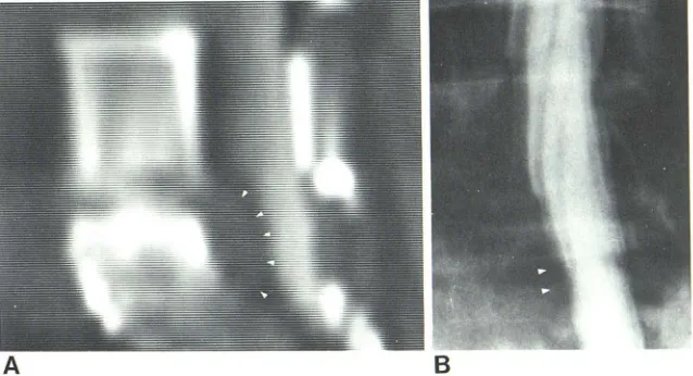
Fig. 9.- A, Metrizamide CT. Extremely large ex -truded disk on oblique format (arrowheads). B,
Oblique view of myelogram. Equivocal compression of S 1 nerve root sleeve (arrowheads).
Complications of metrizamide myelography are well rec-ognized [13-15] and tend to occur several hours after
myelography. The side effects are generally dose-depen-dent. Low doses can be used for metrizamide CT (1-2 ml
170 mg/ml iodine), which make it almost free of side effects.
Mild headaches in one of our patients immediately after the
procedure were most likely related to lumbar puncture
rather than to contrast material in the subarachnoid space.
Radiation exposure of CT examinations (4-5 rad
[0.04-0.05 Gy] skin dose per section) is considerably less
than myelography [16]. In situations where plain or metri-zamide CT is as accurate as myelography, there is ample
justification for selecting the examination with the least side
effects and radiation dosage.
Our study showed no apparent difference in the accuracy
of myelography over CT. In fact, metrizamide CT seemed
slightly superior in some cases (group 3). Thus, it is logical
to conclude that metrizamide CT may be used
interchange-ably with myelography for diagnosis of disk herniations at
specific disk levels. Our study also indicated that the amount
of intrathecal contrast material required for metrizamide CT (1-2 ml 170 mg I/ml) was less than one-eighth of that used
for myelography and was virtually free of side effects. Me-trizamide CT using such low dosages would be expected to be safe for an outpatient procedure provided the patients are kept under observation for 6-8 hr after the procedure.
An additional benefit of metrizamide CT is the low dosage of
radiation (4-5 rad [0.04-0.05 Gy] skin dose/section) as
compared with conventional myelography. We recommend
the following routine for evaluation of lumbar disk disease:
(1) plain CT; if disk is not defined, (2) metrizamide CT
(outpatient); (3) proceed to myelography (inpatient) only if metrizamide CT does not show disk lesion.
REFERENCES
1. Shaprio R. Myelography, 3d ed. Chicago. Year Book Medical, 1975
2. Fort LT, Key JA. Evaluation of myelography in diagnosis of
intervertebral disc lesions in low back. J Bone Joint Surg [Am] 1950;32: 257 -266
3. Scoville WB, Moretz WH, Hankins WD. Discrepancies in
mye-lography; statistical summary of 200 operative cases under go-ing Pantopaque myelography. J Surg Gynecol Obstet 1948;86: 559-564
4. Knutssen F. Experiences with epidural contrast in evaluation of lumbosacral canal in disc prolapse. Acta Radiol (Stockh)
1941 ;22: 694-703
5. Dichiro G, Schellinger D. Computed tomography of spinal cord
after lumbar intrathecal introduction of metrizamide (computer
assisted myelography). Radiology 1976;120: 1 01 -1 04
6. Coin CG, Chan YS, Keranen V, et al. Computer assisted
myelography in disc disease. J Comput Assist Tomogr
1977; 1 : 398-404
7. Lee BCP, Kazam E, Newman AD. Computed tomography of the
spine and spinal cord. Radiology 1978; 128: 95-1 02
8. Coin G, Keranen VJ, Pennink M, et al. Computerized tomog
-raphy of the spine and its contents. Neuroradiology
1978;16: 271-272
9. Meyer GA, Haughton VM, Williams AL Diagnosis of herniated lumbar disc with computer tomography. N Engl J Med 1979;301 : 11 66-1167
10. Federle MP, Moss AA, Margolin FA. Role of computer tomog-raphy in patients with sciatica. J Comput Assist Tomogr 1980;4: 335-341
11. Williams AL, Haughton VM, Syvertsen A. Computed tomogr a-phy in the diagnosis of herniated nucleous pulposus. Radiology
1980; 135: 95-99
12. Kieffer SA, Sherry RG, Wellenstein DE, et al. Bulging lumbar intervertebral disc: myelographic differentiation from herniated disc with nerve root compression. AJNR 1982;3: 51-58.
13. Potts DG, Gomez DG, Abbott GF. Possible causes of compli -cations of myelography with water soluble contrast medium.
Acta Radiol [Suppl] (Stockh) 1977;355:390-402
14. Skalpe 10. Adverse effects of water soluble contrast media in
myelography, cisternography and ventriculography. Acta
Ra-diol [Suppl] (Stockh) 1977;355: 359-370
15. Hindmarsh T. Lumbar myelography with meglumine iocarmate and metrizamide. Acta Radiol [Oiagn] (Stockh)
1975; 16: 209-222
[image:5.612.240.559.94.267.2]