Primary intestinal fibrosarcoma caused by intestinal
perforation in a dog: a case report
H. Avci, R. Yaygingul, M. Gultekin, E.T. Epikmen, K. Ural, A. Belge,
S.S. Birincioglu
Faculty of Veterinary Medicine, University of Adnan Menderes, Aydin, Turkey
ABSTRACT: An 11-year-old male Cocker Spaniel was examined for a palpable abdominal mass located in the jejunum after presenting with a history of anorexia and constipation for several weeks. In a contrast radiogram, a structure with well-defined borders adjacent to the intestine was determined. The intestinal mass, measuring 16 × 9.19 × 8.6 cm and weighing 900 g was surgically removed. At gross examination, when the lumen of a por-tion of the intestine excised together with the tumour mass was exposed, an ulcerated, oval-shaped area 1.2 × 0.6 cm in size was observed on the mucosa. The outer surface of the tumour was homogenous and expanded outwards from the intestinal wall. Histologically, the tumour was composed of fusiform-elongated spindle-shaped to polygonal neoplastic cells forming interlacing fascicles or interwoven bundles in an atypical herringbone pat-tern. Immnunohistochemically, neoplastic cells stained intensely positive for vimentin, and negative for α-SMA, desmin, cyotokeratin (AE1/AE3), S-100 protein, glial fibrillary acidic protein, neuron specific enolase and synap-tophsin. On the basis of the histopathological and immunohistochemical results, the tumour was diagnosed as a fibrosarcoma. The present report is a very rare description of fibrosarcoma of the dog intestine associated with intestinal perforation.
Keywords: dog; fibrosarcoma; intestine; pathology
Fibrosarcoma is a common neoplasm that occurs in dogs, cats and other domestic animals (Kass et al. 1993; Goldschmidt and Hendrick 2002). The skin and subcutis are the primary sites of occurrence of fibrosarcomas in dogs and other domestic animals (Goldschmidt and Hendrick 2002), but these can develop anywhere in the body such as the heart (Speltz et al. 2007), liver (Gallati 1956), kidney (Brown et al. 1975), urinary bladder (Olausson et al. 2005), uterus (Govaere et al. 2010), omentum (Rayner et al. 2010), trachea(Mahler et al. 2006) and, on rare occasions, in the mammary gland (Orr 1984). Primary malignant intestinal tumours are rare in dogs, and they may cause clinical signs of luminal obstruction, intestinal dysfunction and intestinal ulceration and/or perforation, resulting in septic peritonitis (Larock and Ginn 1997; Head et al. 2002). In domestic animals, adenocarcino-ma, leiomyosarcoma and hemangiosarcoma are the malignant intestinal tumours with the highest
incidence (Hayden and Nielsen 1973; Larock and Ginn 1997; Maas et al. 2007). Primary intestinal fibrosarcoma (PIF) in dogs is rarely described in the veterinary literature, and to the best of the authors’ knowledge, there is only one such report in the veterinary literature (Hurov 1962). Similarly, only a few cases of PIF have been described in humans (Shima et al. 2003; Islam et al. 2008).In this pre-sent report a case of PIF in an 11-years-old Cocker Spaniel that originated from the wall of the jejunum is described.
Case description
coincided with a change in the dog’s behaviour and attitude to feed consumption three months previ-ously. The dog typically roamed indoors, but was allowed to roam outside frequently during daylight hours. Following referral to a private veterinary practice a tentative diagnosis of Ehrlichiosis was made on the basis of haematological, and serum biochemical tests, and doxycycline and vitamin B complex (unknown dosages) were administered by the referring veterinary surgeon.
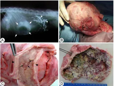
Initial therapy administrations did not achieve clinical remission; therefore, the dog was referred to the University of Adnan Menderes, Veterinary Faculty, Department of Internal Medicine Clinic for further examination and diagnosis. Haematological and serum biochemical analysis solely revealed nor-mocytic-hyperchromic non-regenerative anaemia (RBC 4.23 × 10¹²/l, MCV 70 fl, MCHC 34.2 g/dl) with no serum biochemical abnormalities. Physical examination confirmed the presence of a palpable abdominal mass and abdominal distension. In a contrast radiogram, a structure with well-defined borders adjacent to the intestine but which did not
fill completely with contrast medium was observed (Figure 1A). In an ultrasonographic investigation, a heterogenous cystic structure whose walls were hy-perechoic and which had irregular and a thick dorsal wall and a thin ventral wall was determined at the caudal side of the stomach. Subsequently, the dog was referred to the department of surgery for elec-tive abdominal laparotomy. At the exploration of the abdominal cavity, a mass was seen at the posterior of the stomach and over the jejunum (Figure 1B). The intestinal segment affected by the mass was removed from the abdominal cavity. In response to massaging movements, content passage was observed in the intestine. After the removal of the mass, a resection procedure was carried out at 4 cm distance from the parts of the intestine attached to the mass. After the parts of the intestine were joined by end-to-end anastomosis, a part of the intestine along with the tumour mass was sent to the pathology laboratory for histopathological examination.
For histological evaluation, tissue samples taken from the tumour masses were fixed in 10% neutral buffered formalin, processed routinely, sectioned
[image:2.595.104.477.396.678.2]at 5 µm, and stained with haematoxylin and eosin (H&E) and Masson’s trichrome. To confirm the di-agnosis, additional sections were stained using the streptavidin-biotin-peroxidase complex technique (ABC; Dako, USA) The primary antibodies employed were the following: vimentin (Abcam ab8069), al-pha-smooth muscle actin (α-SMA, Abcam ab18147), desmin (Invitrogen Clone, Zc18), cytokeratin (Dako, Clones AE1/AE3), S-100 protein (Spring Bioscience, clone E2140, USA), glial fibrillary acidic protein (GFAP, Thermo Scientific, clone GA-5), neuron spe-cific enolase (NSE, Spring Bioscience, USA) and syn-aptophsin (Spring Bioscience, clone SP11, USA). For immunohistochemistry, serial sections were fixed in 10% neutral buffered formalin and embedded in paraffin. Five µm thick sections were deparaffin-ised in xylene, dehydrated in graded alcohols, and washed in 0.01M phosphate-buffered saline (PBS), pH 7.2–7.4. Endogenous peroxidase was blocked with 0.3% hydrogen peroxide in absolute methanol for 30 min. Sections were then rinsed in PBS, im-mersed in citrate buffer (2.1 g citric acid
monohy-drate/litre distilled water – DW), pH 6.0, incubated for 10 min in a microwave oven at 750 W, and al-lowed to cool to room temperature (approximately 20 min). To reduce nonspecific staining, the sections were incubated with 10% normal goat serum before exposure to primary antisera. The primary antibod-ies were applied overnight at 4 °C and were followed by a commercial streptavidin-biotin-peroxidase as-say (Invitrogen Histostain plus Detection Kit, USA). After washing the slides with PBS, they were treated with the substrate chromogen (3,3'-diaminobenzi-dine tetrahydrochloride, DAKO), counterstained with haematoxylin, dehydrated, and mounted in a synthetic medium. The immunolabeling procedure included negative control sections incubated in PBS without primary antibody.
[image:3.595.114.482.87.369.2]surface of the tumour mass had a solid appearance, expanded outwards from the intestinal wall, and was homogenous. The tumour had a grayish white thick wall and the cystic structure and its lumen was filled with dark red content. The inner surface of the tumour was haemorrhagic and was covered with dark green pseudomembranes (Figure 1D).
At histopathological examination, the tumour was observed to extend from the submucosa of the intestine, where the ulcer was present, to the se-rosa (Figure 2A). In some areas, infiltrative spread towards the propria mucosa was seen. The tumour was poorly delineated, highly cellular and consisted of fusiform-elongated spindle-shaped to polygo-nal neoplastic cells forming interlacing fascicles or interwoven bundles in an atypical herringbone pattern (Figure 2A, B). Neoplastic cells exhibited eosinophilic cytoplasm and indistinct cell borders with anisocytosis, and the cytoplasm stained blue on Masson’s trichrome stain. The nuclei were large, round to oval, hyperchoromatic and prominent nuclear borders with single to multiple, usually centrally located nucleoli. Multinucleated cells with large, bizarre nuclei were usually scattered throughout the tumour tissue (Figure 2C). Mitotic activity was variable, and averaged about 2–4 per × 40 field. The neoplastic cells were locally invasive, spreading widely into the intestinal muscle layer. Multifocally within the mass there were areas of ne-crosis, haemorrhage and infiltrates of macrophages together with lymphocytes.
Immunohistochemically, the neoplastic cells showed strong positive labelling for vimentin (Figure 2D), and the staining with the antibody was cytoplasmic and diffuse. No immunoreactivity was demonstrable for α-SMA and desmin despite the positive immunoreactivity detected in the tunica media of blood vessels. However, labelling was also not observed with antibodies to cyotokeratins (AE1/AE3), S-100, GFAP, NSE and synaptophsin.
Based on the histopathological and immunohis-tochemical findings, the tumour was diagnosed as a fibrosarcoma. Six months after amputation, local recurrence of the tumour and metastasis was not observed.
DISCUSSION AND CONCLUSIONS
Primary fibrosarcomas of the intestine itself are extremely rare in dogs and other animal species (Goldschmidt and Hendrick 2002), and may
origi-nate anywhere in the body (Orr 1984; Mahler et al. 2006; Speltz et al. 2007; Govaere et al. 2010). To the authors’ knowledge, to date only one case has been reported in the veterinary literature, in which a tumour was described in a 17-year old Cocker Spaniel (Hurov 1962). The present case is thus the second case of fibrosarcoma arising from the intes-tine. Although the number of cases is not enough to evaluate a predisposition to race, the fact that the PIF occurred in a dog of the same race is intriguing.
In dogs and other animals, peritonitis due to rup-tures associated with intestinal ulcers is a com-mon clinical finding (Hinton et al. 2002; Case et al. 2010). However, intestinal tumours generally ap-pear radiographically as masses that are narrowing or obstructing the intestinal lumen (Hurov 1962; Head et al. 2002). In the present case, as tumour development outwards from the intestinal wall and the structure of the intestinal wall was not disrupt-ed, the above mentioned clinical findings were not seen. In addition, at macroscopic examination, a mass with a solid appearance was apparent which had a pyogranulamatous structure surrounded by a thick connective tissue. Therefore, in such masses developing in the abdominal region, clinical and macroscopic differential diagnosis should be firmly supported by histopathological findings.
as they are predisposing factors in the development of malignant tumours.
The tumour in the present report was diagnosed as a fibrosarcoma on the basis of histopathological and immunohistochemical findings (Olausson et al. 2005; Vascellari et al. 2006; Speltz et al. 2007; Rayner et al. 2010). During the diagnostic process, fibrosarcomas must be distinguished from other types of malignant tumours including leiomyosar-comas, hemangiopericytomas, and schmannomas and neurofibrosarcomas (Xu 1986; Kapatkin et al. 1992; Goldschmidt and Hendrick 2002; Cho and Park 2006; Maas et al. 2007). Leiomyosarcomas arise from smooth muscles situated anywhere in the body, and the tumour cells have centrally located and characteristic cigar-shaped nuclei (Kapatkin et al. 1992; Maas et al. 2007). Hemangiopericytomas are characterised by spindle cells containing cy-toplasmic processes arranged in whorls around blood vessels with a fingerprint pattern formed by neoplastic cells (Xu 1986; Cho and Park 2006). Schwannomas and neurofibrosarcomas are charac-terised by interwoven bundles of small wavy spin-dle cells with occasional palisading and whorls and they have positive reactions to S-100 protein, GFAP and NSE (Pumarola et al. 1996; Cullen et al. 2002; Hesaraki et al. 2010). In this case, no histopatho-logical findings of leiomyosarcoma, hemangioperi-cytoma, schmannoma and neurofibrosarcoma were observed, and none of the immunomarkers gave a positive staining reaction except for vimentin, which was diffusely positive, thus confirming the histological diagnosis of a primary intestinal fibro-sarcoma.
As far as we know, this is the second description of PIF originating from jejunum associated with intestinal ulceration, and the clinical and patho-logical findings described above combined with the immunohistochemical results supported the diagnosis of fibrosarcoma in the present case.
RefeReNCeS
Al-Dissi AN, Haines DM, Singh B, Kidney BA (2009): Im-munohistochemical expression of vascular endothelial growth factor and vascular endothelial growth factor receptor in canine cutaneous fibrosarcomas. Journal of Comparative Pathology 141, 229–236.
Brown RJ, Smith AW, Keyes MC (1975): Renal fibrosar-coma in the northern fur seal. Journal of Wildlife Dis-eases 11, 23–25.
Case JB, Fick JL, Rooney MB (2010): Proximal duodenal perforation in three dogs following deracoxib admin-istration. Journal of the American Animal Hospital Association46, 255–258.
Cho HS, Park NY (2006): Primary haemangiopricytoma in the pelvic cavity of a dog. Journal of Veterinary Medicine A 53, 198–201.
Cullen JM, Page R, Misdorp W (2002): An overview of cancer pathogenesis, diagnosis, and management. In: Meuten DJ (ed.): Tumors in Domestic Animals. 4th ed.
Iowa State Press, Iowa. 3–44.
Doddy FD, Glickman LT, Glickman NW, Janovitz EB (1996): Feline fibrosarcomas at vaccination sites and non-vaccination sites. Journal of Comparative Pathol-ogy 114, 165–174.
Gallati WW (1956): Fibrosarcoma associated with the cysticercus of Taenia taeniaeformis in the liver of a muskrat. Ohio Journal of Science 56, 71–75.
Goldschmidt MH, Hendrick MJ (2002): Tumors of the skin and soft tissues. In: Meuten DJ (ed.): Tumors in Domestic Animals. 4th ed. Iowa State University Press,
Iowa. 45–117.
Govaere J, Maes S, Saey V, Blancke W, Hoogewijs M, Deschauwer C, Smits K, Roels K, Vercauteren G, Kruif AD (2010): Uterine fibrosarcoma in a warmblood mare. Reproduction in Domestic Animals 46, 564–566. Hayden DW, Nielsen SW (1973): Canine alimentary
neoplasia. Zentralblatt fur Veterinarmedizin A 20, 1–22.
Head KW, Else RW. Dubielzig RR (2002): Tumors of the alimentary tract. In: Meuten DJ (ed.): Tumors in Do-mestic Animals. 4th ed. Iowa State Pres, Iowa. 401–481.
Hesaraki S, Vaighan AJ, Saraji AA (2010): Gastrointes-tinal autonomic nerve tumor in a Dog-A Case record. Global Veterinaria 5, 11–14
Hinton LE, McLoughlin MA, Johnson SE, Weisbrode SE (2002): Spontaneous gastroduodenal perforation in 16 dogs and seven cats (1982–1999). Journal of the Amer-ican Animal Hospital Association38, 176–187. Hurov L (1962): Perforating ulcers of the small intetine
lymphosarcoma and fibrosarcoma complications. Ca-nadian Veterinary Journal 3, 385–387.
Islam S, Soldes OS, Ruiz R (2008): Primary colonic con-genital infantile fibrosarcoma presenting as meco-nyum peritonitis. Pediatric Surgery International 24, 621–623.
Kapatkin AS, Mullen HS, Matthiesen DT, Patnaik AK (1992): Leiomyosarcoma in dogs: 44 cases (1983– 1988). Journal of the American Animal Hospital As-sociation201, 1077–1079.
casual relation between vaccination and fibrosarcoma tumorigenesis in cats. Journal of the American Animal Hospital Association 203, 396–405.
Larock RG, Ginn PE (1997): Immunohistochemical staining characteristics of canine gastrointestinal stro-mal tumors. Veterinary Pathology 34, 303–311. Lewis HB (2003): Vaccine-associated feline
fibrosar-coma. Veterinary Record 152, 755.
Maas CPHJ, Haar GT, Gaag IVD, Kirpensteijn J (2007): Reclassification of small intestinal and cecal smooth muscle tumors in 72 dogs: clinical, histologic, and im-munohistochemical evaluation. Veterinary Surgery 36, 302–313.
Macy DW, Hendrick MJ. (1996): The potential role of inflammation in the development of postvaccinal sar-comas in cats. Veterinary Clinics of North America-Small Animal Practice 26, 103–109.
Madewell BR, Griffey SM, McEntee MC, Leppert VJ, Munn RJ (2001): Feline vaccine-associated fibrosar-coma: an ultrastructural study of 20 tumors (1996– 1999). Veterinary Pathology 38, 196–202.
Mahler SP, Mootoo NFA, Reece JLM, Cooper JE (2006): Surgical resection of a primary tracheal fibrosarcoma in a dog. Journal of Small Animal Practice 47, 537–540. Martin M (2003): Vaccine-associated fibrosarcoma in a
cat. Canadian Veterinary Journal 44, 660–663. Moizhess TG (2008): Carcinogenesis induced by foreign
bodies. Biochemistry (Moscow) 73, 763–775.
Olausson A, Stieger SM, Loefgren S, Gillingstam M (2005): A urinary bladder fibrosarcoma in a young dog. Veterinary Radiology and Ultrasound 46, 135–138. Orr JP (1984): Fibrosarcoma affecting the mammary
gland of a cow. Canadian Journal of Comparative Medicine 48, 219–222.
Pumarola M, Anor S, Borras D, Ferrer I (1996): Malig-nant epithelioid schwannoma affecting the trigeminal nerve of a dog. Veterinary Pathology 33, 434–436. Rayner EL, Scudamore CL, Francis I, Schoeniger S
(2010): Abdominal fibrosarcoma associated with a retained surgical swab in a dog. Journal of Compara-tive Pathology143, 81–85.
Schumacher J, Watkins JP, Wilson SR, Foreman ME (1983): Burn-induced neoplasia in two horses. Equine Veterinary Journal 18, 410–412.
Shima Y, Ikegami E, Takechi N, Migita M, Hayashi Z, Araki T, Tanaka Y, Sugiyama M, Hashizume K (2003): Congenital fibrosarcoma of the jejunum in a premature infant with meconoim peritonitis. European Journal of Pediatric Surgery 13, 134–136.
Smyth MJ, Crowe NY, Godfrey DI (2001): NK cells and NKT cells collaborate in host protection from meth-ylcholanthrene-induced fibrosarcoma. International Immunology 13, 459–463.
Speltz MC, Manivel JC, Tobias AH, Hayden DW (2007): Primary cardiac fibrosarcoma with pulmonary metas-tasis in a labrador retriever. Veterinary Pathology 44, 403–407.
Vascellari M, Melchiotti E, Mutinelli F (2006): Fibrosar-coma with typical features of postinjection sarFibrosar-coma at site of microchip implant in a dog: Histologic and immunohistochemical study. Veterinary Pathology 43, 545–548.
Xu FN (1986): Ultrastructure of canine hemangioperi-cytoma. Veterinary Pathology 23, 643–645.
Received: 2011–11–10 Accepted after corrections: 2012–07–07
Corresponding Author:
Hamdi Avci, University of Adnan Menderes, Faculty of Veterinary Medicine, Department of Pathology, 09016 Aydin, Turkey