TISSUE DISSOLUTION CAPACITY OF SODIUM HYPOCHLORITE IN COMBINATION WITH
DIFFERENT CHELATING AGENTS
*
Dr. Sharmila Priyanka, B., Dr. Narsimha Rao, V.V. and Dr. Ch.
Department of Pedodontics and Preventive Dentistry, Gitam Dental College and Hospital, Visakhapatnam
ARTICLE INFO ABSTRACT
Background/Introduction
endodontic treatment. Combining different
removing organic and inorganic debris during biomechanical preparation. Aim/Objective
combination of various irriga Materials :
samples were immersed in saline, sodium hypochlorite, ethylene diamine tetra acetic acid(EDTA), etidronic acid/1
tetraacetic acid(EGTA)separately and in combinations of these agents for 5, 10 & 15 minutes. Results:
hypochlorite and HEDP combination, sodium hypochlorite and EGTA combination, sodium hypochlorite and EDTA combination.
did not show any tissue dissolving property. Conclusion:
adequate available chlorine which could be the reason for its high efficiency.
Copyright © 2018, Sharmila Priyanka et al. This is unrestricted use, distribution, and reproduction in any medium,
INTRODUCTION
Success of root canal therapy depends on effective mechanical debridement of pulpal tissue, dentin debris and microorganisms (Irala et al., 2010). Because of complex anatomy of root canals 30-60% remain uninstrumented resulting in insufficient debridement, providing source of nutrition for surviving bacteria (Naenni et al.,
allows for cleaning beyond, which cannot be achieved by instrumentation alone (Agrawal et al.,
endodontic irrigant should be, effective as a germicide and fungicide with prolonged antimicrobial effect, should be non irritating to the periapical tissues, should be stable in solution and active in the presence of blood, serum, and protein derivatives of tissue, having low surface tension to be able to completely remove the smear layer, able to disinfect the dentin/dentinal tubules without any adverse effect on physical properties of exposed dentin, preventing staining of the tooth structure, should be inexpensive, easy to use/apply with no adverse effect on the sealing ability of filling materials.
*Corresponding author: Dr. Sharmila Priyanka, B.,
Department of Pedodontics and Preventive Dentistry, Gitam Dental College and Hospital, Visakhapatnam.
ISSN: 0975-833X
Article History:
Received 22nd January, 2018
Received in revised form 14th February, 2018
Accepted 19th March, 2018
Published online 30th April, 2018
Citation: Dr. Sharmila Priyanka, B., Dr. Narsimha Rao, V.V. and Dr. Ch. Radha Rani
combination with different chelating agents – An invitro study.
Key words:
Sodium hypochlorite, EDTA,
EGTA and HEDP.
RESEARCH ARTICLE
TISSUE DISSOLUTION CAPACITY OF SODIUM HYPOCHLORITE IN COMBINATION WITH
DIFFERENT CHELATING AGENTS – AN INVITRO STUDY
Dr. Sharmila Priyanka, B., Dr. Narsimha Rao, V.V. and Dr. Ch.
Department of Pedodontics and Preventive Dentistry, Gitam Dental College and Hospital, Visakhapatnam
ABSTRACT
Background/Introduction: Irrigation of root canal system is one of the most important steps in endodontic treatment. Combining different irrigants may modify the efficiency of the irrigation in removing organic and inorganic debris during biomechanical preparation.
Aim/Objective: The aim of the study was to determine the chemical interaction that occurs during combination of various irrigating solutions and its effect on tissue dissolution.
Materials : Muscle tissue samples of equal dimensions with their initial weights were taken and the samples were immersed in saline, sodium hypochlorite, ethylene diamine tetra acetic acid(EDTA),
nic acid/1-hydroxyethane 1,1-diphosphonic acid(HEDP), ethylene glycol bis tetraacetic acid(EGTA)separately and in combinations of these agents for 5, 10 & 15 minutes. Results: Maximum tissue dissolving capacity was shown by sodium hypochlorite
hypochlorite and HEDP combination, sodium hypochlorite and EGTA combination, sodium hypochlorite and EDTA combination. HEDP, EGTA, EDTA, and saline solutions when used alone did not show any tissue dissolving property.
Conclusion: Sodium hypochlorite either alone or in combination with HEDP and EGTA maintains adequate available chlorine which could be the reason for its high efficiency.
is an open access article distributed under the Creative Commons medium, provided the original work is properly cited.
Success of root canal therapy depends on effective chemo debridement of pulpal tissue, dentin debris and Because of complex 60% remain uninstrumented ement, providing source of ., 2004). Irrigation allows for cleaning beyond, which cannot be achieved by 2014). Ideally, should be, effective as a germicide and fungicide with prolonged antimicrobial effect, should be non-irritating to the periapical tissues, should be stable in solution and active in the presence of blood, serum, and protein ow surface tension to be able to completely remove the smear layer, able to disinfect the dentin/dentinal tubules without any adverse effect on physical properties of exposed dentin, preventing staining of the tooth o use/apply with no adverse effect on the sealing ability of filling materials.
Dr. Sharmila Priyanka, B.,
Department of Pedodontics and Preventive Dentistry, Gitam Dental
Classification of the commonly used irrigating solutions
Chemical agents
Tissue dissolving agents: NaOCl Antibacterial agents:
Bacteriostatic: CHX, some antibiotics
Bactericidal: some antibiotics, NaOCl
Chelating agents:
Weak: HEDP, EGTA
Strong: EDTA
Combination products (tissue dissolution & antibacterial effect):
MTAD, QMIX, SmearClear, Tetraclean
Natural agents
Antibacterial agents: Green tea, Triphala
Two frequently used irrigants in endodontics
solutions of sodium hypochlorite and ethylene diamine tetraacetic acid.
International Journal of Current Research
Vol. 10, Issue, 04, pp.67784-67788, April, 2018
Dr. Sharmila Priyanka, B., Dr. Narsimha Rao, V.V. and Dr. Ch. Radha Rani, 2018. “Tissue dissolution capacity of sodium hypochlorite in
An invitro study.” International Journal of Current Research, 10, (04), 67784
Available online at http://www.journalcra.com
z
TISSUE DISSOLUTION CAPACITY OF SODIUM HYPOCHLORITE IN COMBINATION WITH
AN INVITRO STUDY
Dr. Sharmila Priyanka, B., Dr. Narsimha Rao, V.V. and Dr. Ch. Radha Rani
Department of Pedodontics and Preventive Dentistry, Gitam Dental College and Hospital, Visakhapatnam
: Irrigation of root canal system is one of the most important steps in irrigants may modify the efficiency of the irrigation in removing organic and inorganic debris during biomechanical preparation.
: The aim of the study was to determine the chemical interaction that occurs during ting solutions and its effect on tissue dissolution.
Muscle tissue samples of equal dimensions with their initial weights were taken and the samples were immersed in saline, sodium hypochlorite, ethylene diamine tetra acetic acid(EDTA), diphosphonic acid(HEDP), ethylene glycol bis –N N N N’ – tetraacetic acid(EGTA)separately and in combinations of these agents for 5, 10 & 15 minutes.
Maximum tissue dissolving capacity was shown by sodium hypochlorite followed by sodium hypochlorite and HEDP combination, sodium hypochlorite and EGTA combination, sodium HEDP, EGTA, EDTA, and saline solutions when used alone
m hypochlorite either alone or in combination with HEDP and EGTA maintains adequate available chlorine which could be the reason for its high efficiency.
Commons Attribution License, which permits
the commonly used irrigating solutions
Tissue dissolving agents: NaOCl
CHX, some antibiotics some antibiotics, NaOCl
Combination products (tissue dissolution & antibacterial
MTAD, QMIX, SmearClear, Tetraclean
Antibacterial agents: Green tea, Triphala
Two frequently used irrigants in endodontics are aqueous solutions of sodium hypochlorite and ethylene diamine INTERNATIONAL JOURNAL OF CURRENT RESEARCH
Sodium hypochlorite is used at concentrations ranging from 0.5 to 5.25% and is known for its good tissue dissolution capacity and dentin disinfecting potential, but because of its limited effect on smear layer removal, it is recommended with chelating agents like EDTA (Grawehr, 2003)
were introduced to endodontics by Nygaard
and they react with Ca2+ ions in dentine and form soluble calcium chelates, thereby decalcifying the dentin.
irrigation of 17% EDTA and NaOCl
recommended regimen for removal of both organic and inorganic components of smear layer (Agrawal
[image:2.595.315.554.88.232.2]Recently introduced and possible alternatives to existing chelating agents are HEDP and EGTA. Etidronic acid or Hydroxy ethylidene bisphosphonate (HEDP) belongs to bisphosphonates family which has been widely used, in the treatment of osteoporosis, Paget’s disease and hypercalcemia associated with malignancies. It was considered as a substitute for traditional chelators because of its fewer adverse effects on dentin (Tartari et al., 2013).
Figure 1. Armamentarium
EGTA or ethylene glycol bis –N N N N’ –
also effective in removal of smear layer, because of its calcium ion specificity (Hegde et al., 2016). Endodontic irrigants with low systemic toxicity should allow optimal disinfection of root canal system. And it has been found that none of the irrigating solutions which are available, can be regarded as optimal. aim of the study was to determine the t
capacity of various irrigating solutions when used alone or in combinations at different time intervals.
MATERIALS AND METHODS
Solutions: 18% HEDP was prepared by dissolving 18 grams
of pure chemical in 100 ml of distilled water.
EDTA was prepared by dissolving 17 grams of disodium EDTA in 100ml of distilled water with aid of sodium hydroxide and then the pH was adjusted to 7 by adding hydrochloric acid. 17% EGTA was prepared by dissolving 17 grams of EGTA in 100ml of distilled water with aid of NaOH and then the pH was adjusted to 7.5 by adding hydrochloric acid. Physiological saline was used as negative control and 2.5% & 5% NaOCl were taken as positive control. All chemical substances were prepared just prior to the
Figure 2.
Tissue dissolution assay: Goat muscle tissue (
hircus) was obtained from slaughter house shown in
and was cut immediately into pieces of 8 × 6 × 4 mm using
67785 Dr. Sharmila Priyanka et al. Tissue dissolution capacity of sodium hypochlorite in combination with different chelating agents
hypochlorite is used at concentrations ranging from 0.5 to 5.25% and is known for its good tissue dissolution capacity and dentin disinfecting potential, but because of its it is recommended with ). Chelating agents were introduced to endodontics by Nygaard – Ostby in 1957 ions in dentine and form soluble calcium chelates, thereby decalcifying the dentin. Alternate has become the recommended regimen for removal of both organic and Agrawal et al., 2014). Recently introduced and possible alternatives to existing chelating agents are HEDP and EGTA. Etidronic acid or ne bisphosphonate (HEDP) belongs to bisphosphonates family which has been widely used, in the treatment of osteoporosis, Paget’s disease and hypercalcemia associated with malignancies. It was considered as a substitute its fewer adverse effects on
tetraacetic acid is also effective in removal of smear layer, because of its calcium Endodontic irrigants with low systemic toxicity should allow optimal disinfection of root canal system. And it has been found that none of the irrigating solutions which are available, can be regarded as optimal. The aim of the study was to determine the tissue dissolution capacity of various irrigating solutions when used alone or in
18% HEDP was prepared by dissolving 18 grams of pure chemical in 100 ml of distilled water. Similarly 17% EDTA was prepared by dissolving 17 grams of disodium EDTA in 100ml of distilled water with aid of sodium hydroxide and then the pH was adjusted to 7 by adding 17% EGTA was prepared by dissolving 17 distilled water with aid of NaOH and then the pH was adjusted to 7.5 by adding hydrochloric acid. Physiological saline was used as negative control and 2.5% & 5% NaOCl were taken as positive control. All chemical substances were prepared just prior to the usage as in
Goat muscle tissue (Capra aegagrus
was obtained from slaughter house shown in Figure 3 and was cut immediately into pieces of 8 × 6 × 4 mm using
stainless steel blade as in the determine their initial weights in
[image:2.595.309.551.94.508.2]Figure 2. Prepared solutions
[image:2.595.44.282.282.428.2]Figure 3. Muscle tissue
Figure 4. Tissue fragments
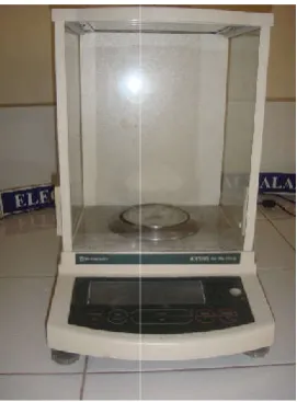
Figure 5. Electronic balance
Tissue dissolution capacity of sodium hypochlorite in combination with different chelating agents
stainless steel blade as in the Figure 4 and were weighed to determine their initial weights in Figure 5.
Prepared solutions
Muscle tissue
Tissue fragments
Electronic balance
[image:2.595.366.502.599.783.2]Based on the irrigant used grouping was done as follows
Group 1 –0.9% physiological saline
Group 2 – 2.5% NaOCl
Group 3 – 17% EDTA
Group 4 – 17% EGTA
Group 5 – 18% HEDP
Group 6 – 2.5% NaOCl + 8.5% EDTA
Group 7 – 2.5% NaOCl + 8.5% EGTA
Group 8 - 2.5% NaOCl + 8.5% HEDP
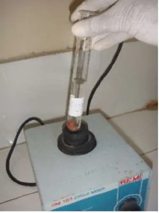
After measuring the pH of test solution in each group with a pH meter, as shown in Figure 6, test tubes were filled with 15ml of test solutions, and the tissue fragments were submerged individually in the solutions and mechanical agitation was performed with CM 101 cyclomixer (REMI) for 15 s per minute for 5 minutes as in Figure 7.
At the end of agitation, the samples were taken out and submerged in distilled water for 30 s to remove the excess test solution. They were then blotted dry and reweighed for comparison with initial values as in Figure 8,9.
[image:3.595.313.552.136.291.2]procedure was repeated for 10 min and 15 min time period also, and initial weight and final weight after subjecting the sample to test solutions, were noted.
Figure 6. Measuring the pH of the solutions
Figure 7. Agitation of tissue immersed in test solution with the help of cyclomixer
67786 International Journal
ed grouping was done as follows
After measuring the pH of test solution in each group with a 6, test tubes were filled with 15ml of test solutions, and the tissue fragments were submerged individually in the solutions and mechanical agitation was performed with CM 101 cyclomixer (REMI) for
of agitation, the samples were taken out and submerged in distilled water for 30 s to remove the excess test solution. They were then blotted dry and reweighed for 8,9. Similarly, this 10 min and 15 min time period also, and initial weight and final weight after subjecting the
Measuring the pH of the solutions sample
ion of tissue immersed in test help of cyclomixer
Statistical analysis: The values were tabulated and subjected
to statistical analysis using analysis of variance (ANOVA) followed by paired t test to assess if there is any difference between specimen weights before and after
[image:3.595.341.526.339.522.2]different time periods and between groups for same time periods.
Figure 8. Submersion in distilled water and blotting dry of sample
Figure 9. Reweighing the tissue sample
RESULTS
Significant decrease (P<0.01)in
all the time periods of immersion was observed for group
2(NaOCl), group 8 (NaOCl+HEDP) and group
7(NaOCl+EGTA). Other groups were not associated with significant loss of weight at any period which were observed from the table no 1. Significant reduction in weight (P<0.01) occurred in the following order according to the table no 2. the end of 5 min G2>G8>G7>G6>G3=G5=G4>G1, min G2>G8>G7>G6>G4>G5=G3>G1 and G2>G8>G7>G6>G3=G4>G5>G1
DISCUSSION
Goal of cleaning and shaping regimen in endodontic therapy is to maximally reduce microbial load and necrotic tissue remnants in root canal system as remnants of organic debris inside root canal system, will serve as substrate for growth of microorganisms that survive the biomechanical preparation and contaminate the root canal after treatment
International Journal of Current Research, Vol. 10, Issue, 04, pp.67784-67788, April, 2018
The values were tabulated and subjected to statistical analysis using analysis of variance (ANOVA) followed by paired t test to assess if there is any difference between specimen weights before and after submersion for different time periods and between groups for same time
Submersion in distilled water and blotting dry of sample
Reweighing the tissue sample
Significant decrease (P<0.01)in weight of tissue fragments for all the time periods of immersion was observed for group
2(NaOCl), group 8 (NaOCl+HEDP) and group
7(NaOCl+EGTA). Other groups were not associated with significant loss of weight at any period which were observed Significant reduction in weight (P<0.01) occurred in the following order according to the table no 2. At the end of 5 min G2>G8>G7>G6>G3=G5=G4>G1, after 10 min G2>G8>G7>G6>G4>G5=G3>G1 and after 15 min G2>G8>G7>G6>G3=G4>G5>G1
cleaning and shaping regimen in endodontic therapy is to maximally reduce microbial load and necrotic tissue remnants in root canal system as remnants of organic debris inside root canal system, will serve as substrate for growth of vive the biomechanical preparation and contaminate the root canal after treatment (Love, 2001).
[image:3.595.53.272.352.519.2] [image:3.595.83.245.559.776.2]Hence biomechanical preparation in conjunction is always done with irrigation for optimal results. Sodium hypochlorite with concentration ranging from 0.5 to 5.25% is the most recommended for its antimicrobial, tissue dissolving and dentin disinfecting potential.
These properties are explained through the following reactions
Saponification reaction: NaOCl acts as an organic and fat
solvent, degrading fatty acids, transforming them into salts (soap) and glycerol (alcohol), that reduces the surface tension of the remaining solution (saponification reaction).
Amino acid neutralization: NaOCl neutralizes amino acids
forming water and salt
Chloramination reaction: Amino acid reacts with
hypochlorous acid to form into chloramines and water. The free available chlorine comprises of hypochlorous acid (HOCl) and hypochlorite ion (OCl-), which exist in equilibrium depending on pH of the solution. Hypochlorite ions (OCl−) exists in alkaline solutions (pH >7), because of its stronger oxidative effect shows higher tissue dissolving capacity. Hypochlorous acid exists in acidic solutions (3< pH < 7), shows powerful bactericidal effect. The disinfecting properties decrease with increase in pH of the solution, paralleling the concentration of disassociated hypochlorous acid.
[image:4.595.110.490.94.182.2]Despite these properties, NaOCl only removes organic structure of smear layer produced during mechanical instrumentation and chelating agents like EDTA are required for effective removal of inorganic part of smear layer, which allows deeper penetration of NaOCl into the dentinal tubules (Grawehr et al., 2003). Chelating agents react with Ca2+ ions in dentine and form soluble calcium chelates, thereby decalcifying the dentin (Agrawal Vineet et al., 2014). Subsequently, use of 17% EDTA and NaOCl has become the recommended regimen for removal of both organic and inorganic components of smear layer. But, whenever EDTA comes in contact with NaOCl, it results in exothermic reaction with complete loss of chlorine gas immediately in the form of bubbles which effects the tissue dissolution and antibacterial activity of NaOCl (Prado et al., 2013). Hence, this study focuses on the use of alternative chelating agents like HEDP & EGTA to be used along with NaOCl. Etidronic acid or Hydroxy ethylidene bisphosphonate (HEDP) was considered as a substitute for traditional chelators as it requires 300 seconds to remove smear layer with fewer adverse effects on dentin, which makes it a weak chelator (Tartari, 2013). EGTA or ethylene glycol bis –N N N N’ – tetraacetic acid is also effective in removal of smear layer, because it binds more specifically to calcium ions without causing dental erosion as seen with EDTA (Hegde et al., 2016). The available chlorine content upon combination of the irrigating solutions, can be assessed indirectly through the tissue dissolution capacity which can be assessed through various methods like the weighing method, total protein assay & hydroxyproline
Table 1. Mean in weight (mg) before submersion (T0) and after 5(T5), 10(T10) & 15(T15) min of submersion into test solutions
Groups pH T0 T5 T10 T15 F value P value G1- SALINE 6.8 353.33 363.33 354.33 338.00 0.50 0.62 G2 – 2.5% NaOCl 11.8 353.33 231.00 232.33 188.00 27.57 <0.01 G3 – 17% EDTA 7.0 352.00 327.67 348.00 331.00 0.62 0.56 G4 – 17% EGTA 7.5 332.67 313.33 344.67 323.67 2.4 0.16 G5 – 18% HEDP 10.8 351.67 331.00 344.00 322.33 1.26 0.34 G6–2.5%NaOCl+ 8.5% EDTA 7.4 346.67 237.67 275.33 278.00 3.9 0.07 G7-2.5%NaOCl+ 8.5% EGTA 8.3 363.00 249.67 228.67 221.33 306.05 <0.01 G8-2.5%NaOCl+ 8.5% HEDP 11.2 376.67 258.00 225.67 198.33 254.82 <0.01
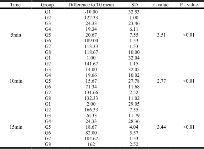
Table 2. Difference to baseline(T0) mean in weight (mg), and standard deviation(SD) after 5(T5),10(T10),15(T15) min of submersion into irrigant solutions
Time Group Difference to T0 mean SD t -value P - value
5min
G1 -10.00 32.53
3.51 <0.01 G2 122.33 1.00
G3 24.33 23.46
G4 19.34 6.11
G5 20.67 7.55
G6 109.00 1.53 G7 113.33 1.53 G8 118.67 10.00
10min
G1 1.00 32.04
2.77 <0.01 G2 141.67 1.15
G3 14.00 32.05
G4 19.66 10.02
G5 15.67 27.78
G6 71.34 11.68
G7 131.66 2.52 G8 132.33 11.02
15min
G1 2.00 29.05
3.44 <0.01 G2 166.33 7.55
G3 26.33 11.79
G4 24.33 28.36
G5 18.67 4.04
G6 82.00 5.57
G7 104.67 1.53
G8 162 2.52
[image:4.595.136.462.222.460.2]determination (Koskinen et al., 1980). In this study, the weighing method is chosen to determine tissue dissolution as it is simple and reliable and also in other assays, irrigant solutions may interfere with the regeants used in those procedures (Irala et al., 2010). Previous studies (Irala et al., 2010; Grawehr et al., 2003; Moorer, 1982; Almeida, 2013) have used different kinds of tissue to determine the tissue dissolution capacity including, porcine muscle, pig palatal mucosa, rabbit liver, bovine muscle and bovine and Human dental pulp tissue. Goat muscle tissue was used in this study due to its easy availability and ease of standardization of surface area of sample. This has shown that, tissue dissolution was greater where NaOCl was used alone in all time periods, followed by, where NaOCl was mixed with HEDP and EGTA in equal parts, with the dissolution not so much seen in the combination of NaOCl and EDTA.
No tissue dissolution was seen when fragments were immersed in saline and chelating agents alone, which was observed in previous studies also (Irala, 2010; Naenni, 2004; Grawehr et al., 2003). Increase in weight of specimens was observed after 5min in saline and after 10min in EDTA, EGTA and in HEDP, which may be because of tissue hydration as a result of absorption of water by the tissue sample in the solutions. Previous studies have shown that tissue dissolution capacity of NaOCl is direct function of free available chlorine which consists of hypochlorous acid and hypochlorite ion (Grawehr
et al., 2003). Hypochlorous acid has powerful bactericidal
activity & hypochlorite ion shows greater tissue dissolving property (Macedo et al., 2010). Interaction of NaOCl with chelating agents results in chlorine gas evaporation and reduction of available chlorine (Zehnder, 2006). The present study confirms the earlier report on reduction of available chlorine in solution because of chlorine gas evaporation, when sodium hypochlorite interacts with acidic hydrogen of chelating agents. Hence there is least dissolution of the tissue upon combination of these irrigants. Greater tissue dissolution potential observed in mixture of NaOCl with HEDP or EGTA, compared to EDTA, might be due to the fact that being the weak chelators they did not actively interfere with chlorine of NaOCl, thereby maintaining the available chlorine in the solution which was required for dissolution (Tartari et al., 2013; Sayin et al., 2007). Also previous studies have shown that, weak chelators also remove smear layer efficiently without much erosion of peritubular and intertubular dentin maintaining sufficient root dentin hardness (Sayin et al., 2007).
Limitations of the study
The antibacterial efficacy of NaOCl when combined with various weak chelators as well as the pH potential of irrigants at various intervals could not be determined which are the limitations of this study.
Conclusion
Combining NaOCl with weak chelators like EGTA, HEDP gives the advantage of removing both organic and inorganic parts of the smear layer, making it a better irrigant than when used alone.
Acknowledgment
Department of Biotechnology, Gitam University,
Visakhapatnam.
Conflicts of interest: None
REFERENCES
Agrawal Vineet S, Rajesh M, Sonali K and Mukesh P. 2014. A Contemporary Overview of Endodontic Irrigants– A Review. J Dent App.,1(6): 105-115.
Almeida LH, Gomes AP, Giardino L, Souza EM, Pappen FG. 2013. Pulp tissue dissolution capacity of sodium hypochlorite combined with cetrimide and polypropylene glycol. Brazilian Dental Journal., 24(5):477-81.
Grawehr M, Sener B, Waltimo T, Zehnder M. 2003. Interactions of ethylenediamine tetraacetic acid with sodium hypochlorite in aqueous solutions. International
Endodontic Journal, 36(6):411–417.
Haapasalo M, Shen Y, Qian W, Gao Y. 2010. Irrigation in endodontics. Dental Clinics of North America Apr
30;54(2):291-312.
Hegde RJ, Bapna K. 2016. Comparison of removal of endodontic smear layer using ethylene glycol bis (beta-amino ethylether)-N, N, N’, N’-tetraacetic acid and citric acid in primary teeth: A scanning electron microscopic study. Contemp Clin Dent., 7(2):216-20.
Irala LE, Grazziotin-Soares R, Salles AA, Munari AZ, Pereira, JS. 2010. Dissolution of bovine pulp tissue in solutions consisting of varying NaOCl concentrations and combined with EDTA. Brazilian Oral Research 24(3): 271–6. Koskinen KP, Stenvall H, Uitto VJ. 1980. Dissolution of
bovine pulp tissue by endodontic solutions. European
Journal of Oral Sciences., 88(5):406-11.
Love RM. 2001. Enterococcus faecalis–a mechanism for its role in endodontic failure. International Endodontic Journal 2001;34(5): 399-405.
Macedo RG, Wesselink PR, Zaccheo F, Fanali D, Van Der Sluis LW. 2010. Reaction rate of NaOCl in contact with bovine dentine:effect of activation, exposure time, concentration and PH. International Endodontic Journal.,
43(12):1108-15.
Moorer WR, Wesselink PR. 1982. Factors promoting the tissue dissolving capability of sodium hypochlorite. International
Endodontic Journal, 15(4):187-196.
Naenni N, Thoma K, Zehnder M. 2004. Soft tissue dissolution capacity of currently used and potential endodontic irrigants. Journal of Endodontics 30(11):785–7.
Prado M, Santos Júnior HM, Rezende CM, Pinto AC, Faria RB, Simao RA, Gomes BP. 2013. Interactions between irrigants commonly used in endodontic practice: a chemical analysis. Journal of Endodontics., 39(4):505–10.
Sayin TC, Serper A, Cehreli ZC, Otlu HG. 2007. The effect of EDTA, EGTA, EDTAC, and tetracycline-HCl with and without subsequent NaOCl treatment on the microhardness of root canal dentin. Oral Surg Oral Med Oral Pathol Oral
Radiol Endod., 104(3):418-24.
Tartari T, Duarte Junior AP, Silva Junior JO, Klautau EB, Silva ESJMH, Silva ESJPA 2013. Etidronate from medicine to endodontics: Effects of different irrigation regimes on root dentin roughness. J Appl Oral Sci.,
21(5):409-15.
Zehnder M. 2006. Root canal irrigants. Journal of
Endodontics, 32(5):389-98.
67788 International Journal of Current Research, Vol. 10, Issue, 04, pp.67784-67788, April, 2018