Crystal structure of methyl
1-methyl-2-oxospiro[indoline-3,2
000-oxirane]-3
000-carboxylate
M. P. Savithri,aP. S. Yuvaraj,bB. S. R. Reddy,bR. Rajacand A. SubbiahPandic*
aDepartment of Physics, Queen Mary’s College (Autonomous), Chennai 600 004, India,bUniversity of Madras, Industrial Chemistry Laboratory, Central Leather Research Institute, Adyar, Chennai 600 020, India, andcDepartment of Physics, Presidency College (Autonomous), Chennai 600 005, India. *Correspondence e-mail: [email protected]
Received 10 February 2015; accepted 29 March 2015
Edited by W. T. A. Harrison, University of Aberdeen, Scotland
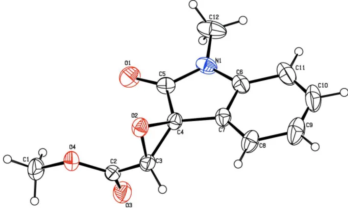
In the title compound, C12H11NO4, the dihedral angle between
the indole ring system (r.m.s. deviation = 0.019 A˚ ) and the oxirane ring is 88.8 (2). The oxirane O atom and the bridging
ester O atom are in an approximate syn conformation [O— C—C—O =25.4 (3)] In the crystal, inversion dimers linked
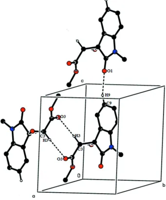
by pair of C—H O hydrogen bonds generate R2 2
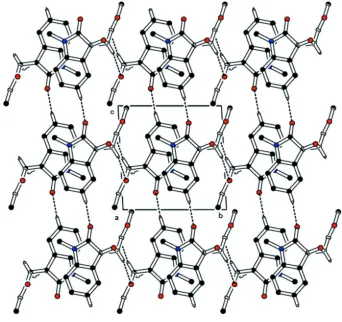
(8) loops, where the C—H donor group forms part of the oxirane ring. A second C—H O interaction arising from one of the C—H groups of the benzene ring links the dimers into [001] double chains.
Keywords:crystal structure; ester; spiro compound; indoline; oxirane; hydrogen bonding.
CCDC reference:1056692
1. Related literature
For the bioactivity of indole derivatives, see: Di Fabio et al. (2007); Sharma & Tepe (2004). For a related structure, see: Savithriet al.(2015).
2. Experimental
2.1. Crystal data
C12H11NO4
Mr= 233.22 Triclinic,P1
a= 7.1401 (4) A˚
b= 8.7787 (4) A˚
c= 9.0678 (4) A˚
= 91.517 (3) = 104.227 (3)
= 94.714 (3) V= 548.44 (5) A˚3
Z= 2
MoKradiation
= 0.11 mm1
T= 293 K
0.350.300.30 mm
2.2. Data collection
Bruker Kappa APEXII CCD diffractometer
Absorption correction: multi-scan (SADABS; Bruker, 2004)
Tmin= 0.963,Tmax= 0.969
11311 measured reflections 1927 independent reflections 1480 reflections withI> 2(I)
Rint= 0.037
2.3. Refinement
R[F2> 2(F2)] = 0.055
wR(F2) = 0.173
S= 1.06 1927 reflections 158 parameters
H atoms treated by a mixture of independent and constrained refinement
max= 0.30 e A˚
3
min=0.27 e A˚
3
Table 1
Hydrogen-bond geometry (A˚ ,).
D—H A D—H H A D A D—H A
C3—H3 O3i
0.95 (3) 2.52 (3) 3.414 (3) 157 (2) C9—H9 O1ii
0.93 2.43 3.335 (4) 163
Symmetry codes: (i)xþ1;y;zþ1; (ii)x;y;zþ1.
Data collection: APEX2 (Bruker, 2004); cell refinement: SAINT
(Bruker, 2004); data reduction:SAINT; program(s) used to solve structure:SHELXS97(Sheldrick, 2008); program(s) used to refine structure: SHELXL97 (Sheldrick, 2008); molecular graphics:
ORTEP-3 for Windows(Farrugia, 2012); software used to prepare material for publication:SHELXL97andPLATON(Spek, 2009).
Acknowledgements
The authors thank Dr Babu Vargheese, SAIF, IIT, Madras, India, for the data collection.
Supporting information for this paper is available from the IUCr electronic archives (Reference: HB7369).
data reports
o274
Savithriet al. doi:10.1107/S2056989015006398 Acta Cryst.(2015).E71, o274–o275Bruker (2004).APEX2,SAINT,XPREPandSADABS. Bruker AXS Inc., Madison, Wisconsin, USA.
Di Fabio, R., Micheli, F., Alvaro, G., Cavanni, P., Donati, D., Gagliardi, T., Fontana, G., Giovannini, R., Maffeis, M., Mingardi, A., Tranquillini, M. E. & Vitulli, G. (2007).Bioorg. Med. Chem. Lett.17, 2254–2259.
Savithri, M. P., Yuvaraj, P. S., Reddy, B. S. R., Raja, R. & SubbiahPandi, A. (2015).Acta Cryst.E71, o188–o189.
Sharma, V. & Tepe, J. J. (2004).Bioorg. Med. Chem. Lett.14, 4319–4321. Sheldrick, G. M. (2008).Acta Cryst.A64, 112–122.
supporting information
sup-1
Acta Cryst. (2015). E71, o274–o275
supporting information
Acta Cryst. (2015). E71, o274–o275 [https://doi.org/10.1107/S2056989015006398]
Crystal structure of methyl 1-methyl-2-oxospiro[indoline-3,2
′
-oxirane]-3
′
-carboxylate
M. P. Savithri, P. S. Yuvaraj, B. S. R. Reddy, R. Raja and A. SubbiahPandi
S1. Experimental
To a solution of the catalyst of diphenyl[(R)-2-pyrrolidinyl]methanol (0.15mmol) and trans-α-ylideneoxindoles 1 (0.5
mmol) in nHexane for HPLC grade (2.7ml) was added TBHP (5.5M in decane solution, 0.6mol) at room temperature
(25°C). The resultant heterogeneous mixture was maintained under stirring until the reaction completion (TLC
nHexane/EtOAc). After wards, the crude reaction mixture was purified by flash chromatography on silica gel
(nHexane/EtOAc) to furnish the epoxy oxindoles trans-2 and cis-3. Colourless blocks were obtained by slow evaporation
of a solution of the title compound in ethyl acetate at room temperature.
S2. Refinement
All H atoms were fixed geometrically and allowed to ride on their parent C atoms, with C-H distances fixed in the range
[image:3.610.133.474.399.613.2]0.93-0.98 Å with Uiso(H) = 1.5Ueq(C) for methyl H 1.2Ueq(C) for other H atoms.
Figure 1
Figure 2
The molecular packing as viewed along the a axis. Dashed lines shows the C—H···O hydrogen bonds. H atoms not
supporting information
sup-3
[image:5.610.135.476.76.488.2]Acta Cryst. (2015). E71, o274–o275
Figure 3
A partial view of the hydrogen-bond interactions C9—H9···O1 and C3—H3···O3 along a axis.
Methyl 1-methyl-2-oxospiro[indoline-3,2′-oxirane]-3′-carboxylate
Crystal data
C12H11NO4 Mr = 233.22
Triclinic, P1 Hall symbol: -P 1 a = 7.1401 (4) Å b = 8.7787 (4) Å c = 9.0678 (4) Å α = 91.517 (3)° β = 104.227 (3)° γ = 94.714 (3)° V = 548.44 (5) Å3
Z = 2 F(000) = 244 Dx = 1.412 Mg m−3
Mo Kα radiation, λ = 0.71073 Å Cell parameters from 1927 reflections θ = 2.3–25.0°
Bruker Kappa APEXII CCD diffractometer
Radiation source: fine-focus sealed tube Graphite monochromator
ω and φ scans
Absorption correction: multi-scan (SADABS; Bruker, 2004) Tmin = 0.963, Tmax = 0.969
11311 measured reflections 1927 independent reflections 1480 reflections with I > 2σ(I) Rint = 0.037
θmax = 25.0°, θmin = 2.3° h = −8→8
k = −10→10 l = −10→10
Refinement
Refinement on F2
Least-squares matrix: full R[F2 > 2σ(F2)] = 0.055 wR(F2) = 0.173 S = 1.06 1927 reflections 158 parameters 0 restraints
Primary atom site location: structure-invariant direct methods
Secondary atom site location: difference Fourier map
Hydrogen site location: inferred from neighbouring sites
H atoms treated by a mixture of independent and constrained refinement
w = 1/[σ2(F
o2) + (0.102P)2 + 0.172P]
where P = (Fo2 + 2Fc2)/3
(Δ/σ)max < 0.001
Δρmax = 0.30 e Å−3
Δρmin = −0.27 e Å−3
Special details
Geometry. All esds (except the esd in the dihedral angle between two l.s. planes) are estimated using the full covariance matrix. The cell esds are taken into account individually in the estimation of esds in distances, angles and torsion angles; correlations between esds in cell parameters are only used when they are defined by crystal symmetry. An approximate (isotropic) treatment of cell esds is used for estimating esds involving l.s. planes.
Refinement. Refinement of F2 against ALL reflections. The weighted R-factor wR and goodness of fit S are based on F2,
conventional R-factors R are based on F, with F set to zero for negative F2. The threshold expression of F2 > 2sigma(F2) is
used only for calculating R-factors(gt) etc. and is not relevant to the choice of reflections for refinement. R-factors based on F2 are statistically about twice as large as those based on F, and R- factors based on ALL data will be even larger.
Fractional atomic coordinates and isotropic or equivalent isotropic displacement parameters (Å2)
x y z Uiso*/Ueq
supporting information
sup-5
Acta Cryst. (2015). E71, o274–o275
C1 0.2456 (5) −0.0803 (4) 0.0221 (3) 0.0692 (9) H1A 0.1393 −0.1255 −0.0577 0.104* H1B 0.3391 −0.1534 0.0521 0.104* H1C 0.3056 0.0081 −0.0141 0.104* C12 0.2681 (5) 0.6277 (3) 0.3066 (5) 0.0774 (10) H12A 0.2979 0.7114 0.3813 0.116* H12B 0.1528 0.6440 0.2303 0.116* H12C 0.3742 0.6214 0.2597 0.116* C10 0.2921 (5) 0.5344 (5) 0.7951 (4) 0.0845 (12) H10 0.3223 0.6068 0.8761 0.101* C9 0.2516 (5) 0.3848 (5) 0.8237 (4) 0.0796 (11) H9 0.2537 0.3576 0.9225 0.096* H3 0.273 (4) 0.024 (3) 0.488 (3) 0.052 (7)*
Atomic displacement parameters (Å2)
U11 U22 U33 U12 U13 U23
O2 0.0454 (10) 0.0440 (9) 0.0450 (9) −0.0086 (7) 0.0076 (7) 0.0005 (7) O4 0.0565 (11) 0.0523 (11) 0.0456 (10) 0.0033 (8) 0.0026 (8) −0.0115 (8) O3 0.0524 (13) 0.0730 (13) 0.0597 (12) 0.0065 (9) 0.0068 (9) −0.0112 (9) O1 0.0859 (15) 0.0673 (12) 0.0455 (11) 0.0096 (10) 0.0247 (10) 0.0167 (9) N1 0.0464 (12) 0.0339 (11) 0.0738 (15) 0.0021 (8) 0.0206 (10) 0.0087 (9) C7 0.0352 (12) 0.0495 (14) 0.0418 (13) 0.0004 (10) 0.0099 (9) −0.0061 (10) C2 0.0477 (15) 0.0335 (12) 0.0425 (13) 0.0041 (10) 0.0020 (10) 0.0010 (9) C4 0.0380 (12) 0.0387 (12) 0.0351 (11) −0.0034 (9) 0.0078 (9) 0.0011 (9) C3 0.0477 (14) 0.0373 (12) 0.0382 (13) −0.0010 (10) 0.0024 (10) 0.0032 (10) C6 0.0333 (13) 0.0470 (14) 0.0650 (17) 0.0033 (10) 0.0125 (11) −0.0104 (11) C5 0.0425 (13) 0.0441 (13) 0.0476 (14) 0.0047 (10) 0.0154 (10) 0.0087 (10) C8 0.0495 (15) 0.0805 (19) 0.0392 (14) −0.0009 (13) 0.0128 (11) −0.0032 (12) C11 0.0491 (17) 0.0595 (18) 0.100 (3) 0.0038 (13) 0.0116 (16) −0.0328 (17) C1 0.077 (2) 0.082 (2) 0.0455 (15) 0.0188 (16) 0.0083 (13) −0.0170 (14) C12 0.0574 (18) 0.0453 (16) 0.137 (3) 0.0067 (13) 0.0348 (18) 0.0312 (17) C10 0.061 (2) 0.110 (3) 0.074 (2) 0.0127 (19) 0.0073 (16) −0.052 (2) C9 0.062 (2) 0.128 (3) 0.0476 (17) 0.0067 (19) 0.0138 (14) −0.0251 (18)
Geometric parameters (Å, º)
C2—C3 1.484 (4) C10—C9 1.367 (5) C4—C3 1.466 (3) C10—H10 0.9300 C4—C5 1.509 (3) C9—H9 0.9300
C3—O2—C4 61.80 (14) O1—C5—C4 126.6 (2) C2—O4—C1 116.2 (2) N1—C5—C4 106.3 (2) C5—N1—C6 111.2 (2) C7—C8—C9 117.8 (3) C5—N1—C12 122.4 (3) C7—C8—H8 121.1 C6—N1—C12 126.4 (2) C9—C8—H8 121.1 C8—C7—C6 121.6 (2) C6—C11—C10 116.7 (3) C8—C7—C4 131.6 (2) C6—C11—H11 121.7 C6—C7—C4 106.7 (2) C10—C11—H11 121.7 O3—C2—O4 125.0 (2) O4—C1—H1A 109.5 O3—C2—C3 121.3 (2) O4—C1—H1B 109.5 O4—C2—C3 113.7 (2) H1A—C1—H1B 109.5 O2—C4—C3 58.71 (14) O4—C1—H1C 109.5 O2—C4—C7 123.07 (19) H1A—C1—H1C 109.5 C3—C4—C7 125.9 (2) H1B—C1—H1C 109.5 O2—C4—C5 116.78 (18) N1—C12—H12A 109.5 C3—C4—C5 121.0 (2) N1—C12—H12B 109.5 C7—C4—C5 105.51 (19) H12A—C12—H12B 109.5 O2—C3—C4 59.48 (14) N1—C12—H12C 109.5 O2—C3—C2 119.95 (19) H12A—C12—H12C 109.5 C4—C3—C2 121.3 (2) H12B—C12—H12C 109.5 O2—C3—H3 117.1 (15) C9—C10—C11 122.8 (3) C4—C3—H3 116.4 (16) C9—C10—H10 118.6 C2—C3—H3 112.8 (15) C11—C10—H10 118.6 C11—C6—C7 120.9 (3) C10—C9—C8 120.3 (3) C11—C6—N1 128.8 (3) C10—C9—H9 119.9 C7—C6—N1 110.3 (2) C8—C9—H9 119.9 O1—C5—N1 127.2 (2)
supporting information
sup-7
Acta Cryst. (2015). E71, o274–o275
C5—C4—C3—C2 4.2 (3) C6—C7—C8—C9 0.9 (4) O3—C2—C3—O2 157.5 (2) C4—C7—C8—C9 176.2 (3) O4—C2—C3—O2 −25.4 (3) C7—C6—C11—C10 0.5 (4) O3—C2—C3—C4 87.1 (3) N1—C6—C11—C10 −178.5 (2) O4—C2—C3—C4 −95.8 (2) C6—C11—C10—C9 −0.3 (5) C8—C7—C6—C11 −0.8 (4) C11—C10—C9—C8 0.5 (5) C4—C7—C6—C11 −177.1 (2) C7—C8—C9—C10 −0.7 (4) C8—C7—C6—N1 178.4 (2)
Hydrogen-bond geometry (Å, º)
D—H···A D—H H···A D···A D—H···A
C3—H3···O3i 0.95 (3) 2.52 (3) 3.414 (3) 157 (2)
C9—H9···O1ii 0.93 2.43 3.335 (4) 163