Original Article
Application of improved fixed nerve retractor in
posterior surgery of lumbar disc herniation
Jinfeng Liang1, Yuantian Qin2, Zhan Peng1, Jin Li1, Pu Wang1, Wenjun Huang1, Guangye Wang1
1Department of Orthopedics, People’s Hospital of Baoan District in Shenzhen, Shenzhen, China; 2College of Astronautics, Nanjing University of Aeronautics and Astronautics, Nanjing, China
Received January 23, 2018; Accepted July 2, 2018; Epub November 15, 2018; Published November 30, 2018
Abstract: This study aimed to discuss the potential intraspinal surgery value of fixed nerve retractor. This retrospec -tive analysis was performed on a total of 80 patients with monosegment lumbar disc herniation (LDH) combined with lumbar spine instability, from December 2013 to December 2015. Patients in group A (n = 40) were treated
using traditional nerve retractor to obstruct nerve roots. Patients in group B (n = 40) were treated using fixed nerve retractor to fix nerve roots. Visual analogue scale (VAS) and Japanese Orthopaedic Association (JOA) scores were used to estimate potential differences in patients in pre-operation, post-operation, and follow up visits at the first
month, third month, sixth month, and twelfth month, respectively. Simultaneously, detailed operation times of
ac-tual tractive, intraoperative hemorrhages, and operation times in each patient were recorded. No significant differ
-ences in age, gender, distribution of lesion segments, illness course, VAS, and JOA scores before the operation were detected between groups A and B. In group B, in the first month and third month, VAS and JOA scores were improved compared to group A, but no significant differences were detected at sixth months and twelve months. Moreover,
tractive times of nerve roots in group B were shorter than in group A. Amount of intraoperative hemorrhages and
operation times were also less. Compared to the traditional method, fixed nerve retractor are more effective in the treatment of segment LDH. The main reason may be significantly reduced tractive times, amount of bleeding, and
operation times, which contribute to early recovery.
Keywords: Fixed nerve retractor, lumbar disc herniation (LDH), visual analogue scale (VAS), Japanese Orthopaedic Association (JOA) scores
Introduction
Lumbar disc herniation (LDH) is a syndrome with main symptoms of stimulating and oppressing nerve roots and cauda equina, caused by lumbar degenerative disease, annu-lar disruption, and herniation of the nucleus pulposus. LDH is a common and frequently-occurring disease. It is one of the main reasons leading to back and leg pain [1]. This disease is usually given conservative treatment, but treat-ment cycles are long and the disease is prone to relapse. With the development of living stan-dards, increasing numbers of patients have selected operative treatment [2, 3]. Since Miter
and Barr first operatively verified a cure of LDH
in 1934, operative treatment of LDH has been performed for over 70 years, being largely developed and innovated [4]. LDH manifests as low back pain radiating to the lower limbs with a distribution area corresponding to
derma-tomes of the nerve roots. In industrialized coun-tries, back pain due to LDH or other degenera-tive osteodiscal changes is the leading cause of occupational absenteeism. It is one of the main reasons for consultation in primary care and one of the most prevalent causes of chronic pain, second only to headaches. The usual clini-cal manifestation of LDH is sciatica. Some 30-40% of the population suffers from sciatica, at some point, especially between the fourth
may be part of a syndrome of neurogenic clau-dication. Currently, combined methods of
pos-terior interbody fusion and pedicle screw fixa -tion have been widely applied in clinic treat-ment of LDH. Its security and effectiveness have been validated by many clinical studies [2]. Exposing nerve roots and remove disc materials, the nerve root retractor has been a useful surgical instrument.
However, the operation has certain risks [5]. Nerve injury is one of the most common
compli-cations in fixed fusion surgery [6]. Nerve injury
may be a transient paralysis or may be conti- nual damage. To remove outstanding nucleus pulposus or implant fusion cage in interverte-bral spaces after excision of nucleus pulposus, nerve root retractors are used to stretch nerve roots. This can cause nerve tractive damage [7]. Krishna et al. reported that nerve damage
caused by excessive traction possessed 56%
nerve damage and it was the most common reason [8]. Therefore, avoiding tractive nerve damage has been an urgent problem for spinal surgery.
aimed to discuss the operation value of this method in intraspinal operations.
Materials and methods
Patients
A total of 80 patients with monosegment LDH, accompanied by illnesses that were unstable and suitable for PLIF treatment, from December 2013 to December 2015, in Shenzhen Baoan Hospital, were selected at last. They were ran-domly divided into 2 groups, group A and gro- up B (n = 40 in each group). These patients were examined, diagnosed, and operated on by the same spine surgeons. Some indexes were examined before operation, including history-taking, neurological examinations, lumbar posi-tive side, and hyperextension as well as X-
ray examinations of hyperflexion, CT, and MRI
examination. According to inclusion and exclu-sion criteria, these patients were obviously diagnosed.
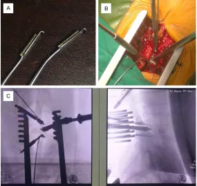
[image:2.612.89.374.72.341.2]Inclusion criteria: (1) Imaging results indicated monosegment LDH and unstable illnesses. Figure 1. A. The improved nerve retractor has prefabricated pipeline in
the surface of the nerve stripper or hook, and the kirschner wire can pass
through it to fix the retractor on the vertebral body. B. The intervertebral space was exposed by the fixed retractors by the side of two pedicle. C. It
showed the anteroposterior and lateral radiographs during operation.
Nerve root retractors are a necessary instrument in sur-gery of nucleus pulposus re- moval [7]. However, the old ver-sion of nerve root retractors, once commonly used in clinics, can cause nerve root injuries due to various reasons, includ-ing excessive traction [9, 10]. Previous studies have shown that improved nerve tractive methods may ideal in reducing nerve damage [7, 9, 11]. At present, widely used new-style nerve root retractors, such as the Love nerve root retractor, have not been recommended for clinical use due to their complex structure and poor clinical utility. Therefore, dis-covering a safe and effective method to retract nerve roots and expose disc space would be of great value.
This present study used an
improved nerve hook to fix
12335 Int J Clin Exp Med 2018;11(11):12333-12339 According to FryMoyer criteria [12], X slice of
anteflexion-rear protraction indicated shifting
more than 3 mm or adjacent vertebral body enlarged, L5S1 segment was larger than 20°, and other segments more than 15° were unstable; (2) Clinical features were serious lum-bocrural pain. Patients had obvious radiculopa-thy symptoms, no lumbar injury, or history of lumbar spine surgery. These patients were
cured and left the hospital via first regular oper -ation (such as lumbar surgery) or they needed operative treatment because they had poor therapeutic effects after half a year via conser-vative treatment; and (3) Patients were required to coordinate follow up scores according to the study design. Exclusion criteria: (1) Patients diagnosed as protrusion of LDH combined with lumbar spondylolisthesis and lumbar spinal stenosis; (2) Patients diagnosed with LDH com-bined with other serious internal medicine dis-eases and patients with poor physical quality;
and (3) Included subjects that could not finish
clinical research and follow up.
The trial was approved by the Shenzhen Baoan Hospital Ethics Committee, Guangdong, China.
Operative technique
All patients were given general anesthesia in
the prone position. Vertebral plates and zygop -ophysis of patients were exposed by separating paravertebral muscles in two sides via layer-by-layer cutting using a posterior midline approach based on the center of lesion segments. Four pedicle screws were implanted into interverte-bral space of verteinterverte-bral lesions. They were vali-dated under C-Arm X-medical equipment.
Vertebral plates were cut, narrow lateral recess
was enlarged, and dural sac and relevant nerve roots were exposed by stripping intervertebral discs from nerve roots and dural sac using nerve hooks. About 1.5 cm of nerve root was released to obtain suitable nerve root tension.
Posterior-lateral fibrous rings were exposed
after slightly opening the dural sac and nerve
roots. Knives were used to cut fibrous rings and
the incision indicated square. Nucleus pulpo-sus clamps were used to chop intervertebral tissues. Annular scrapers were used to chop cartilage plates from intervertebral discs and subchondral cortical bone was sustained and chopped to rough surface. In these processes, annular curets were ensured not to exceed to
forward fibrous rings, avoiding damage to blood
vessels and nephric ducts in front of the lumbar spine. Interbody fusion cages were used to
con-firm suitable type. Appropriate crushbones
were implanted into intervertebral discs and then interbody fusion with crushbones were implanted into intervertebral discs. The two sides were suppressed to approved depths using an embedder. The detailed cage position
was perspective identified and pedicle screws
were screwed into the centrum and pressurized
fixed. Finally, cases were further perspective identified to verify stable cages and no broken
crushbones or nucleus pulposus and other compressive things existing in the canalis spinalis.
During surgery, there were two methods to pull nerve roots and dural sacs to expose interver-tebral discs. In group A, traditional nerve retrac-tors including nerve hooks and nerve
dissec-tors were used to expose annulus fibrosus fibrocartilaginis intervertebralis by stretching
nerve roots and dural sacs to the spinal canal line. In group B, two improved nerve retractors were used to slightly pull nerve roots and dural
sacs. Next, two kirschner wires were fixed in
two vertebral bodies, respectively, via prefabri-cated pipelines in the end of nerve dissectors or hooks. Nerve roots and dural sacs were split with intervertebral discs. Terminus of kirschner
wires were fixed in an operation towel using
hemostatic forceps (Figure 1). Brain cottons were put between nerve retractors and nerve roots. Real tractive times were reduced as far as possible. Tractive times, intraoperative hem-orrhages, and operative times were detailed and recorded. After the operation, hormone, trophic nerve, and improved microcirculation drugs were given. Drainage tubes were removed if the amount of drainage was less than 50 mL/24 hours. Patients could resume ground activity after 3-5 days with protection in the waistline. After 12-14 days, patients stitches were removed. They began to train back mus-cles after 2 weeks. Waistline protection was worn for 1 month and aggravating activities were avoided for 3 months.
Clinical evaluation
Patients were required to provide background
of the spine. VAS and JOA scores were recorded before the operation and in the first month, 3rd
month, 6th month, and 12th month after the
operation.
Statistical analysis
SPSS 13.0 software was used to perform sta-tistical analysis. Tractive times, intraoperative
hemorrhages, and VAS and JOA scores after
the operation were analyzed using t-test. VAS and JOA scores before and after the operation
at different time points were analyzed using
AMOVA analysis. Inspection level α was set
0.05 and P < 0.05 is considered a statistically
significant difference.
compared. Compared with group A, group B had less tractive time, intraoperative hemor-rhages, and operative time (P < 0.05, Table 2). In the two groups, no obvious post-operative complications were found.
All patients were performed outpatient reviews
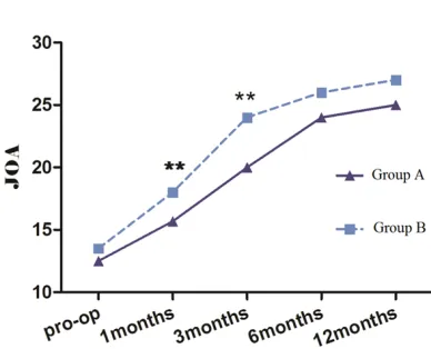
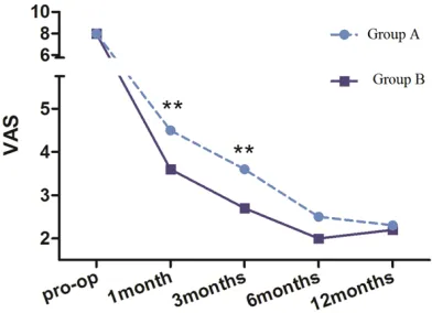
and finished follow ups via telephone or e-mail. Compared with group A, JOA and VAS scores were significantly improved in group B in the first and 3rd month after operation. However, in
the 6th month and 12th month, no significant
differences were detected. In every group, JOA and VAS scores after the operation were signifi -cantly changed (P < 0.05, Figures 2, 3).
Discussion
[image:4.612.90.325.96.190.2]Incidence of LDH has gradually increased with increasing population age. Strict conservative treatment can respite symptoms, but this does not satisfy patients because it does not respite the origin of the symptoms. Therefore, opera-tions are a necessary treatment method [2]. Since lamina resection of nucleus pulposus with excision, reported by Nagayama et al. [12], was successfully performed, it has been the most common surgical method in spine sur-gery. Most patients can be improved regarding nerve root pain using this surgery. However, the effects of surgery are not always perfect. Some patients may experience postoperative residu-al low back pain, numbness of lower limbs, and muscle weakness. This may be due to tractive
Table 1. General information about patients in the study
Indexes Group A Group B Age (Years old) 41±12 44±13 Gender (male/female) 23/17 25/15 Segmental lesions
L4-L5 19 23
L5-S1 21 17
[image:4.612.88.324.237.300.2]Course of disease (m) 66±9 63±12
Table 2. Comparison of tractive time, intraoperative hemorrhage, and operation time in the two groups
Indexes Group A Group B Tractive time of nerve root (min)* 11.45±1.56 9.05±2.07 Intraoperative hemorrhage (ml)* 425±45 376±43 Operation time (min)* 140±21 125±18 *: Group A compared with Group B, p < 0.05.
Results
Operations were successfully completed
in all patients and detailed operation pro-cesses were recorded. Serious complica-tions, such as nerve injury, large vascular injuries, embolisms, and perioperative death, were not detected. Subdural leak-age was found in 2 patients in group A and 1 patient in group B. The situation was covered with gelatin sponges. Drainage tubes were removed after 5-7 days. Drain entrances were sewn and notches were I-level healed.
Statistical analysis showed that the two
groups had no significant differences in
age and gender composition, distribution of lesion segments, and course of disease before the operation (P > 0.05, Table 1), indicating that the two groups could be
Figure 2. Comparison of pre- and post-operative JOA
[image:4.612.90.284.309.466.2]12337 Int J Clin Exp Med 2018;11(11):12333-12339 damage in nerve roots [13, 14]. Compared with
peripheral nerves, nerve roots lack protection of epineurium and beam membranes. Both strength and stiffness are lower than peripher-al nerves, therefore, nerve roots are prone to
be influenced by mechanical stretching [15, 16]. How to reduce tractive damage in nerve
roots has attracted much attention, including intraoperative nerve detection, intraoperative awakening experiment [17], and
redevelop-ment of nerve hooks [7, 11]. In this study, fixed
nerve retractors improved from the nerve
strip-pers and hooks were used to fix nerve roots to
expose intervertebral discs. This study aimed to discuss the potential operative value of these retractors in canalis spinalis.
In clinic, lumbar disc herniation with lumbar vertebrae unsteadiness is common, due to degeneration of lumbar intervertebral discs [18]. With more lumbar disc degeneration, the higher the incidence of lumbar instability will be [19]. Simple nucleus pulposus removal may generally aggravate instability of the lumber spine, further leading to long-term lower back pain [20]. Therefore, in treatment, effective de- compression and rebuilt stability of the spine are ideal operative methods of improving long-term effects [21]. In this study, patients with LDH and lumbar instability were involved. They were performed using PLIF to lumbar interbody fusion. The aim of this operation was to remove compression factors of nerve roots or dural sac factors, simultaneously stabilizing the spine. In PLIF surgery, it is necessary that the assis-tant pulls the nerve roots in epidural capsule to
the offside using nerve retractors to expose
intervertebral disc fiber rings, then dealing with
intervertebral space. Generally, hook indicates L type and front end indicates plate right hook.
It can be tractive in the surface of fibrous rings
and have tractive and protection roles in epi-dural sacs and nerve roots [7]. However, opera-tions in intervertebral space may be performed with nerve hooks, which may lead to movement of nerve hooks and damage nerves. Therefore, the assistant must keep their posture and con-tinuously pull the nerve hooks. It is very strenu-ous. The process may cause movement of nerve hooks and pulling of nerves, causing damage if the assistant feels arm numbness due to long-term maintenance of the same pos-ture. Addressing this problem, this study used two improved nerve retractors to replace the traditional hook. Kirschner wires were used to
fix them in two vertebral bodies by surface
rebuilt pipelines. Thus, nerve roots and dural sacs can be split with intervertebral discs. No additional pulling was needed, compared with
traditional the hook, as kirschner wires can fix
the responding positions after nerve epidural is
pulled to specific positions. The position of
hooks cannot be moved by human factors. The
operating position is exact and fixation is reli -able and secure. Moreover, this method can help assistants to concentrate on other tions, effective hemostatic, and reduce opera-tive times. This present study found that
improved nerve retractors can significantly
reduce tractive times, intraoperative hemor-rhages, and operative times.
Matsui et al. [22] reported that the degree of damage via nerve pull is related to tractive time and intensity. This conclusion was by studying relationships of nerve roots pull and neural symptoms after operations. Gentle operations and reduced tractive times may be crucial in reducing nerve damaging symptoms. However, Feltes et al. [23] found that, although larger nerve root pulling intensities were involved than that in Matsui’s study, follow up results
showed that no significant nerve damaging
symptoms were detected. Feltes et al. believed that the difference may be derived from the shorter time of pulling nerves [23]. Based on these, this study speculated that reducing trac-tive time will have a common role in improving nerve symptoms. This study showed that both
[image:5.612.91.287.72.214.2]VAS and JOA scores in group B were improved
Figure 3. Comparison of pre and post-operative VAS
in the first month and 3rd month after the
opera-tion, indicating that improved nerve retractors can reduce tractive times of nerve roots and contribute to nerve functional recovery by opti-mizing surgical processes. Interestingly, no
significant differences in these scores were detected in the 6th month and 12th month
between the two groups. This may be due to
the fact that regeneration of nerve fibers is always detected within 6 months after being
damaged [24].
Although this study showed the advantages of
using modified nerve root retractors, some limi -tations should be noted. For example, the tech-nique used in this study only applies to the PLIF operation, not the more widely used TLIF opera-tion. Also, whether it can be used in percutane-ous procedures that use tubular retraction sys-tems requires further investigation. In addition, the sample size in the present study was small. A further study with a larger cohort will be need-ed to further corroborate the present
observa-tions. The long-term benefits of modified retrac -tors require further study.
Compared with the traditional method, im-
proved fixed nerve retractors are more effec
-tive in treating LDH, thanks to significantly
reduced tractive times, intraoperative hemor-rhages, and operative times. These can pro-mote early recovery.
Acknowledgements
This study was supported by a Basic Research Project of Science and Innovation Commission
in Shenzhen (No. JCYJ20160427193559599)
and Basic Research Project of Science and Technology Planning of Baoan Region in
Shenzhen (No. 2016CX166).
Disclosure of conflict of interest
None.
Address correspondence to: Guangye Wang, De-
partment of Orthopedics, People’s Hospital of
Baoan District in Shenzhen, Shenzhen 518000,
China. Tel: +86 181 26270378; Fax: +86 755 27788311; E-mail: [email protected]
References
[1] Altun I and Yuksel KZ. Lumbar herniated disc: spontaneous regression. Korean J Pain 2017; 30: 44-50.
[2] Delgado-Lopez PD, Rodriguez-Salazar A, Mar-
tin-Alonso J and Martin-Velasco V. Lumbar disc
herniation: natural history, role of physical ex-amination, timing of surgery, treatment
op-tions and conflicts of interests. Neurocirugia
(Astur) 2017; 28: 124-134.
[3] Cahill KS, Chi JH, Groff MW, Mcguire K,
Afendulis CC and Claus EB. Outcomes for sin
-gle-level lumbar fusion. Spine 2011; 36: 2354-2362.
[4] Kalichman L and Hunter DJ. The genetics of intervertebral disc degeneration. Familial pre-disposition and heritability estimation. Joint Bone Spine 2007; 75: 383-387.
[5] Taylor RS, Ryan J, O’Donnell R, Eldabe S,
Kumar K and North RB. The cost-effectiveness of spinal cord stimulation in the treatment of failed back surgery syndrome. Clin J Pain
2010; 26: 463-469.
[6] Juricek M, Rehak L, Tisovsky P and Horvath J. The effect of complications on the quality of life after surgery for lumbar spine
degenera-tive disease. Acta Chir Orthop Traumatol Cech
2010; 77: 112-117.
[7] Cui ZM, Bao GF, Li WD, Xu GH and Zhang JB. Application of self-made nerve root r etr actor in poster ior lumbar interbody fusion. Zhongguo Zuzhi Gongcheng Yanjiu Yu Linchuang Kangfu
2007; 11: 7044-7046.
[8] Krishna M, Pollock RD and Bhatia C. Incidence,
etiology, classification, and management of
neuralgia after posterior lumbar interbody
fu-sion surgery in 226 patients. Spine Journal
2008; 8: 374-379.
[9] Feltes C, Fountas K, Davydov R, Dimopoulos V
and Robinson JS Jr. Effects of nerve root re-traction in lumbar discectomy. Neurosurg
Focus 2002; 13: E6.
[10] Nagayama R, Nakamura H, Yamano Y, Yamamoto T, Minato Y, Seki M and Konishi S. An experimental study of the effects of nerve root retraction on the posterior ramus. Spine
(Phila Pa 1976) 2000; 25: 418-424.
[11] Li Y, Zhang X, Kong Z, Huang W and Wang G. Kirschner wire is more effective than the nerve root retractor in treating patients with disc her-niation. Clin Neurol Neurosurg 2015; 139: 51-55.
[12] Frymoyer JW. Low back pain. The role of spine fusion. Neurosurg Clin N Am 1991; 2: 933-954.
[13] Hayashi N, Tamaki T and Yamada H. Experi- mental study of denervated muscle atrophy following severance of posterior rami of the
lumbar spinal nerves. Spine 1992; 17: 1361.
[14] Paajanen H, Erkintalo M, Parkkola R, Salminen J and Kormano M. Age-dependent correlation of low-back pain and lumbar disc
12339 Int J Clin Exp Med 2018;11(11):12333-12339
[15] Beel JA, Stodieck LS and Luttges MW. Struc- tural properties of spinal nerve roots:
biome-chanics. Exp Neurol 1986; 91: 30-40.
[16] Stodieck LS, Beel JA and Luttges MW. Struc- tural properties of spinal nerve roots: protein
composition. Exp Neurol 1986; 91: 41-51.
[17] Bindal RK and Ghosh S. Intraoperative electro-myography monitoring in minimally invasive transforaminal lumbar interbody fusion. J
Neurosurg Spine 2007; 6: 126-132.
[18] Fujiwara A, Tamai K, An HS, Kurihashi T, Lim TH, Yoshida H and Saotome K. The relation-ship between disc degeneration, facet joint osteoarthritis, and stability of the degenerative lumbar spine. J Spinal Disord 2000; 13: 444-450.
[19] Hao-Peng L, Yang BH and Zhang GA. The clini-cal research in correlation factors of degenera-tive lumbar instability. Journal of Practical
Orthopaedics 2009; 5: 339-341.
[20] Siepe CJ, Heider F, Haas E, Hitzl W, Szeimies U, Stäbler A, Weiler C, Nerlich AG and Mayer
MH. Influence of lumbar intervertebral disc
degeneration on the outcome of total lumbar disc replacement: a prospective clinical, histo-logical, X-ray and MRI investigation. Eur Spine J 2012; 21: 2287-2299.
[21] Ito Z, Imagama S, Kanemura T, Hachiya Y, Miura Y, Kamiya M, Yukawa Y, Sakai Y, Katayama Y, Wakao N, Matsuyama Y and Ishiguro N. Bone union rate with autologous iliac bone versus local bone graft in posterior lumbar interbody fusion (PLIF): a multicenter
study. Eur Spine J 2013; 22: 1158-1163.
[22] Matsui H, Kitagawa H, Kawaguchi Y and Tsuji H. Physiologic changes of nerve root during posterior lumbar discectomy. Spine 1995; 20:
654-659.
[23] Feltes C, Fountas K, Davydov R, Dimopoulos
V and Robinson JS Jr. Effects of nerve root
retraction in lumbar discectomy. Neurosurg
Focus 2002; 13: E6.