MSc thesis in Technical Medicine
Electrical brain responses during
processing of nociceptive stimuli
around the detection threshold
:
an explorative study in pain-free subjects and failed back surgery syndrome patients
Tom Berfelo
Technical Medicine
University of Twente
Enschede, The Netherlands
Electrical brain responses during processing of
nociceptive stimuli around the detection
threshold:
an explorative study in pain-free subjects and failed back surgery syndrome patients
A dissertation submitted to the University of Twente for the degree of
Master of Science in Technical Medicine
by
Tom Berfelo
7 February 2019
Graduation committee:
Prof. dr. ir. P.H. Veltink (chairman)
Acknowledgements
When I started working on my thesis, my experience with the high-tech experiment for monitoring nociceptive processing was limited to participation in an earlier study about this technique by Boudewijn at the University of Twente. From that moment I was enthusiastic about the possibilities for giving new insights into the neurophysiologic mechanisms in human. Fortunately, I was asked to continue his work to the next step by applying this technique in medicine, knowing that I was to face significant challenges. Apart from being familiar with the technique, I had to prepare the process in detail, including writing a study protocol approved by the medical ethical research committee and leading the study project in St. Antonius Hospital. On the way, I have learned to develop academic skills, but also gained more experience in clinical skills by spending a lot of time in Operation Room. The many possibilities offered by the St. Antonius Hospital was huge. I grew in the role of the technical physician, which is all because I was driven by passionate people around me this year.
First of all, Boudewijn, I would like to thank you for being the hero who helped me with technical and intellectual advises about the experiment. I have learned a lot from you because your abstract ideas were always inspiring me. Additionally, I would like to thank the Nociceptive and Somatosensory Processing team for the meetings and discussions about this Nocicept-project.
Jan, your relentless spirit for innovation and technology helped me to get energy from research. I had never expected that I would delve into this direction. Thank you for facilitating this position at the Nocicept project and being critical and open-minded during the meetings. These efforts contributed technically the details in this project which are made.
During translocating the experiment from the university lab to the hospital environment, I was happy to collaborate with enthusiastic people. In general, everyone was open to innovation. Lieke and Nynke, it was my pleasure to work with you at St. Antonius Hospital. Thank you for having fun during the lunches, walks and coffee breaks. You were motivating me to work hard and still walk 10.000 steps every day. In addition, I would like to thank all the colleagues of St. Antonius Hospital. Your (personal) accessibility and help for participating in my study were great. Without you, I had never obtained all these data. This means the same for the pain specialists who enabled me to recruit chronic pain patients. It was instructive to work with these patients. Moreover, I would like to thank all the courageous pain patients who participated in the study, despite the pain was sometimes unbearable. Without you, we would never achieve the development of this future diagnostic with clinical relevance.
Rian, thank you for supervising the process during the last two years. You have pushed me to develop reflection skills, which helped me to keep control of priorities in my own work. I grew fascinated by asking myself every time: What do you want for yourself? Additionally, I would like to thank Bernice and Tessa for their input during the reflection meetings. I think it was useful to discuss our competencies and ambitions.
Thanks to all my friends who contributed to having fun in my spare time. I liked the (winning) squash games, soccer training evenings and city trips in the weekends. Also, I would like to thank the friends who enjoyed drinking a beer or them who just wanted to talk with some coffee.
Acronyms
ACC anterior cingulate cortex
AIC anterior insular cortex
Ag/AgCl silver chloride
BMI body mass index
BPI brief pain inventory
BSS biomedical signal and systems
CNS central nervous system
CRPS complex regional pain syndrome
CSI central sensitivity inventory
D stimulus detection
DN4 Douleur neuropathic 4,
EEG electroencephalography
EP evoked potential
eQST electrical quantitative sensory testing
FBSS failed back surgery syndrome
GCT gate control theory
GLMM generalized linear mixed model
IASP international association for the study of pain
IES intra-epidermal electrocutaneous stimulation
IPI inter-pulse-interval
LMM linear mixed model
MTT multiple threshold tracking
MTT-EP multiple threshold tracking – evoked potential
N2 second negative evoked potential component
NDT nociceptive detection threshold
NMDA N-methyl-D-aspartate
NoP number of pulses
NPQ neurophysiology of pain questionnaire
NRS numeric rating scale
P1 first positive evoked potential component
PAG periaqueductal gray
PFS pain-free subjects
pQST pressure quantitative sensory testing
PW pulse width
QST quantitative sensory testing
S1 primary somatosensory cortex
S2 secondary somatosensory cortex
SD standard deviation
SNR signal-to-noise-ratio
SP1 single-pulse stimuli
SP2_10 double-pulse stimuli with 10 ms inter-pulse-interval
SP2_40 double-pulse stimuli with 40 ms inter-pulse-interval
StA St. Antonius hospital
TENS transcutaneous electrical nerve stimulation
TRL number of trials/number of received stimuli
UT University of Twente
VAS visual analog score
Abstract
Multiple threshold tracking (MTT) has been shown to be effective in measuring the effect of stimulus parameters on stimulus detection. In addition, the evoked potential (EP) has been shown to reflect neurophysiological activity related to stimulus processing. Therefore, a combination of both techniques, known as the MTT-EP experiment, is a promising diagnostic method which may provide objective insight into the processing of nociceptive stimuli. Stimulus-related EPs were recently investigated using the MTT-EP experiment in pain-free subjects at the University of Twente, but its applicability has not been explored yet in a hospital environment and in chronic pain patients.
Firstly, therefore, we explored the replicability of the MTT-EP experiment in twenty pain-free subjects at St. Antonius Hospital. Secondly, we observed the neurophysiological responses during processing of nociceptive stimuli around the detection threshold in seven failed back surgery syndrome (FBSS) patients.
Results show that (initial) NDTs and EPs present profiles and phenomena (such as habituation and paired-pulse facilitation), which are in line with results from the University of Twente. Also, it is seen that the EP is rather modulated by stimulus detection, amplitudes and the number of received stimuli. Strikingly, we found higher NDTs in FBSS patients, in whom we assumed they suffered from a central sensitization syndrome (CSI-score = 49.0), comparing to results of pain-free subjects (CSI-score = 14.6). These NDTs in FBSS patients may implicate that additional facilitating effects occurred in the central nervous system. However, the influence of analgesics is uncertain. Additionally, an early phase component of the EP was found at CPz-A1A2 for detected stimuli, which might indicate that it can be a potential biomarker of brain processing in FBSS patients.
Table of contents
ACKNOWLEDGEMENTS ... VI
ACRONYMS ... VIII
ABSTRACT ... X
1 INTRODUCTION ... 1
1.1 PROBLEM STATEMENT ... 1
1.2 RESEARCH OBJECTIVE ... 2
1.3 THESIS OUTLINE ... 2
2 BACKGROUND ... 3
2.1 PAIN ... 3
2.2 PATHOPHYSIOLOGY OF PAIN ... 6
2.3 OBSERVATION OF NOCICEPTIVE PROCESSING... 7
2.4 ELECTRO-ENCEPHALOGRAPHY ... 10
2.5 MTT-EP EXPERIMENT ... 12
2.6 IMPLICATIONS ... 14
3 METHODS ... 17
3.1 SUBJECTS ... 17
3.2 DESIGN ... 17
3.3 MATERIALS AND METHODS OF MEASUREMENT... 18
3.4 DATA ANALYSIS ... 20
3.5 REPLICABILITY IN PAIN-FREE SUBJECTS ... 21
3.6 BEHAVIOR OF NEUROPHYSIOLOGICAL EFFECTS IN FBSS PATIENTS ... 21
4 RESULTS ... 23
4.1 SUBJECT CHARACTERISTICS ... 23
4.2 REPLICABILITY IN PAIN-FREE SUBJECTS ... 24
4.2 BEHAVIOR OF NEUROPHYSIOLOGICAL EFFECTS IN FBSS PATIENTS ... 26
5 DISCUSSION ... 28
5.1 NDTS AND EPS ... 29
5.2 BEHAVIOR OF NEUROPHYSIOLOGICAL EFFECTS ... 30
5.3 DETECTED STIMULI RELATED TO EVOKED POTENTIAL ... 31
5.4 ALTERED BEHAVIOR OF NEUROPHYSIOLOGICAL EFFECTS RELATED TO CENTRAL SENSITIZATION ... 31
5.4 INTERPRETATION IN TERMS OF A FACILITATED CONDITIONED PAIN MODULATION ... 33
5.5 STRENGTHS AND LIMITATIONS ... 34
6 CONCLUSION ... 37
7 RECOMMENDATIONS ... 39
7.1 SHORT-TERM RECOMMENDATIONS ... 39
7.2 FUTURE PERSPECTIVES ... 39
REFERENCES ... 43
APPENDIX ... 47
A1 AVERAGE NDTS AND SD ... 47
A2 NDTS OF PAIN-FREE SUBJECTS ... 48
A3 NDTS OF FBSS ... 48
1 Introduction
1.1 Problem statementChronic pain is a health issue with a dramatic impact on European society. Since the pathophysiology of malfunctioning nociceptive systems is still poorly understood, roughly one out of five adults suffer from moderate-to-severe non-cancer chronic pain in Europe1. Moreover, more than two million people in the Netherlands continue to suffer from chronic pain. Besides the fact that chronic pain has an impact on patient-perceived health status, such as significantly affected everyday activities, personal relationships and depressive symptoms, it also entails a significant economic burden on society. Annual costs are about 20 billion euros and. Notably, 40% of chronic pain patients do not receive adequate treatment for their pain2. This emphasizes the importance of improving treatments for chronic pain syndromes. Current therapies are mostly based on symptom spreading and are made to provide pain relief. However, this approach is rarely successful3. More important, there is a lack of reliable methods monitoring nociceptive processing which can explain the nature of chronic pain disorders objectively. Conventional pain monitoring is based on subjective pain reports, analgesic intakes or questionnaires (e.g. DN4, NPQ or CSI). These methods are limited to clinical diagnostics because changes in neuroplasticity are not recognized. Early detection of maladaptive mechanisms in the nociceptive system should decrease the inadequate treatments. Consequently, identification of nociceptive system properties enables mechanism-based monitoring of chronic (low back) pain disorders.
For this reason, we need to investigate possibilities to improve prevention, diagnosis, and treatment of chronic pain. How and why chronic pain is caused and maintained is often unclear. Therefore, it is important to study the underlying mechanisms (central and peripheral), and how they are altered in chronic pain patients compared to healthy subjects. Clinical studies reveal that a changed sensitivity can be interpreted as an important contribution of central sensitization to chronic pain patients4. Exploring objective biomarkers for central sensitization will be extremely helpful. One major obstacle is the lack of an objective measure of central and peripheral sensitization. Recently, a method for measuring nociceptive detection thresholds (NDTs) has been developed. These are stimulus amplitude thresholds for a detectable sensation, which use intra-epidermal electrocutaneous stimulation (IES) of the skin. IES preferentially activates nociceptive nerve fibers in the superficial skin (pin-prick sensation at detection level) without initial activation of tactile nerve fibers (a non-painful sensation at detection level). Therefore, IES can be used to estimate pain sensitivity measuring the NDT. The NDTs can be determined using a multiple threshold tracking (MTT) algorithm, which was developed in earlier studies5,6. Tracking NDTs can facilitate the investigation of the underlying mechanisms
A possible objective measure of nociception related activity in the central nervous system (CNS) is electroencephalography (EEG). Multiple-trial averages of this EEG signal, referred to as evoked potentials (EPs), have been shown to be sensitive to changes in stimulus parameters such as the number of pulses9,10 or number of trials11. Firstly, MTT has been shown to be effective in measuring the effect of stimulus parameters on stimulus detection. Secondly, the EP has been shown to reflect neurophysiological activity related to stimulus processing. Therefore, a combination of both techniques, known as the MTT-EP experiment, might provide insight into the relationship between neurophysiological activity and nociceptive stimuli. Recently, this relationship has been investigated in a study from the Biomedical Signals and Systems (BSS) research group at the University of Twente. That study showed that components of the EP were closely related to the stimulus detection and stimulus amplitudes12. The next step is to investigate the applicability of the MTT-EP experiment in a hospital. We need to explore if these results are a replication of results from university lab and observe if the experiment is feasible to be performed by patients.
1.2 Research objective
Therefore, the aim of this explorative study is (1) to explore whether results of the MTT-EP experiment in pain-free subjects at St. Antonius hospital are a replication of results in pain-free subjects at the University of Twente and (2) to observe neurophysiological responses during processing of nociceptive stimuli around the detection threshold in chronic pain patients.
1.3 Thesis outline
2 Background
2.1 PainPain is defined as ‘An unpleasant sensory and emotional experience associated with actual or potential tissue damage, or described in terms of such damage’, according to the International Association for the Study of Pain (IASP)13. It plays an important role in normal defense mechanisms, warning of potentially damaging environment actions and initiating behavioral strategies14. Pain is always subjective and is best regarded as an experience involving both a physiologic sensation and an emotional reaction to sensation13,14.
Pain can be categorized into nociceptive, neuropathic and mixed pain. Nociceptive pain is pain arising by activation of specialized peripheral sensory receptors (nociceptors)15. Neuropathic pain is pain initiated by a direct consequence of a lesion or disease affecting the somatosensory system15. If both nociceptive and neuropathic pain occur in the same patient, it is known as mixed pain.
2.1.1 Nociceptive processing of pain
Nociception is a primary physiologic mechanism of pain, which consists of the process of transduction, transmission, central modulation and perception (Figure 1)14. Processing of pain
[image:17.595.211.384.503.719.2]signals is a complex process in the nociceptive system, which is roughly regulated by ascending and descending pathways. The route of signals from peripheral nociceptors through the spinal cord going up to the brain are referred to as the ascending pathway. Neurons of the descending pathway are connected from the brain stem through the spinal cord to the dorsal horn. While the ascending pathway is responsible for transmitting the pain signal up to the brain, the descending pathway is responsible for controlling and inhibiting the ascending pathway essentially.
Peripheral processing of nociceptors
The first process of nociception is transduction by which noxious thermal, mechanical or chemical stimuli are selectively converted to electrical signals in the nociceptors. The specialized free nerve endings reside mostly in the epidermis and function to protect tissue to injury. Nociceptor stimulus thresholds have a relatively high threshold of activation. Pain receptors can be activated by several neurotransmitters (acetylcholine, serotonin, histamine, bradykinin, substance P, cholecystokinin, adenosine, glutamate, bombesin, neuropeptide-Y, prostaglandin E and endogenous opioids) and other influences (acidity and temperature). Activation of afferent nociceptors results in generating action potentials transmitting to their synapses in the dorsal horn. This second process is also called a transmission of information. Nociceptors are responsible for transmission from the periphery to the spinal cord. These first-order neurons are classified into unmyelinated C-fibers and finely myelinated Að-fibers, with small diameter axons (2 – 5 µm and < 5 µm, respectively)17,18. C-fibers have a conduction
velocity of 0.5 to 2 m/s, which characterize as a slow, diffuse, dull and aching pain sensation. Að- fibers have a conduction velocity of 5 to 15 m/s and show a rapid, pricking and well-localized pain sensation18.
Dorsal horn neurons
Cell bodies of both afferent nociceptive fibers are included in the dorsal root ganglia, which contain connections to synapses with dorsal horn neurons in the grey matter in the dorsal horn. The dorsal horn is a complex relay station, which can be seen as the first decision point. Once the nociceptive signal arrives the dorsal horn of the spinal cord, transduction of the first-order neuron to the second-order neuron takes place by neurochemistry. Several neurotransmitters are involved in this process, such as cholecystokinin, substance P, glutamate and γ-Hydroxybutyric acid (GHB). The N-methyl-D-aspartate (NMDA) receptor plays an important role in transduction as well18.
[image:18.595.380.525.503.673.2]The dorsal horn is divided into several laminae with multiple interconnections (Figure 2). C-fibers terminate in lamina I and II (substantia gelatinosa of Rolando), whereas Að-fibers terminate in lamina I and V and non-nociceptive Aβ-fibers terminate in lamina III to V17–19. Dorsal horn neurons are either classified as nociceptive-specific neurons, wide dynamic range (WDR) neurons, low threshold mechanic (LTM) neurons or interneurons. Nociceptive-specific neurons synapse with Að- and C-fibers in lamina I, WDR neurons synapse with Að- and fibers in lamina V. LTM neurons synapse with (tactile) Aβ-fibers in laminae IV. Interneurons are situated in and connected with all laminae and receive input from afferent fibers, as well as from descending pathways. Interneurons can be subdivided into exciting and inhibiting interneurons, which influence e.g. other interneurons (processing) or ascending neurons (sensation)20. Descending fibers terminate in (several laminae of) the dorsal horn, which modulate pain signals by inhibition21.
Central modulation
Subsequently, the nociceptive process is continued by central modulation. The dorsal horn is an important area in the CNS, which can be determined as a gate control system (GCT)22. This
GCT, described by Melzack and Wall in 1965, supposes that a network of nociceptive neurons in the dorsal horn can modulate sensory input and therefore influences the transmission of pain signals to the brain. They describe in a simplistic view that activation of inhibitory interneurons in the substantia gelatinosa can be caused by stimulation of non-painful large afferents (Aβ-fibers) that would suppress transmission in small afferents (C-fiber)18. This mechanism could describe why rubbing the painful area decreases pain.
Ascending spinal tracts
The spinothalamic tract is the main second-order neuron which is responsible for carrying the nociceptive signals for pain and temperature to higher centers of the brain (Figure 1). This tract is located in the anterolateral white matter of the spinal cord18. The spinothalamic tract consists
of a lateral (neospinothalamic) and medial (paleospinothalamic) tract for fast and slow pain, respectively.
Supraspinal centers
Supraspinal centers are reached using parallel distributed systems. One of the centers is the reticular formation consisting of complex core groups at the brainstem. This area is important for pain experience, as nociceptive input has a deep effect on reticular activity. It plays a role in consciousness and arousal, autonomic functions and pain suppression via the periaqueductal gray (PAG) matter of the midbrain. Besides pain suppression via descending modulatory tracts, PAG delivers ascending projections to the hypothalamus and thalamus. The thalamus is the key area for nociceptive processing, which serves as relay point. The tracts terminate in their respective lateral and medial located thalamic nuclei and from here the neurons project to regions of the cerebral cortex14,18,23. The amygdala is an important part of the limbic system. Also, the hippocampus, septal nuclei, preoptic region, hypothalamus, and some thalamic parts belong to limbic structures. The limbic system supports functions including emotion and motivation, which determine purposeful behavior14.
Cerebral cortex
The primary somatosensory cortex (S1) has a prominent and highly modulated role for perception of pain24. Also, the secondary somatosensory cortex (S2), insula, orbitofrontal cortex, dorsal-lateral prefrontal cortex, extended amygdala, and cingulate cortex activate by painful stimuli14,18,23. The S1 is build up by somatotopic organization following Penfield’s homunculus pattern23. Note, that activation of the cortical area is related to the origin of the peripheral nociceptive stimulus location.
Descending modulatory pathways
medullary reticular formation, locus coeruleus, and raphe nuclei. Serotonin and noradrenaline are key neurotransmitters involved in descending inhibition. Descending tracts can be activated by endogenous opioid peptides, such as enkephalins and β-endorphins. Activation of these tracts results in a decreased release of substance P, and therefore inhibition of pain signal transmission.
2.2 Pathophysiology of pain
2.2.1 Chronic pain
Chronic pain is described as that pain that persists beyond the normal healing13,26. It is characterized by an enhanced perception of pain to a nociceptive stimulus (hyperalgesia) and the novel perception of a normally innocuous stimulus as being painful (allodynia)16. Chronic pain is a pathophysiological function of the peripheral and/or central sensory pathways, which results in an altered sensitization. The exact pathophysiology underlying chronic pain problems are largely unknown. The prevalence of chronic pain depends on when, where and how it is measured. Three months are often taken as the point beyond the normal healing. The back is the commonest location of chronic pain. A large survey of chronic pain in Europe shows that nineteen percent of the responders suffer from pain for more than six months2. The impact of chronic pain is often determined by extent and duration. It is correlated with poor (psycho)physical and social aspects of health. General factors associated with chronic pain are female gender, increasing age, acute uncontrolled pain and deprivation of household income, education and cultural- and geographical properties27.
2.2.2 Central sensitization
[image:20.595.179.413.605.754.2]Since the early 1980s, it was discovered that central sensitization plays a role in chronic pain. After decades of research, Latremoliere and Woolf defined central sensitization as ‘an enhancement in the function of neurons and circuits in nociceptive pathways caused by increases in membrane excitability and synaptic efficacy as well as to reduced inhibition and is a manifestation of the remarkable plasticity of the somatosensory nervous system in response to activity, inflammation, and neural injury’28. In other words, central sensitization is use-dependent plasticity of neural signaling within the CNS that is associated with development and maintenance of chronic pain4,29 (Figure 3). It is suggested that the central sensitization is altered in chronic pain patients, such as FBSS patients4.
2.2.3 Failed back surgery syndrome
Chronic low back pain is a socioeconomic burden with a prevalence in general adult population of 37% and a lifetime prevalence of between 60% and 80%30,31. Increasing rates of spine
surgeries have increased the number of patients suffering from FBSS. FBSS is a diagnosis that describes persistent or recurrent low back pain following spine surgery31,32 and has a prevalence of 10-40%33. Patients suffering from FBSS are supposed to have an altered central sensitization forced by constant stimulation of nociceptive circuits3,4,33–36. Recent literature shows that the sympathetic nervous system is overstimulated in FBSS patients37. This may be a contributing factor in maintenance of pain. Although the complex pathophysiology is poorly understood, it involves both nociceptive and neuropathic factors38. One of the mechanisms involved is the abnormal ectopic activity in neurons, caused by disturbed expression and distribution of ion channels3,39. Another change is the loss of inhibitory mechanisms, resulting in increased activity of second- and third-order neurons3.
Many of these patients are seen by pain specialists every year at the outpatient pain clinic of St. Antonius Hospital. Symptoms of (low) back pain radiating to the leg(s) are common for FBSS patients. They are often treated by strong opioids, such as morphine and pregabalin, because other interventions and pharmacologic strategies have failed. Since FBSS is a complex condition, it is difficult to treat. These patients are often suffering multiple years from pain. Some of them are eligible for neuromodulation, which is seen as a last-resort treatment. However, this does not work always effective for everyone. In practice, there are about 60 FBSS patients out of 120 chronic pain patients scheduled for neuromodulation each year, of which not all of them are new because these interventions include also battery replacements or retreatments due to broken leads or infections. Nevertheless, the waiting list is between three and six months. Previous experience at St. Antonius Hospital shows that this category is very helpful to contribute to clinical research. These FBSS patients have tried all the possible treatments and understand how important it is to investigate the underlying pain mechanisms.
2.3 Observation of nociceptive processing
To understand the pain mechanisms, quantification of pain perception is needed. Unfortunately, there is no ‘golden standard’ available for determination of pain. For clinical application, pain perception is currently often observed by visual analog scores (VAS) and numeric rating scales (NRS). However, it is hard to quantify exactly pain perception, because of the subjective nature. Questionnaires are often used for clinical application. For example, the Brief Pain Inventory (BPI) is a pain questionnaire used to evaluate the severity of pain and the impact on daily life.
2.3.1 Psychophysical methods
While questionnaires are useful, psychophysical methods provide more objective information about nociceptive processing of pain. Quantitative sensory testing (QST) is an upcoming method for quantifying changes in somatosensory neural function. This psychophysical method can be subdivided into pressure QST (pQST), electrical QST (eQST) and nociceptive detection threshold (NDT) measurements. Each method is based on stimulation of specific peripheral nerve fibers. However, NDT measurements are mainly usual for quantifying nociceptive pain processing in clinical research. NDT experiments are used in combination with IES of the skin, in case of nociceptive nerve fiber stimulation.
2.3.2 Stimulation of nociceptive pathways
Intra-epidermal electrical stimulus electrode
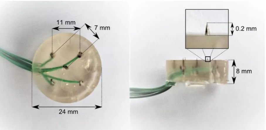
Nociceptive fibers can be activated by IES applied using an IES-5 electrode, which contains an array of 5 micro-needles (Figure 4)41. These electrodes protrude only 0.2 mm through the stratum
[image:22.595.73.527.484.706.2]corneum of the skin. The electrodes do not penetrate the epidermis and are therefore considered non-invasive. Such a superficial intrusion in the epidermis permits specific activation of superficial (Aδ) nociceptive skin fibers, which has been shown by Inui et al.42,43 and confirmed independently by Mouraux et al.44. Making use of this specificity, Steenbergen et al. have been using BiModEl electrodes (similar to the IES-5 and also produced at the University of Twente), to study the somatosensory topography of Aδ fibers in human subjects45–47. Similarly, Doll et al. have been using the IES-5 electrodes to characterize peripheral and central changes of the nociceptive system with respect to stimulus parameters6,7. Also, van den Berg et al. have been using the IES-5 electrodes for analyzing stimulus-related evoked potentials around the nociceptive detection threshold. The IES-5 electrode is a medical accessory of the stimulator, which supplies stimuli to the electrodes.
Stimulator
The stimulator is an AmbuStim 1-channel stimulator, developed and thoroughly tested by the BSS group at the University of Twente. A desktop computer running a custom computer program written in LabVIEW 2013, SP1 controls all stimulation procedures and registers the applied stimulus amplitudes (in mA) and their trigger codes, the responses to stimuli, and the stimulus times in milliseconds. In addition to registering stimulus and threshold data, all communication between software and stimulator is logged.
2.3.3 Psychometric curve
The psychometric curve (Figure 5,) was estimated by the stimulus-response pairs from the experiment, which was based on the psychometric function. The psychometric function is a constructive model applied for psychophysical data. It explains the relationship between electrical stimuli and the responses of the subject expressed in detection probability. This model provides information about detection thresholds related to different stimulus settings.
Figure 5. A). Detected (closed marker) and undetected (open marker) stimuli can be shown for each stimulus setting, when the stimulus was perceived or unperceived, respectively. B). The psychometric curve was estimated by the stimulus-response pairs from the experiment. Adapted from Doll et al. (2016).
2.3.4 Multiple threshold tracking
[image:23.595.78.518.314.483.2]2.3.5 Nociceptive detection threshold
NDT values (Figure 6) can be determined using the MTT paradigm by detecting the subject’s response (detected or not detected) to multiple stimuli with different amplitudes and subsequently derived using the psychometric function. The stimulus amplitude with a detection probability of 0.5 is generally used as NDT value in clinical research. Values of the NDT estimate the degree of pain perception and is a psychophysical parameter reflecting nociceptive processing.
2.4 Electro-encephalography
2.4.1 Physiology
The physiology of the EEG is based on voltage differences across cell membranes. Neurons and myocytes are specialized to generate rapidly voltage differences by opening and closing ion channels. Neurons have a resting membrane potential between approximately -50 and -70 mV. This is caused by concentration gradients of sodium, potassium, and calcium across cell membranes, and semipermeable membrane properties. Communication between neurons is mainly driven by chemical synapses, in which neurotransmitters interact with ionotropic receptors. Neurons are excitable cells, in which the membrane potential can be modified by activity of voltage-dependent ion channels or interactions of neurotransmitters. In case of sufficient voltage change, an action potential can be generated. Electrical rhythms occur in multiple spatial scales by neuronal interactions. The EEG reflects mainly activity of cortical pyramidal cells, because of countless partly synchronous excitatory and inhibitory postsynaptic currents that give rise to voltage differences between 10 and 100 µV on the skull49.
Electrical activity of the EEG signal can be determined by an electrical dipole consisting of three characteristics: (1) the dipole is localized at a specific location in the brain, (2) has a specific size and (3) has a specific direction, which is given in a three-dimensional space50. A
[image:24.595.144.449.193.365.2]dipole can be described by a potential field, which overviews the temporary field strengths. These potential fields can be determined by analyzing the derivations of the EEG. Derivation of the EEG signal is characterized by a difference in measurement of bio-electricity. Voltage differences between channels describe a potential field.
Figure 7. Grand average evoked potential from 12 pain-free subjects at the University of Twente. The evoked potential was derived at CPz-A1A2 (with a band-pass filter of 0.1 Hz to 40 Hz) in response to a nociceptive stimulus. Adapted from B. van den Berg (2018)12.
2.4.2 Evoked potentials
An evoked potential (EP, Figure 7) is defined as a time-locked neurophysiological signal in response to a stimulus of peripheral nerve fibers. EPs are objectively physiological markers derived from EEG signals, which can be used for clinical purposes (sensor and motor systems). EPs can be characterized by waveform amplitude (µV) and latency (ms)51. The EP is a
psychophysical parameter reflecting nociceptive processing. Each peak (positive or negative) is given a letter (P or N) and a number (1, 2, 3, etc.) in its name. The number describes the order of the peak. For example, P1 is the first positive wave and N2 is the second negative wave. Another way to describe peaks in the EP is by its latency. For example, P100 is a positive wave at a latency of about 100 milliseconds and N100 is a negative wave. Peak to peak and interpeak changes can reflect clinically relevant characteristics.
2.5 MTT-EP experiment
EPs can be registered using an EEG recording system. Earlier studies from the University of Twente used the MTT-EP experiment to measure nociceptive processing during electrical stimulation12,56. The MTT-EP set-up was created by Schooneman et al. and adapted by B. van den Berg12,56, which combines nociceptive stimulation with EEG registration. The experiment uses the procedure of the MTT set-up developed by Doll et al. to perform MTT5.
2.5.1 Experiment set-up
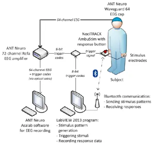
[image:26.595.139.454.390.688.2]The subject sits in a chair and manually controls the NociTrack stimulator (Figure 8). The stimulator is connected by cables with the cathodic IES-5 stimulus electrode and anodic transcutaneous electrical nerve stimulation (TENS) electrode on the subject’s hand. Using Bluetooth, the stimulator receives information from a dedicated laptop running software for stimulus pattern generation and triggering stimuli. Thereafter, the stimulator sends the recording response data back to the computer. The NociTrack is connected by a cable with the EEG amplifier to send trigger codes. At the same time, the scalp EEG records continuously cortical activity using an ANT Neuro Waveguard 64 EEG cap containing 64 Ag/AgCl electrodes. This 64 channel EEG is connected with an ANT Neuro 72-channel Refa EEG amplifier. Subsequently, this amplifier is connected by optical fibers to communicate with a dedicated laptop containing software for EEG recording.
2.5.2 Data analysis
Nociceptive detection probabilities and thresholds estimated the average NDTs and probability curve by including all data in a generalized linear mixed model (GLMM). This statistical model analyzed successfully average thresholds for this longitudinal data set, which was researched by B. van den Berg in previous research.
Due to bad signal-to-noise-ratio (SNR) of the EP, averaging does not provide noise reduction. Van den Berg studied that linear mixed models (LMM) can improve the analysis of EPs during MTT. LMM is useful because the longitudinal data from MTT-EP experiments consists of repeated-measurements which is clustered within subjects.
LMM can be used to estimate relationships within data. This is useful in a design with multiple measures per subject. Linear and random structures are formulated in an LMM, which ensures that all data can be used in a single regression. Fixed effects are parameters that do not vary. Random effects, also known as stochastic part of the model, are parameters which cannot be controlled experimentally. Using a complex mathematical computation based on linear regression, it is possible to estimate the outcome variable and fixed effects.
EP is the outcome variable of the LMM in the MTT-EP experiment. Stimulus detection (D), amplitudes of the three stimulus types (SP1, SP2_10, and SP2_40), and the total amount of received stimuli (TRL) are defined as fixed effects. The experiment of the subject is set as a random effect. The full mathematical background behind LMMs is out of the scope of this thesis, so please refer to literature of Jiang57. A summarized mathematic version for this MTT-EP experiment is elaborated by B. van den Berg12.
2.5.3 Results of pain-free subjects at the University of Twente
Nociceptive detection thresholds
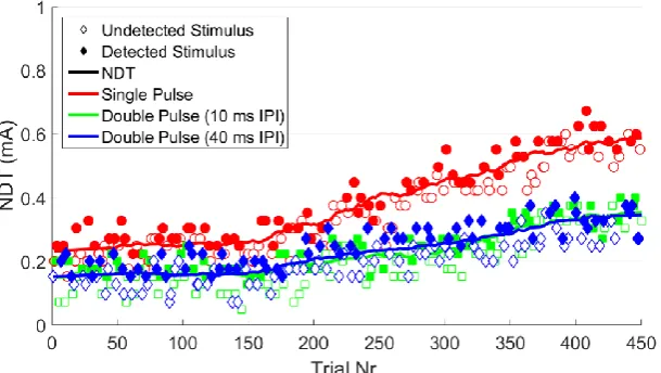
Results of NDTs from 25 pain-free subjects demonstrated that average initial NDTs for single-pulse stimuli were 0.2 mA and increased to approximately 0.5 mA during the last trial (Figure 11, left upper panel). At the same time, initial NDTs for double-pulse stimuli were around 0.1 mA and increased gradually to approximately 0.2 mA. Note that NDTs for single-pulse stimuli are higher than for double-pulse stimuli and that the difference in IPI did not affect the NDTs. It was observed that habituation and paired-pulse facilitation played a role during the MTT-EP experiment.
Evoked potentials
2.6 Implications
Research on underlying pathophysiology of chronic pain diseases is needed because 19% of the European people are still suffering from chronic pain1. The number of ineffective treatments is enormous, which results in high costs and social-economic burden. Great steps should be taken in the field of diagnostics and treatments for chronic pain. Although the underlying pathophysiology is still not completely understood, literature describes that central sensitization is seen in FBSS patients. At St. Antonius Hospital, approximately 60 patients diagnosed with FBSS are placed on the waiting list for neuromodulation each year. These chronic pain patients have passed all the possible therapies and are still suffering from pain, so it is assumed that these patients are certainly suffering from a central sensitization syndrome.
Since earlier studies from the University of Twente showed that the MTT-EP experiment might be promising for objective observation of nociceptive processing in pain-free subjects. Therefore, the first step is to dislocate the experiment from the university lab to the hospital environment. Then, it is recommended to explore whether results of the experiment at St. Antonius Hospital are a replication of results from the previous study and observe if it is feasible to perform by FBSS patients. At the same time, results of pain-free subjects the MTT-EP experiment at St. Antonius can be helpful to use as healthy control group for results of chronic pain patients in the future.
The MTT-EP experiment is hypothesized to be applicable when phenomena of the NDTs and EPs measured in the hospital are in line with results from the previous study at the University of Twente. Furthermore, it is hypothesized that neurophysiological responses might be different in chronic pain patients compared to pain-free subjects due to an altered central sensitization.
2.6.1 Primary research objectives
The primary objective is to explore whether results of the MTT-EP experiment in pain-free subjects at St. Antonius hospital are a replication of results in pain-free subjects at the University of Twente. Also, the objective is to describe how NDTs and EPs for electrocutaneous stimuli using an MTT paradigm behave in both pain-free subjects and FBSS patients.
Primary research questions:
a. Are results of the MTT-EP experiment in pain-free subjects replicable in a hospital environment?
• How present the average NDT and EP profiles?
• How behave neurophysiological effects such as habituation and paired-pulse facilitation?
• How are detected nociceptive stimuli related to the EP?
2.6.2 Secondary research objectives
Secondary objectives are (1) to see if differences in behavior of NDTs and EPs can be found between FBSS patients and pain-free subjects, and (2) to analyze how the NDT and EP are related to central sensitization in FBSS patients.
Secondary research questions:
a. Is an altered behavior of NDTs and EPs found in FBSS patients in comparison to pain-free subjects at St. Antonius Hospital?
3 Methods
3.1 Subjects
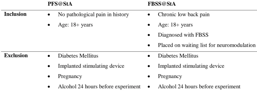
[image:31.595.69.520.313.474.2]Twenty pain-free subjects (PFS@StA) and seven FBSS patients (FBSS@StA) were enrolled in the study between September 2018 and November 2018. The subjects were included according to the inclusion- and exclusion criteria (Table 1). Verbal and written informed consent was obtained prior to inclusion. None of the pain-free subjects took analgesic medication. FBSS patients were allowed to continue the medication intake, if necessary. Not completing the MTT-EP experiment or analyzing EEG electrode ‘M1’, ‘M2’, ‘CPz’, ‘FPz’, ‘T7’, or ‘T8’ impedance higher than 5 kOhm was the exclusion criterion. The study was approved by the Medical research Ethics Committees United (MEC-U, file number: NL66136.100.18).
Table 1. Inclusion/exclusion criteria for pain-free subjects (PFS) and failed back surgery syndrome (FBSS) patients at St. Antonius Hospital (@StA).
PFS@StA FBSS@StA
Inclusion • No pathological pain in history
• Age: 18+ years
• Chronic low back pain
• Age: 18+ years
• Diagnosed with FBSS
• Placed on waiting list for neuromodulation
Exclusion • Diabetes Mellitus
• Implanted stimulating device
• Pregnancy
• Alcohol 24 hours before experiment
• Diabetes Mellitus
• Implanted stimulating device
• Pregnancy
• Alcohol 24 hours before experiment
3.2 Design
The study was a mono-center, explorative cross-sectional study, which was carried out in the Pain Clinics department at St. Antonius Hospital Nieuwegein, The Netherlands. This study monitored electrical brain responses during processing of nociceptive stimuli around the detection threshold. Each subject underwent one session of the MTT-EP experiment, consisting of two measurements (Figure 9).
[image:31.595.117.477.593.745.2]3.3 Materials and methods of measurement
3.3.1 Stimuli
[image:32.595.71.529.346.415.2]The AmbuStim 1-channel stimulator was used for stimulation, which was connected to a cathodic electrode. This sterilized IES-5 electrode contained an array of five 0.2 mm needles. The electrode was placed gently on the dorsal hand and fixed with tape. Nociceptive (Að) fibers in the epidermis were specifically activated by IES6,7,42–47. A rectangular 9 x 5 cm TENS electrode served as an anode and was placed proximal to the IES-5 electrode at the wrist. A personal computer was wirelessly connected to the stimulator using Bluetooth. Moreover, the computer ran a custom computer program written in LabVIEW 2013. All stimulation procedures, stimulus amplitudes (and their trigger codes), responses to the stimuli and stimulus times were controlled and registered by the program code.
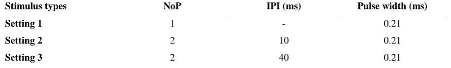
Table 2. Three stimulus types were executed: (1) the stimulus consisted of one cathodic square-wave electrical current pulse with a pulse width of 0.21 ms (setting 1); (2) the stimulus consisted of two cathodic square-wave electrical current pulses with a pulse width of 0.21 ms and an IPI of 10 ms (setting 2); (3) the stimulus consisted of two cathodic square-wave electrical current pulses with a pulse width of 0.21 ms and an IPI of 40 ms (setting 3). IPI of 10 ms and 40 ms were chosen because of the electrophysiology of synapses in the nociceptive system. The stimulus amplitude was limited to a maximum current of 2.0 mA in the stimulation software, because the detection threshold was expected between 0.0 and 1.0 mA.
Stimulus types NoP IPI (ms) Pulse width (ms)
Setting 1 1 - 0.21
Setting 2 2 10 0.21
Setting 3 2 40 0.21
3.3.2 Multiple threshold tracking paradigm
During both measurements, a total of 450 stimuli consisting of 150 stimuli for each stimulus type (setting 1, setting 2 and setting 3) were applied to the subject. The MTT paradigm tracked NDTs for these three types of stimuli (Table 2)5,7. Thresholds for each combination of NOP and
IPI was tracked simultaneously by measuring the subject’s response (detected or not detected) to a randomized set of stimulus amplitudes. All types of stimuli were selected the same number of times, but in a random order. Observer and subject bias were decreased by varying the stimulus type randomly.
3.3.3 EEG recording
Simultaneously to the stimulation, electrical brain activity was recorded continuously with a sampling frequency of 1 kHz. This was performed using an ANT Neuro Waveguard EEG cap containing 64 Ag/AgCl electrodes in combination with a TMSi 72-channel Refa EEG amplifier. The EEG and the trigger codes were recorded on a dedicated computer running TMSi Polybench (Polybench Designer 1.30.0) software. The EEG cap was adjusted to the size of the head, before it wasapplied on the head. The Cz electrode was set in the middle between the
nasion and inion and between both mastoids. A ground electrode was placed on the forehead and earlobe electrodes were also applied (A1A2) for CPz-A1A2 analysis. All cap electrodes were
Figure 10. Timeline of the procedure. The CSI was part of the questionnaires. Preparation of the EEG cap was dependent of the subjects hair, and therefore preparation time varied to meet the required electrode impedances (<5 kOhm). The MTT-EP experiment consisted of two measurements (in which the order was determined by randomization). The whole session took about 130 minutes.
3.3.4 Procedure
First, the subject was informed about the purpose of the study (Figure 10). After completion of the informed consent, the subject was asked to fill in a set of questionnaires, including the CSI to quantify central sensitization. One session was divided into two measurements: measuring NDTs and EPs during IES of the dominant hand followed by IES of the nondominant hand, in which the order was determined by randomization. Mobile phones were not allowed in the room of the MTT-EP experiment to prevent artifacts in the EEG signal. The subject was asked to sit in the chair during the measurements. Before starting the first measurement, the software of the EEG system was prepared, and the EEG electrodes were placed. All scalp-electrode impedances were verified and recorded to be less than 5 kOhm. Next, the stimulator system was prepared, and the stimulation electrodes were attached on the dorsal hand while the stimulator was held in the other hand. Then, the subject was familiarized with test stimuli and detection tasks before the start of each measurement.
Familiarization
Nociceptive detection tasks during both measurements
The subject was instructed to initiate a perceived stimulus by pressing the response button and releasing it as soon as any sensation was felt, which was ascribed to the application of a stimulus. The subject was asked to repeat the task after about half a second. If the button was released within 1000 ms after the stimulus, the stimulus was labeled as ‘detected’. The stimulus was labeled as ‘undetected’ if the button was still pressed. Simultaneously, the EEG signals were recorded when the button was pressed. Therefore, the subjects were asked to focus their eyes on one point and avoid muscular face movements (e.g. talking and swallowing).
3.4 Data analysis
3.4.1 Nociceptive detection threshold
Individual NDT values were determined using the psychometric function, which estimated the subjects’ detection threshold for each stimulus setting. Trials consisting twice the previous detection threshold were removed from threshold analysis. Nociceptive detection probabilities and thresholds were analyzed for every stimulus type by including the information from all subjects in a generalized linear mixed model (GLMM). The estimated average threshold was determined by a linear predictor consisted of fixed effects and random effects. The intercept, stimulus amplitude (for each stimulus type) and total received stimuli were defined as fixed effects. Between-measurement random effects were applied for every fixed effect. A moving window of 30 stimulus-response pairs was used. The GLMM approached the subject’s response using a logit link function which was regulated by the stimulus amplitude and habituation. The predicted model response was approached by Equation 1:
Logit(D) ~ 1 + SP1 + SP2_10 + SP2_40 + TRL + (1 + SP1 + SP2_10 + SP2_40 + TRL | Measurement) (1)
The subject’s response was described by the stimulus detection (D). The stimulus was specified by the amplitude of a single-pulse stimulus (SP1), and a double-pulse stimulus with an IPI of 10 ms (SP2_10) and an IPI of 40 ms (SP2_40). Habituation was included with respect to the total amount of received stimuli (TRL). To determine the significance, the coefficient of the GLMM were statistically tested against the null-hypothesis using a Wald t-test. The predicted psychophysical curve based on probability summation of two pulses was calculated by the probability of the detection threshold (from available data) using logistic regression.
Pre-processing of EEG data
The offline EEG data were re-referenced to the A1A2-electrodes. Second, EOG components from the raw EEG-signals were filtered by the application of an independent component analysis. Then, the EEG was preprocessed on the clean MTT trials using FieldTrip, which is a MATLAB toolbox for signal processing58. Trials for the EP analysis were segmented using a
3.4.2 Evoked potential
The central and lateral component of the EPs were respectively derived from two derivations: (1) CPz-A1A2 and (2) Tc-FPz (contralateral channel, T7-FPz or T8-FPz). Grand average EPs over all stimuli were displayed for both derivations. Whether grand average EPs from detected stimuli differed from undetected stimuli were assessed by nonparametric statistical testing58,59. An overview of grand average EPs from all channels was illustrated using a butterfly plot. Timestamps for grand average scalp topographies were based on the N2 and P2 peaks in both pain-free subjects and FBSS patients. In this study, the N2 peak was defined as the maximal negative peak approximately 200 ms post-stimulus at Tc-FPz. The P2 peak was defined as the first maximal positive peak approximately 400 ms post-stimulus at CPz-A1A2.
The variation of EPs was analyzed using a linear mixed model (LMM). The EP was approached by Equation 2:
EP ~ 1 + D + SP1 + SP2_10 + SP2_40 + TRL + (1 + D + SP1 + SP2_10 + SP2_40 + TRL | Measurement) (2)
Stimulus detection (D), which reflected the subject’s response (detected or not detected), was used as a fixed effect in this statistical model. Besides this parameter, three stimulus amplitudes (SP1, SP2_10, and SP2_40) and total amount received stimuli (TRL) were selected also as fixed effects. Again, between-measurement random effects were included for these fixed effects.
Grand average EPs described by these fixed LMM coefficients were displayed at CPz-A1A2 and Tc-FPz. To determine the significant components of the grand average EPs, the coefficient of the LMM were statistically tested against the null-hypothesis using a t-test59.
3.5 Replicability in pain-free subjects
The replicability of the MTT-EP experiment at St. Antonius Hospital was explored by multiple parameters; average NDTs, psychometric curves, grand average EPs and their scalp topographies, and fixed coefficients of the LMM which influenced the detected grand average EPs, were observed in both pain-free subjects’ groups. All the data of pain-free subjects at the University of Twente (PFS@UT) was imported from the previous study12.
3.6 Behavior of neurophysiological effects in FBSS patients
4 Results
Table 3. Characteristics of pain-free subjects (PFS) at the University of Twente (UT) and St. Antonius Hospital (StA) and characteristics of failed back surgery syndrome (FBSS) patients at St. Antonius Hospital. Note that the number of measurements is twice times the number of subjects included for analysis at St. Antonius Hospital, because they were measured at both hands. Differences in age, BMI, NRS and CSI between PFS@StA and FBSS@StA were tested using an independent sampled t-test. Significance level p < 0.05.
PFS@UT PFS@StA FBSS@StA p-value
Number of subjects 25 17 7 -
Number of measurements 25 34 14 -
Sex (M/F) 16/9 3/14 3/4 -
Age (mean ± SD) 23.0 ± 3.6 35.9 ± 11.9 54.3 ± 11.3 0.003*
Handedness (R/L) 24/1 15/2 5/2 -
BMI - 22.2 ± 2.8 24.7 ± 3.4 0.070
NRS last week (mean ± SD) - 1.4 ± 0.7 7.4 ± 1.6 0.001*
NRS during MTT-EP experiment - 1.0 ± 0.0 6.7 ± 2.1 0.001*
CSI score (mean ± SD) - 14.6 ± 8.8 49.0 ± 15.5 0.001*
Medication intake (Yes/No) - 1/16 6/1 -
Duration of FBSS in months (mean ± SD) - - 35.0 ± 21.2 -
Duration of pain in years (mean ± SD) - - 18.6 ± 17.5 -
4.1 Subject Characteristics
4.1.1 Pain-free subjects
In total 17 pain-free subjects at St. Antonius Hospital (PFS@StA) were analyzed. Three other participants were excluded due to one of the following reasons: M1/M2 electrodes showed impedances higher than 5 kOhm or the subject could not complete the MTT-EP experiment because of uncertain reasons. The group characteristics are summarized in Table 3. Three men and fourteen women are shown as pain-free subjects. The mean and standard deviation (SD) of these subjects’ age is 35.9 ± 11.9 years, ranging from 18 to 63 years. The CSI scores (14.6 ± 8.8) demonstrate that none of the subjects suffered from a central sensitization syndrome (CSI-score >40). The body mass index is 22.2 ± 2.8 kg/m2. Fifteen subjects (88%) reported being right handed. Four subjects mentioned suffering from pain in the week before the MTT-EP experiment (NRS: 1.4 ± 0.7) and one of them took pain analgesic medication (paracetamol) on the day of the MTT-EP experiment. None of the subjects reported pain on the questionnaires before the MTT-EP experiment (NRS: 1.0 ± 0.0).
4.1.2 FBSS patients
they were placed on the waiting list for a neurostimulator, but they did not have an implanted stimulation device on the day of the MTT-EP experiment. The patients suffered 18.6 years from pain on average, ranging from 3 to 54 years. In addition, these chronic pain patients were diagnosed with FBSS for 35.0 ± 21.2 months. Among the patients, the mean and standard deviation age is 54.3 ± 11.3 years, varying from 39 to 76 years. Five patients noted being right handed. Table 1Table 3 presents that age (p = 0.003), NRS-score (before (p = 0.001) and during the MTT-EP experiment (p = 0.001) and CSI-score (p = 0.001) are significantly higher in the FBSS patients compared to the pain-free subjects at St. Antonius Hospital. BMI does not show significant (p = 0.070) differences between both groups.
4.2 Replicability in pain-free subjects
4.2.1 Nociceptive detection thresholds
The most striking observation to emerge from the estimated average NDT data is the effect of habituation, which is similar to previous results from the University of Twente (Figure 11, upper panel). Next to this, it is crucial to note that single-pulse stimuli show higher NDTs than double-pulse stimuli. The estimated average initial thresholds for single-double-pulse stimuli are approximately 0.2 mA and 0.15 mA for double-pulses. These are in line with previous results.
Psychometric curves of pain-free subjects
The most striking result to emerge from the psychometric curve is the effect of paired-pulse facilitation, which occurred in both pain-free subject groups (Figure 11, lower panel). Last trial curves show higher stimulus amplitudes than first trials for all stimuli. The predicted double-pulse curve estimates the threshold purely based on probability summation. Since single-double-pulse curves show higher stimulus amplitudes than the predicted double-pulse curves, it is seen that there is a facilitating effect involved due to the paired-pulse. Furthermore, the steepness of the curves suggests that double-pulse stimuli are more reliably detected than single-pulse stimuli.
4.1.3 Evoked potentials
4.2 Behavior of neurophysiological effects in FBSS patients
4.2.2 Nociceptive detection thresholds
Average NDTs estimated by GLMM analysis are higher in FBSS patients than in pain-free subjects (Figure 11, upper panel). The average initial threshold for single-pulse stimuli is more than 0.3 mA, and for double-pulse stimuli, it is more than 0.2 mA. Although the NDTs increase generally over time in both groups, detection thresholds rise faster in FBSS patients. Also, detection thresholds in FBSS patients are less constant than in pain-free subjects. For all participants, stimulation by single-pulses shows higher NDTs than by double-pulses. Thresholds for single-pulse stimuli rise faster over time as well. Varying of IPIs present almost identical detection thresholds.
Psychometric curves of FBSS patients
Altered behavior of psychometric curves can be observed in chronic pain patients (Figure 11, lower panel). In general, it is seen that the stimulus amplitude shifts from the left to the right in a period between the first and last trial, which implies that the stimulus intensity increases during the measurement. Also, these curves demonstrate that higher stimulus amplitudes were required for stimulus detection in chronic pain patients compared to pain-free subjects. Additionally, a lower steepness of the curves is shown in these patients.
4.2.3 Evoked potentials
Fig ure 12 . B utte rfly plo ts ( up pe r p an els ), in clu din g g ra nd av era ge sc alp to po gra ph ies an d g ra nd av era ge E Ps of th e c on tra late ra l c om po ne nt ( m idd le p an els ) a nd gr an d a ve ra ge sc alp to po gra ph ies an d g ra nd av era ge EP s o f th e c en tra l c om po ne nt (lo w er pa ne ls) fro m p ain -fre e s ub jec ts at U niv ers ity o f T w en te (P FS @ U T), p ain -fre e s ub jec ts at St. A nto niu s H os pita l (P FS @ StA ) a nd fa ile d b ac k s urg ery sy nd ro m e p atie nts a t S t. An ton ius H os pita l (F BS S@ StA ). N ote th at t he co ntr ala ter al c om po ne nt i s d eriv ed fro m T c-F Pz (T 7 o r T 8 ch an ne l o n th e c on tra late ra l s ide of stim ula tio n) an d th e c en tra l c om po ne nt i s d eriv ed fro m C Pz -A 1A 2. T he N 2 pe ak is se en a t th e c on tra late ra l c om po ne nt, w he re as th e P 2 p ea k is se en a t th e c en tra l c om po ne nt. Th e s ca lp t op og ra ph ies illu str ate th e d ipo le d es crib ed b y a pot en tia l fie ld a ro un d th e p ea ks w ith a tim e w ind ow of 20 m s. W he n th e E P f ro m de te cte d s tim uli (g re en lin e) a re sig nifi ca ntly (p < 0.0 5) d iffe re nt f ro m un de te cte d s tim uli (re d li ne ), th e c ha nn el i s m ark ed by a s ta r (* ) in th e to pog ra ph y a nd th e c om po ne nt i s c olo re d in gr ee n on th e g ra nd a ve ra ge g ra ph .
Central component EP and scalp topography Contralateral component EP and scalp topography Butterfly plot
[image:41.595.51.559.72.766.2]
Figure 13. Fixed coefficients of the linear mixed model related to the grand average EPs for detected stimuli. Stimulus detection (D), the amplitude of single pulse stimuli (SP1), the amplitude of double-pulse stimuli with 10 ms IPI (SP2_10) and 40 ms IPI (SP2_40), and total received number of stimuli (TRL) were set as fixed coefficients. The EPs were derived from the contralateral component (Tc-FPz) upper panel) and central component (CPz-A1A2, lower panel) in pain-free subjects at University of Twente (PFS@UT, left), and both pain-free subjects (PFS@StA, middle) and FBSS patients (FBSS@StA, right) at St. Antonius Hospital.. Components of the EP for detected stimuli which were significantly different from the EP for undetected stimuli are colored in green. This implicates that the EP is modulated by the coefficient.
Contralateral component: Tc-FPz
PFS@UT PFS@StA FBSS@StA
Central component: CPz-A1A2
5 Discussion
In this study, we explored the replicability of the MTT-EP experiment in pain-free subjects at St. Antonius Hospital. Secondly, we observed the neurophysiological responses during processing of nociceptive stimuli around the detection threshold in chronic pain patients. The study yielded two main findings. (1) Results of the MTT-EP experiment in pain-free subjects at St. Antonius Hospital are a replication of results from the previous study at the University of Twente based on observations of NDTs and EPs during nociceptive stimulation. (2) Behavior of neurophysiological effects seems to be altered in FBSS patients compared to pain-free subjects.
5.1 NDTs and EPs
The estimated average (initial) NDT thresholds in pain-free subjects were quite similar to previous results for all stimulus settings. Results of NDTs from 17 pain-free subjects demonstrated that average NDTs for single-pulse stimuli started around 0.2 mA and increased to 0.4 mA during the last trial (Figure 11, upper panel). At the same time, initial NDTs for double-pulse stimuli were around 0.15 mA and increased gradually to approximately 0.2 mA. Strikingly, in all cases the NDTs were increasing over the number of trials and, moreover, NDTs for single-pulse stimuli were higher than for double-pulse stimuli. The difference in IPI did not affect the NDTs. These results suggest that the MTT-EP experiment can replicate similar NDTs in pain-free subjects at St. Antonius Hospital using a changed observer, (pain-free) population and environment.
Namely, the contralateral component was mainly modulated by stimulus detection and the central component was influenced by all fixed effects (stimulus detection, amplitudes of the single and double-pulse stimuli and the number of received stimuli) in both pain-free subject groups.
5.2 Behavior of neurophysiological effects
5.2.1 Habituation
The effect of habituation was found in virtually all participants, which was based on the rising NDTs during the measurements (Figure 11, upper panel). Interestingly, the coefficient for a total number of received stimuli (TRL) showed a negative effect on the grand average EP in pain-free subjects (Figure 13, lower panel), indicating that EP amplitude decreases for repeated stimuli due to habituation. This effect was also observed in the previous study at the University of Twente and earlier studies11,12,52. However, it is uncertain why thresholds in FBSS patients
increased faster over time than in pain-free subjects (Figure 11, upper panel). Controversially, based on results of the LMM, it was seen that habituation does not significantly affect the EP in FBSS patients (Figure 13, lower panel). In addition, it is not likely that habituation occurred more extremely in FBSS patients compared to results from Vossen et al., because they concluded that habituation was less in FBSS11. Therefore, the strengthened habituating effect might possibly be a result of other reasons (i.e. medication intake, as discussed below). Since other literature studied different populations, using different stimulation paradigms and other analysis methods, it is in general difficult to compare the effect of habituation.
5.2.2 Paired-pulse facilitation
Paired-pulse facilitation was observed in pain-free subjects, which was in line with results from the University of Twente. In addition, paired-pulse facilitation was discovered in FBSS patients (Figure 11, lower panels). The presence of such a facilitating effect is in our knowledge a novel finding in FBSS patients, however, long-term potentiation is known in chronic pain patients60. Paired-pulse facilitation can be explored by the fact that estimated thresholds for double-pulse stimuli were clearly lower than predicted thresholds based on probability summation (Figure 11, lower panels). Therefore, this study showed that pure probability summation does not fully explain the decrease in threshold when the number of pulses is increased. Thus, it was suggested that the detection probability of the second pulse is facilitated by the first pulse6 because R.J. Doll et al. also observed earlier this effect by processing of temporal stimulus properties on nociceptive detection probability in pain-free subjects6.