ORIGINAL RESEARCH
INTERVENTIONAL
Evaluation of the Pontine Perforators of the Basilar Artery
Using Digital Subtraction Angiography in High Resolution and
3D Rotation Technique
S. Lescher, T. Samaan, and J. Berkefeld
ABSTRACT
BACKGROUND AND PURPOSE: Compromise of perforating branches of the basilar artery resulting in brain stem infarctions has been described as a major complication of intracranial stent placement for basilar artery stenosis or after implantation of endovascular flow diverters. Descriptions of pontine arteries are mainly based on examinations of injection specimens; however, there is a lack of consistent presentation of the small branches of the basilar artery in the imaging literature. Therefore, we retrospectively analyzed DSA images and 3D rotational angiography with a review of literature for an imaging definition of microvascular anatomy of the brain stem.
MATERIALS AND METHODS: We retrospectively analyzed 2k DSA images (detector format 32⫻32 cm; image matrix 2480⫻1920 pixels) and 3D rotational angiography reconstructions (5 second DSA, subtraction technique) obtained on Axiom Artis zee biplane neuroradio-logic angiography equipment using standard protocol.
RESULTS: On 2D and 3D DSA images, small arterial side branches of the basilar artery can be demonstrated in each of the cases but with a wide variation in the visibility of these vessels. Compared with 2D DSA images, 3D DSA reconstructions allow superior visualization of the small branches of the basilar artery.
CONCLUSIONS: Our results demonstrate that 2D DSA and 3D-rotation techniques are able to reliably visualize the penetrating branches of the brain stem in vivo. No zone of basilar artery is free from important side branches. Collateral pathways between circumferential and perforating arteries are occasionally detectable. In the future, further refinement of imaging techniques is necessary to increase the reliability of small vessel angiography to use this data for risk assessment before stent placement and aneurysm treatment.
ABBREVIATIONS:3DRA⫽3D rotational angiography; 2k matrix DSA⫽detector format 32⫻32 cm; image matrix 2480⫻1920 pixels
C
ompromise of perforating branches of the basilar artery has been described as a major complication of intracranial angio-plasty and stent placement or implantation of endovascular flow diverters.1,2Because of the small diameter of these side branches of 383m, visualization by different means of angiographic imaging like CT- or MR-based angiography is difficult and uncertain.3High-resolution DSA images with 2k matrix (detector format 32⫻32 cm; image matrix 2480⫻1920 pixels) and especially flat detector CT with pixel sizes of 308m⫻308m (2⫻2 binning), using a reconstructed section thickness of 0.1 mm and high-contrast resolution of CT and 3D reconstructions, provide a new
basis to display these small vessels on recent flat detector neuroan-giography systems.4We hypothesized that the 3D-rotation tech-nique allows superior visualization of the small branches of the basilar artery than 2D DSA images do.
The microvascular anatomy of the vertebrobasilar junction and the distal basilar tip has been described in detail in the neu-rosurgical and neuroradiologic literature.5-7Remarkably, there are no comparable descriptions of the proximal and middle por-tion of the basilar artery in the imaging and neurosurgical litera-ture. As such, anatomic knowledge is based on examinations of post mortem injection preparations.8,9
A continuation of patterns of the spinal cord vascular supply has been postulated for the brain stem with additional circumfer-ential branches for the pons and cerebellum.7
The presence of perforating branches is known; however, in the past, angiographic imaging was far away from reliable pattern recognition and display of potential collateral pathways.
The lack of consistent descriptions in the imaging literature raised the question of whether the latest generations of flat
detec-Received November 3, 2013; accepted after revision February 6, 2014.
From the Institute of Neuroradiology, Hospital of Goethe University, Frankfurt am Main, Germany.
Please address correspondence to Stephanie Lescher, MD, Institute of Neuroradi-ology, Hospital of Goethe University, Schleusenweg 2–16, 60528 Frankfurt am Main, Germany; e-mail: [email protected]
Indicates open access to non-subscribers at www.ajnr.org
tor neuroangiography allow for more reliable visualization of the pontine branches of the basilar artery with the use of 2k matrix DSA images and 3D rotational angiography.
Therefore, we retrospectively analyzed 2k DSA images and 3D rotational angiography reconstructions together with a review of the literature for an imaging definition of the microvascular anatomy.
MATERIALS AND METHODS
For retrospective analysis, we identified 41 consecutive cases with indications for selective vertebral angiography and 3D rotational angiography (3DRA) in a period from January 2011 until March 2013. Twenty-one datasets with minor image quality due to in-complete contrast filling or movement artifacts were excluded. Twenty datasets with adequate image quality were selected for further evaluation.
The protocol was authorized by the ethics committee of Frankfurt.
All angiograms and 3DRA data were obtained on an Axiom Artis zee biplane (Siemens, Erlangen, Germany) neuroradiologic angiography system. The angiographic system is equipped with a flat panel detector with 2480⫻1920 pixels (2k matrix) and a pixel pitch of 154m⫻154m.
For the evaluation of pontine arteries, 2k DSA images using high-resolution technique and 3DRA runs of the basilar artery be-tween the vertebrobasilar and the pontomesencephal junction were analyzed. Indications for DSA and especially 3DRA were justified by suspected pathologies like aneurysms (ruptured/unruptured), arte-riovenous malformations, stenosis, infarctions, or vasculitis.
DSA with 2k Matrix
Compared to former angiographic systems equipped with an 1k image matrix of 1240⫻960 pixels, the newest generations of angiographic systems provide improved and higher spatial reso-lution using a 2k image matrix of 2480⫻1920 pixels and a flat detector zoom format of 32⫻32 cm in each plane. For retrospec-tive evaluation of DSA images in standard Towne and lateral projec-tions, we selected 20 consecutive cases with normal findings in the intracranial vertebrobasilar circulation and good image quality. DSA images were obtained using a standard program with a variable frame rate with 4 frames per second in the arterial phase, a small focus with a focal spot size of 0.3 mm, and edge enhancement reconstruction algorithm. A flat detector zoom format of 32⫻32 cm in each plane was used. In addition, 4 – 6 mL of non-ionic contrast material was applied via hand injection after selective catheterization of the dom-inant vertebral artery. The average effective dose of this examination was 2.7 mSv measured in an anthropomorphic Alderson-Rando phantom with thermoluminescence dosimetry.
Image evaluation was done retrospectively by consensus of 2 experienced reviewers who counted the number of visible side branches at each side of the basilar artery and distinguished be-tween circumferential arteries and AICA variants. They described whether circumferential arteries were displayed in a segmental or more irregular network pattern and whether common trunks or anastomoses between adjacent branches were present.
The reviewers also looked for direct paramedian pontine perforators.
3D Rotation Technique
In the 20 patients with indications for 3DRA of the posterior circulation, we reconstructed the stored raw data of 3D DSA runs with a scan time of 5 seconds. A native rotational run was followed by contrast-enhanced images after mechanical intra-arterial in-jection of 20 mL of nonionic contrast material (Ultravist 240; Bayer-Schering, Berlin, Germany) into the dominant vertebral artery with a flow of 3 mL/s. Injection was started 2 seconds before the contrast-enhanced run. In each run, 133 images were ob-tained with a scan time of 5 seconds. The average effective dose of this examination was 0.9 mSv measured in an anthropomorphic Alderson-Rando phantom with thermoluminescence dosimetry. The spatial resolution of flat detector CT was reported to be 3.0 lp/mm with no binning and 1.5 lp/mm with 2⫻2 binning for high-resolution modes.4,10
The 3DRA raw data were transferred to a dedicated angio-graphic workstation (Siemens). The 3D datasets and CT-like im-ages of subtraction angiograms were reconstructed using stan-dard volume rendering and flat detector CT algorithms (DynaCT-arterial). MPR with section thicknesses of 0.1 mm and MIP with section thicknesses between 6 and 10 mm as well as volume rendering reconstructions with bicolored window and threshold settings were used for analysis of small vessels.
Datasets with a basilar artery stenosis and a midbrain AVM were analyzed as illustrative cases and additional proof of the value of our technique.
The number and pattern of pontine arteries was determined retrospectively by consensus of 2 reviewers. Analog to 2k DSA evaluation, the 2 reviewers counted the number of visible side branches at each side of the basilar artery and looked for direct paramedian midline perforators to determine origin and course of the small brain stem vessels. Especially in pathologic cases with stenotic lesion, we looked for potential collaterals between the small brain stem vessels according to rotating reconstructions. Perforators originating directly from the basilar artery were eval-uated according to subtracted flat detector CT images.
RESULTS
Biplane 2k DSA Images in Standard Projections
Circumferential pontine arteries can be regularly demonstrated on DSA images of the basilar artery obtained with recent technol-ogy. The most common pattern is a segmental pattern of up to 10 circumferential arteries with slightly descending course around the pons. The origins from the dorsal surface of the basilar artery are best seen on lateral projection in an average distance of 1.5–3.5 mm between 2 “segmental” vessels and some variations between left and right. Common trunks bearing origins of several arteries, longitudinal anastomoses between different arteries, or midline perforators to the brain stem are occasionally seen.
3D and Flat Panel CT Angiography Reconstructions
Compared with DSA images, 3DRA and flat detector CT recon-structions are superior in demonstrating anatomic patterns of circumferential arteries and direct pontine perforators (Table).
visualized more clearly in 3DRA compared with 2k DSA images. The number of the segmental arteries on each side of the basilar artery is the same in both 3DRA and 2k DSA images (Table). The
distance between the origins of the cir-cumferential branches ranged from 2.6 – 4.0 mm. Transversal flat detector CT re-constructions (DynaCT arterial mode) show that the circumferential arteries during their downward-curving route to-ward the tegmentum of the pons give off peripheral perforating branches extend-ing to the pontine parenchyma. Asymme-tries between left and right and variations in size have been revealed as common variation.
In 10 of 20 cases, several circumferen-tial arteries arose from a common trunk (Table).
Descending course and tortuosities of circumferential arteries may mimic longi-tudinal anastomoses or arcade-like con-nections that are truly present in 6 cases with a more pial-like network and in a fur-ther 11 cases with longitudinal anastomoses between adjacent circumferential arteries close to the origin and in no cases in the dorsolateral periphery of the brain stem (Table; Fig 2). 3DRA dataset reconstruc-tions are able to differentiate between true anastomosis and vessel overlap.
Analogous to the sulco-comissural ar-teries in the spinal cord7,8direct perfora-tors to the pons could be identified as small paired vessels arising from the dor-sal surface of the basilar artery and ex-tending into the parenchyma. Direct pon-tine perforators could be identified on 3 levels (Fig 3): at the pontomesencephal junction (rostral), midbasilar (central), and vertebrobasilar junction (caudal). These 3 groups could be defined in all the evaluated images.
The AICA as the main branch of the midbasilar artery was seen in all cases. A large caliber of the AICA seems to be as-sociated with smaller and less numerous other circumferential arteries.
Cases with Pathologies
In 2 cases with hemodynamically relevant stenosis of the basilar artery and the intra-dural part of the vertebral artery (V4), we identified small collat-erals connecting circumferential arteries of the basilar artery bridging the stenosis (Fig 4).
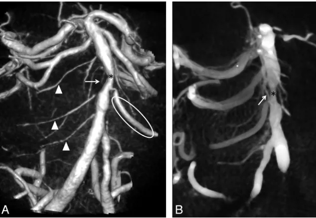
FIG 1. Segmental pattern and descending course of circumferential pontine arteries (arrows) on volume rendering reconstructions of a DynaCT dataset of the basilar artery in Towne and lateral projection.
FIG 2. Another case shows a less regular pial network pattern (*) with connections (arrow) between adjacent arteries that are better demonstrated on DynaCT reconstructions (A), than on high resolution DSA with 2k matrix (B).
Basic patterns of the small side branches of the basilar artery in 2k DSA and 3DRA imaging studies Number of Segmental
Arteries Right
Number of Segmental Arteries Left
Direct Pontine Perforators
Collateralsa
Common Trunks of Circumferential
Arteries
Pial Network
0–3 4–7 8–10 0–3 4–7 8–10 Caudal Medial Rostral
2k DSA 10 7 3 5 11 4 0 0 0 6 8 1
3DRA 6 13 1 5 13 2 20 20 20 11 10 6
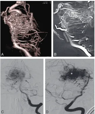
[image:3.594.52.372.135.389.2] [image:3.594.54.373.435.648.2]In one case with an arteriovenous malformation, the circum-ferential and perforating brain stem arteries were clearly hypertrophic as they were part of the AVM-feeding vessels. In comparison to the other cases, the quantity of vessels was also considerably higher and they could be displayed more clearly (Fig 5).
DISCUSSION
Current neuroradiologic angiographic equipment is able to detect the main anatomic features of small pontine arteries. Volume rendering and CT-like reconstructions of 3D rotational datasets are superior to conventional DSA. A larger number of projec-tions, like the subtraction technique with individual adjustment of threshold levels of flat panel CT and 3D reconstructions, in-crease the possibilities of image processing. In particular, trans-verse and parasagittal sections of volume rendering datasets with a section thickness between 5 to 10 mm include larger parts of the
curved and descending course of the cir-cumferential arteries. The visualization of the smaller direct pontine perforators de-manded thinner sections around 3 mm.
MPR with thick sections displayed the best results of the basic anatomic patterns of the small side branches of the basilar artery because of the general orientation of these penetrating vessels with their characteristic downward-curving route toward the pontine tegmentum. The con of using thicker reconstructed sections is a decline of spatial resolution in small vessel angiography. Some of the side branches cannot be displayed free from superimpo-sitions, and the correct allocation to the left or right side is difficult.
Compared with the single frames of the 2k DSA series, the continuous acqui-sition of the 3D rotational angiography runs with a scan time of 5 seconds pro-vides more reliable vessel detection (Figs 2 and 5). In 3D reconstructions, the small branches can be visualized free from su-perimpositions because of the increased number of projections and the longer ac-quisition time during pulsative vessel filling of these small branches.
Contrary to what might be expected from consideration of the total image acquisition count only, the patient radiation dose (re-ported in literature before) for a 3D rotational angiography ac-quisition is lower than for a biplanar DSA series.11
Venous contamination was widely excluded with early start and short scan durations of the scans and visual proof that small arteries originate directly from the basilar artery.
Imaging findings are coincident with previous anatomic de-scriptions based on examinations of injection specimens.8,9 How-ever, compared with the anatomic literature, angiographic detect-ability of the small vessels of the basilar artery seems to be incomplete. With existing angiography techniques and recon-struction tools, it is not possible to display all circumferential
FIG 3.Axial cuts of DynaCT dataset with thin sections of 3 mm reconstructed from 3D rotational angiogram shows ventral pontine perforators (bright arrows) originating from the basilar artery (*) at the junction between mesencephalon and pons (A), at the midpontine level (B), and at the vertebrobasilar junction (C).
[image:4.594.57.535.47.198.2] [image:4.594.54.371.244.463.2]arteries and anastomoses for an exact anatomic definition of this region as found in descriptions based on anatomic preparations. In addition, our results give rise to the assumption that there are variations with deviations from the common segmental pat-tern: in several cases, we observed irregular settings with common trunks and anastomoses between adjacent branches resembling a pial network similar to findings at the spinal cord (Fig 2). Due to limitations in spatial resolution, anastomoses as potential path-ways for local collaterals are only proved in occasional cases (Fig 2). In the presence of a basilar or vertebral artery stenosis, only 1 small collateral could be detected in each of the 2 cases (Fig 4). More circumferential arteries and anastomoses were seen in an AVM case with hypertrophy of arterial feeders (Fig 5).
Similar to the sulco-comissural arteries in the spinal cord, which enter the spinal cord only in greater intervals, we could regularly detect 3 groups of direct pontine perforators extending from the dorsal surface of the basilar artery entering the brain stem at the ventral surface (Fig 3).
The practical value of our findings is unknown at the moment. However, in the context of endovascular interventions, it
be-comes clear that almost no segment of the basilar artery is free from important side branches and display of these small vessels may contribute for risk assessment before angioplasty and stent placement. In the SAMMPRIS trial, most ischemic strokes after angiographically successful angio-plasty and stent placement in the basilar artery in perforator territory.12In addi-tion, perforator infarcts after treatment of fusiform aneurysms of the basilar artery raise doubts about sufficient consider-ation of vascular anatomy. In conclusion, efforts at reducing complications from endovascular treatments of aneurysm, in-farctions, or stenosis with intracranial an-gioplasty and stent placement should fo-cus on reducing the risks of regional perforator infarction.12
For the sake of completeness, some basilar artery pathologies have no impor-tant branch vessels in the involved seg-ment, eg, sidewall aneurysms between the origins of the circumferential pontine ar-teries or aneurysms arising from the ven-tral surface of the basilar artery, which is free from side branches.
Limitations
Variation between the cases may be at-tributed to anatomic variants or a lack of detectability of the small vessels. Incom-plete or delayed filling with contrast ma-terial and limitation in the resolution of smaller and terminal branches may be reasons for the incomplete display of an-atomic features.9,13,14 There are only a few descriptions of pontine arteries in the existing angiographic literature.7Only with the use of high-field MR imaging were Kang et al15able to detect similar patterns.
Incomplete contrast filling of the small branches in 2k DSA images depends on hand injection. Although details of technical safeguards for neuroangiography are seldom discussed in existing literature and contrast application via injector seems to be the standard in many institutes performing neuroangiography, in our opinion, hand injection in 2D DSA is safer and therefore the stan-dard in our institute.16-18Contrast application via injector was only selected for 3D DSA. The caliber of the vertebral arteries varies quite often and hand injection may avoid complications, for example, dissection or vessel rupture.
CONCLUSIONS
Our study proves the concept that modern neuroangiographic imaging with 3DRA and flat detector CT reconstructions is able to display small pontine arteries with patterns described in the ana-tomic literature.
Potential furture developments should focus on a continuous improvement of image quality and further refinement of
techno-FIG 5. Volume rendering reconstruction (A) and parasaggital cut of a DynaCT dataset (B) show multiple hypertrophied circumferential pontine arteries (arrows) involved as feeders in the supply of a mesencephalic AVM (*). Compared with the volume rendering reconstructionsAand
[image:5.594.52.373.45.427.2]logical advances or concepts to increase reliability of small vessel angiography. Further development and improvement of applica-tions are necessary. Limited spatial and contrast resolution of flat panel angiography as well as suboptimal protocols for injection of contrast materials are the main limitations of our approach. Pos-sible avenues for future improvements should address the devel-opment of flat detectors with even higher resolution and faster and more flexible detector readout, allowing for faster scanning to improve image quality of small vessel angiograms.4
Remarkably, our findings were generated from historical da-tasets using standard clinical protocols. Dedicated angiographic protocols with optimization of scan time, iodine concentration, and injection parameters of contrast materials may provide future options to use this data for risk assessment of patients with arterial occlusive or stenotic disease or aneurysms before endovascular treatment with angioplasty and stent placement.
This was a proof of concept study about the feasibility to visu-alize the small side branches of the basilar artery by use of modern angiographic equipment and reconstructions tools. The sample size of this study is too low for an exact anatomic definition of this anatomic region. Currently, further research on small vessel an-giography will be necessary and should address technical optimi-zation to improve image quality as well as clinical validation in larger studies to become a routine procedure in neuroangiogra-phy. Future studies should be evaluated blinded to avoid exagger-ation of the results and should compare different angiographic techniques.
Disclosures: Stephanie Lescher received support for travel to the RSNA 2013 meeting from Siemens Health Care AG, related to the study. Joachim Berkefeld received a consulting fee or honorarium* and travel support* related to the study; the project is part of a scientific cooperation between Siemens and the Institute of Neuroradi-ology at the University Frankfurt, Germany, and travel expenses for presentation of the project are covered by Siemens.*Money paid to institution.
REFERENCES
1. Piotin M, Blanc R, Kothimbakam R, et al.Primary basilar artery stenting.AJR Am J Roentgenol2000;175:1367– 69
2. Chimowitz MI, Lynn MJ, Derdeyn CP, et al.Stenting versus aggres-sive medical therapy for intracranial arterial stenosis.N Engl J Med
2011;365:993–1003
3. Lang J.Clinical Anatomy of the Posterior Cranial Fossa and its Foram-ina.Stuttgart: Thieme; 1991
4. Kalender WA, Kyriakou Y.Flat-detector computed tomography (FD-CT).Eur Radiol2007;17:2767–79
5. Mercier PH, Brassier G, Fournier HD, et al.Vascular microanatomy of the pontomedullary junction, posterior inferior cerebellar arter-ies, and the lateral spinal arteries.Interv Neuroradiol2008;14:49 –58 6. Mercado R. Santos-Franco J, Ortiz-Velazquez I, et al.Vascular anat-omy of the foramen of Vicq d’Azyr: a microsurgical perspective.
Minim Invasive Neurosurg2004;47:102– 06
7. Lasjaunias P, Berenstein A, Ter Brugge KG.Surgical Neuroangiogra-phy.Berlin-Heidelberg: Springer-Verlag; 2001
8. Marinkovic S, Gibo H.The surgical anatomy of the perforating branches of the basilar artery.Neurosurgery1993;33:80 – 87 9. Duvernoy HM. Human Brain Stem Vessels. Berlin Heidelberg:
Springer-Verlag; 2010
10. Kyriakou Y, Struffert T, Do¨rfler A, et al.Basic principles of flat de-tector computed tomography (FD-CT).Radiologe2009;49:811–19 11. Schueler B, Kallmes D, Cloft H.3D cerebral angiography: radiation
dose comparison with digital subtraction angiography.AJNR Am J Neuroradiol2005;25:1898 –901
12. Derdeyn CP, Fiorella D, Lynn MJ, et al.Mechanisms of stroke after intracranial angioplasty and stenting in the SAMMPRIS trial. Neu-rosurgery2013;72:777–95
13. Pai BS, Varma RG, Kulkarni RN, et al.Microsurgical anatomy of the posterior circulation.Neurol India2007;55:31– 41
14. Pais D, Arantes M, Casal D, et al.Brain stem arteries in Canis familiaris–implications in experimental procedures.Braz J Morphol Sci2009;26:8 –11
15. Kang CK, Park CA, Kim KN, et al.Non-invasive visualization of basilar artery perforators with 7T MR angiography.JMRI2010; 32:544 –50
16. Yousem DM, Trinh BC.Injection rates for neuroangiography: re-sults of a survey.AJNR Am J Neuroradiol2001;22:1838 – 40 17. Fox AJ.Technical aspects of neuroangiography: are risks and
safe-guards understood in the same way? AJNR Am J Neuroradiol
2001;22:1809 –10