ORIGINAL RESEARCH
PEDIATRICS
Prevalence and Spectrum of In Utero Structural Brain
Abnormalities in Fetuses with Complex Congenital
Heart Disease
M. Brossard-Racine, A.J. du Plessis, G. Vezina, R. Robertson, D. Bulas, I.E. Evangelou, M. Donofrio, D. Freeman, and C. Limperopoulos
ABSTRACT
BACKGROUND AND PURPOSE: Brain injury is a major complication in neonates with complex congenital heart disease. Preliminary evidence suggests that fetuses with congenital heart disease are at greater risk for brain abnormalities. However, the nature and frequency of these brain abnormalities detected by conventional fetal MR imaging has not been examined prospectively. Our primary objective was to determine the prevalence and spectrum of brain abnormalities detected on conventional clinical MR imaging in fetuses with complex congenital heart disease and, second, to compare the congenital heart disease cohort with a control group of fetuses from healthy pregnancies.
MATERIALS AND METHODS: We prospectively recruited pregnant women with a confirmed fetal congenital heart disease diagnosis and healthy volunteers with normal fetal echocardiogram findings who underwent a fetal MR imaging between 18 and 39 weeks gestational age.
RESULTS:A total of 338 fetuses (194 controls; 144 with congenital heart disease) were studied at a mean gestational age of 30.61⫾4.67 weeks. Brain abnormalities were present in 23% of the congenital heart disease group compared with 1.5% in the control group (P⬍.001). The most common abnormalities in the congenital heart disease group were mild unilateral ventriculomegaly in 12/33 (36.4%) and increased extra-axial spaces in 10/33 (30.3%). Subgroup analyses comparing the type and frequency of brain abnormalities based on cardiac physi-ology did not reveal significant associations, suggesting that the brain abnormalities were not limited to those with the most severe congenital heart disease.
CONCLUSIONS: This is the first large prospective study reporting conventional MR imaging findings in fetuses with congenital heart disease. Our results suggest that brain abnormalities are prevalent but relatively mild antenatally in fetuses with congenital heart disease. The long-term predictive value of these findings awaits further study.
ABBREVIATIONS:CHD⫽congenital heart disease; GA⫽gestational age; HLHS⫽hypoplastic left-heart syndrome
B
rain injury is a major complication in neonates with complex congenital heart disease (CHD).1,2During the past decade, agrowing body of evidence has demonstrated the high prevalence
of preoperative brain imaging abnormalities in neonates with CHD, suggesting an antenatal origin in many cases.3-6More
re-cently, our group has used quantitative MR imaging to demon-strate a progressive third-trimester deceleration in volumetric brain growth and metabolism in fetuses with CHD,7reduced
vol-umes of gray (cortical and subcortical) and white matter, and significant delays in cortical gyrification and surface area in fe-tuses with hypoplastic left-heart syndrome (HLHS).8To date,
there is only 1 published report of brain findings in fetuses with CHD by using conventional MR imaging.9 This retrospective
chart review of 53 cases reported a 39% incidence of brain anom-alies in fetuses with CHD. Taken together, preliminary evidence suggests that fetuses with CHD are at risk of brain abnormalities, including delayed maturation and brain injury. However, to date, no study has prospectively ascertained the frequency and nature of brain abnormalities by conventional MR imaging in fetuses with CHD. The primary objective of this study was to determine
Received November 20, 2013; accepted after revision December 27.
From the Advanced Pediatric Brain Imaging Research Laboratory (M.B.-R., I.E.E., D.F., C.L.), Division of Diagnostic Imaging and Radiology (M.B.-R., G.V., D.B., I.E.E., D.F., C.L.), Fetal and Transitional Medicine (M.B.-R., A.d.P., M.D., C.L.), and Division of Car-diology (M.D.), Children’s National Medical Center, Washington DC; and Depart-ment of Radiology (R.R.), Children’s Hospital Boston and Harvard Medical School, Boston, Massachusetts.
This study is supported by the Canadian Institutes of Health Research (MOP-81116); Marie Brossard-Racine was financially supported by the Fonds de Recherche en Sante du Quebec postdoctoral fellowship at the time of manuscript preparation. Paper previously presented at: Annual Meeting of the Pediatric Academic Societ-ies, May 4 –7, 2013; Washington, DC.
Please address correspondence to Catherine Limperopoulos, PhD, Advanced Pedi-atric Brain Imaging Research Laboratory, Division of Diagnostic Imaging and Radiol-ogy/Fetal and Transitional Medicine, Children’s National Medical Center, 111 Michi-gan Ave NW, Washington D.C., 20008; e-mail: [email protected]
Indicates open access to non-subscribers at www.ajnr.org
the prevalence and spectrum of brain abnormalities detected on conventional clinical MR imaging in fetuses with complex CHD and, second, to compare the CHD cohort with a control group of fetuses from healthy pregnancies.
MATERIALS AND METHODS
SubjectsAs part of an ongoing prospective observational study that started in September 2007, pregnant women with a fetal CHD diagnosis confirmed by echocardiography at Boston Children’s Hospital7
and Children’s National Medical Center were recruited as pa-tients. A fetal echocardiography was performed by one of the cen-ter’s experienced fetal echocardiographers on the day of the mother’s appointment. Fetal CHD diagnoses were subsequently categorized as cyanotic-versus-acyanotic and single- versus 2-ventricle physiology as determined by oxygen saturation and anatomic classification. During the same study timeframe, healthy controls were also recruited from healthy volunteers with normal fetal echocardiography findings or pregnant women for whom an echocardiography was performed for a previous family history of CHD. Patients and controls with multiple pregnancies, abnormalities on fetal sonography, congenital infection, docu-mented chromosomal abnormalities, and/or multiorgan dysmor-phic conditions were excluded from the study.7Thereafter,
en-rolled pregnant women completed a fetal MR imaging during the second or third trimester of pregnancy. Additionally, we amended our original protocol in 2013 to include serial fetal MR imaging studies (ie, 2 fetal MR imaging studies) as part of this prospective study, to examine fetal brain development during the second and third trimesters. For the subset of fetuses that underwent 2 fetal MR imaging scans, both studies were included in our analyses. Clinical and demographic information for each subject was col-lected through medical record reviews or questionnaires. Written informed consent was obtained for every participant, and the study was approved by the institutional review boards of Boston Children’s Hospital and Children’s National Medical Center.
MR Imaging Data Acquisition
Each fetal MR imaging study was performed on a 1.5T scanner either on an Achieva (Philips Healthcare, Best, Netherlands) de-vice by using a 5-channel phased-array coil (Boston Children’s cohort) or on a Discovery MR450 scanner (GE Healthcare, Mil-waukee, Wisconsin) by using an 8-channel phased-array coil (Children’s National Medical Center cohort). Multiplanar single-shot fast spin-echo imaging sequences were performed as follows: on the Philips scanner, single-shot fast spin-echo TE⫽120 ms, TR⫽971 ms/section, 330-mm FOV, 256⫻204 acquisition ma-trix; for the GE scans, TE⫽160 ms, TR⫽ 1110 ms/section, 320-mm FOV, 256⫻192 acquisition matrix. Diffusion tensor imaging was acquired in 12 directions with the following param-eters: TE⫽minimum, TR⫽8000 ms, and 4-mm section thick-ness. The FOV was 330 mm on the GE scanner versus 280 mm on the Philips scanner. Finally, a T1 spoiled gradient-recalled acqui-sition was performed with TE⫽minimum, TR⫽100 ms/section, 360-mm FOV, 256⫻160 acquisition matrix on the GE scanner. No contrast or sedation was used for any of the MR imaging studies.
All MR imaging studies were reviewed for clinical findings by one of the center’s expert pediatric neuroradiologists, both of whom had⬎15 years of experience with fetal brain MR imaging (R.R. at Boston Children’s Hospital and G.V. at Children’s Na-tional Medical Center). The neuroradiologists were blinded to patient-versus-control status, the subject’s medical history, and type of CHD. All studies were reviewed for the presence of devel-opmental malformations, acquired abnormalities, and brain mat-uration following the clinical guidelines in place at the time of the study. Mild ventriculomegaly was diagnosed when the maximum width of the lateral ventricles at the level of the atrium measured between 10 and 15 mm.10Increased extra-axial space was
diag-nosed when the distance between the inner margin of the cranium and the outer margin of the frontal, temporal, parietal, and/or occipital regions was considered enlarged for gestational age (GA).11,12Finally, images were also examined for signal
abnor-mality on T2/T1 images and the presence of restricted diffusion on diffusion imaging acquisitions.
Statistical Analysis
Descriptive statistics by using means, SDs, and frequencies were used to characterize the sample and the nature of the brain abnor-malities described on conventional MR imaging. Independent samplettests and2analyses were used to explore differences and
associations between groups, CHD diagnoses, cyanotic CHD ver-sus acyanotic, single- verver-sus 2-ventricle physiology, sex, GA, and MR imaging abnormalities. All analyses were performed by using the Statistical Package for the Social Sciences, Version 20 (IBM, Armonk, New York).
RESULTS
Group Characteristics
A total of 338 pregnant women (144 with fetuses with CHD, 194 controls) successfully completed second- or third-trimester MR imaging. Of these fetuses, 182 (53.8%) were male (82 with CHD, 100 controls). As described in our “Materials and Methods” sec-tion, in 2013, a second fetal MR imaging was added to our MR imaging protocol (ie, 2 fetal MR imaging studies) to examine fetal brain development serially. Consequently, 28 women (10 fetuses with CHD, 28 controls) completed 2 fetal MR imaging studies, the first during the second trimester and the second during the third trimester, resulting in 376 MR imaging studies. The mean GA of these 376 studies was 30.61⫾4.67 weeks (range, 18.29 –39.29 weeks), of which 273 (72.6%) were performed during the third trimester. Although a significantly smaller proportion of studies was performed during the second trimester in the control group compared with the CHD group (2⫽12.75,P⬍.001), the mean
GA at MR imaging for the control group (29.94⫾5.14 weeks) was significantly lower compared with that in the CHD group (31.57⫾3.71 weeks;t⫽3.57;P⬍.001).
Abnormal Conventional MR Imaging Findings
Brain abnormalities detected on MR imaging were significantly more frequent among patients with CHD (2⫽36.87,P⬍.001)
and were detected in approximately one-quarter (33/144, 22.9%) of the fetuses with CHD compared with only 1.5% (3/194) in the controls. The type and distribution of brain abnormalities on fetal MR imaging in the CHD group are summarized in Table 2. Mild unilateral ventriculomegaly (Fig 1) was the most common abnor-mality reported in 8.3% (12/144). Of these cases, left-sided ven-triculomegaly was twice as common (n⫽8) as right-sided (n⫽
4). Ventriculomegaly was isolated in all cases, with the exception of 2 fetuses, one of which also presented with increased extra-axial CSF spaces and the second with a frontal subependymal cyst. Only 1 fetus presented with mild symmetric ventriculomegaly. The presence of increased extra-axial CSF spaces (Fig 2) was the sec-ond most common MR imaging abnormality in our cohort, re-ported in 6.9% (10/144) of CHD subjects. Three fetuses (2.1%) with CHD were diagnosed with isolated inferior vermian hyp-oplasia on MR imaging (ie, no other associated cerebral malfor-mations were identified). Three other fetuses (2.1%) presented with immature brain appearance defined as an underdevelop-ment of the anatomic structures, such as fissures and sulci being less developed than expected for the estimated GA. Finally 4 pa-tients presented with white matter abnormalities, which included cysts in 2 fetuses (1.4%) and signal hyperintensity on T2-weighted
imaging in 2 others (1.4%), one of which also demonstrated glob-ally decreased diffusion in the white matter.
The 3 brain abnormalities detected in the control group in-cluded the following: right-sided mild unilateral ventriculo-megaly (0.5%), left germinal matrix hemorrhage (0.5%), and mild cerebral underdevelopment (0.5%). A fourth fetus had 2D brain biometrics that were slightly less than expected for GA on a second-trimester MR imaging; however, these normalized on a third-trimester MR imaging, and the brain was considered nor-mal. As indicated in our inclusion criteria, no brain abnormalities were previously reported on fetal sonography in our cohort.
Fetal Brain Anomalies and Type of CHD
There was no significant association between type of brain abnor-mality and CHD diagnosis, GA at MR imaging, or sex. Unilateral ventriculomegaly was more frequent in the fetuses with HLHS (5/12, 41.7%), while increased extra-axial CSF spaces were more common in fetuses with a double-outlet right ventricle (3/10, 30.0%). Of the 3 fetuses with isolated inferior vermian hypoplasia, the first had HLHS, the second was diagnosed with tetralogy of Fallot, and the third had truncus arteriosus. One fetus with tetral-ogy of Fallot presented with cysts in the periventricular white matter, and another fetus with transposition of the great arteries demonstrated isolated left-frontal subependymal cysts. Immature brain appearance was reported in 3 fetuses, one with tetralogy of Fallot, the second with double-outlet right ventricle, and the third with transposition of the great arteries. Finally, in 2 fetuses with transposition of the great arteries, there was evidence of diffuse white matter hyperintensity in the first and a focal hyperintense signal in the deep frontal white matter in the second.
Subgroup analyses based on cardiac physiology (ie, singversus 2-ventricle physiology and cyanotic-singversus-acyanotic le-sions) were performed, but no significant association was found (2⫽1.91,P⫽.662). However, increased extra-axial CSF spaces
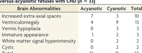
[image:3.594.54.284.379.554.2]were primarily diagnosed in fetuses with acyanotic heart disease (7/10), while unilateral ventriculomegaly was 3 times more fre-quent in fetuses with cyanotic CHD. White matter signal abnor-malities, cysts, and vermian hypoplasia were exclusively described in the subgroup with cyanotic lesions. The distribution of brain abnormalities between fetuses with cyanotic-versus-acyanotic CHD is reported in Table 3.
Table 1: CHD diagnoses distribution (nⴝ144)
Diagnosis Counts (%)
Abnormal MRI Findings (%) Hypoplastic left-heart syndrome 32 (22.2) 7 (21.21) Transposition of the great artery 27 (18.8) 5 (15.2) Double-outlet right ventricle 16 (11.1) 5 (15.2)
Tetralogy of Fallot 17 (11.8) 5 (15.2)
Pulmonary atresia 12 (8.3) 1 (3.0)
Atrioventricular septal defect 7 (5.9) 4 (12.1)
Aortic stenosis 6 (4.2) 2 (6.1)
Tricuspid atresia 6 (4.2) 0 (0.0)
Ventricular septal defect 4 (2.8) 0 (0.0) Hypoplastic right-heart syndrome 4 (2.8) 1 (3.0)
Pulmonary stenosis 2 (1.4) 1 (3.0)
Truncus arteriosus 2 (1.4) 2 (6.1)
Ebstein anomaly 1 (0.7) 0 (0.0)
Severe coarctation of the aorta 1 (0.7) 0 (0.0)
Total 144 (100) 33 (100)
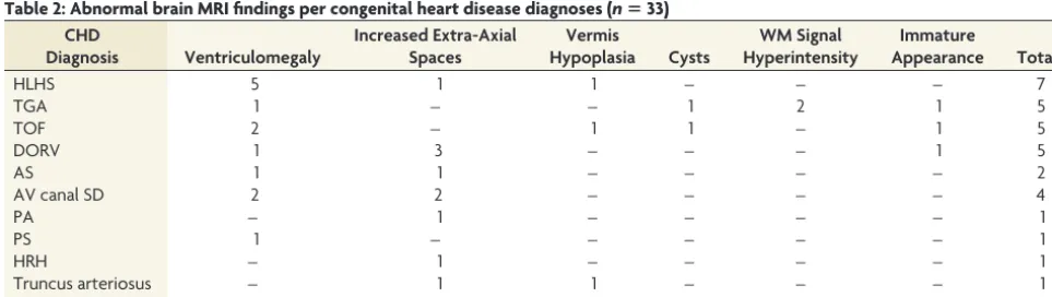
Table 2: Abnormal brain MRI findings per congenital heart disease diagnoses (nⴝ33) CHD
Diagnosis Ventriculomegaly
Increased Extra-Axial Spaces
Vermis
Hypoplasia Cysts
WM Signal Hyperintensity
Immature
Appearance Total
HLHS 5 1 1 – – – 7
TGA 1 – – 1 2 1 5
TOF 2 – 1 1 – 1 5
DORV 1 3 – – – 1 5
AS 1 1 – – – – 2
AV canal SD 2 2 – – – – 4
PA – 1 – – – – 1
PS 1 – – – – – 1
HRH – 1 – – – – 1
Truncus arteriosus – 1 1 – – – 1
Total 13 10 3 2 2 3 33
[image:3.594.52.534.578.714.2]Fetal Brain Abnormalities and Gestational Age
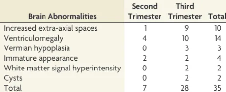
The mean GA of fetuses with CHD with brain abnormalities was 31.15⫾3.46 weeks and was not significantly different compared with fetuses with CHD with normal findings on brain MR imag-ing studies (mean GA⫽31.78⫾3.78 weeks;t⫽0.748,P⫽.44). Abnormalities were not found to be significantly more frequent during the second or third trimester (2⫽1.91,P⫽.662), and
their distribution is reported in Table 4. Among the 35 MR imag-ing studies with abnormal findimag-ings in the CHD group, 2 fetuses were scanned twice (ie, in the second and third trimesters). One fetus presented with mild bilateral ventriculomegaly, while the second presented with immature brain appearance. For both fe-tuses, findings remained consistent at both MR imaging time points.
In the control group, the 3 brain ab-normalities were noted on second-tri-mester MR imaging studies. The fetus with a germinal matrix hemorrhage in the left ventricle underwent a second MR imaging during the third trimester, and mild enlargement of the lateral ven-tricle secondary to the hemorrhage was reported.
DISCUSSION
This is the first prospective study to de-scribe the prevalence of brain abnormal-ities by conventional clinical MR imag-ing in a large, consecutive cohort of fetuses with CHD and to compare them with a control group of fetuses from healthy pregnancies. We found that structural brain abnormalities were sig-nificantly more frequent in fetuses with CHD, with approximately one-quarter (23%) demonstrating in utero structural brain abnormalities compared with
⬍2% in the control group. Mild unilat-eral ventriculomegaly and increased ex-tra-axial CSF spaces were most com-monly reported, together representing almost 70% (22/33) of all antenatal brain abnormalities in fetuses with CHD. Other findings among fetuses with CHD included vermian hypoplasia (n ⫽ 3), cerebral cortical immaturity (n⫽3), periventricular cysts (n⫽2), white matter signal hyperintensity (n⫽
2), and mild bilateral ventriculomegaly (n⫽1). Finally, we found no significant difference in the prevalence of brain abnormalities in fetuses with single- versus 2-ventricle physiology or in cyanot-ic-versus-acyanotic lesions.
Unilateral ventriculomegaly was found in 12 (8%) of the fe-tuses with CHD. Our findings corroborate the retrospective find-ings reported by Mlczoch et al,9who described mild asymmetric
dilation of the lateral ventricles as the most common brain anom-aly in this population. Unilateral mild ventriculomeganom-aly is, in fact, one of the most common brain abnormalities referred for fetal MR imaging and counseling in clinical fetal centers11,13and is estimated
to affect 0.07% of all pregnancies.10Only 1 fetus in our control group
(0.05%) presented with mild unilateral ventriculomegaly.
Increased extra-axial CSF spaces were uniquely reported in fetuses with CHD and were present in 10 cases (9 reported in the third trimester), making this the second most common abnor-mality in this group. Enlarged extra-axial CSF spaces were mild and isolated in all cases, except in 1 fetus that also had mild uni-lateral ventriculomegaly. The finding of increased extra-axial CSF spaces could be due to delayed parenchymal brain development or disturbed CSF dynamics (possibly due to the fetal cardiac anomaly) or both. Additionally, immature cerebral cortical devel-opment was detected in 2 fetuses with CHD (2%) compared with
FIG 1. A,T2-weighted axial view of the brain of a healthy control at 32.55 weeks’ GA.B, T2-weighted axial view of the brain of a patient with unilateral left ventriculomegaly at 32.00 weeks’ GA.
[image:4.594.56.375.48.205.2]FIG 2. A,T2-weighted axial view of the brain of a healthy control at 26.56 weeks’ GA.B, T2-weighted axial view of the brain of a patient with extra-axial spaces at 27.28 weeks’ GA.
Table 3: Distribution of abnormal brain MRI findings in cyanotic-versus-acyanotic fetuses with CHD (nⴝ33)
Brain Abnormalities Acyanotic Cyanotic Total
Increased extra-axial spaces 7 3 10
Ventriculomegaly 4 9 13
Vermis hypoplasia 0 3 3
Immature appearance 1 2 3
White matter signal hyperintensity 0 2 2
Cysts 0 2 2
[image:4.594.55.375.251.410.2] [image:4.594.55.285.465.551.2]only 1 (0.5%) in the control group. Together, these findings sup-port the notion that delayed brain maturation is likely a direct consequence of the cardiac defect. In our previous studies of fe-tuses with CHD by using quantitative brain MR imaging tech-niques, we described a progressive third-trimester deceleration in cerebral growth7and delayed cortical maturation (in fetuses with
HLHS)8compared with healthy fetuses. In the current study, 3 of
these cases of suspected delay in brain maturation were detected by clinical MR imaging readings before the third trimester. Ten women in our CHD sample underwent repeated fetal MR imag-ing studies in the second and third trimesters, and in all cases, the findings were enduring within cases. Nevertheless, a larger sample with serial fetal MR imaging studies is needed to better delineate the onset and progression of delayed brain development in fetuses with CHD.
Disturbances in oxygen/substrate supply present a major threat to brain development in fetuses with CHD.14,15 Using
Doppler sonography, Donofrio et al15compared the
cerebral-placental ratio between fetuses with CHD and healthy controls and showed that the normal intrinsic cerebral compensatory re-sponse to insufficient oxygen-substrate delivery (ie, cerebral va-sodilation) was activated in fetuses with CHD. The cerebral-pla-cental ratio was significantly lower overall in fetuses with CHD, with values⬍1.0 most prevalent in fetuses with a single-ventricle physiology. Reduced cerebral-placental ratio was significantly as-sociated with decreased head circumference, supporting the no-tion that the “brain-sparing” response was insufficient to sustain brain growth. In addition, we have previously reported a signifi-cant association between smaller total brain volume and lower combined ventricular output from the aorta in fetuses with CHD.7In the current study, evidence of brain abnormalities,
including delayed brain development, was present among fetuses with various CHD diagnoses, suggesting that the risk of disturbed brain maturation in fetuses with CHD is not restricted to those with the most complex cardiac lesions, such as HLHS. This is also consistent with Mlczoch et al,9who found no association between
brain abnormalities and the type of CHD diagnosis. Future stud-ies that relate brain abnormalitstud-ies to specific Doppler ultrasound measures are needed to address this important question.
One fetus in the control group had a germinal matrix hemor-rhage detected on MR imaging at 23 weeks’ GA. Germinal matrix hemorrhage is a common complication of premature birth but has been rarely described in the fetus in utero. The history of the pregnancy was unremarkable. Secondary enlargement of the
af-fected ventricle was reported on the follow-up MR imaging at 32 weeks and was consistent with the few in vivo cases reported in the literature.16The patient was referred for clinical neurologic
con-sultation and follow-up.
Three fetuses in our CHD cohort were diagnosed with inferior vermian hypoplasia, an anomaly that may represent delayed or arrested development of the cerebellar vermis. In previous stud-ies, we have described an overall favorable prognosis for infants and school-aged children with a fetal MR imaging diagnosis of isolated vermian hypoplasia.17However, the impact of inferior
vermian development on subsequent neuropsychologic develop-ment in fetuses with CHD is unknown and awaits further investigation.
White matter abnormalities were detected in 4 fetuses with CHD and in none of the control fetuses. These abnormalities included T2-weighted signal abnormality and cysts in the white matter. Acquired white matter signal hyperintensity was exclu-sively found in 2 fetuses with transposition of the great arteries. Subependymal cysts (possibly the residua of ischemic-hemor-rhagic injury) were present in 2 fetuses, one with transposition of the great arteries and another with tetralogy of Fallot. Although the available literature is limited, the prognosis of isolated sub-ependymal cysts diagnosed in utero has been good.18,19White
matter abnormalities, thought to be a form of periventricular leu-komalacia, have been reported in fetuses with HLHS20on
post-mortem examination and in a number of postnatal studies of infants with CHD.5,21,22The relation between the fetal WM
ab-normalities seen in our population and the periventricular leu-komalacia described after birth in infants with CHD is not clear but could support an antenatal onset in some cases. The prognos-tic significance of fetal white matter signal intensity in this popu-lation remains to be determined. The current limitations of con-ventional fetal MR imaging may have hindered our ability to detect other cases of white matter abnormalities in utero. Al-though diffusion-weighted imaging is increasingly used in fetal MR imaging, this acquisition is particularly sensitive to fetal mo-tion that results in image degradamo-tion.23A recent case report
sug-gested that differences in the white matter microstructural orga-nization could also be detected in utero during the third trimester of 3 fetuses with CHD compared with healthy fetuses.24However,
further studies that assess white matter integrity in fetuses with CHD are needed to better understand the relationship between these macro- and microstructural preliminary findings and their long-term consequences.
In postnatal preoperative studies of infants with CHD, the most prevalent brain lesions included ischemic lesions21and
dif-fuse and focal white matter injury,6,25,26including periventricular
leukomalacia.5,27The brain MR imaging findings in our fetal
co-hort with CHD differed in both type and severity from these post-natal studies of infants with CHD. In fact, with the exception of 1 fetus with diffuse white matter injury, the brain anomalies de-scribed in our sample were all mild in severity. This difference in lesion severity could be explained, in part, by hemodynamic in-sults occurring during the complex transition from a fetal to post-natal circulation, a potentially hazardous period for infants with CHD.28Moreover, recent evidence suggests that brain injuries
[image:5.594.54.285.65.159.2]found in neonates with CHD preoperatively are associated with
Table 4: Distribution of abnormal brain MRI findings among fetuses with CHD per trimester of pregnancy (nⴝ35)a
Brain Abnormalities
Second Trimester
Third Trimester Total
Increased extra-axial spaces 1 9 10
Ventriculomegaly 4 10 14
Vermian hypoplasia 0 3 3
Immature appearance 2 2 4
White matter signal hyperintensity 0 2 2
Cysts 0 2 2
Total 7 28 35
a
microstructural and metabolic markers of brain immaturity.29
Follow-up postnatal MR imaging studies and neurodevelopmen-tal outcome assessments are needed to determine whether the prenatal findings described herein are transient or persistent in nature and their impact on long-term outcome. These studies are currently underway.
To our knowledge, this study is the first prospective study to describe conventional clinical MR imaging findings in a large sample of fetuses with CHD during the second and third trimes-ters of pregnancy and to compare them with a large control group of fetuses from healthy pregnancies. Moreover, our CHD sample included a representative range of cardiac diagnostic categories and was not limited to those with the most severe forms of CHD (eg, HLHS). However, given that we carefully excluded cases with multiorgan dysmorphic conditions, as well as chromosomal or genetic conditions, we may have underestimated the true preva-lence of brain abnormalities in the overall fetal CHD population. Structural abnormalities of the brain may result as part of a spec-trum of other congenital malformations including CHD. Finally, our overall goal was to describe the prevalence and spectrum of brain abnormalities detected on conventional MR imaging in fe-tuses with complex CHD. This objective was carried out by deter-mining the frequency with which neuroradiologists made the clinical diagnosis of brain abnormality in fetuses with CHD com-pared with healthy control fetuses. Therefore, we did not perform repeat blinded MR imaging interpretations to assess intra- and inter-reader reliability. Nevertheless, our study was conducted in 2 major referral centers for congenital heart disease, and all MR imaging studies were reviewed by an experienced fetal neuroradi-ologist following clinical best practice standards. Consequently, we are confident that the findings reported in this study are rep-resentative of the current findings in clinical settings.
CONCLUSIONS
During the past decade, in vivo fetal MR imaging has contributed enormously to our understanding of fetal brain abnormalities in the living fetus. Our findings demonstrate that structural brain abnormalities are much more prevalent in fetuses with complex CHD than in controls and are not confined to those with the most critical forms of CHD.
However, although prevalent, the findings in our study were relatively mild in extent and were often suggestive of delayed or arrested brain maturation. These data contribute to a growing body of evidence suggesting that an important com-ponent of neurologic dysfunction in survivors of CHD is pre-natal in origin.
Disclosures: Dorothy Bulas—RELATED:Grant: Canadian Institutes of Health Re-search.*UNRELATED:Consultancy: unpaid consultant to GE Healthcare and Philips Healthcare on ultrasound technology not relevant to this research,Royalties: UpTo-Date chapter royalties. Mary Donofrio—RELATED:Grant: Canadian Institutes of Health Research,* Catherine Limperopoulos—RELATED:Grant: Canadian Institutes of Health Research.**Money paid to the institution.
REFERENCES
1. Owen M, Shevell M, Majnemer A, et al.Abnormal brain structure and function in newborns with complex congenital heart defects before open heart surgery: a review of the evidence.J Child Neurol
2011;26:743–55
2. Donofrio MT, Massaro AN.Impact of congenital heart disease on brain development and neurodevelopmental outcome.Int J Pediatr
2010;2010. pii:359390
3. Block AJ, McQuillen PS, Chau V, et al.Clinically silent preoperative brain injuries do not worsen with surgery in neonates with congen-ital heart disease.J Thorac Cardiovasc Surg2010;140:550 –57 4. Licht DJ, Shera DM, Clancy RR, et al.Brain maturation is delayed in
infants with complex congenital heart defects.J Thorac Cardiovasc Surg2009;137:529 –36, discussion 536 –37
5. Licht DJ, Wang J, Silvestre DW, et al.Preoperative cerebral blood flow is diminished in neonates with severe congenital heart defects.
J Thorac Cardiovasc Surg2004;128:841– 49
6. Miller SP, McQuillen PS, Hamrick S, et al.Abnormal brain develop-ment in newborns with congenital heart disease.N Engl J Med
2007;357:1928 –38
7. Limperopoulos C, Tworetzky W, McElhinney DB, et al.Brain vol-ume and metabolism in fetuses with congenital heart disease: eval-uation with quantitative magnetic resonance imaging and spec-troscopy.Circulation2010;121:26 –33
8. Clouchoux C, du Plessis AJ, Bouyssi-Kobar M, et al.Delayed cortical development in fetuses with complex congenital heart disease.
Cereb Cortex2013;23:2932– 43
9. Mlczoch E, Brugger P, Ulm B, et al.Structural congenital brain dis-ease in congenital heart disdis-ease: results from a fetal MRI program.
Eur J Paediatr Neurol2013;17:153– 60
10. Wax JR, Bookman L, Cartin A, et al. Mild fetal cerebral ventriculomegaly: diagnosis, clinical associations, and out-comes.Obstet Gynecol Surv2003;58:407–14
11. Girard NJ, Raybaud CA.Ventriculomegaly and pericerebral CSF collection in the fetus: early stage of benign external hydrocepha-lus?Childs Nerv Syst2001;17:239 – 45
12. Watanabe Y, Abe S, Takagi K, et al.Evolution of subarachnoid space in normal fetuses using magnetic resonance imaging.Prenat Diagn
2005;25:1217–22
13. Glenn OA, Barkovich AJ.Magnetic resonance imaging of the fetal brain and spine: an increasingly important tool in prenatal diagno-sis, part 1.AJNR Am J Neuroradiol2006;27:1604 –11
14. Donofrio MT.The heart– brain interaction in the fetus: cerebrovas-cular blood flow in the developing human.Prog Pediatr Cardiol
2006;22:41–51
15. Donofrio MT, Bremer YA, Schieken RM, et al.Autoregulation of cerebral blood flow in fetuses with congenital heart disease: the brain sparing effect.Pediatr Cardiol2003;24:436 – 43
16. Fukui K, Morioka T, Nishio S, et al.Fetal germinal matrix and intra-ventricular haemorrhage diagnosed by MRI. Neuroradiology
2001;43:68 –72
17. Tarui T, Limperopoulos C, Sullivan NR, et al.Long-term develop-mental outcome of children with a fetal diagnosis of isolated inferior vermian hypoplasia. Arch Dis Child Fetal Neonatal Ed
2014;99:F54 –58
18. Bats AS, Molho M, Senat MV, et al.Subependymal pseudocysts in the fetal brain: prenatal diagnosis of two cases and review of the literature.Ultrasound Obstet Gynecol2002;20:502– 05
19. D’Addario V, Selvaggio S, Pinto V, et al.Fetal subependymal cysts with normal neonatal outcome: a case report.Fetal Diagn Ther
2003;18:170 –73
20. Hinton RB, Andelfinger G, Sekar P, et al.Prenatal head growth and white matter injury in hypoplastic left heart syndrome.Pediatr Res
2008;64:364 – 69
21. Mahle WT, Tavani F, Zimmerman RA, et al.An MRI study of neu-rological injury before and after congenital heart surgery. Circula-tion2002;106(12 suppl 1):1109 –14
22. Andropoulos DB, Hunter JV, Nelson DP, et al.Brain immaturity is associated with brain injury before and after neonatal cardiac sur-gery with high-flow bypass and cerebral oxygenation monitoring.
J Thorac Cardiovasc Surg2010;139:543–56
24. Berman JI, Hamrick SEG, McQuillen PS, et al.Diffusion-weighted imaging in fetuses with severe congenital heart defects.AJNR Am J Neuroradiol2011;32:E21–22
25. McQuillen PS, Hamrick SE, Perez MJ, et al.Balloon atrial septos-tomy is associated with preoperative stroke in neonates with trans-position of the great arteries.Circulation2006;113:280 – 85 26. Partridge SC, Vigneron DB, Charlton NN, et al.Pyramidal tract
mat-uration after brain injury in newborns with heart disease.Ann Neu-rol2006;59:640 –51
27. Petit CJ, Rome JJ, Wernovsky G, et al.Preoperative brain injury in transpo-sition of the great arteries is associated with oxygenation and time to sur-gery, not balloon atrial septostomy.Circulation2009;119:709–16 28. Friedman AH, Fahey JT.The transition from fetal to neonatal
circulation: normal responses and implications for infants with heart disease.Semin Perinatol1993;17:106 –21