ORIGINAL RESEARCH
ADULT BRAIN
Performance of CT ASPECTS and Collateral Score in Risk
Stratification: Can Target Perfusion Profiles Be Predicted
without Perfusion Imaging?
XS. Dehkharghani, XR. Bammer, XM. Straka, XM. Bowen, XJ.W. Allen, XS. Rangaraju, XJ. Kang, XT. Gleason, X C. Brasher, and XF. Nahab
ABSTRACT
BACKGROUND AND PURPOSE: Endovascular trials suggest that revascularization benefits a subset of acute ischemic stroke patients with large-artery occlusion and small-core infarct volumes. The objective of our study was to identify thresholds of noncontrast CT–ASPECTS and collateral scores on CT angiography that best predict ischemic core volume thresholds quantified by CT perfusion among patients with acute ischemic stroke.
MATERIALS AND METHODS: Fifty-four patients with acute ischemic stroke (⬍12 hours) and MCA/intracranial ICA occlusion underwent NCCT/CTP during their initial evaluation. CTP analysis was performed on a user-independent platform (RApid processing of PerfusIon and Diffusion), computing core infarct (defined as CBF of⬍30% normal). A target mismatch profile consisting of infarction core ofⱕ50 mL was selected to define candidates with acute ischemic stroke likely to benefit from revascularization.
RESULTS:NCCT-ASPECTS ofⱖ9 with a CTA collateral score of 3 had 100% specificity for identifying patients with a CBF core volume of ⱕ50 mL. NCCT-ASPECTS ofⱕ6 had 100% specificity for identifying patients with a CBF core volume of⬎50 mL. In our cohort, 44 (81%) patients had an NCCT-ASPECTS ofⱖ9, a CTA collateral score of 3, or an NCCT-ASPECTS ofⱕ6.
CONCLUSIONS: Using an NCCT-ASPECTS ofⱖ9 or a CTA collateral score of 3 best predicts CBF core volume infarct ofⱕ50 mL, while an NCCT-ASPECTS ofⱕ6 best predicts a CBF core volume infarct of⬎50 mL. Together these thresholds suggest that a specific population of patients with acute ischemic stroke not meeting such profiles may benefit most from CTP imaging to determine candidacy for revascularization.
ABBREVIATIONS:AIS⫽acute ischemic stroke; RAPID⫽RApid processing of PerfusIon and Diffusion
R
evascularization aims to prevent progression of ischemic injury in acute ischemic stroke (AIS).1-4Recent success intrials of endovascular AIS therapy, while restoring motivation for acute stroke intervention, has left the subject of an optimal patient-selection paradigm largely unaddressed.5-8While the
primary goals in this setting include timely revascularization, the relative merits of expedited triage versus identification of target imaging profiles remain the subject of ongoing inquiry.
Contemporary guidelines on AIS management, therefore, remain inconclusive as to the role of multimodal imaging selection.9
We recently reported the benefits of a high-speed computing tool for CT perfusion analysis over qualitative approaches to im-aging triage for prognostication among patients with anterior cir-culation AIS.10The findings therein suggested that a user- and
vendor-independent computational tool may outperform purely qualitative approaches in outcome prediction. Similar imple-mentations of this tool in recent, prospective endovascular tri-als suggested strong results as an approach to patient selection; however, the relative contribution of CTP-based selection cri-teria, among other trial-specific features, remains uncertain in light of the overall favorable outcomes reported across dispa-rate trial designs.5-8
The objective of our study was to identify thresholds of NCCT-ASPECTS and collateral score on CT angiography that best pre-dict ischemic core volume thresholds quantified by CTP among patients with AIS.
Received November 20, 2015; accepted after revision January 10, 2016. From the Departments of Radiology and Imaging Sciences (S.D., M.B., J.W.A., T.G.) and Neurology (S.D., J.W.A., S.R., C.B., F.N.), Emory University Hospital, Atlanta, Georgia; Department of Radiology (R.B.), Stanford University Hospital, Stanford, California; Institut fu¨r Radiologie und Nuklearmedizin (M.S.), Kantonsspital Winter-thur, WinterWinter-thur, Switzerland; and Department of Biostatistics (J.K.), University of Michigan, Ann Arbor, Michigan.
Please address correspondence to Seena Dehkharghani, MD, Department of Radiology and Imaging Sciences and Neurology, Neuroradiology Division; Attention: Mary Davis, Department of Radiology and Imaging Sciences, Emory University Hospital, 1364 Clifton Rd NE, Atlanta, GA 30322; e-mail: [email protected]
MATERIALS AND METHODS
The population examined was previously studied in a report of the relative predictive value of quantitative and qualitative acute stroke imaging analysis in prognostication and clinical outcome prediction.10Briefly, 62 continuous patients (36 women; median
age, 70 years; range, 33–94 years) with AIS (⬍12 hours) and MCA or intracranial ICA occlusion were identified from a prospectively collected, single-institution stroke registry and radiologic infor-matics query of 815 patients with ischemic stroke, spanning Feb-ruary 1, 2011, to December 31, 2013, with Emory University Hos-pital review board approval. All patients were evaluated initially by a dedicated vascular neurologist in the emergency setting, with initiation of institutional stroke protocol facilitating expedited triage, imaging, interpretation, and treatment when appropriate. All patients underwent comprehensive stroke imaging at presen-tation, including NCCT, CTA, and CTP. Patients were included in the analysis on the basis of successful completion of the imaging protocol, absence of motion or other artifacts rendering imaging nondiagnostic, and the absence of large hemorrhages such as pa-renchymal hematomas (types 1 and 2) potentially confounding final infarction measurement. Exclusion criteria were an inability to undergo multimodal CT, a history of renal failure, and patient age younger than 18 years. A subset of patients received intrave-nous and/or intra-arterial thrombolytic therapy as per institu-tional protocol and at the discretion of the treating vascular neu-rologist and neurointerventionalist.
Imaging Protocol
All patients underwent an institutional stroke imaging protocol including NCCT, CTP, and CTA. CT was performed on a 40-mm, 64 – detector row clinical system (LightSpeed VCT; GE Health-care, Milwaukee, Wisconsin). Helical NCCT (120 kV[peak], 100 – 350 auto-mAs) was performed from the foramen magnum through the vertex at 5.0-mm section thickness. In the absence of visible intracranial hemorrhage during real-time evaluation by a radiologist and stroke neurologist, 2 contiguous CTP slabs were obtained for 8-cm combined coverage of the supratentorial brain, obtained at 5-mm sections per slab. Cine mode acquisition (80 kVp, 100 mAs) permitting high-temporal-resolution (1-second sampling interval) dynamic bolus passage imaging was performed following the administration of 35 mL of iodinated contrast (io-pamidol, Isovue 370; Bracco Diagnostics, Princeton, New Jersey), power injected at 5 mL/s through an 18-ga or larger antecubital IV access. Contrast administration was followed by a 25-mL saline flush at the same rate. For both slabs, the same acquisition and injection protocol was used (ie, a total of 70 mL of iodine was used for CTP). Last, helical CTA (120 kVp, 200 –350 auto-mAs) was performed from the carina to the vertex (section thickness/inter-val, 0.625 /0.375 mm) following IV administration of 70 mL of iodinated contrast injected at 5 mL/s and followed by a 25-mL saline flush.
Follow-up imaging in all patients included brain MR imaging for documentation of final infarct size within 3 days of CTP, per-formed on a 3T (Tim Trio; Siemens, Erlangen, Germany) clinical whole-body system with local signal reception by a dedicated 12-channel head coil. All images were transferred to a separate work-station for analysis (Mac Pro; Apple, Cupertino, California) by
using a third-party DICOM viewer (OsiriX Imaging Software; http:// www.osirix-viewer.com). The details of the postprocessing pipeline were reported previously.10
Imaging Analysis
NCCT-ASPECTS. ASPECTSs were assigned by 2 experienced vas-cular neurologists (S.R., F.N.) blinded to all other imaging and clinical outcomes. ASPECTS uses a 10-point visual inspection scale estimating ischemic burden in the supratentorial brain as detailed previously by Barber et al.11
CTA Collateral Score. A CTA-derived collateral vessel-scoring methodology was used as detailed previously.10,12Briefly, 2
expe-rienced neuroradiologists, both with subspecialty certification and experienced in stroke and neurovascular imaging, assigned CTA collateral scores using a visual inspection methodology to quantify surface leptomeningeal collaterals in response to proxi-mal arterial compromise, compared with the contralateral side.12
The neuroradiologists assigned scores blinded to clinical and out-comes data and all other imaging.13,14A collateral score was
as-signed by using an ordinal, visual grading system estimating col-lateral flow, scored 0 –3 as follows: Colcol-lateral flow was assigned a score of zero for absent surface vasculature, 1 for⬎0 butⱕ50% vasculature, 2 for⬎50 but⬍100% vasculature, and 3 for normal or supranormal surface vasculature of the MCA territory. CTA analysis was performed by using 20-mm axial sliding maximum intensity projection and 0.625-mm axial source images and or-thogonal and curved multiplanar reformats as needed.
CT Perfusion. All perfusion imaging was postprocessed by using a custom, noncommercial version of a vendor-independent soft-ware platform (RAPID) provided by Stanford University.15
RAPID is an automated computational tool designed for timely analysis of CTP data as used recently in the stroke trial setting.3-5,7
Details of the perfusion postprocessing pipeline were discussed previously.10,15Briefly, following preprocessing steps correcting
rigid-body motion, arterial input function selection is performed and deconvolved from the voxel time-attenuation course using a delay-insensitive algorithm for isolation of the tissue residue function. The time to maximum of the tissue residue function is determined on a voxelwise basis, and time-to-maximum maps are incrementally thresholded between 4 and 10 seconds at 2-second intervals with penumbral maps overlaid on the source CTP data.10,15
Cerebral blood flow maps expressed in milliliters/100 g/min-ute were compg/min-uted as outlined elsewhere. Relative CBF maps have been used in the stroke trial setting as estimates of irrevers-ibly infarcted (core) tissues by using thresholds of relative CBF of
⬍30% contralateral normal tissues.16Parametric maps were
au-tomatically generated and overlaid on source images for review purposes.
Statistical Analysis
ANOVA analysis was performed on the fitted linear regression, and anF-test was applied. Because the NCCT-ASPECTS was con-sidered a dummy variable in the regression and ANOVA analyses, the Kendallcorrelation was used to test the strength of the cor-relation. Given the previously high interreader correlation for the NCCT-ASPECTS ofⱖ7 (0.93) in our study population, all vari-ables were assessed as the unweighted mean of combined reader scores for qualitative variables.10
An operationally defined CTP profile predicting favorable outcome was assigned as prescribed in a recently reported, pro-spective endovascular therapy trial.5Specifically, a target
mis-match profile consisting of an infarction core ofⱕ50 mL was selected as the target relative CBF infarction core volume deter-mined across ASPECTS and at a dichotomized NCCT-ASPECTS ofⱖ7. The 50 mL threshold was selected as a reference volume as reported by the investigators of the recent Solitaire With the Intention for Thrombectomy as Primary Endovascular Treatment (SWIFT PRIME) trial and as used in the recent devel-opment of a benchmarking software environment for quality control in stroke perfusion imaging.5,17The Fisher exact test was
further applied to determine the association of ASPECTS ofⱖ7 and infarction core ofⱕ50 mL. A prediction error model for correct classification of patients as having greater or less than 50 mL CBF infarction core was determined across NCCT-ASPECTS and at a dichotomized ASPECTS ofⱖ7. Sensitivity, specificity, and positive and negative predictive values for various thresholds of ASPECTS and collateral scores were calculated and reported.
Statistical analysis was performed in R statistical and comput-ing software (http://www.r-project.org/).
RESULTS
Sixty-two patients (36 women; median age, 70 years; range, 33–94 years) with AIS (⬍12 hours) and MCA or intracranial ICA occlu-sion constituted the study population as previously reported. In-complete or degraded imaging necessitated exclusion of 8 pa-tients, leaving 54 patients for analysis. An ICA or M1 occlusion was present in 41 (76%) patients, with proximal M2 segment occlusion in the remainder. No patients had bilateral arterial occlusions.
As shown inTable 1, the median NIHSS score at admission was 15 (interquartile range, 16); the mean duration from the time of onset/last known healthy to imaging was 210 minutes. Twenty-three (43%) patients received IV tPA, and 9 (17%) underwent endovascular treatment with intra-arterial tPA (n⫽3) or throm-bectomy (n⫽6). The median NCCT-ASPECTS was 9
(interquar-tile range, 1). Median final infarction volume as measured by MR imaging was 37 (interquartile range, 96) mL.
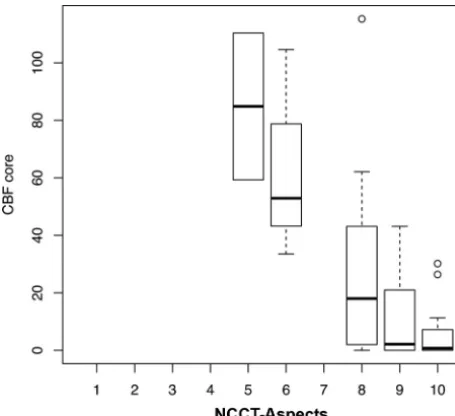
While a significant association was detected between CBF core estimates and NCCT-ASPECTS (Kendallcorrelation,⫺0.51; P ⬍ .01), large variability was found across 2-reader mean ASPECTS values (Fig 1). For example, across patients with an NCCT-ASPECTS of 8, the CBF core volume ranged from 0 to 115 mL with a median of 23 mL (interquartile range, 42 mL). Ranges of core volume increased further at lower ASPECTS.Table 2 pres-ents sensitivity, specificity, negative predictive values, and positive predictive values of the candidate predictors, NCCT-ASPECTS and collateral scores. For the analysis of dichotomized ASPECTS ofⱖ7, the CBF core volume ranged from 0 to 115 mL with a median of 4 mL (interquartile range, 15 mL). An NCCT-ASPECTS ofⱖ9 had 100% specificity (95% CI, 60 –100) for iden-tifying patients with CBF core volume of ⱕ50 mL, while an NCCT-ASPECTS ofⱕ6 had 100% specificity (95% CI, 90%– 100%) for identifying patients with a CBF core volume of⬎50 mL. The prediction error model for correct identification of in-farction core ofⱕ50 mL among ASPECTS ofⱖ7 demonstrated significant associations but low specificity relative to a CBF core of ⱕ50 mL (prediction error, 9%;P⫽.025; sensitivity, 0.98; speci-ficity, 0.50; negative predictive value, 0.80; positive predictive value, 0.92).
[image:3.594.304.532.46.254.2]Our cohort included 28 patients with a collateral score of 1, 10 patients with a score of 2, and 15 patients with a score of 3. A collateral score of 3 on CTA had 100% specificity (95% CI, 47%– 99%) but only 33% sensitivity (95% CI, 20%– 49%) for identify-ing patients with AIS with a CBF core volume ofⱕ50 mL. A collateral score ofⱕ1 had an 88% sensitivity (95% CI, 47%–99%) and a 53% specificity (95% CI, 38%– 68%) for identifying pa-tients with AIS with a CBF core volume of⬎50 mL. The collateral Table 1: Patient characteristicsa
Characteristics
Admission NIHSS 15 (16)
Time of onset/last known healthy to imaging (min) 210 (252)
IV tPA (No.) (%) 23 (43)
Endovascular treatment (No.) (%) 9 (17)
IA tPA 3
Thrombectomy 6
NCCT-ASPECTS 9 (1)
Final infarction volume (mL) 37 (96)
Note:—IA indicates intra-arterial.
a
Data are reported as median (interquartile range) unless otherwise noted.
[image:3.594.52.287.53.153.2]score demonstrated a significant association with CBF ischemic core volumes (P⬍.01).
DISCUSSION
Our study found significant variability in CBF core volumes among patients with AIS with similar NCCT-ASPECTS, we iden-tified thresholds of NCCT-ASPECTS ofⱖ9 and collateral scores of 3 with high specificity for finding patients with AIS with core volumes considered ideal for revascularization. We also found that NCCT-ASPECTS ofⱕ6 had a high specificity for identifying patients with AIS with core volumes that made them suboptimal candidates for revascularization.
Recent successful AIS trials have used disparate methodologies for patient selection. These have differed primarily in their use of either fast but potentially insensitive methodologies (eg, NCCT-ASPECTS) versus more rigorous approaches to estimating tissue viability with CTP, permitting operational tissue classification,
segmentation, and volume measures.3,4,7,18These specific factors
have been emphasized as targets for optimization and general requirements in the stroke-research setting in recent expert con-sensus.19 NCCT-ASPECTS aims to qualitatively identify early
ischemic changes modulated by ischemic bulk water shifts (ie, edema). The speed and nearly invariable access to NCCT-ASPECTS are clearly advantageous; however, reproducibility and interrater agreement are reportedly variable.11,20-22The
insensi-tivity of NCCT to initial water shifts, primarily those from the interstitial to the intracellular compartment preceding progres-sive vasogenic edema, may preclude accurate estimation of neu-ronal injury in the very early aftermath of infarction.14,20-23
NCCT-ASPECTS may furthermore be limited by its tendency to cluster largely variable volumes of injury across its coarsely chang-ing scale, as illustrated in Fig 2, in which identical NCCT-ASPECTS between 2 subjects can belie considerable differences in the actual volume of injury. Such challenges may underlie existing reports of greater agreement and predic-tive accuracy for CTP in comparison with NCCT-ASPECTS.13,14,24,25
Not-withstanding these features, we previ-ously reported a high interrater agree-ment in the assignagree-ment of dichotomized ASPECTS of⬎7, despite more marginal agreement across all ASPECTSs.
We selected an infarction core threshold of 50 mL as a reference vol-ume against which NCCT-ASPECTS and collateral scores were studied, as re-ported by the investigators of the recent SWIFT PRIME trial.5 The 50-mL
threshold has furthermore been
[image:4.594.53.378.277.422.2]pro-FIG 2.CT perfusion ischemic core estimates and complementary NCCT-ASPECTS in 2 patients (AandB). Selected images from panels of RAPID-derived CBF core maps (white overlays) and NCCT-ASPECTS in 2 subjects, both with an ASPECTS of 8. Large differences in the estimated volume of irreversible ischemic core are noted despite high ASPECTS in both patients presenting with acute stroke-like symptoms. Patient 1 is an 83-year-old woman (NIHSS score⫽29) with NCCT-ASPECTS hypoattenuation suspected within the anterior left insular region and lateral lentiform; Patient 2 is an 83-year-old man (NIHSS⫽28) with NCCT-ASPECTS abnormality suspected within the lateral perirolandic parietal lobe and the lateral temporal lobe (not shown). rCBF indicates relative CBF.
Table 2: Accuracy of NCCT-ASPECTS and collateral score in prediction of CBF infarction core of<50 mL
Threshold Sensitivity Specificity PPV NPV
NCCT-ASPECTS (CBF core volumeⱕ50 mL)
ⱖ4 100 0 84.9 –
ⱖ5 100 25 88.2 100
ⱖ6 100 37.5 90.0 100
ⱖ7 97.8 50 91.7 80.2
ⱖ8 91.1 62.5 93.2 55.5
ⱖ9 68.9 100 100 36.4
ⱖ10 26.7 100 100 19.5
Collateral score (CBF core volumeⱕ50 mL)
ⱖ0 100 0 84.9 –
ⱖ1 53.3 87.5 96.0 25.0
ⱖ2 33.3 100 100 21.0
ⱖ3 0 100 – 15.1
[image:4.594.53.531.443.669.2]posed in the development of a recently reported benchmarking tool derived from pooled, prospectively acquired stroke trial data to test the accuracy of perfusion-processing software for future trial use.17We thus propose that this threshold is relevant and
reflective of current viewpoints in stroke imaging. Specifically with regard to such CTP selection criteria in AIS trials, we ob-served large ranges and variability in CBF infarction core volumes across ASPECTS.5
Within this population, the CTA collateral score demon-strated a strong statistical association with CTP ischemic core volumes, but low sensitivity and specificity for threshold infarc-tion predicinfarc-tion. While these findings could reflect statistical lim-itations related to sample size, we hypothesize that collateral score and other static measures of surface vascularity may be unable to capture the dynamic nature of collateral enhancement, while also lacking in their ability to identify the truly nutritive capacity of surface vessels. Recent advances in CT angiography, particularly the development of timing-invariant CTA derived from CTP dy-namic bolus-passage source data, offer some promise in mitigat-ing the timmitigat-ing sensitivity of standard CTA in identifymitigat-ing collateral vessels.26
We acknowledge several study limitations, particularly those inherent in the retrospective nature of the analysis. Het-erogeneity in the study cohort precluded subselection of treat-ed-versus-untreated patients. However, we contend that bias related to treatment selection had a negligible impact on the study conclusions because the primary aim of our study was to examine the variability between contemporaneously acquired imaging triage strategies. The qualitative parameters in this study were generated from 2 independent, experienced read-ers, in whom variability may bias results; however, as previ-ously reported, interreader agreement was high across vari-ables in this study population.10The relative standard in this
study, against which the qualitative variables were compared, was the RAPID software environment. While other such soft-ware solutions are available, we recently reported the strengths of the RAPID tool as a fully automated, user- and vendor-independent means of semi-quantitative perfusion analysis. As a semi-quantitative CTP computing tool, RAPID has been shown to perform well, matching or exceeding the accuracy of similar software environments relative to a ground truth digi-tal perfusion phantom in a recent study, and the use of similar iterations of the RAPID tool in recent multicenter trials may further support the generalizability of our findings.3,4,5,7,27
Patient selection criteria likely modulate success in achieving a favorable clinical response following revascularization in acute ischemic stroke. The era of contemporary revascularization tech-nologies now permits timely and dependable restoration of flow in most cases; however, optimal identification of a target popula-tion for treatment remains critical, and the ideal selecpopula-tion strategy remains inconclusively established. These findings suggest that readily available and expedited approaches to selection such as ASPECTS correlate with commonly used perfusion parameters but may lack sensitivity to inform accurate and quantitative esti-mations of core volumes.
CONCLUSIONS
Using an NCCT-ASPECTS ofⱖ9 or a CTA collateral score of 3 best predicts a CBF core volume infarct ofⱕ50 mL, while an NCCT-ASPECTS ofⱕ6 best predicts a CBF core volume infarct of⬎50 mL. Together these thresholds suggest that a specific pop-ulation of patients with AIS not meeting such profiles may benefit most from CT perfusion to determine their candidacy for revascularization.
Disclosures: Matus Straka—UNRELATED:Employment: iSchemaView;Stock/Stock
Options: iSchemaView. Srikant Rangaraju—UNRELATED:Grants/Grants Pending:
National Institutes of Health,*Comments: Dr Rangaraju is a trainee under the Na-tional Institute of Neurological Disorders and Stroke T32 training grant (2T32 NS 007480-15, Principal Investigator, Allan I. Levey). *Money paid to the institution.
REFERENCES
1. Saver JL, Jahan R, Levy EI, et al.Solitaire flow restoration device versus the Merci retriever in patients with acute ischaemic stroke (SWIFT): a randomised, parallel-group, non-inferiority trial. Lan-cet2012;380:1241– 49CrossRef Medline
2. Nogueira RG, Lutsep HL, Gupta R, et al.Trevo versus Merci retriev-ers for thrombectomy revascularisation of large vessel occlusions in acute ischaemic stroke (TREVO 2): a randomised trial.Lancet
2012;380:1231– 40CrossRef Medline
3. Lansberg MG, Straka M, Kemp S, et al; DEFUSE 2 study investigators. MRI profile and response to endovascular reperfusion after stroke (DEFUSE 2): a prospective cohort study.Lancet Neurol2012;11: 860 – 67CrossRef Medline
4. Albers GW, Thijs VN, Wechsler L, et al; DEFUSE Investigators. Mag-netic resonance imaging profiles predict clinical response to early reperfusion: the diffusion and perfusion imaging evaluation for understanding stroke evolution (DEFUSE) study.Ann Neurol2006; 60:508 –17CrossRef Medline
5. Saver JL, Goyal M, Bonafe A, et al; SWIFT PRIME Investigators. Stent-retriever thrombectomy after intravenous t-PA vs. t-PA alone in stroke.N Engl J Med2015;372:2285–95CrossRef Medline
6. Goyal M, Demchuk AM, Menon BK, et al; ESCAPE Trial Investiga-tors.Randomized assessment of rapid endovascular treatment of ischemic stroke.N Engl J Med2015;372:1019 –30CrossRef Medline
7. Campbell BC, Mitchell PJ, Kleinig TJ, et al; EXTEND-IA Investiga-tors.Endovascular therapy for ischemic stroke with perfusion-im-aging selection.N Engl J Med2015;372:1009 –18CrossRef Medline
8. Berkhemer OA, Fransen PS, Beumer D, et al.A randomized trial of intraarterial treatment for acute ischemic stroke.N Engl J Med2015; 372:11–20CrossRef Medline
9. Powers WJ, Derdeyn CP, Biller J, et al; American Heart Association Stroke Council.2015 American Heart Association/American Stroke Association Focused Update of the 2013 Guidelines for the Early Management of Patients With Acute Ischemic Stroke Regarding Endovascular Treatment: A Guideline for Healthcare Professionals From the American Heart Association/American Stroke Associa-tion.Stroke2015;46:3020 –35CrossRef Medline
10. Dehkharghani S, Bammer R, Straka M, et al.Performance and pre-dictive value of a user-independent platform for CT perfusion analysis: threshold-derived automated systems outperform exam-iner-driven approaches in outcome prediction of acute ischemic stroke.AJNR Am J Neuroradiol2015;36:1419 –25CrossRef Medline
11. Barber PA, Demchuk AM, Zhang J, et al.Validity and reliability of a quantitative computed tomography score in predicting outcome of hyperacute stroke before thrombolytic therapy: ASPECTS Study Group—Alberta Stroke Programme Early CT Score.Lancet2000; 355:1670 –74CrossRef Medline
13. Sillanpaa N, Saarinen JT, Rusanen H, et al.The clot burden score, the Boston Acute Stroke Imaging Scale, the cerebral blood volume ASPECTS, and two novel imaging parameters in the prediction of clinical outcome of ischemic stroke patients receiving intravenous thrombolytic therapy. Neuroradiology 2012;54:663–72 CrossRef Medline
14. Sillanpaa N, Saarinen JT, Rusanen H, et al.CT perfusion ASPECTS in the evaluation of acute ischemic stroke: thrombolytic therapy per-spective.Cerebrovasc Dis Extra2011;1:6 –16CrossRef Medline
15. Straka M, Albers GW, Bammer R.Real-time diffusion-perfusion mismatch analysis in acute stroke.J Magn Reson Imaging2010;32: 1024 –37CrossRef Medline
16. Campbell BC, Christensen S, Levi CR, et al.Cerebral blood flow is the optimal CT perfusion parameter for assessing infarct core.Stroke
2011;42:3435– 40CrossRef Medline
17. Cereda CW, Christensen S, Campbell BC, et al.A benchmarking tool to evaluate computer tomography perfusion infarct core predic-tions against a DWI standard.J Cereb Blood Flow Metab2015 Oct 19. [Epub ahead of print]CrossRef Medline
18. Davis SM, Donnan GA, Parsons MW, et al; EPITHET Investigators. Effects of alteplase beyond 3 h after stroke in the Echoplanar Imag-ing Thrombolytic Evaluation Trial (EPITHET): a placebo-con-trolled randomised trial.Lancet Neurol2008;7:299 –309CrossRef Medline
19. Wintermark M, Albers GW, Broderick JP, et al.Acute stroke imaging research roadmap II.Stroke2013;44:2628 –39CrossRef Medline
20. Weir NU, Pexman JH, Hill MD, et al.How well does ASPECTS pre-dict the outcome of acute stroke treated with IV tPA?Neurology
2006;67:516 –18CrossRef Medline
21. Puetz V, Dzialowski I, Hill MD, et al.The Alberta Stroke Program Early CT Score in clinical practice: what have we learned?Int J Stroke
2009;4:354 – 64CrossRef Medline
22. Dzialowski I, Hill MD, Coutts SB, et al.Extent of early ischemic changes on computed tomography (CT) before thrombolysis: prognostic value of the Alberta Stroke Program Early CT Score in ECASS II.Stroke2006;37:973–78CrossRef Medline
23. Aviv RI, Mandelcorn J, Chakraborty S, et al.Alberta Stroke Program Early CT Scoring of CT perfusion in early stroke visualization and assessment. AJNR Am J Neuroradiol 2007;28:1975– 80 CrossRef Medline
24. Lin K, Rapalino O, Law M, et al.Accuracy of the Alberta Stroke Program Early CT Score during the first 3 hours of middle cerebral artery stroke: comparison of noncontrast CT, CT angiography source images, and CT perfusion.AJNR Am J Neuroradiol2008;29: 931–36CrossRef Medline
25. van Seeters T, Biessels GJ, Niesten JM, et al; Dust Investigators. Reli-ability of visual assessment of non-contrast CT, CT angiography source images and CT perfusion in patients with suspected isch-emic stroke.PLoS One2013;8:e75615CrossRef Medline
26. Smit EJ, Vonken EJ, van der Schaaf IC, et al.Timing-invariant recon-struction for deriving high-quality CT angiographic data from ce-rebral CT perfusion data. Radiology 2012;263:216 –25 CrossRef Medline
27. Kudo K, Christensen S, Sasaki M, et al; Stroke Imaging Repository (STIR) Investigators.Accuracy and reliability assessment of CT and MR perfusion analysis software using a digital phantom.Radiology