ORIGINAL RESEARCH
PEDIATRICS
Development of a Standardized MRI Scoring Tool for
CNS Demyelination in Children
L.H. Verhey, H.M. Branson, S. Laughlin, M.M. Shroff, S.M. Benseler, B.M. Feldman, D.L. Streiner, J.G. Sled, and B. Banwell
ABSTRACT
BACKGROUND AND PURPOSE: The degree to which MR imaging is useful in the diagnosis of MS is predicated on standardized and reliable evaluation of MR imaging parameters. We aimed to devise items for an MR imaging scoring tool that would have high inter-rater agreement and would be straightforward to apply.
MATERIALS AND METHODS:On the basis of a literature search and consensus of an expert panel, we identified 48 parameters that describe acute CNS demyelination, predict MS diagnosis, or characterize demyelinating disorder mimics. MR images of children with clinically confirmed MS, monophasic ADEM, and angiography-negative biopsy-positive small-vessel primary angiitis of the CNS were scored by 2 neuroradiologists independently, using the preliminary 48-parameter tool. Parameters with Cohen ⱖ0.6 and deemed important in predicting diagnosis were retained. Parameters not visualized on routine clinical imaging or not important in differentiating MS, ADEM, and SV-cPACNS were discarded.
RESULTS: Of 65 eligible patients, 55 children were enrolled (16 with monophasic ADEM, 27 with MS, 12 with SV-cPACNS); 10 were excluded (6 had hard-copy films, 4 did not meet MR imaging quality requirements). Of the 48 parameters, 16 were retained in the final scoring tool. The remaining 28 parameters were discarded: 4 had⬍0.6 and were not deemed useful in predicting diagnosis; 9 were not visible on routinely acquired clinical images; and 15 had inter-rater agreementⱖ0.6 but were not useful in differentiating monophasic ADEM, MS, and SV-cPACNS.
CONCLUSIONS: We propose a 16-parameter MR imaging scoring tool that is straightforward to apply in the clinical setting and demon-strates high inter-rater agreement.
ABBREVIATIONS:ADEM⫽acute disseminated encephalomyelitis; CI⫽confidence interval; ICC⫽intraclass correlation coefficient; SV-cPACNS⫽small-vessel childhood primary angiitis of the CNS
O
wing to its high sensitivity, MR imaging is an invaluable tool for the diagnosis and management of patients with MS. MR imaging plays a key role in confirming an MS diagnosis before asecond clinical attack in patients with an incident CNS demyelinating event1-3; in excluding alternate diagnoses; in monitoring response to MS-targeted therapy by evaluation of lesion accrual on serial imag-ing; and in monitoring disease progression seen by formation of con-fluent lesions and atrophy. The value of MR imaging in MS diagnosis and in informing clinical management largely rests on the extent to which standard acquisition protocols and consistent terminology are used. The Consortium of MS Centers published a consensus-based MR imaging acquisition protocol recommended for patients with MS.4A standard lexicon of the MR imaging features of CNS demy-elination would further increase the consistency with which MR im-ages are reported and would facilitate pooling of datasets in multi-centered studies. We aimed to devise items for a standard MR imaging scoring tool for pediatric-onset CNS demyelination that demonstrates high inter-rater reliability and is straightforward to ap-ply in the clinical setting.
Received July 10, 2012; accepted after revision August 13.
From the Program in Neuroscience and Mental Health (L.H.V., B.B.); Division of Pediat-ric Neuroradiology (H.M.B., S.L., M.M.S.), Department of Diagnostic Imaging; Divisions of Rheumatology (S.M.B., B.M.F.) and Neurology (B.B.), Department of Pediatrics, and Pro-gram in Physiology and Experimental Medicine (J.G.S.), The Hospital for Sick Children, Toronto, Ontario, Canada; Department of Psychiatry (D.L.S.), Department of Medical Biophysics (J.G.S.), Institute of Medical Science (L.H.V., B.B.), Department of Medical Imaging (H.M.B., S.L., M.M.S.), Institute of Health Policy, Management and Evaluation (S.M.B., B.M.F.); Department of Medicine (B.M.F.); and Dalla Lana School of Public Health (B.M.F.), University of Toronto, Toronto, Ontario, Canada; and Departments of Clinical Epidemiology and Biostatistics (D.L.S.) and Psychiatry and Behavioural Neurosciences (D.L.S.), McMaster University, Hamilton, Ontario, Canada.
This work was supported by the Canadian Multiple Sclerosis Scientific Research Foun-dation, Canadian Institutes of Health Research, and Multiple Sclerosis Society of Canada.
Please address correspondence to Brenda Banwell, MD, Division of Neurology, Department of Pediatrics, The Children’s Hospital of Philadelphia, Rm. 10018 Colket Research Building, 3501 Civic Center Blvd., Philadelphia, PA, 19104; e-mail: [email protected]
Indicates open access to non-subscribers at www.ajnr.org
Indicates article with supplemental on-line appendix and tables
MATERIALS AND METHODS
Participants and DefinitionsChildren and adolescents younger than 18 years of age with MS and monophasic ADEM were identified from the Pediatric De-myelinating Disease Registry (The Hospital for Sick Children– SickKids, Toronto, Ontario, Canada), and those with SV-cPACNS were identified from a single-center prospective co-hort of children followed at SickKids.5,6Participant selection was based on the following inclusion criteria: 1) availability of an MR imaging scan acquired within 30 days of the initial clinical presentation; 2) children with MS diagnosed on the basis of at least 2 demyelinating episodes7and followed from the first attack for a minimum of 2 years; 3) children with monophasic ADEM (defined as polyfocal neurologic deficits and encephalopathy)8followed for at least 3 years without fur-ther clinical or MR imaging evidence of new or recurrent de-myelination; and 4) children with SV-cPACNS having met the Calabrese diagnostic criteria (no evidence of systemic vasculi-tis, normal cerebral angiography findings, and brain biopsy confirmation of isolated small-vessel vasculitis).9Participants with SV-cPACNS were included in this study because their presenting clinical and MR imaging features often overlap those of acute CNS demyelination and thus they were thought to be informative in delineating MR imaging features suited to future studies in which the MR imaging tool will be evaluated for specificity across different CNS disorders. Ethics approval was obtained for this study.
MR Imaging Scoring Tool
A comprehensive PubMed search for articles published between January 1, 1980, and January 1, 2012, was performed by using combinations of the following search terms: “magnetic resonance imaging” OR “MRI,” “multiple sclerosis” OR “MS,” “pediatric,” “inflammatory demyelination,” “acute disseminated encephalo-myelitis” OR “ADEM,” and “small-vessel CNS vasculitis.” The search was restricted to English language publications. References cited in original and review articles were also assessed. Fifty-one articles were reviewed, from which 48 MR imaging param-eters were identified. On-line Table 1 lists the MR imaging parameters that formed the initial iteration of the scoring tool. A panel of experts in pediatric CNS demyelinating and inflam-matory disease that included a neurologist (B.B.), a rheuma-tologist (S.M.B.), and 3 neuroradiologists (H.M.B., S.L., M.M.S.) was convened (moderated by L.H.V.) to create work-ing definitions for each of the 48 parameters on the basis of the literature and panel consensus. As detailed in On-line Table 1, all parameters with the exception of lesion count were binary (present/absent). Following the panel session, a dictionary was created to document the definition of each parameter. A sec-ond follow-up panel meeting was held to further revise the parameters and definitions.
Two investigators (H.M.B., S.L.) independently applied the 48-parameter scoring tool, blinded to clinical information, to a training set of 9 randomly ordered MR imaging scans (3 with MS, 3 with ADEM, 3 with SV-cPACNS). These 9 scans were not in-cluded in the test set described subsequently.
MR Imaging Analysis
MR images were acquired at 1.5T according to clinical protocol and archived at SickKids. At minimum, T1- and T2-weighted or FLAIR images were required for each patient. Postcontrast T1-weighted and diffusion-T1-weighted images were evaluated when available. MR images were reviewed for image quality by a pedi-atric neuroradiologist (H.M.B. or S.L.) blinded to clinical infor-mation; hard-copy films and scans degraded by dental hardware or patient motion artifacts were not evaluated. All MR images were copied from the PACS, anonymized, and subsequently ana-lyzed on an eFilm (Version 1.5.3; https://estore.merge.com/na/ index.aspx) DICOM viewer workstation.
The MR image acquired at presentation was evaluated by the 2 trained raters (H.M.B., S.L.) independently and blinded to pre-senting symptoms and diagnosis. One individual (L.H.V.) was present during all scoring sessions to ensure consistent use of the parameters by the 2 trained raters and to perform data entry. A dictionary of the 48 parameters was available to both raters during the scoring sessions. An Access (Version 2003; Microsoft, Red-mond, Washington) database was created, into which the 2 trained raters’ responses to the 48 parameters were entered.
A lesion was defined as a T2-weighted or FLAIR hyperintensity with a minimum diameter of 3 mm in either the axial, sagittal, or coronal plane. Adjacent lesions were classified as distinct when sep-arated by at least 1 mm of normal-appearing tissue. T1 hypointense lesions were defined as hypointense to cortical gray matter on T1-weighted imaging and were correlated with hyperintense lesions on T2-weighted imaging. T1 hypointense lesions were confirmed as nonenhancing on postcontrast T1-weighted imaging.
Statistical Analysis
The level of inter-rater agreement for each of the 48 parameters was determined by calculating Cohen10or the ICC11as appro-priate. Strength of agreement was arbitrated asⱕ0⫽poor, 0.01– 0.20⫽slight, 0.21– 0.40 ⫽fair, 0.41– 0.60⫽ moderate, 0.61– 0.80⫽substantial, 0.81–1⫽almost perfect.12For determining whether parameters were retained or discarded, the expert panel proposed the following a priori rules: 1) Parameters with inter-rater agreementⱖ0.6 and the lower limit of the 95% CIⱖ0.5 would be retained, provided the parameters were deemed diagnostically useful on the basis of the panel expertise and relevant literature. 2) Param-eters deemed by the panel as important on the basis of the literature in discriminating monophasic ADEM, MS, and SV-cPACNS for whichor ICC was⬍0.6 or the lower limit of the 95% CI was⬍0.5 would be re-evaluated independently by both raters on all scans after refinement of the parameter definition; if inter-rater agreement in-creased toⱖ0.6 after re-scoring, the parameter would be retained. 3) Parameters not visualized on routine clinical MR imaging sequences would be discarded, irrespective of the level of inter-rater agreement. 4) Parameters not contributory to differentiating ADEM, MS, and SV-cPACNS would be discarded.
A 1-way ANOVA with post hoc Bonferroni pair-wise compar-isons was used to compare age at first attack in children with monophasic ADEM, MS, and SV-cPACNS.
Statistical analyses were performed by using STATA version 12 (StataCorp, College Station, Texas) and the Statistical Package for the Social Sciences, Version 20.0 (IBM SPSS Statistics for Win-dows, Armonk, New York).
RESULTS
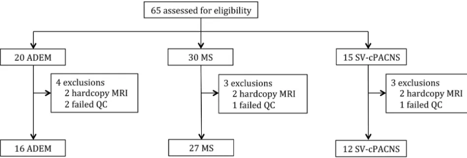
ParticipantsSixty-five children and adolescents were assessed for eligibility. MR images for 10 participants were excluded because either they were of insufficient quality (n⫽4) or the images were on hard-copy film (n⫽6). MR images were evaluated for the 55 children (16 with ADEM, 27 with MS, and 12 with SV-cPACNS) included (Fig 1). Children with ADEM were younger (mean age, 6.2⫾4.4 years) at the time of first attack than those with MS (mean age, 12.8⫾3.5 years;P⬍.0001) and SV-cPACNS (mean age, 10.3⫾ 4.3 years;P⫽.028). Onset age did not differ between children with MS and those with SV-cPACNS (P⫽.209).
Retained Parameters
Of the 48 parameters, 10 (demonstrating an inter-rater agreement statistic ofⱖ0.6 with a lower limit of the 95% CIⱖ0.5) could be readily evaluated on routine clinical sequences and were deemed diagnostically useful on the basis of panel consensus and the
lit-erature (Table 1). A post hoc decision was to collapse “midline brain stem,” “left brain stem,” and “right brain stem” into 1 pa-rameter (brain stem) because any 1 lesion commonly co-occurred in all 3 locations. The panel agreed to rename the parameter “fin-gerlike projections” as “gyral projections,” a more accurate de-scriptor of the feature. Of the 10 parameters, 2 that were not deemed by the panel to be MR imaging features of acute CNS demyelination were retained for their perceived utility in distin-guishing CNS demyelination and SV-cPACNS or other mimics: 1) leptomeningeal enhancement (⫽0.85; 95% CI, 0.55–1.0) was deemed by the panel as an important feature in differentiating CNS inflammatory demyelination from SV-cPACNS in cases in which presenting symptoms were nondiscriminatory; and 2) dif-fusion restriction visible on DWI (⫽1.0) was deemed by the panel as a specificity parameter, given its reported sensitivity for arterial ischemia.
The panel attributed the low inter-rater agreement of 3 of the 48 parameters (caudate:⫽0.49, 95% CI, 0.25– 0.73; putamen: ⫽0.44, 95% CI, 0.16 – 0.71; globus pallidus:⫽0.39, 95% CI, 0.13– 0.65) to the challenge of precisely delineating the borders of the lentiform nuclei and caudate on conventional MR imaging. The panel also deemed that the radiologic distinction among the nuclei was not relevant to acute CNS demyelination. Therefore, the panel agreed to collapse these 3 parameters into 1, basal ganglia.
Of the 48 parameters, 6 (1 of which included the collapsed parameter “basal ganglia”) were deemed by the panel as impor-tant in predicting chronic demyelination as opposed to a mono-phasic demyelinating illness but had inter-rater reliability statis-tics⬍0.6 following first-pass scoring (Table 2). The panel agreed that ambiguity in the definitions of 5 of the 6 parameters hindered their reliable application across raters. Revisions were made to the definition of the 5 parameters as follows: 1) “bilateral lesion dis-tribution,” refers to both the supratentorial and infratentorial re-gions; 2) “juxtacortical,” a lesion must involve the subcortical U-fibers to be scored as juxtacortical; 3) “intracallosal,” provision of anatomic landmarks to define borders of the corpus callosum in the transverse plane (On-line Appendix, Parameter 1) and specification that 1 mm of normal-appearing white matter sur-rounding a lesion required to define a lesion as being “intracal-losal” (to distinguish such lesions from periventricular lesions);
[image:3.594.56.531.49.210.2]FIG 1. Description of participants. QC indicates quality control.
Table 1: Inter-rater agreement of parameters retained
Parameter 95% CI
Lesion count 0.81a 0.68–0.89
Gyral projectionb 0.74 0.54–0.93
Diffusion restriction of lesion 1.0 1.0–1.0
Lesion enhancement 0.92 0.81–1.0
Leptomeningeal enhancement 0.85 0.55–1.0
Black hole 0.76 0.54–0.98
Periventricular lesion 0.80 0.66–0.94
Internal capsular lesion 0.74 0.58–0.91
Brain stem lesionc 0.72 0.57–0.88
Cerebellar lesion 0.73 0.56–0.91
a
ICC reported.
b
Formerly fingerlike projection.
c
[image:3.594.53.285.248.365.2]and 4) “thalamic” and “basal ganglia,” encompass lesions that involve the thalamus or basal ganglia even if the lesions are not entirely contained within these regions.
The poor inter-rater agreement of the sixth parameter, “sub-cortical lesions,” was due to a difference in opinion among raters on what represented subcortical white matter. One rater referred to all supratentorial white matter extending between the cortical ribbon and the lateral ventricles as subcortical; therefore, a lesion located in the supratentorial white matter that did not abut the cortex or lateral ventricle was scored as subcortical. The other rater viewed the supratentorial white matter as divided into deep (adjacent to the lateral ventricles) and superficial (adjacent to the cerebral cortex) white matter and therefore scored only those le-sions in the white matter that were adjacent to, but not contiguous
with, the cortex as “subcortical lesions.” To ensure that the subcortical lesion parameter was interpreted as involving all supratento-rial (nonjuxtacortical and nonperiven-tricular) white matter, the definition was revised and the parameter was renamed “cerebral white matter-other.”
After the definitions were revised to eliminate ambiguity, the 6 parameters were independently re-evaluated on all scans by both trained raters. Inter-rater agreement increased toⱖ0.6, permitting their reten-tion in the final tool (Table 2).
In total, 16 parameters were retained in the final tool (Tables 1 and 2). A textual and pictographic atlas of the final 16-parameter scoring tool was created and published in our recent work (On-line Appendix).13In the atlas, anatomic landmarks have been delineated for parameters that rely on accu-rate identification of anatomic structures.
Parameters Discarded
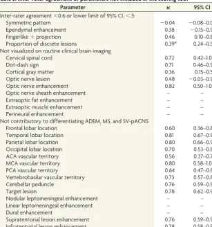
As shown in Table 3, of the 48 parameters, we excluded 28: Four due to low inter-rater agreement, 9 due to poor visualization of the parameter on routine clinical imaging, and 15 that the expert panel deemed to be not diagnostically valuable.
Specifically, “symmetric pattern,” “ependymal enhancement,” “fingerlike⫹ projection,” and “proportion of discrete le-sions” had⬍0.6 (or the lower limits of the 95% CI⬍0.5) and, even if redefined, were not deemed by the panel to be diag-nostically useful.
Nine parameters could not be accu-rately scored on routine clinical sequences. Although well-recognized as features of MS, the evaluation of “intracortical,” “cer-vical spinal cord,” and “optic nerve le-sions,” requires targeted cortical, spinal cord, or orbital imaging sequences—all of which are not routinely acquired in a clinical brain MR imaging protocol. One parameter (the “dot-dash” sign34) has been de-scribed in 1 study as an early MR imaging feature of MS; however, scoring the parameter requires thin sagittal T2-weighted or FLAIR imaging through the midline. The remaining 5 parameters (“optic nerve enhancement,” “optic nerve sheath enhancement,” “extraoptic fat enhancement,” “extraoptic muscle enhancement,” and “perineural enhancement”) require specialized fat-sup-pressed orbital imaging.
Finally, 15 parameters, despite demonstrating acceptable in-ter-rater agreement, did not aid in differentiating ADEM, MS, and SV-cPACNS (On-line Table 2) and were, therefore, excluded. The formation of demyelinating and SV-cPACNS lesions does not respect lobar (4 parameters) or vascular territory (4
parame-Table 2: Inter-rater agreement of parameters retained following rescoring
Parameter
First Pass Second Pass
95% CI 95% CI
Bilateral distribution 0.66 0.45–0.87 0.73 0.50–0.95
Intracallosal 0.65 0.42–0.87 0.90 0.76–1.00
Thalamic 0.60 0.40–0.80 0.85 0.71–0.99
Basal gangliaa 0.37 0.16–0.58 0.77 0.60–0.94
Cerebral white matter-otherb 0.38 0.16–0.59 0.69 0.46–0.91
Juxtacortical 0.46 0.28–0.63 0.64 0.41–0.86
aBasal ganglia collapsed from caudate (⫽0.49; 95% CI, 0.25– 0.73), putamen (⫽0.44; 95% CI, 0.16 – 0.71), and
globus pallidus (⫽0.39; 95% CI, 0.13– 0.65).
[image:4.594.54.368.55.145.2]bFormerly subcortical lesion.
Table 3: Inter-rater agreement of parameters not included in the scoring tool
Parameter 95% CI
Inter-rater agreement⬍0.6 or lower limit of 95% CI,⬍.5
Symmetric pattern ⫺0.04 ⫺0.08–0.01
Ependymal enhancement 0.38 ⫺0.15–0.91
Fingerlike⫹projection 0.46 0.10–0.82
Proportion of discrete lesions 0.39a 0.24–0.54
Not visualized on routine clinical brain imaging
Cervical spinal cord 0.72 0.42–1.0
Dot-dash sign 0.71 0.46–0.97
Cortical gray matter 0.36 0.15–0.58
Optic nerve lesion 0.48 ⫺0.03–0.98
Optic nerve enhancement 0.82 0.50–1.0
Optic nerve sheath enhancement – –
Extraoptic fat enhancement – –
Extraoptic muscle enhancement – –
Perineural enhancement – –
Not contributory to differentiating ADEM, MS, and SV-pACNS
Frontal lobar location 0.60 0.36–0.85
Temporal lobar location 0.81 0.67–0.94
Parietal lobar location 0.80 0.66–0.94
Occipital lobar location 0.70 0.53–0.86
ACA vascular territory 0.56 0.37–0.75
MCA vascular territory 0.80 0.58–1.0
PCA vascular territory 0.64 0.47–0.82
Vertebrobasilar vascular territory 0.73 0.57–0.89
Cerebellar peduncle 0.76 0.59–0.93
Target lesion 0.78 0.62–0.94
Nodular leptomeningeal enhancement – –
Linear leptomeningeal enhancement – –
Dural enhancement – –
Supratentorial lesion enhancement 0.76 0.59–0.94
Infratentorial lesion enhancement 0.78 0.58–0.98
Note:—ACA indicates anterior cerebral artery; PCA, posterior cerebral artery; –,invalid due to low frequency of occurrence inⱖ1 cell of the 2⫻2 table.
a
[image:4.594.58.366.193.521.2]ters) boundaries, and the location of contrast-enhancing lesions (ie, supratentorial and infratentorial) was not discriminatory. While the presence of leptomeningeal enhancement was retained in the scoring tool, the more specific parameters (“nodular lepto-meningeal enhancement,” “linear leptolepto-meningeal enhance-ment,” and “dural enhancement”) were too infrequently noted to permit computation of inter-rater reliability and were not deemed by the panel to be discriminatory. The parameter called “cerebellar peduncle lesions” was discarded due to its co-occur-rence with brain stem lesions and cerebellar lesions and the chal-lenge of deciphering the margins of the cerebellar peduncles. “Target lesions” was discarded because the panel could not reach consensus on a consistent definition.
DISCUSSION
We created an MR imaging scoring tool consisting of 16 param-eters that demonstrate substantial inter-rater reliability. The ra-tionale for each of the parameters included in the tool is detailed in the Supplementary Panel of the On-line Appendix and is based on evidence for their utility in characterizing the MR imaging features of acute demyelination and for their utility in discrimi-nating acute CNS demyelination and SV-cPACNS. For a clinically useful tool to be generally accepted, it must be practical to use in the clinical setting without the need for rigorous training. Thus, we intentionally created the tool to be binary-response (with the exception of lesion count, which has an upper limit lesion count of 15) to minimize the quantitative requirement and maximize its efficient use in clinical practice.
The first component of developing the MR imaging tool was to devise the parameters themselves. We used established methods of item identification, including a literature search and expert consensus,14to formulate a comprehensive list of potential pa-rameters. The rationale for performing a literature search was so that the tool would comprise parameters that have been empiri-cally demonstrated to be MR imaging characteristics of acute CNS demyelination and relapsing-remitting MS or features that might discriminate demyelination and SV-cPACNS. This method of de-vising tool parameters has been used in other instances, such as in a scale for differentiating irritable bowel syndrome and organic bowel disease, in which the parameters represented clinical fea-tures and laboratory values that were found to distinguish the 2 patient groups.15
We also used expert consensus as a second method for devis-ing potential parameters. We selected the expert panel from neu-roradiology, neurology, and rheumatology staff working in our established pediatric demyelinating disease and CNS vasculitis programs, ensuring extensive clinical and radiologic experience with pediatric-onset MS and SV-cPACNS. The expert panel played a key role in defining the comprehensive list of parameters identified from the literature and in devising definitions for each parameter that were objective and were straightforward to apply. Similar utility of expert opinion has been reported elsewhere, such as the Patient-Reported Outcomes Measurement Information System initiative funded by the National Institutes of Health, in which experts contribute potential parameters to be used in eval-uating patient-reported outcomes across multiple health conditions.16
Following identification of relevant MR imaging parameters for the diseases under evaluation, we created definitions for each parameter to enable consistency in application of the parameters among raters.17,18Therefore, a key component of our work was to assess the inter-rater reliability of the potential parameters iden-tified by the literature search and expert opinion; only parameters that demonstrated substantial or good inter-rater agreement ( ⱖ 0.6)10,12were retained in the final tool. Several MR imaging pa-rameters demonstrated poor inter-rater agreement on first eval-uation but were deemed important discriminatory parameters by the panel. These parameters were redefined; then, all scans were rescored, and only when the inter-rater agreement increased to ⱖ0.6 was the parameter retained.
A second noteworthy aspect is that we focused on the inter-rater reliability of our parameters and not intrainter-rater reliability. Because the error contributing to intrarater reliability is consid-ered contained within inter-rater reliability, demonstration of high inter-rater reliability is sufficient.17Of the 16 parameters retained in the final tool, 14 demonstrated inter-rater agreement that was considerably higher (ⱖ0.72) than our a priori cut-point of 0.6. The use of inter-rater agreement to guide the selection of parameters included in a scoring tool is not unique to our study. Similar methodology was used during the creation of the well-established Glasgow Coma Scale, a clinical scale for evaluating the depth and duration of impaired consciousness and coma,19and the International Standards for Neurologic Classification of Spi-nal Cord Injury of the American SpiSpi-nal Injury Association, a prac-tice guideline for classifying the degree of neurologic impairment due to spinal cord injury.18,20,21
A key aspect of the diagnostic criteria for multiple sclerosis is the exclusion of mimics of CNS demyelination.22,23In creating our proposed tool, due to the clinical challenge of distinguishing SV-cPACNS from MS, we included MR images of children with SV-cPACNS because this disease was thought to have a similar but potentially distinct inflammatory pattern on MR imaging. We also included parameters that have been reported in CNS infec-tion and malignancy. Specifically, leptomeningeal enhancement has been described in children with SV-cPACNS6and is well-documented in infectious24,25and neoplastic26-28processes. Lep-tomeningeal enhancement is not a feature of MS, and its presence was highlighted by an international consensus panel as a “red flag” to consider other nondemyelinating etiologies.23In contrast, the MR imaging parameter of “acute diffusion restriction,” while highly characteristic of vascular occlusion such as stroke,29-31has also been a feature recently reported in acute demyelinating le-sions, particularly tumefactive lele-sions,32,33and therefore was re-tained in the final tool.
in-curring higher cost in the context of research studies. The yield of orbital or spinal imaging in the absence of clinical evidence of acute or remote optic nerve or spinal cord involvement has not been established in children subsequently diagnosed with MS; further studies will be required, with appropriate consideration of scanning time.
We created a manual that contains an atlas and definition for each parameter and highlights important anatomic delineations to ensure that the tool can be accurately used by radiologists and clinicians who were not part of the creation of the tool. Not only will this aid in use of the tool across clinical centers, it also serves as a model for MR imaging scoring tools to be applied in pediatric MS clinical trials in which inclusion in the trial will be predicated on accurate MS diagnosis.
The intended future application of our proposed MR imaging tool is multifaceted and requires validation. The first priority will be to determine the ability of the MR imaging tool to identify distinct features of different CNS inflammatory disorders, such as monophasic CNS demyelination, MS, ADEM, and SV-cPACNS because such disorders share clinical features rendering accurate diagnosis challenging at onset. The MR imaging tool will then be evaluated for specificity in identifying noninflammatory CNS dis-orders, such as inherited or metabolic diseases that also have onset during childhood and impact CNS white matter. Finally, we need to evaluate user satisfaction with the tool because application in a busy clinical environment will require endorsement of utility and applicability.
CONCLUSIONS
We developed an MR imaging scoring tool consisting of 16 items that demonstrate substantial inter-rater agreement. Using estab-lished methods of item identification, including a literature search and expert consensus, the parameters are based on evidence for their utility in characterizing the MR imaging features of acute CNS inflammation and demyelination. The binary-response na-ture of the parameters and the tool manual will facilitate utility of the tool in clinical practice without the need for rigorous training. The scoring tool will inform the creation of struc-tured reporting that is increasingly being proposed for use in diagnostic radiology.
ACKNOWLEDGMENTS
We thank Drs Adrian Fawcett and Sunita Venkateswaran for their contributions to the initial phase of this work, the staff of the Pediatric Demyelinating Disease Program at The Hospital for Sick Children (Toronto, Ontario, Canada), and the partic-ipating children and their families for their commitment to our research program.
Disclosures: Leonard H. Verhey—RELATED:Grant: Canadian Multiple Sclerosis Sci-entific Research Foundation,*Comments: This study was funded under a research grant from the Canadian Multiple Sclerosis Scientific Research Foundation. L.H. Ver-hey receives scholarship support from the Canadian Institutes of Health Research, Multiple Sclerosis Society of Canada, University of Toronto, and The Hospital for Sick Children,Support for Travel to Meetings for the Study or Other Purposes: Canadian Multiple Sclerosis Scientific Research Foundation,Comments: Travel to present at scientific meetings was covered under a research grant from the Canadian Multiple Sclerosis Scientific Research Foundation. Helen M. Branson—RELATED: Grant: Canadian Multiple Sclerosis Scientific Research Foundation,*Support for Travel to Meetings for the Study or Other Purposes: Canadian Multiple Sclerosis
Scientific Research Foundation,*Comments: I did not personally receive any funding but other authors received funding for travel to meetings. Suzanne Laughlin— RE-LATED:Grant: Canadian Multiple Sclerosis Scientific Research Foundation.* Mano-har M. Shroff—RELATED:Grant: Canadian Multiple Sclerosis Scientific Research Foundation,*Comments: This study was conducted under a research grant (paid to SickKids) from the Canadian Multiple Sclerosis Scientific Research Foundation, Sup-port for Travel to Meetings for the Study or Other Purposes: Canadian Multiple Sclerosis Scientific Research Foundation,*Comments: The grant covered travel ex-penses for fellows or postdoctorates with whom I worked, when presenting this work at scientific meetings. I personally was never paid out of this grant for travel expenses to present at any meeting. Brenda Banwell—RELATED:Grant: Canadian Multiple Sclerosis Scientific Foundation,*UNRELATED:Consultancy: Biogen-IDEC, Merck-Serono,Comments: no relevance to present work,Grants/Grants Pending: Canadian Institute of Health Research,* MS Society of Canada,*Payment for Lec-tures (including service on Speakers Bureaus): Novartis, Biogen-IDEC, Serono, Trav-el/Accommodations/Meeting Expenses Unrelated to Activities Listed: visiting pro-fessorships. *Money paid to the institution.
REFERENCES
1. McDonald WI, Compston A, Edan G, et al.Recommended diagnos-tic criteria for multiple sclerosis: guidelines from the International Panel on the Diagnosis of Multiple Sclerosis.Ann Neurol2001;50: 121–27
2. Polman CH, Reingold SC, Edan G, et al.Diagnostic criteria for mul-tiple sclerosis: 2005 revisions to the “McDonald Criteria.”Ann Neu-rol2005;58:840 – 46
3. Polman CH, Reingold SC, Banwell B, et al.Diagnostic criteria for multiple sclerosis: 2010 revisions to the McDonald criteria.Ann Neurol2011;69:292–302
4. Simon JH, Li D, Traboulsee A, et al.Standardized MR imaging pro-tocol for multiple sclerosis: Consortium of MS Centers consensus guidelines.AJNR Am J Neuroradiol2006;27:455– 61
5. Hutchinson C, Elbers J, Halliday W, et al.Treatment of small vessel primary CNS vasculitis in children: an open-label cohort study.
Lancet Neurol2010;9:1078 – 84
6. Elbers J, Halliday W, Hawkins C, et al.Brain biopsy in children with primary small-vessel central nervous system vasculitis.Ann Neurol
2010;68:602–10
7. Poser CM, Paty DW, Scheinberg L, et al.New diagnostic criteria for multiple sclerosis: guidelines for research protocols.Ann Neurol
1983;13:227–31
8. Krupp LB, Banwell B, and Tenembaum S.Consensus definitions proposed for pediatric multiple sclerosis and related disorders.
Neurology2007;68:S7–12
9. Cellucci T, Benseler SM.Diagnosing central nervous system vascu-litis in children.Curr Opin Pediatr2010;22:731–38
10. Sim J, Wright CC.The kappa statistic in reliability studies: use, in-terpretation, and sample size requirements.Phys Ther 2005;85: 257– 68
11. Shrout PE, Fleiss JL.Intraclass correlations: uses in assessing rater reliability.Psychol Bull1979;86:420 –28
12. Landis JR, Koch GG.The measurement of observer agreement for categorical data.Biometrics1977;33:159 –74
13. Verhey LH, Branson HM, Shroff MM, et al.MRI parameters for prediction of multiple sclerosis diagnosis in children with acute CNS demyelination: a prospective national cohort study.Lancet Neurol2011;10:1065–73
14. Streiner DL, Norman GR.Health Measurement Scales: A Practical Guide to Their Development and Use.New York: Oxford University Press; 2008:17–29
15. Kruis W, Rindfleisch GE, Weinzierl M.Zinc deficiency as a problem in patients with Crohn’s disease and fistula formation. Hepatogas-troenterology1985;32:133–34
16. Cella D, Yount S, Rothrock N, et al.The Patient-Reported Outcomes Measurement Information System (PROMIS): progress of an NIH Roadmap cooperative group during its first two years.Med Care
2007;45:S3–11
Guide to their Development and Use.New York: Oxford University Press; 2008:167–207
18. Marino RJ, Jones L, Kirshblum S, et al.Reliability and repeatability of the motor and sensory examination of the international stan-dards for neurological classification of spinal cord injury.J Spinal Cord Med2008;31:166 –70
19. Teasdale G, Jennett B. Assessment of coma and impaired consciousness: a practical scale.Lancet1974;2:81– 84
20. Jonsson M, Tollback A, Gonzales H, et al.Inter-rater reliability of the 1992 international standards for neurological and functional classifi-cation of incomplete spinal cord injury.Spinal Cord2000;38:675–79 21. Priebe MM, Waring WP.The interobserver reliability of the revised
American Spinal Injury Association standards for neurological classification of spinal injury patients. Am J Phys Med Rehabil
1991;70:268 –70
22. Charil A, Yousry TA, Rovaris M, et al.MRI and the diagnosis of multiple sclerosis: expanding the concept of “no better explana-tion.”Lancet Neurol2006;5:841–52
23. Miller DH, Weinshenker BG, Filippi M, et al.Differential diagnosis of suspected multiple sclerosis: a consensus approach.Mult Scler
2008;14:1157–74
24. Baumann M, Birnbacher R, Koch J, et al.Uncommon manifesta-tions of neuroborreliosis in children. Eur J Paediatr Neurol
2010;14:274 –77
25. Jang S, Suh SI, Ha SM, et al. Enterovirus 71-related
encephalo-myelitis: usual and unusual magnetic resonance imaging findings.
Neuroradiology2012;54:239 – 45
26. Singh SK, Leeds NE, Ginsberg LE.MR imaging of leptomeningeal metastases: comparison of three sequences.AJNR Am J Neuroradiol
2002;23:817–21
27. Pang J, Banerjee A, Tihan T.The value of tandem CSF/MRI evalua-tion for predicting disseminated disease in childhood central ner-vous system neoplasms.J Neurooncol2008;87:97–102
28. Chamberlain MC.A review of leptomeningeal metastases in pediat-rics.J Child Neurol1995;10:191–99
29. Warach S, Gaa J, Siewert B, et al.Acute human stroke studied by whole brain echo planar diffusion-weighted magnetic resonance imaging.Ann Neurol1995;37:231– 41
30. De Vries LS, Van der Grond J, Van Haastert IC, et al.Prediction of outcome in new-born infants with arterial ischaemic stroke using diffusion-weighted magnetic resonance imaging.Neuropediatrics
2005;36:12–20
31. Kirton A, Shroff M, Visvanathan T, et al.Quantified corticospinal tract diffusion restriction predicts neonatal stroke outcome.Stroke
2007;38:974 – 80
32. Kiriyama T, Kataoka H, Taoka T, et al.Characteristic neuroimaging in patients with tumefactive demyelinating lesions exceeding 30 mm.J Neuroimaging2011;21:e69 – e77