Virtual Reaction Chambers as a tool for
DNA sequencing
Dissertation
zur Erlangung des Grades
der Doktorin der Naturwissenschaften
der Naturwissenschaftlich-Technischen Fakultät
der Universität des Saarlandes
von
Ana V. Almeida
Saarbrücken
Tag des Kolloquiums: 16.10.2017
Dekan: Univ.-Prof. Dr. rer. nat. Guido Kickelbick
Mitglieder des Prüfungsausschusses:
- Gutachter (Betreuer): Prof. Dr. Andreas Manz
- Gutachter (nicht Betreuer): Prof. Dr. Ralf Seemann
- Vorsitzenden: Prof. Dr.-Ing. Georg Frey
Bitte unterschrieben als Einzelblatt einreichen
und in die Arbeit eingebunden, ebenfalls unterschrieben.
Eidesstattliche Versicherung
Hiermit versichere ich an Eides statt, dass ich die vorliegende Arbeit selbstständig und ohne Benutzung anderer als der angegebenen Hilfsmittel angefertigt habe. Die aus anderen Quellen oder indirekt übernommenen Daten und Konzepte sind unter Angabe der Quelle gekennzeichnet. Die Arbeit wurde bisher weder im In- noch im Ausland in gleicher oder ähnlicher Form in einem Verfahren zur Erlangung eines akademischen Grades vorgelegt.
Ort, Datum
Abstract
The topic of this thesis refers to the contribution of genetic variations, in which single nucleotide polymorphisms (SNP) can be included, to the manifestation and cause of disease. The facilitation of the treatment and cure of such conditions is done through identification and study of the cascade of biological mechanisms and processes; however the achievement of such goals is hampered by the formidable complexity and scale of the data sets involved in nucleic acid and protein analysis and thus the development of faster and cheaper methods is desirable. An intuitive trajectory for this development is the miniaturization of analytical methods with the incorporation of microfluidics which entails the manipulation of fluid analytes through channels and chambers on the microscale. This young field is projected to be instrumental in the realization of low temporal and financial cost analytical techniques.
This thesis demonstrates the application of open-surface microfluidics to sequence DNA with the use of pyrosequencing. This method facilitates a reduction in instrument complexity and size and thus allows for functional integration or device disposability. Following incubation of the DNA with superparamagnetic particles, it was placed on a hydrophobic glass substrate. The DNA was then moved through microliter-volumes of mineral oil-coated water droplets via manipulation of the nanoparticles using a magnetic field; thus, these droplets could then be used either for pyrosequencing or for washing of the DNA strands. The reaction performance was determined, using the resequencing protocol with a 34 base pair (bps) of single-stranded DNA, to be highly linear for all 4 homopolymers events tested. De novo sequencing was performed with 51 and 81 bps while it was also verified that up to 7 homopolymers could be determined. This method displays full compatibility with previously demonstrated open surface steps for sample preparation and so confirms PCR on a flat glass substrate as an integrated sample-to-answer protocol.
All assays were based on primer extension via DNA polymerase. A microfluidic device consisting of microliter-volume droplet-to-droplet DNA transport via manipulation of magnetic particles was used in this work. The
difference in reaction kinetics for matched and mismatched configurations at the
3’-ends of the primer-template complex was employed in sample differentiation. The assay combined pyrosequencing technology with a sequence-by-synthesis bioluminometric DNA sequencing probe on one common microfluidic platform. Base-by-base sequencing was performed to obtain accurate single nucleotide polymorphism (SNP) scoring data with microliter volumes. The application of magnetically actuated beads to facilitate virtual chamber reactions for chip based DNA analysis was presented. Single base incorporation was seen to be detectable with the use of this pyrosequencing assay.
Keywords: DNA sequencing, single nucleotide polymorphisms, superparamagnetic particles, open surface microfluidics, magnetic actuation.
Zusammenfassung
Mit Abschluss des Humangenomprojekts verlagert sich der Fokus darauf, zu verstehen, wie genetische Variationen, wie z. B. Einzelnukleotid-Polymorphismus (SNP, single nucleotide polymorphism), zu Erkrankungen führen. Die enorme Menge an Sequenzinformationen müssen mit schnelleren, wirtschaftlicheren und leistungsfähigeren Technologien für die Analyse von RNA, DNA und Proteinen analysiert werden, um die die biologischen Mechanismen lebender Organismen zu verstehen und zu kontrollieren. Ein Ansatz ist die Miniaturisierung analytischer Methoden durch die Anwendung der Mikrofluidik, wobei Ströme in Kanälen auf der Mikrometer-Skala manipuliert werden. Es wird erwartet, dass Fortschritte in der Mikrofluidik-Chip-Technologie eine wichtige Rolle bei der Entwicklung wirtschaftlicher und schneller DNA-Analysemethoden spielen werden.
In dieser Doktorarbeit wird die Anwendung von Mikrofluidik an offenen Oberfläche für die Sequenzierung von DNA mittels Pyrosequenzierung demonstriert. Dies bietet Vorteile bezüglich der Instrumentengröße, der Einfachheit, Verfügbarkeit und Funktionsintegration, insbesondere in Kombination mit den vielfältigen und flexiblen Möglichkeiten der Mikrofluidik an offenen Oberflächen. Die DNA wurde auf superparamagnetischen Partikeln inkubiert und auf ein Glassubstrat mit hydrophobischer Schicht platziert. Die Partikel mit gebundener DNA wurden mithilfe von Magnetkraft über Tropfen in Mikroliter-Größe, beschichtet mit Mineralöl zur Verhinderung von Verdunstung, bewegt. Diese Tropfen dienten als Reaktionsstationen für die Pyrosequenzierung sowie als Waschstationen. Das Sequenzierungsprotokoll mit 34 Basenpaaren (bps) einzelsträngiger DNA wurde zur Bestimmung der Reaktionsleistung verwendet und zeigte eine ausgezeichnete Linearität für alle 4 Homopolymere. Diese De-novo-Sequenzierung wurde mit 51 und 81 bps durchgeführt. Wir haben außerdem geprüft, dass bis zu 7 Homopolymere bestimmt werden können. Dieses Verfahren ist vollständig mit bisher verwendeten Schritten zur Probenvorbereitung an offenen Oberflächen kompatibel. Daher kann eine PCR als vollständig integriertes System von der
Probe bis zum Ergebnis auf einem flachen Glassubstrat zusammengestellt werden.
In dieser Doktorarbeit werden Mikrofluidik-Anwendungen für verschiedene Genotypisierungsassay vorgestellt. Das übergeordnete Ziel ist die Kombination des Potentials des Chiplabor-Konzepts der Mikrofluidik mit Biochemie, um Verfahren für die DNA-Analyse zu entwickeln uns derzeit verfügbare Verfahren zu verbessern. Drei Genotypisierungsassays werden hier mithilfe von miniaturisierten Mikrofluidik-Methoden überprüft.
Alle Assays basieren auf der Verlängerung von Primeren durch DNA-Polymerase. Ein Mikrofluidik-Instrument mit Handhabung von Tropfen zu Tropfen für Magnetpartikel für Volumen im Mikroliterbereich wurde in diesen Studien verwendet. Das Mikrofluidik-Verfahren nutzt die Vorteile der unterschiedlichen Reaktionskinetik für komplementäre und
nicht-komplementäre Konfigurationen am 3‘-Ende des Primer-Template-Komplexes. Insgesamt beinhalteten die Assays die Anpassung der Pyrosequenzierungstechnologie, eines bioluminometrischen DNA-Sequenzierungsassays basierend auf Sequenzierung durch Synthese, an dieselbe Mikrofluidik-Plattform. Basenweise Sequenzierung wurde in einem Mikrofluidik-Instrument durchgeführt, um genaue SNP-Scoring-Daten für Volumina im Mikroliterbereich zu erzielen. In dieser Doktorarbeit wird die Anwendung virtueller Reaktionskammern von Partikel, die magnetisch ausgelöst werden, für die Chip-basierte DNA-Analyse präsentiert. Die Inkorporation von einzelnen Basen mithilfe der Pyrosequenzierungsreaktion wurde auf diesen Partikel beobachtet.
Stichworte: DNA-Sequenzierung, Einzelnukleotid-Polymorphismus, Superparamagnetischpartikel, offene Mikrofluidik, Mikrosystem, magnetische Betätigung
Acknowledgments
I would like to thank everyone who has contributed to this thesis and has made these last few years so enjoyable.
I am especially grateful to:
Andreas Manz for accepting me as a PhD student and for being a true visionary with a fantastic skill for attracting good people.
Pavel Neuzil, Microfluidics group leader, for his guidance during this scientific journey.
My colleagues, fellow students and friends not only in KIST-Europe but overall in Saarbrücken and beyond:
Zeynep and Mark: the shorty-spicy-lady and the tally-gentleman. Wouldn’t think
of better companions to start this journey with. You guys rock!
Matthias: starting off with the labs and kicking off with the projects on a fresh group and becoming a permanent member of my family…so much to say and
so little space. Because of you, Saarland will always be home to me. Thank you for all the inspiring and fruitful discussions on microfluidics and life in general, for being so enthusiastic and, last but not least, for being you.
Jang Mi: my favorite power Frau! Determination and fantastic energy to keep all of those boys under control.
Jukyung and Seung Jae…such a long jurney! Thank you for being part of it!
Tijmen: my climbing partner and honeybear!
Thomas: no bull**** modus operandi and one of the fairest people I know.
Marc: my chuchu! For life! Let’s keep on growing and getting distracted with
Life.
André and Eric: Perhaps one day I’ll be as cool as you.
Leon Abelmann: To prove me on a daily basis that Science is a playground for our curious hearts.
Everyone in KIST-europe: for interesting discussions on a variety of subjects
(some of which involved science…) and for the very funny and moments; for
being great and enthusiastic colleagues and friends …
KIST-europe for hosting me for these years, to introduce me to such different and beautiful culture and enabling me in Science.
All collegues, former and present, at KIST-Europe for being so nice and helpful and for contributing (everyone in his or her own special way) to the fantastic atmosphere of this place. I feel very privileged to have had the opportunity to get to know you and to work with you, and even if all names aren’t listed, you
are in my heart.
My friends, in and out of science, for being such great persons and for all the good times we have shared.
My family for always being there for me with good advice, skype calls full of affection or whatever will do the trick to keep me motivated
My parents for endless support and encouragement and for believing in me and
wishing me happiness whatever I do. They are my giants (“The reason why I
am seeing so far, it is because I am standing on the shoulders of giants”, Isaac Newton).
Ferdia, the love of my life, my life-partner, for loving me and being my rock in stormy weather. We made it!
Table of contents
Eidesstattliche Versicherung III
Abstract V Zusammenfassung VII Acknowledgements IX Table of contents XI Abbreviations XV 1. Introduction: DNA 1
1.1 Chemical structure of DNA and organization 1
1.2 Genomic variatons 3
1.2.1 Mutations 3
1.2.2 Single nucleotide polymorphisms 7
1.3 DNA sequencing techniques 8
1.3.1 Polymerase chain reaction 9
1.3.2 Dideoxy DNA sequencing by chain termination 11 1.3.3 Non-electrophoretic DNA sequencing methods 14
1.3.4 Pyrosequencing technology 18
1.4 SNP genotyping technologies 22
1.5 Detection 24
1.6 Next stage for DNA sequencing technologies 25 2. Introduction: Microfluidics and micro total systems (TAS) 27
2.1 Microfluidics 27
2.2 Microfabrication 29
2.3 Diffusion 30
2.4 Microparticles 30
2.4.1 Particle handling on chip 32
2.5 Miniaturized platforms for DNA analysis 32
2.5.1 Microarray technologies 33
2.5.2 Microfluidics based technologies 36
2.5.3 Bead-based methods 43
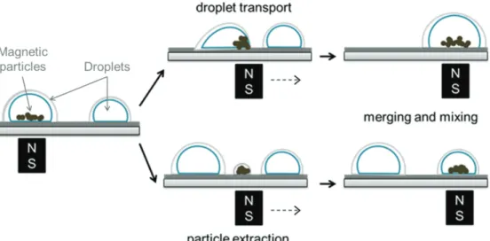
3 Open surface microfluidics (OSM) 45
3.2 Droplet actuation platform 47
3.3 Droplet manipulation 48
3.4 Important parameters 51
3.5 OSM for DNA analysis 54
4 Overview 59
4.1 Motivation 59
4.2 Objectives 64
5 OSM and virtual reaction chambers 65
5.1 Microfluidic superheating for peptide analysis 65 5.2 Superheating for protein thermal stability analysis 66 5.3 qPCR with VRCs for different applications 67
5.4 My published research 67
5.5 Wrap up 67
6 Materials and Methods 69
6.1 Glass substrate 69
6.2 Droplets 70
6.3 xyz-stage 71
6.4 Detection system 73
6.5 SPP/ssDNA/primer complex preparation 74
6.6 Buffers and solutions preparation 75
6.7 Glass spotting 75
6.7.1 X-design 76
6.7.2 Circle/linear-design 77
6.7.3 Enzymes and substrates design 78
6.8 DNA preparation 78
7 Results Discussion - 81
7.1 Experimental design 81
7.2 First experiments 83
7.3 VRCs placement 87
7.4 Optimization of reagents and buffers 89
7.5 Washing efficiency 91
7.6 Mixing improvement 92
7.7 Effect of DNA concentration 93
7.9 Resequencing 99
7.10 De novo sequencing 101
7.11 Longer sequencing testing 103
7.12 Comments on methods 105
8 Conclusions and outlook 109
List of abbreviations
Abbreviation Description
A adenine
AIDS acquired immuno defficiency syndrome AMASE apyrase-mediated allele-specific extension ARMS amplification refractory mutation system ASA allele specific amplification
ASPCR allele-specific PCR ATP adenosine triphosphate AuNPs gold nanoparticles
bp base pairs
BSA bovine serum albumin
C cytosine
CBC complete blood count CCD charge coupled device CE capillary electrophoresis COC cyclic olefin copolymer
Cys cysteine
DASH dynamic allele specific hybridization dATP deoxyadenosine triphosphate dCTP deoxycytosine triphosphate ddC dideoxycytosine ddG dideoxyguanine ddNTP dideoxynucleotide ddT dideoxythymine dGTP deoxyguanine triphosphate DI deionized dNTP nucleoside triphosphates
dsDNA double-stranded deoxyribonucleic acid dTTP deoxythymine triphosphate
FRET fluorescence resonance energy transfer
G Guanine
GFP green fluorescent protein
His Histidine
HIV human immuno deficiency virus
Ile Isoleucine
IPA isopropyl alcohol LCR ligase chain reaction
Leu Leucine
LNA locked nucleic acid
LOC lab-on-a-chip
MEMS Microelectromechanical
Met Methionine
MPs magnetic particles
OLA oligonucleotide ligation assay OSM open surface microfluidics
PASA PCR amplification of specific alleles
PC poly(carbonate)
PCR polymerase chain reaction PDMS polydimethylsiloxane PMMA poly(methyl methacrylate)
POC point-of-care
PPi pyrophosphate
PTFE polytetrafluoroethylene RCA rolling circle amplification SBE single base extension
SBH sequencing-by-hybridization SBS sequencing-by-synthesis Ser Serine Ser Serine SNP single-nucleotide polymorphism SPP superparamagnetic particles
ssDNA single-stranded deoxyribonucleic acid SU-8 epoxy-based negative photoresist
T thymine Thr threonine TNP triple-nucleotide polymorphisms Tyr tyrosine UV ultraviolet Val valine
Chapter 1
Introduction: DNA
The human body consists of approximately 3,72x10¹³ cells[1], most containing the hereditary information of the genome in the form of 6x10⁹ base pairs of deoxyribonucleic acid (DNA).[2] DNA molecules are the libraries in living cells where the information required for building a cell and an organism are stored.
This subchapter 1.1 briefly describes relevant information over DNA and corresponding sequencing technologies and the microfluidics field, emphasing the inter-relation between both topics.
1.1 Chemical structure of DNA and organization
DNA is a linear polymer composed of single chemical units called nucleotides.[3-4] The number of nucleotides in a cellular DNA molecule can exceed a hundred million.[2] A nucleotide has three parts: a phosphate group, a deoxyribose and an organic base. The bases are adenine, guanine, cytosine and thymine; abbreviated A, G, C and T, respectively.[4] When the nucleotides
polymerize to form DNA, the hydroxyl group attached to the 3’ carbon of the
sugar group of one nucleotide forms an ester bond to the phosphate attached to
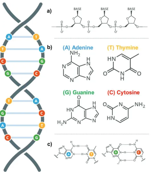
the 5’ carbon of another nucleotide. This dictates the extremely important property of the orientation of the polynucleotide strand.[5] DNA consists of two associated polynucleotide strands forming a double helix. The sugar-phosphate backbones are on the outside of the double helix and the bases project into the interior. The orientation of the two strands is antiparallel and complementary to each other. The strands are held together by the cooperative energy of many hydrogen bonds in addition to hydrophobic interactions.[6-7] The opposite strands are held in precise register by a regular base pairing between the two strands: A is paired with T by two hydrogen bonds and G is paired with C by three hydrogen bonds.[7]
Figure 1: Chemical structure of DNA. DNA stores and transmits genetic information in all multicellular life forms. The sequences formed by the bps code for proteins which execute a wide range of physiological functions. a) The
“backbone” made up of phosphate sugars which support the subunits of the
polymer, each nucleotide consists of a phosphate group, a sugar group and a base. b) Adenine, thymine, guanine and cytosine are the bases present in DNA. c) These bases form base pairs (bps) with the second strand in the double helix; Adenine (A) always pairs with Thymine (T), whilst guanine (G) always pairs with cytosine (C).
1.2 Genomic variations
This DNA is arranged into n chromosomes or in a plasmid depending on the organism (Figure 2) and the composition of the DNA bases within these macromolecules determines the genotype and contributes to the phenotype of an individual[8] (Figure 1). Consequently, alterations in chromosomes may lead to genetically related diseases (Figure 3). Cells are constantly exposed to agents and events that can be harmful to the DNA. Depurination, deamination, oxidation and methylation of bases are endogenous events that can change the base composition of DNA, while exogenous agents such as UV light, ionizing radiation and genotoxic substances may damage DNA by creating pyrimidine dimers, double strand breaks and adducts.[9-14] Several different gatekeeper and DNA repair systems exist to maintain DNA integrity, but occasionally these backup systems fail and mutations arise.
1.2.1 Mutations
Mutations can be divided into two classes; gross alterations and subtle alterations. Gross alterations involve changes in chromosome number, partial deletions of chromosomes, chromosomal translocations and gene duplications, while subtle alterations comprise of single base substitutions, insertions and deletions.[15-16] Mutations are common events in cancer and the ability to monitor them is therefore of great interest for diagnostic and prognostic purposes as well as in understanding gene function.[17]
Therefore, detecting genomic variations is fundamental in genetic research. For example, in cancer research, mutation detection is vital for diagnosis but also gives insight into gene function[18]. Strains of microorganisms can also be identified by point variations in their genome, and, in recent years, the pharmaceutical companies have shown a rising interest in single nucleotide
Figure 2: DNA configuration. DNA is contained within the cells of every living thing though its organization varies within different life form. A binary distinction can be made between the two main DNA organizational conformations with organisms being either prokaryotes or eukaryotes. a) Prokaryotes. Bacteria are prokaryotic cells. They do not exhibit membrane-bound nucleus with their DNA, instead existing freely in the cytoplasm. Their monochromosomal DNA sequence, which is circular in aspect and located at the center of the cell, contains the genetic information required to support all cellular functions. Additional rings of bacterial DNA known as plasmids. Certain genes within these plasmids can code for antibiotic resistance which increases the probability of survival. Interbacterial exchange of plasmids is also possible through hair-like
extensions from each cell’s surface known as pili. b) Eukaryotes: animal, plant and fungi cells are eukaryotes. All contain a membrane-bound nucleus in which
the cell’s chromosomes are linear. Chromosomal DNA is tightly coiled around
special proteins, known as histones, shown below. (adapted from http://www.bbc.co.uk/education/guides/z36mmp3/revision/2).
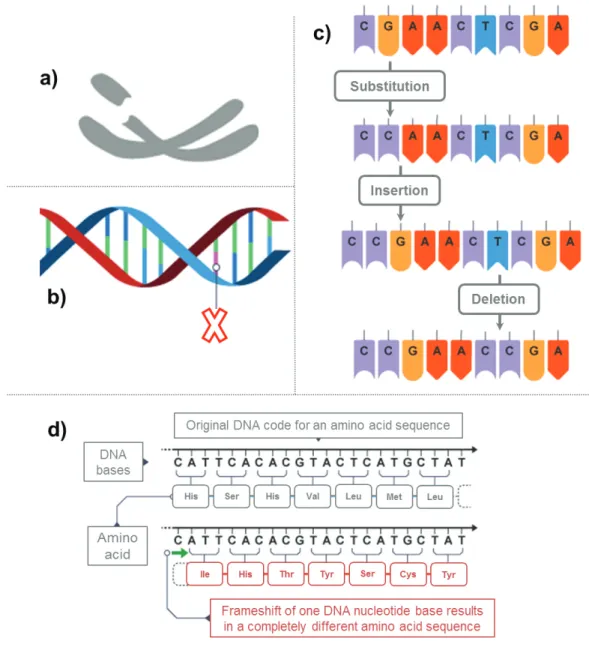
Figure 3: Figure 3: Mutations: During cell division, the DNA replication process is stringently controlled in order to preserve the genetic code held within the sequence of nucleotides. Despite this, encoding errors do evade this mechanism and exist in the copied genome as mutations, from which no protein or an altered protein can be expressed. Structure mutations affect the structure of one or more chromosomes (a) while single gene mutations affect only a small number of bases (b). Point mutations refer to the substitution of one codon alone which can result in missense, nonsense or a splice-site mutation (c). Frame shift mutations occur when insertions or deletions are not corrected (d).This implies that when nucleotides are grouped in threes to form codons the reading frame is shifted by one so that completely unrelated amino acids are outputted resulting in a most commonly junk protein (adapted from http://www.bbc.co.uk/education/guides/zc499j6/revision/2).
Gross alterations involve chromosomal number, chromosomal translocation, and partial deletion of chromosomes. These types of genomic alterations can be detected by rather simple techniques, such as microscopy and fragment analysis of implicated chromosomal regions.[20] Mutation detection of subtle sequence alterations, however, requires more sensitive techniques, is time-consuming and is more expensive. This perhaps explains why a wide range of techniques have been developed for this purpose. The growing number of methods indicates that a perfect technique which fulfills the required criteria and is able to detect all possible mutations/variations has yet not been described.[21]
The techniques for detecting subtle sequence changes can be divided into three groups. The first is known as scanning methods and is used to search for unknown mutations in pre-defined sequences.[22] The second group is known as diagnostic methods and is used to search for analysis of mutations and variations at defined positions, such as hot-spot sequences and single nucleotide polymorphisms (SNPs)[23]. Since SNPs are much more frequent than hot-spot mutations and because most recent efforts have been focused on detecting single nucleotide variations, the diagnostic techniques will hereafter be referred to as methods for analysis of SNPs[24]. The third group of method is sequencing technologies that are used to reveal the exact nature of a mutation, regardless of being unknown or pre-defined[25].
Some of these techniques and their application fields will briefly be reviewed here. However, irrespective of the mutation status (known or unknown), all the techniques have advantages and disadvantages. Some are simple but do not detect all mutations while others are more complex and detect almost all mutations. Often, factors such as laboratory and personnel experience, required accuracy, required throughput, cost and project type have to be taken into account prior to selection of a method.[26-27]
1.2.2 Single nucleotide polymorphisms (SNPs)
The DNA content of one individual is 99.9 percent identical to any other
person’s DNA. Differences in the sequence of DNA among individuals are called genetic variation. Genetic variation accounts for some of the differences between individuals, such as eye color, length, and blood group. However, genetic variation may also predispose some people to disease and explain why some respond better to certain drugs than to others. Therefore, detecting genetic differences between individuals and determining their impact on human health are fundamental in genomic research. The most common genetic variations are called single-nucleotide polymorphisms (SNPs). SNPs are single base-pair positions in genomic DNA at which different sequence alternatives, alleles, exist in normal individuals in a population (Figure 4). By definition, a position is referred to as a SNP when it exists in at least two variants for which the least abundant allele is present at or above a frequency of 1% in the tested population. Variations that occur at lower frequencies are not considered useful for genetic studies simply because they are not likely to occur in enough individuals. They are referred to as rare variants of the locus instead of SNPs. SNPs occur on 3 per 1000 base pairs in the human genome on average [28].
Figure 4. a) SNP position with two alternative DNA sequences (alleles); b) SNP
incidence vs mutation incidence. The percentages shown (6% for SNPs and 0,1% for mutation refer to occurrence in a given population).
There has been a large academic and industrial effort to discover and catalogue SNPs. To date, nearly 10 million SNPs have been catalogued and entered into public databases, such as the SNP Consortium (TSC) and the Human Genome Variation database (HGVbase). Recently, the haplotype mapping (HapMap) project was initiated in a joint effort by leading academic and commercial teams. A haplotype is defined as a series of closely linked alleles in a unit on the same chromosome. The haplotypes can contain a large number of SNPs, but a few SNPs are enough to uniquely identify the haplotypes in a block. The aim is to characterize the structure of sequence variation throughout the genome. SNP maps promise to increase significantly our ability to understand and treat complex diseases such as cancer, diabetes, asthma, vascular disease, and psychiatric disorders. Furthermore, their high prevalence of distributions across the genome [29-30] makes SNPs suitable for serving as genetic markers in linkage studies. Because DNA segments that lie near each other on a chromosome tend to be inherited together, SNPs can be used as an indirect way of tracking the inheritance pattern of a gene that has not yet been identified but whose approximate location is known. Therefore, pharmaceutical companies may also use SNPs as markers to discover new and better genes for identifying new drug targets for a number of common and complex diseases.
As the number of identified SNPs increases, the demand will increase for efficient methods to score them to fully explore their impact on human health. Several methods that have been developed to meet the demand are reviewed in the subchapter 1.4 SNP genotyping technologies.
1.3 DNA sequencing techniques
Despite the wide range of techniques available for mutation detection, DNA sequencing is the most accurate method to determine the exact nature of a mutation or a variable position. DNA sequencing is considered to be the golden standard for mutation and SNPs detection, and for this reason, mutations and SNPs determined by scanning methods (described in subchapter 1.4) must be confirmed by DNA sequencing. Among DNA sequencing methods,
conventional Sanger DNA sequencing has been used extensively, but techniques, such as sequencing by hybridization, mass spectrometry and pyrosequencing, have recently gained attention.
In this subchapter, the focus will be aimed at [31] sequencing methods and technical parameters, such as sequencing speed, read length, and base-call precision.
1.3.1 Polymerase chain reaction (PCR)
Despite not being a sequencing technique, many of such techniques are strongly based or dependent on polymerase chain reaction.
The polymerase chain reaction (PCR) is an in vitro molecular biology technique used to amplify nucleic acids (DNA). It was originally developed by Kary Mullis (Nobel Prize, Chemistry 1993) during his time working at the Cetus Corporation in the 1980s. This technique is now standard in the majority of biology laboratories throughout the world and has been incrementally improved from its introduction in 1983 [32].
The reaction embodied in the PCR technique is a primer extension with serves to augment nucleic acid sequences in vitro. Its application in the field of molecular diagnostics has grown to the degree that its adoption as a tool for nucleic acid detection is nigh on universal and has made the technique a key tool in research, development and clinical diagnostics.
Thermostable polymerase (most commonly derived from the thermophilic bacterium Thermus aquaticus and called Taq) has facilitated the isolation of freshly synthesized complimentary DNA with negligible reduction in enzymatic function. As the PCR amplication process is a power function, the increase can be as high as a billion fold in only a short number of runs which allows for the rapid determination of qualative and quantitative information about the sequence of interest. Since its advent, substantial improvements and modificaitons have been reported, including multiplex PCR[33], asymmetric PCR[34], hot-start PCR[35], nested PCR[36], RT-PCR[37], touchdown PCR[38], real-time PCR[39-41], and miniaturization[42-43].
The reaction entails the hybridization of the strands of a DNA molecule of interest to two synthetic oligonucleotides or primer sequences. The hybridized synthetic sections can then act as a substrate for a DNA polymerase to build a complimentary strand through sequential incorporation of the appropriate nucleotide. The whole process can be simplified to three steps (Figure 5) which are (i) the separation of dsDNA into its two complimentary strands at a temperature of =/>90ºC, (ii) annealing of the primer occurs at 50-75ºC and (iii) the extension optimally occurs between 72 and 78ºC [44]. The rate at which the changes in temperature occur, referred to as the ramp rate, and the duration at which the reaction remains static at each temperature and the number of times these cycle runs are repeated are controlled by the thermocycler. The progression of technological capacity throughout the intervening years have led to a considerable reduction in the ramp times via the use of electronically mediated heating blocks or forced convection heat exchangers for rapid temperature change.
Figure 5: Schematic drawing of the PCR cycle (a) denaturation: DNA strands separate at temperature of 95ºC; (b) annealing takes places at circa 60ºC when the primer binds to the complementary bases at the end of the sequence of each strand; (c) elongation: at 72ºC a heat resistant DNA polymerase forms the complimentary sequence to the template strand thus forming a new double stranded DNA molecule. This process can be repeated for 25 – 30 cycles to amplify the sequence of interest
1.3.2. Dideoxy DNA sequencing by chain termination (Sanger DNA sequencing)
The advent of the Sanger nucleic acid sequencing technique [45] has cause nothing short of a revolution within the biological sciences. A testament to the worth of this facile protocol, as already stated above, is the fact that it is still in use to this day, more than a quarter century after its inception, having amassed numerous improvements and advancements over this time. The modulus operandi of this procedure is the termination of enzymatically synthesized DNA. It is not possible for the dideoxynuleotide (ddNTP) to form a proceeding bond in a DNA sequence and thus, the synthetic process is disabled when a ddNTP is inserted into the forming chain. Four separate reactions are executed, one for each base after which each reaction can then be separated by molecular weight using electrophoresis. The sequence can be determined with the combination of the four electrophoretograms with gaps in one being filled with complimentary bands from the other three reactions. This principal is shown in Figure 6.
Even with its labor intensive, low throughput tedium, Sanger sequencing remains in use to this day. One factor which can be attributed to its notable
resilience within the modern laboratory is the technique’s ability to detect
virtually any and all mutations as well as describe the specific nature of the alteration form the original. Additionally, it has an average read length of 500 bases and is thus applicable to the genome sequencing and re-sequencing.
The demand for long read-length, or the number of bases read per run, a key necessity for a DNA sequencing technique along with short analysis duration, low cost and high accuracy has obliged several modifications to the original technique. Automation in particular has been a key enhancer to the original method by increasing throughput and freeing up resources to facilitate the sequencing of extended lengths of DNA.
Within the modern automated sequencing protocol, the electrophoretic separation of the strand lengths is combined with a fluorescent detection process via incorporation of a fluorescing moiety into the ddNTP markers and thus can be observed in real time through excitation of the strands by a laser beam (Figure 6). The sequencing techniques applied to this setup can
incorporate either one dye only or four-dye labeling system. The former employs one single dye moiety functionalized with one of the four bases creating, ddA, ddC, ddG and ddT; as each of these molecules have identical emission spectra, they must be separated in to four lanes for electrophoretic identification of their locations [46-47] (e.g. the Amersham Pharmacia Biotech’s
A.L.F. DNA sequencer). The latter four-dye labeling system avails of the use of fluorescent moieties with dissimilar emission wavelengths thus enabling the simultaneous detection of the location of each base in one lane only [48] (e.g.
Perkin Elmaer’s ABI DNA sequencer) which makes high-through put screening feasible. It is indeed the development of such a high throughput system that has facilitated the creation of software tools for the rapid quantification of polymorphisms and mutations within a sequence. However, the simultaneous detection of four partially overlapping emission peaks obliges the use of a computer algorithm to interpret and resolve the raw signal to its associated base with the accuracy of this sometimes hampering and delaying the identification of mutations and differences between sequences (Figure 7).
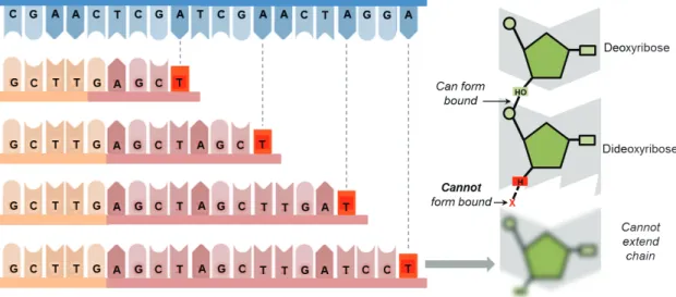
Figure 6: Dideoxynucleotide: Dideoxynucleotides (ddNTPs) lack the 3’-hydroxyl
group necessary for forming a phosphodiester bond. Consequently, ddNTPs prevent further elongation of a nucleotide chain and effectively terminate replication. The resulting length of a DNA sequence will reflect the specific nucleotide position at which the ddNTP was incorporated. For example, if a ddGTP terminates a sequence after 8 nucleotides, then the 8th nucleotide in the sequence is a cytosine
Figure 7: Sequencing: Dideoxynucleotides can be used to determine DNA sequence using the Sanger method. Four PCR mixes are set up, each containing stocks of normal nucleotides plus one dideoxynucleotide (ddA, ddT, ddC or ddG). As a typical PCR will generate over 1 billion DNA molecules, each PCR mix should generate all the possible terminating fragments for that particular base. When the fragments are separated using gel electrophoresis, the base sequence can be determined by ordering fragments according to length. If a distinct radioactive or fluorescently labelled primer is included in each mix, the fragments can be detected by automated sequencing machines. If the Sanger method is conducted on the coding strand (non-template strand), the resulting sequence elucidated will be identical to the template strand.
There are three different approaches for dye labeling of the sequencing fragments. The first approach is the use of a 5’-end dye labeled sequencing primer [46, 48]. The second approach is to label the 3’-ends of the sequencing fragment by using dye-labeled dideoxy nucleotides (dye-terminators) [47]. Alternatively, fluorescently labeled nucleotides in an extension-labeling step can internally label the fragments [49].
Acrylamide slab gel electrophoresis has been the most widely adopted format for Sanger sequencing. Relying merely on slab gel technology was not sufficient to accomplish the challenges set by the Human Genome Project. The continual and unrelenting drawbacks of slab gel electrophoresis and the demand for more rapid sequencing and higher throughput led to the development of capillary electrophoresis (CE) [50]. The completion of the human genome was only possible due to several technological advances offered by CE.
Electrophoresis is performed basically in an approach similar to slab gels with the advantage that each capillary contains a single DNA sample and therefore tracking problems are eliminated.
1.3.3. Non-electrophoretic DNA sequencing methods
A few DNA sequencing methods which are not electrophoretic based have been conceived in the last decades. These techniques have advantages and disadvantages compared to electrophoretic separation methods, depending on the type of applications performed and have promising applicability into miniaturized systems.
Sequencing-by-hybridization
The sequencing-by-hybridization (SBH) technique is in fact based on the same principle as that of Southern blotting [51] and was simultaneously described by two independent research groups in 1988 [52]. It key premise was its ability to facilitate de novo sequencing with the development of hybridization arrays[53-54]. The technique entails the labeling and subsequent annealing of an unknown strand of DNA to an array of short length oligonucleotides (e.g. all
65,536 combinations of 8-mers) which is followed by the analysis of the sequence by the pattern formed from its hybridization with the array. For this a computer assisted assembly program is essential for the ultimate reconstruction of the correct order and sequence of the original strand (Figure 8).
Because of the sheer scale of the data set from which the sequence is determined, the SBH technique has been prone to produce ambiguous results due to repetitive regions within the unknown sequence. Additionally, the formation of secondary structures within the target also reduces the accuracy of this technique. False positives are also caused by single base mismatches resulting from variations within the stability of the duplex of a complete match; this mostly occurs at the 3’-terminus. The effect of these issues can be minimized with the use of expression analysis or comparative sequencing between SBH microarrays though these issues have still not been solved in de novo sequencing.
Figure 8 Sequencing-by-hybridization (SBH) (i.e. the reverse dot-blot format). An unknown target is labeled and hybridized with an array of known octamer oligonucleotides, with the formed hybridization pattern being interpreted to determine the sequence of the targeted DNA sequence.
Sequencing-by-synthesis
The approach referred to as sequencing-by-synthesis (SBS) was first described in 1985 by Robert Melamede [55]. This technique consists of the
sequential addition and incorporation of nucleotides in an extension reaction directed by a polymerase primer in such a manner that a strand complementary to the original forms through interactive addition of the four dNTPs. Figure 9 shows the principle of SBH.
The detection of incorporation of nucleotides in the primed DNA strand can be done either directly or indirectly. In the direct method, fluorescent labels are attached to the dNTPs to allow their detection with an appropriate detection method [56-57]. A draw back to this technique is the low number of bases that can be added due to the high failure rate of nucleotide incorporation which can in turn lead to desynchronization of the sequence being copied [57]. The post-synthesis removal of the fluorophore acts as one further step in the process during which errors can be introduced.
The indirect approach to detection employs natural nucleotides for the incorporation process. The pyrophosphate (PPi) released during the condensation polymerization reaction of the growing chain is then detected as an indirect indication of successful nucleotide incorporation [58-59]. The principal advantage of this approach is the ability to use unadulterated, naturally occurring nucleotides which in turn exhibits superior incorporation efficiency in the assay. The enzymatic process laid out in the scheme below details is based on the luminometric detection of ATP through the conversion of PPi molecules through an enzymatic cascade.
(DNA) + dNTP!"#$/000000000001 2DNA3%&'()*,-.* 45+ $PPi$ (1)
PPi$ + $APS#67$.8'%98,('-.*/0000000000001 ATP$ + $ SO:;< (2)
ATP$ + $luciferin$ + $ O<
'8=>?*,-.*
Figure 9. An illustration of the underlying principal of sequencing by synthesis consisting on the primed DNA template being subjected to iterative additions of nucleotides while in the vicinity of a DNA polymerase molecule, represented in the schematic as an oval shape. If a complimentary nucleotide is added, incorporation of it into the sequence can be conducted through a condensation reaction, releasing a PPi molecule and polymerizing the nucleotide to the chain.
With the addition of dNTP to the reaction mixture, the DNA polymerase
incorporates a complimentary nucleotide onto the 3’terminus of the strand which
releases a PPi molecule into the solution. The free PPi molecule can then be observed via a coupled enzymatic reaction consisting of the ATP sulfurylase-mediated conversion of PPi to ATP which is in turn consumed by the firefly luciferase enzyme to produce detectable fluorescence [58, 60]. Detection of the signal is done with a photon multiplier or a charge coupled device (CCD) camera. This method does not, however, lend itself to the robust sequencing of DNA and thus no further progression of the technique is reported in the literature.
In progressive stages over the course of time, the sequencing-by-synthesis method has been developed into a robust, easy-to-use sequencing method for DNA through improvements in the removal efficiency of nucleotides and additional modification of the sequencing-by-synthesis principal. The innovation of the Pyrosequencing technology developed and described by Nyrén and colleagues is a direct result of this [61-63].
1.3.4 Pyrosequencing technology
The negation of unincorporated or excess dNTPs through total removal or degradation has been a key factor in the application of the sequencing-by-synthesis technique to DNA sequencing. In the Pyrosequencing method, the removal of nucleotides is executed in one of two ways (i) the solid-phase approach uses a three-coupled enzyme-mediated reaction with interspersed washing steps and (ii) the liquid-phase approach which uses a combination of four enzymes in a cascade reaction which requires no additional washing step.
Pyrosequencing by the solid-phase approach
The solid-phase approach localizes the sequencing-be-synthesis method to the surface of a solid-phase. All four nucleotides are added and then removed in dispensing and washing steps with one additional nucleotide being added to the sequence each time. There are several solid-phases currently in use; magnetic beads are on more recent approach [64] where a DNA template and annealed primer is labeled with biotin and immobilized on a magnetic bead which is coated with streptavidin. The primed strand of DNA is incubated with the three enzymes, DNA polymerase, ATP sulfurase and luciferase. Following the addition of each nucleotide, the DNA template is immobilized through the application of a magnetic field to the magnetic particles, holding them stationary while the washing cycle is completed.
There are several drawbacks to this method including the stripping of the DNA templates during the washing step, repetitive addition of enzymes and difficulties achieving a steady baseline; in addition, by its technical nature, this method does not lend itself to facile automation. A more promising approach is the use of a flow system to detect PPi and ATP on streptavidin coated substrates on which are biotin-mediated immobilization of ATP sulfurylase and luciferase. The use of a dynamic fluid flow should result in a more stable base line and in turn improve the detection of the sequence signal. This modified method is also more economical as the enzyme mixture does not need to be replenished after each addition of dNTP; additionally the constant fluid flow
serves to remove all inhibitory products in real time, thus rendering the reaction un-attenuated as well as making the system applicable to a microfluidic format.
The solid phase Pyrosequencing method was successfully applied to the microfluidic paradigm by 454 Lifesciences (since acquired by Roche) [65].
Pyrosequencing by the liquid-phase approach
The introduction of a nucleotide-degrading enzyme, called apyrase, was responsible for the breakthrough of liquid-phase Pyrosequencing [60-61, 63]. The introduction of this enzyme into the Pyrosequencing system negated the use of solid-phase separation and thus rendered obsolete the additional washing and enzyme addition steps synonymous with the latter. Apyrase exhibits high catalytic activity with low amounts of it efficiently degrading unincorporated nucleoside triphosphates in the reaction system first to nucleoside diphosphates and then to nucleoside monophosphate. Apyrase also serves to stabilize the base line with no fluctuations in the procedure as the enzyme catalysis reaction proceeds.
This liquid-phase Pyrosequencing method utilizes a combination of four enzymes in a cascade reaction while the sequencing of the DNA strand is observed in real time. The reaction is begun with the annealing of a primer onto a single DNA strand serving as the template. Figure 10 presents a schematic description of the sequencing of a partially amplified DNA strand of the human papillomavirus (HPV) with Pyrosequencing devices. The four enzyme-catalyzed cascade reaction is shown whereby APS indicates adenosine 5’- phosphosulphate and hv is the light photons emitted by the bioluminescent enzymatic reaction.
Figure 10. The liquid-phase Pyrosequencing method is a non-electrophoretic real-time DNA sequencing method which relies employs the release of light from the luciferase-luciferin reaction as a detection signal to indicate the incorporation of a nucleotide onto the target strand of DNA. All four nucleotides are added in iteration to a mixture of four enzymes (1). The pyrophosphate (PPi) which is released in the DNA polymerase-catalyzed reaction is converted quantitatively to ATP by ATP sulfurylase (2); this provides the required energy to facilitate the oxidation of liciferin by firefly luciferase to generate a light (hu) (3). This light can be then detected by a photon detection device and observed in real time through the use of an appropriate piece of computer software (4a). Apryase then catalyzes the degradation of the unincorporated nucleotides at which point the cycle is complete and the system is ready for the addition of the next nucleotide (4b)
In the liquid-phase Pyrosequencing method, the primer is hybridized with the template strand of DNA and the combined with the reaction enzymes, DNA polymerase, ATP sulfurylasem luciferase and apyrase, along with the substrates APS and luciferin. The four dNTPs are then sequentially added to the reaction. When the appropriate, complimentary, nucleotide is incorporated
by DNA polymerase onto the 3’-end of the DNA template, PPi can then be released in an equimolar quantity to the incorporated nucleotide.
The conversion of PPi to ATP is done quantitatively in the presence of APS by ATP sulfurylase. The ATP produced in this reaction the drives the luciferase-mediated production of oxyluciferin through conversion from luciferin
which in turn produces visible light proportionally to the original quantity of ATP. This light can then be detected by a photon multiplier tube or CCD camera and displayed as a peak in a pyrogram. Each signal of light is directly proporational to the number of nucleotides incorporated by the preceding reaction. Apyrase constantly degrades ATP and the remaining excess of dNTPs. Once the degradation process is completed, the systematic addition process is begun again. As this proceeds, a DNA strand complimentary to the template is formed with the sequence being interpreted through the displayed pyrogram peaks.
The Pyrogram, produced by the Pyrosequencing process is analogous to the electropherogram in the Sanger sequencing method. A pyrogram can be read and observed in real-time as the reaction is ongoing and is the de facto display of all the sequence signal peaks, displaying all nucleotide additions, the dispensation order of nucleotides and the incorporated and unincorporated dNTPs which produce the base line.
The modulus operandi of pyrosequencing relies upon the cooperation of several enzymes to allow the monitoring of the synthesis of DNA. Stability, fidelity, specificity, sensitivity, KM, and kcat are all critical parameters to the optimal performance of reaction. The enzyme kinetics can be studied in real-time and are given in Figure 6. The ascending pyrogram curve displays a slope indicative of the activity of DNA polymerase and ATP sulfurylase while the height of the signal is indicative of luciferase activity. The slope at which the final curve decends indicates the of nucleotide degradation
As described above, apyrase is the catalyst for the degradation of ATP with unincorporated or excess nucleotides being reduced to form nucleotide monophosphates and inorganic phosphate; this results in a steady and uniform decrease in the luminescence of the light signal to an intensity below that of the baseline of the pyrogram. When this point is reached, there is an additional internal lasted less than a minute to allow the degradation of the nucleotides to be completed so that the next dNTP can be added. The addition of the nucleotides can be repeated several times without any necessity to interrupt the process with a washing step.
The enzymes employed in the Pyrosequencing technique are naturally expressed by various organisms. The chemistry standard to Pyrosequencing
polymerase [66]. The ATP sulfurylase which is used in the technique is a recombinant version originating from the yeast cell Saccharomyces cerevisiae
[67] while the luciferase enzyme is taken from the American firefly Photinus
pyralis. The apyrase is taken from Solanum tuberosum (Pimpernel variety)
[68-69]. The global time scale for the event from polymerization to light detection is
between 3 and 4 seconds at room temperature.
The conversion of PPi to ATP by ATP sulfurylase occurs in approximately 1.5 seconds while the generation of light from the production of luciferase is executed in less than 0.2 seconds [70]. The light generated exhibits a maximum wavelength of 560 nanometers which can be readily detected by a photodiode, photon multiplier tube, or a CCD-camera.
There are many unique advantages to the Pyrosequencing technique amongst contemporary DNA sequencing technologies. One of these advantages is that mutations, deletions and insertions can be directly observed in the pyrgram (discussed in the next chapter) and that the order of dispensation of the nucleotides can be facilely programed. The ability to perform the sequencing process directly downstream of the primer, beginning with the first base after the annealed primer makes the design of the primer more flexible and thus makes this technique highly advantageous. The real-time production of the pyrogram as the pryosequencing process progresses, allows nucleotide incorporations and base callings to be immediately and continuously observed for each sample sequence. The Pyrosequencing method is finally and most importantly for scalable methods, easily automated for high throughput screening events.
1.4 SNP genotyping technologies
The first DNA sequencing method was described in 1977 by Sanger and co-workers [45]. This method, which came to be called Sanger DNA sequencing, is based on enzymatic chain termination, a set of single-stranded DNA molecules is generated, that is size-separated by gel electrophoresis. This method is by far the most common sequencing approach and has been further developed and optimized. Sanger DNA sequencing was the workhorse in the genome project and has been widely used in SNP discovery. However, its use
in SNP genotyping is limited by its low throughput and relatively high cost per sample. Pyrosequencing, an alternative to Sanger DNA sequencing, was developed in 1998 [61]. It is a real-time DNA sequencing method based on a four-enzyme mixture reaction that uses luciferase-luciferin light release to detect nucleotide incorporation by DNA polymerase into a target DNA This method will be described in detail in Chapter 5.
Over the last ten years, significant efforts have been made to improve SNP analysis with alternative techniques, so a growing number of new technologies have been introduced. The alleles are distinguished on the basis of different reaction principles, and the methods are then combined with general detection methods to reveal the sequence information. With some few exceptions, the methods require a pre-amplification step by polymerase chain reaction (PCR) to produce enough DNA for testing. A number of high quality reviews have been published on the ever increasing number of SNP scoring technologies [23, 71-72]. Most of the methods can be divided between hybridization-based-methods [39, 73-76] (Figure 11), ligation-based methods [77-84], primer extension by DNA polymerase[29, 85-93] and invasive cleavage-based method [94-95].
Figure 11. The principle of TaqMan assay. Two fluorescent dyes, a reporter (R) and a quencher (Q), are attached to the probes used to allele-specific hybridize with the target DNA. When both dyes are attached to the probe, reporter dye emission is
quenched due to FRET
(fluorescence energy transfer) from the reporter dye to the quencher dye. During the PCR reaction cycle, the exonuclease activity of the polymerase fragments the probe, and the reporter molecule (R) is released. The PCR is followed in real time and an increase in fluorescence indicates the presence of the matching allele.
1.5. Detection
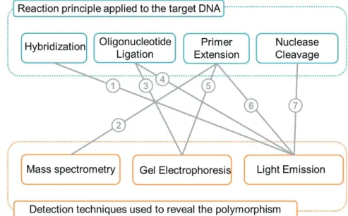
A variety of techniques is used to generate signals and determine the allelic status of SNPs and are combined with general detection methods to obtain sequence information. The multiplicity of methods currently available is due in part to the combination of reaction principles with different detection systems. Figure 12 shows the different combinations used to analyze various reaction principles.
Most of the common detection methods are based on the detection of light emission (fluorescence or chemiluminescence) with, in some cases, separation by gelelectrophoresis or on mass spectrometry. Fluorescence detection methods have greatly increased the sensitivity and ease of analyzing DNA. The availability of different fluorescent dyes and the diversity of their application have made them the most widely applied detection methods today. Fluorescence resonance energy transfer (FRET), in which energy is transferred from one molecule to another, is becoming useful in applications such as the DASH and TaqMan assay. Notable alternatives to fluorescence include chemiluminescence in the pyrosequencing system and mass spectrometric detection.
Figure 12. SNP genotyping methods categorized by reaction principles and detection.
1.6 Next stage for DNA sequencing platforms
DNA sequencing is considered the gold standard method for discovering new SNPs, but many alternative methods have been developed for known SNPs. Each method has unique ways of distinguishing a particular SNP position and is coupled with a detection method. There are at least 20 different SNP genotyping methods currently available [96]. A list of some of these SNP genotyping technologies is given in Table 1. The list does not cover all the methods but does give a representative picture of what is offered on the market. Many of these methods have been commercialized with a 384-well format and automated, such as the single-base extension based SNPstream, MALDI-TOF
MS based Massarray System and Pyrosequencing’s high-throughput system
[96]
.
Each SNP genotyping method has advantages and disadvantages and is suitable for a different range of applications [72]. Ultimately, choosing which method to use is based on the type of data to be generated, the number of samples to be processed, and the cost. That there is no single universal method for all situations promises further development of revolutionary methods. There is clearly a need for DNA analysis methods that are more specific, sensitive, fast, simple, automatable, and cost effective than those presently available.
These demands are driving the rapid evolution of a diverse range of new technologies. Some of these technologies have been developed on miniaturized platforms, which in themselves offer numerous advantages. Some of these platforms will be described in the following Chapter 3.
Table 1. Selected SNP genotyping methods.
Method Main advantage Company
Hydridization
DASH Inexpensive labelling Dynametrix, Ltd TaqMan No post-PCR treatment Applied Biosystems Oligonucleotide Ligation
SNPlex Multiplexing capacity Applied Biosystems Primer extension
SNPs stream Simple, versatile Orchid Bioscience SNaPshot Sensitive, no labeling Applied Biosystems MassArray Sensitive, no labeling Sequenom
Pyrosequencing Informative, denovo
sequencing Biotage
Nuclease cleavage
Chapter 2
Introduction: Microfluidics and micro total
systems (TAS)
Microfluidics involve the manipulation, transport, and analysis of fluids in micrometer volumes. The field has bloomed and branched off into many different areas, for which a number of excellent general reviews are available (e.g. biological and chemical analysis [97-99], point-of-care testing [100], clinical and forensic analysis [101], molecular diagnostics [102] and medical diagnostics
[103]). LOC systems are compact, often stand-alone systems featuring full
process integration and automation to perform complex tasks.
This subchapter 2.1 briefly describes relevant information over the microfluidics field, emphasizing the inter-relation between the chapters 1 and 2.
2.1 Microfluidics
Microfluidics involve the manipulation, transport, and analysis of fluids in micrometer sized volumes (Figure 13). Microfabrication technology is used to build microfluidic devices that exploit the inherent properties of liquids and gases in the microscale domain. These devices can accurately control minute volumes of fluid on a scale of nanoliters or picoliters. Making explicit use of the unique microfluidic effects, the microfluidic concept has today evolved towards the development of promising tools for analyzing biological samples in a way
commonly referred as ”Lab-on-a-chip” (LOC). The concept of LOC or
miniaturized total analysis systems (μTAS) as it is known today was proposed in
early 1990s by Manz et al. [104]. Since that time the field has bloomed and branched off into many different areas, for which a number of excellent general reviews are available (e.g. biological and chemical analysis [97-99], point-of-care testing [100], clinical and forensic analysis [101], molecular diagnostics [102] and medical diagnostics [103]). LOC systems are compact, often stand-alone systems featuring full process integration and automation to perform complex tasks.
Figure 13 An example of laminar flow in a junction of microfluidic channels containing flows of blue, yellow and colorless dyes. The design of the shown chip consists of 500 µm by 100 µm channels with the direction of flow being from left to right as indicated by the red and blue arrows. The main advantages of microfluidic systems over conventional systems include: minimized consumption of reagents, rapid response times (Table 2), well-controlled reaction conditions, parallel processing, speed of separation, excellent mass and heat transfer, low dead volumes, small power consumption, reduced manufacturing costs, portability, and disposability. The concept is still in its infancy, and significant technical challenges have to be overcome before the full potential of this technology is realized in biomedical research. Some of the challenges include fluid transport and the interfaces between the macroscopic and the microscopic environment, which include sample introduction and extraction as well as interrogation.
Table 2: Comparison of time and costs for the complete screen using traditional
methods and in microfluidic emulsions.
Robot Microfluidic drops
Total reactions 5 x 10⁷ 5 x 10⁷
Reaction volume 100 µL 6 pL
Total volume 5,000 L 150 µL
Reactions/day 73,000 1 x 10³
Total time ~2 years ~7 h
Number of plates/devices 260,000 2
Cost of plates/devices $520,000 $1.00
Cost of tips $10 million $0.30
Amortized cost of instruments $280,000 $1.70
Substrate $4.75 million $0.25
Total cost $15.81 million $2.50
Adapted from Agresti J. J. et al, Ultrahigh-throughput screening in drop-based microfluidics for directed evolution, PNAS 2010, 107:4004-4009.
With these numerous advantages it is clear why microfluidic devices have received such interest in recent times, having great potential for revolutionizing chemical and biological processes.
2.2 Microfabrication
Microfluidic platforms can be fabricated involving different materials, with the function and ideal properties of the device playing the center role concerning the choice of substrate. Glass and silicon are usually processed using a photolithography and wet etching. [105-106] Glass offers advantages in terms of its optical transparency and stability with strong chemicals. Silicon is used less commonly than glass due to its lack of transparency but tend to be easier to mass produce.
Hard polymers have gained attention as substrates, being examples poly(carbonate) (PC), [107] poly(methyl methacrylate) (PMMA), [108] and cyclic olefin copolymer (COC). [109-110] Their disadvantages is that they are usually not as stable to harsh chemicals and reaction conditions as glass, hence their use in chemical processes is more limited than with glass. [111-112] A big advantage though is that replication technologies can be applied to polymers enabling cuts in cost and time of production of disposable systems, important for example for point-of-care applications. Methods in use for fabrication for polymers comprehend injection moulding, [110, 113] imprinting, [108] hot embossing, [108, 114], and laser ablation. [115]
Poly(dimethylsiloxane) (PDMS) [116] is definitely the most used material for chip fabrication in research environment because of its rapid and simple fabrication that enables fast prototyping, though it is not so useful for mass fabrication. PDMS devices can be prepared via soft lithography. [116-117]Shortly,
a “master” is prepared with characteristics from the chip design as positive relief achieved with a photocurable epoxy. Next, the elastomer precursor and curing agent are mixed and placed over the master. The device is then left to cure, then peeled off the master and secured to a cover plate such as glass or PDMS. If the sealing is well performed, the channel network is then ready to be used.
2.3 Diffusion
In the closed channels and chambers networks of microfluidic systems, the diffusion of molecules needed for certain reactions can be estimated using the Einstein-Smoluchowski equation (Equation 1). [118]
= !2"# Equation 1
where x = distance (m), D = diffusion coefficient (m2.s-1), and t = time (s). This equation enables the enhancement or reduction of mixing in the microchannels while designing the chips, depending on the purpose of the system.
Microfluidic systems have been proven to have many advantages over so called conventional systems and they have been particularly very successfully in areas such as separations and chemical or biological assays. In the case of biological assays, the big surface-to-volume ratio of a microchannel enables surface-based assays, in which for example, antibodies attached to the channel surface are used to capture antigens, which is overall carried out efficiently due to the reduced diffusion distances of antigens to the surface. In addition to these advantages, the surface-to-volume ratio can be even more increased by using functionalized microparticles, allowing further increases in reaction efficiency. [119] Subchapter 1.2.4 Microparticles explains how such particles are incorporated into these devices and the mechanisms by which they can be manipulated for performing a number of procedures, including bioassays and separations.
2.4 Microparticles
Polymers, glass, silica, gold, and inorganic crystals, are a few examples of materials particles can be fabricated from. depending on the, and their purpose and are available in a wide range of sizes (1 to 100000 nm). [120-121] Particles with diameters of 1 nm to 1 µm suspended in solution are known as colloidal suspensions, while particles between 1 µm and 100 µm are commonly referred to as microparticles, and when in suspension form coarse suspensions.