571 J. Gen. Physiol. © The Rockefeller University Press • 0022-1295/2002/06/571/10 $5.00 Volume 119 June 2002 571–580
Absence of Proton Channels in COS-7 Cells Expressing Functional
NADPH Oxidase Components
Deri Morgan,1 Vladimir V. Cherny,1 Marianne O. Price,2 Mary C. Dinauer,2
and Thomas E. DeCoursey1
1Department of Molecular Biophysics and Physiology, Rush Presbyterian St. Luke’s Medical Center, Chicago, IL 60612
2Herman B. Wells Center for Pediatric Research, Department of Pediatrics (Hematology/Oncology) and Medical and Molecular
Genetics, James Whitcomb Riley Hospital for Children, Indiana University Medical Center, Indianapolis, IN 46202
a b s t r a c t Nicotinamide adenine dinucleotide phosphate (NADPH) oxidase is an enzyme of phagocytes that produces bactericidal superoxide anion (O2) via an electrogenic process. Proton efflux compensates for the charge movement across the cell membrane. The proton channel responsible for the H efflux was thought to be contained within the gp91phox subunit of NADPH oxidase, but recent data do not support this idea (DeCoursey,
T.E., V.V. Cherny, D. Morgan, B.Z. Katz, and M.C. Dinauer. 2001. J. Biol. Chem. 276:36063–36066). In this study, we investigated electrophysiological properties and superoxide production of COS-7 cells transfected with all NADPH oxidase components required for enzyme function (COSphox). The 7D5 antibody, which detects an
extra-cellular epitope of the gp91phox protein, labeled 96–98% of COS
phox cells. NADPH oxidase was functional because
COSphox (but not COSWT) cells stimulated by phorbol myristate acetate (PMA) or arachidonic acid (AA) produced superoxide anion. No proton currents were detected in either wild-type COS-7 cells (COSWT) or COSphox cells
stud-ied at pHo 7.0 and pHi 5.5 or 7.0. Anion currents that decayed at voltages positive to 40 mV were the only currents observed. PMA or AA did not elicit detectable H current in COSWT or COSphox cells. Therefore, gp91phox does not
function as a proton channel in unstimulated cells or in activated cells with a demonstrably functional oxidase.
k e y w o r d s : phagocytes • gp91phox • H channels • superoxide • respiratory burst
I N T R O D U C T I O N
Nicotinamide adenine dinucleotide phosphate (NADPH)* oxidase is an important antibacterial en-zyme of granulocytes that produces superoxide anion (Babior, 1999). This multicomponent complex com-prises several cytosolic subunits and the membrane-bound cytochrome b558, which is composed of two sub-units (gp91phox and p22phox) that coordinate FAD and
two heme moieties (Yu et al., 1998). Upon stimulation, the cytosolic components, p67phox, p47phox, and Rac, a
small GTP binding protein, translocate to the mem-brane and interact with cytochrome b558 (DeLeo and
Quinn, 1996). The assembled complex catalyses the
ox-idation of NADPH and the transfer of electrons across the membrane to extracellular oxygen, which is an electrogenic process (Henderson et al., 1987). The en-zyme can be activated by the phorbol ester PMA and by arachidonic acid (Babior, 1999). Voltage-gated proton currents are also greatly enhanced by these stimuli (De-Coursey et al., 2000, 2001a; Cherny et al., 2001). H ef-flux is thought to compensate electrically for electron movement through NADPH oxidase (Henderson et al., 1987, 1988).
It has been proposed that a proton channel exists within the gp91phox subunit of the NADPH oxidase. This
conclusion was based originally on studies of CHO cells transfected with gp91phox in which H channel function
was deduced from pH changes monitored by fluores-cent pH dyes (Henderson et al., 1995, 1997; Hen-derson, 1998). Using only indirect measurements it is difficult to assess the existence and properties of H channels and to rule out the involvement of other mechanisms that transport H equivalents. Two recent studies provide direct voltage-clamp evidence of H currents in CHO, HEK-293, and COS-7 cells heterolo-gously transfected with gp91phox (Henderson and
Meech, 1999; Maturana et al., 2001). However, neither study is convincing. Transfection of gp91phox into
HEK-293 cells simply increased the endogenous H currents fourfold (Maturana et al., 2001). The putative H cur-Address correspondence to Tom DeCoursey, Department of
Molecu-lar Biophysics and Physiology, Rush Presbyterian St. Luke’s Medical Center, 1750 W Harrison, Chicago, IL 60612. Fax: (312) 942-8711; E-mail: [email protected]
*Abbreviations used in this paper: AA, arachidonic acid; CGD, chronic granulomatous disease; DPI, diphenylene iodonium chlo-ride; NADPH, nicotinamide adenine dinucleotide phosphate; pHi,
intracellular pH; pHo, extracellular pH; PMA, phorbol
12-myris-tate 13-ace12-myris-tate; SITS, 4-acetamido-4-isothiocyanostilbene-2,2’-disul-phonic acid; SOD, superoxide dismutase; Vrev, reversal potential; WT,
wild-type.
rents in COS-7 cells transiently transfected with gp91phox
and studied in whole-cell configuration (Maturana et al., 2001) appear to reverse 100 mV positive to the Nernst potential for H, and thus were not proton se-lective. The currents in CHO cells reported by Hen-derson and Meech (1999) differ profoundly from H currents in all phagocytes studied to date: they are much less sensitive to inhibition by Zn2, Zn2 fails to slow act, no H current was detectable at pHi 7.5 even though the currents at pHi 6.9 were larger than native voltage-gated proton currents reported in any cell, and activation was at least an order-of-magnitude more rapid.
Nanda et al. (1994) found normal voltage-gated proton currents in human monocytes from chronic granulomatous disease (CGD) patients who lacked cy-tochrome b558, strong evidence that neither gp91phox nor p22phox is a proton channel in unstimulated
pha-gocytes. We demonstrated recently that gp91phox does
not mediate voltage-gated proton currents in the my-elocytic PLB-985 cell line (DeCoursey et al., 2001b). Upon induction by dimethylformamide, PLB-985 cells express functional NADPH oxidase and are capable of producing superoxide anions (Zhen et al., 1993). PLB-985 cells in which gp91phox was knocked out by
gene targeting had large voltage-gated proton cur-rents that were normal in every respect. Furthermore, PMA stimulation increased the H conductance to a similar extent in wild-type (WT) and knockout cells. Similar results were obtained in neutrophils from CGD patients lacking gp91phox expression. Therefore,
gp91phox does not mediate H currents in resting or
activated cells. However, these results do not com-pletely exclude the possibility that gp91phox might
function as a proton channel. Because of the endog-enous H currents in PLB-985 cells and CGD neu-trophils, a small contribution by gp91phox might have
been missed. Furthermore, one might propose that some other molecule might be up-regulated to com-pensate for the loss of gp91phox, as frequently occurs in
genetic knockout studies (Yang et al., 2001). There-fore we performed the converse of the knockout experiment by introducing gp91phox into COS-7 cells,
which have no endogenous H currents (Maturana et al., 2001). Although the goal of this study was to search for proton currents, there were large anion currents in most COS-7 cells and these are described briefly. We report here that neither WT COS-7 cells (COSWT) nor COS-7 cells transfected with the four main NADPH oxidase components, including gp91phox
(COSphox), have detectable voltage-gated proton
cur-rents, nor were any induced by PMA stimulation. We conclude that under conditions in which NADPH oxi-dase activity can be demonstrated, gp91phox does not
function as a proton channel.
M A T E R I A L S A N D M E T H O D S
Cell Preparation and Maintenance
Green monkey kidney cells (COSWT) were obtained from
Ameri-can Type Culture Collection (CRL-1651). COS-7 cells stably transfected with the four main subunits of NADPH oxidase: gp91phox, p22phox, p47phox, and p67phox (COS
phox cells) were devel-oped by Price and colleagues (Price et al., 2002). First, COS-7 cells were stably transfected with p22phox and gp91phox. These cells were then further modified by stable transfection with p47phox, and p67phox to produce COS
phox cells. Cells were maintained in Dulbecco’s MEM (Sigma-Aldrich) as described previously (Yu et al., 1997), with the exception that media for COSphox cells also contained three antibiotics to ensure selection of transformed cells: 2 g ml1 puromycin (Sigma-Aldrich), 0.8 mg ml1 G418
(Sigma-Aldrich), and 175 g ml1 hygromycin (Sigma-Aldrich).
COSphox cells were split when 70% confluent. COS-7 cells are ad-herent and were harvested by incubation with 0.25% trypsin in 1 mM EDTA (GIBCO-BRL) for 5 min at 37C. For electrophysio-logic recordings, cells were allowed to attach to small glass cover-slips. The cells were kept in suspension for the cytochrome c as-says.
Electrophysiology
Whole cell and permeabilized patch recordings were performed as described previously (Cherny and DeCoursey, 1999; DeCour-sey et al., 2000) using micropipettes pulled from 7052 glass (Gar-ner Glass). Although COS cells tend to flatten in culture, we se-lected rounded cells for recording, because voltage-clamp con-trol is better and because the superoxide assays were done with nonadherent (and hence rounded) cells. Some cells were nearly spherical and others were irregular in shape; we did not attempt to record from flat cells. The cell diameter was 15–25 m, and the capacity in whole-cell measurements was 14.4 4.5 pF (mean SD) in 20 sequentially studied COSphox cells. Whole-cell solutions (pipette and bath solutions) contained 100 mM buffer, 1 mM EGTA, 70 mM tetramethylammonium (TMA) methane-sulfonate (MeSO3), and 2 mM MgCl2 or 3 mM CaCl2, with an
os-molarity of 300 mOsm. Buffers were Mes for pH 5.5 and BES (N,N-bis[2-Hydroxyethyl]-2-aminoethanesulfonic acid) for pH 7.0 solutions. In some experiments the Cl concentration was in-creased by replacing TMAMeSO3 with TMACl or with Ringer’s
solution (in mM: 160 NaCl, 4.5 KCl, 2 CaCl2, 1 MgCl2, and 5
HEPES). Permeabilized patch recordings were performed in so-lutions containing 100 mM TMAMeSO3, 25 mM (NH4)2SO4, 2
mM MgCl2, 5 mM BES, 1 mM EGTA titrated to pH 7.0 with
TMAOH. The purpose of the 50 mM NH4 in the bath and
pi-pette solutions was to clamp pHi near pHo (pH 7.0) (Grinstein et
al., 1994; DeCoursey et al., 2000). Pipette solutions also con-tained 0.5–1 mg ml1 amphotericin B (Sigma-Aldrich). Pipettes
were filled after dipping the tip in amphotericin free solution. Experiments were done at 21C or at room temperature (21– 24C). Currents were amplified using either Axopatch 200B (Axon Instruments, Inc.) or List EPC-7 (List Electronic) amplifi-ers and filtered at 100 or 200 Hz. PClamp8 (Axon Instruments, Inc.) or lab-generated software and an Indec Laboratory acquisi-tion and display system (Indec Corporaacquisi-tion) were used for data acquisition and manipulation.
Superoxide Anion (O2) Production
performed by incubating cells (2.5 105 cells well1) at 37C in
250 l HBSS containing 75 M cytochrome c. Kinetic assays were performed on a Ceres UV900 plate reader (Biotek Instruments) and superoxide production was quantified using an extinction coefficient of 21.1 mM cm1 for cytochrome c. The superoxide
assays were performed on the same batches of cells and during the same time period that the electrophysiological studies were done.
7D5 Antibody Staining
Cells were harvested by brief trypsinization, pelleted (1,200 rpm for 6 min at 4C), resuspended in PBS as a wash, and pelleted. Cells were resuspended in PBS containing 10% normal goat se-rum and incubated for 30 min on ice to block nonspecific bind-ing sites. Cells were pelleted again then resuspended in PBS at 107 cells/ml. One aliquot of cells (100 l) was incubated with 100
l 7D5 supernatant for 30 min on ice while a second aliquot was incubated with 2 l of isotype control CD-117 antibody. PBS con-taining 0.1% BSA (1 ml) was added as a wash and the cells were pelleted. Cells were resuspended in PBS and incubated for 30 min on ice with FITC conjugated goat-mouse secondary anti-body (1:100 dilution). PBS containing 0.1% BSA (1 ml) was added as a wash, and the cells were pelleted and then resus-pended in 500 l PBS for fluorescence-activated cell sorter
analy-sis using a FACSCalibur machine (Becton Dickinson) equipped with CellQuest software. A total of 10,000 cells were counted for each condition. Cells with very low fluorescence (comprising 7% of the total, and falling below the lower limit of the graph) were considered dead and were gated out. Staining with 7D5 was compared with staining with isotype control. Also, levels of 7D5 staining were compared in COSphox cells and COSWT cells.
R E S U L T S
COS-7 Cells Have Cl Currents
COSWT and COSphox cells were studied in whole-cell
con-figuration in solutions lacking most permeant ions at pHi 5.5 and pHo 7, conditions favorable for detect-ing proton currents. Many cells appeared to become “leaky” within a few seconds of breaking the membrane patch. This “leak” reflected the appearance of a large time-independent conductance at the holding poten-tial (–60 mV) and at all potenpoten-tials negative to 40 mV. The properties of this conductance were similar in COSWT (Fig. 1, A–C) and COSphox cells (Fig. 1, D–F).
Figure 1. Rundown of whole-cell currents in COSWT (A–C) and COSphox (D–F) cells. Families of currents were recorded at various times after establishing whole cell configuration at pHi 5.5 and pHo 7.0 in TMAMeSO3 solutions. Insets illustrate the voltage pulses, which were
During large depolarizing pulses the currents turned off with time. This decay reflected a genuine decrease in conductance because it was reversed upon repolar-ization; the inward currents were initially small and turned on with time. As evident in Fig. 1, the conduc-tance decreased progressively after establishing whole-cell configuration in both COSWT and COSphox cells.
The rundown produces a progressive decrease in hold-ing current that is evident in some of the families. Eventually, only very small time-independent currents remained (Fig. 1, C and F).
The behavior of the currents in COSWT and COSphox
cells, including the rundown phenomenon, was remi-niscent of Cl currents in Chinese hamster ovary cells studied in similar solutions (Cherny et al., 1997). These currents resemble volume-regulated anion currents in several other epithelial cell types (McCann et al., 1989; Solc and Wine, 1991; Arreola et al., 1995). Fig. 2 shows the inhibition of whole-cell currents in a COSphox cell
by the traditional Cl channel blocker 4-acetamido-4’-isothiocyanostilbene-2,2’-disulfonic acid (SITS). The conductance decreased substantially at all potentials
upon addition of SITS (Fig. 2 B), and was at least par-tially restored after wash out (unpublished data). To confirm the Cl selectivity of the conductance we ramped the voltage from 100 to 80 mV in solutions with different Cl concentrations (Fig. 2 C). Trace 1 shows ramp currents in symmetrical 5 mM Cl where the reversal potential (Vrev) was near 0 mV. Substituting the bath solution with 166 mM Cl increased the out-ward current at depolarizing voltages and shifted Vrev negative by 31 mV. Upon returning to the original solu-tion, Vrev shifted back and the currents were reduced below the original levels, probably due to rundown (compareFig. 1). The shift in Vrev is consistent with a relative permeability of 0.5 for MeSO3 compared with Cl, calculated by the Goldman-Hodgkin-Katz volt-age equation after correction for liquid junction poten-tials. The outward current increased consistently in high [Cl]. The shift in Vrev was often less pronounced than in Fig. 2 C, but the shift in Vrev was larger when the anion conductance was large (i.e., shortly after estab-lishing whole cell configuration). The predominant conductance in COS-7 cells was anion selective.
COS-7 Cells Lack H Currents
To identify putative voltage-gated proton currents, we waited for the anion conductance to subside and then looked for currents that increased with time during de-polarizing pulses. In some cells time-dependent out-ward currents were seen, although typically this behav-ior was observed only after the input resistance had de-creased markedly. An example is shown in Fig. 3. Normal behavior was observed early in this experiment on a COSWT cell. The family of currents in Fig. 3 A was recorded starting 4 min after whole-cell configuration was established. Anion currents that are time indepen-dent except at large positive voltages were observed. The current at Vhold60 mV decreased progressively, in part because of progression of the rundown of anion currents during the 3.5 min required to record this family of long pulses. After the pulses to 60 and 80 mV that elicited decaying outward currents, the inward cur-rent upon repolarization was greatly reduced, reflect-ing the channel closreflect-ing at positive voltages that is re-sponsible for the current decay. The family of currents in Fig. 3 B was recorded in the same cell immediately after the previous family. By this time, the anion
con-ductance had run down substantially and the currents at all voltages were scaled down, but otherwise exhib-ited similar time and voltage dependence. Near the end of the pulse to 60 mV (pulse 6), the current sud-denly increased. The current at Vhold increased as well, and no recovery was observed before the next pulse, to 80 mV. During this last pulse (pulse 7), the current was much larger and there was a time-dependent increase in outward current during the pulse. Under normal cir-cumstances, we would interpret the sudden increase in conductance to indicate damage to the cell membrane or loss of the seal. Such cells are usually considered to be “dead” and are discarded. However, because the time-dependent increasing outward currents superfi-cially resemble voltage-gated proton currents, we con-tinued recording. In cells with currents like these, changing pHo did not change either Vrev or the thresh-old voltage for activating time-dependent outward cur-rent. These currents thus were not H selective, nor did they display the pronounced pHo sensitivity of gat-ing of all voltage-gated proton channels (Eder and De-Coursey, 2001). Addition of 100 M ZnCl2 had no ef-fect on the amplitude or kinetics of these currents. In summary, there was no credible evidence of voltage-Figure 3. Sudden appearance of time-dependent outward current in a COSWT cell, studied at pHo 7.0 and pHi 5.5. (A) A family of
cur-rents recorded starting 4 min after establishing whole-cell configuration displays behavior typical of the anion conductance. The pulses were applied in 20-mV increments up to 80 mV, at 30-s intervals. The entire family required 3.5 min to complete. The first and last pulses are numbered, showing the progressive decrease in current at 60 mV. (B) A family recorded in the same cell shortly after the end of the previous family. The currents at all voltages including Vhold are scaled down, reflecting the rundown typically observed in these cells. In this
cell during the pulse to 60 mV (pulse 6), there was a sudden catastrophic increase in leak current. The inward current upon repolarization was greatly increased. Interpolation of the current at the end of the pulse to 60 mV and that immediately after repolarization to 60 mV gives an estimated Vrev of 4 mV, suggesting a nonselective leak conductance. The subsequent pulse to 80 mV elicited an outward current
that increased with time. Subsequent pulse families evoked time-independent leak current at all voltages, with a time-dependent increas-ing current at large positive voltages. Changincreas-ing pHo from 7.0 to 5.5 did not shift Vthreshold or elicit changes in gating kinetics normally seen
in cells with voltage-gated H currents. Addition of 100 M ZnCl2 did not inhibit the time-dependent outward currents. Although such
gated proton currents in any COSWT or COSphox cell
studied.
To investigate whether stimuli that activate NADPH oxidase might induce or enhance proton currents, as has been observed in human neutrophils and eosino-phils (DeCoursey et al., 2000, 2001a; Cherny et al., 2001) we used the permeabilized patch configuration, which preserves NADPH oxidase function (DeCoursey et al., 2000). Many unstimulated COSphox cells in permeabilized
patch configuration exhibited Cl currents that disap-peared with time (Fig. 4, A–C), similar to the currents in whole-cell studies. Increasing [Cl]o increased the out-ward current and tended to shift Vrev to more negative voltages (unpublished data). No time-dependent cur-rents were seen in COSphox cells after the Cl currents ran
down. In about one-third of COSphox cells the anion
con-ductance was small or absent throughout the experi-ment, and the currents were similar to those in Fig. 4 C.
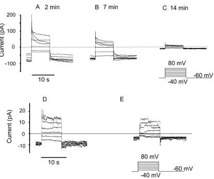
Fig. 4, D and E, respectively, show families of currents at high gain before and 10 min after the addition of 60 nM
PMA. No time-dependent currents appeared at any voltage after the addition of PMA in this cell. In Fig. 4, D and E, dis-crete current steps consistent with a single channel con-ductance of 30 pS can be seen at some voltages. These currents are most likely conducted through anion chan-nels similar to volume-sensitive anion chanchan-nels reported previously (Solc and Wine, 1991). Single voltage-gated pro-ton channel currents are too small to record directly (By-erly and Suen, 1989; DeCoursey and Cherny, 1993). Fur-thermore, 100 M Zn2 did not inhibit any component of the currents in PMA-treated COSphox cells. Thus, both
un-stimulated (31 cells studied in whole-cell or permeabilized-patch configuration) and PMA- or AA-stimulated COSphox
cells (n 45) lack detectable proton currents.
COSphox Cells Express gp91phox and Produce Superoxide Anion (O2)
To assess the efficiency of gp91phox expression in COS phox
[image:6.612.104.522.59.409.2]cells, we used the 7D5 antibody, which is directed Figure 4. Lack of effect of PMA on membrane currents COSphox cells in permeabilized patch configuration. (A–C) Families of voltage-clamp currents in a COSphox cell in permeabilized patch configuration at the indicated times after beginning recording. The bath solution contained TMAMeSO3 at pH 7.0, and both pipette and bath contained 50 mM NH4 to clamp pHi near pHo. Currents at high gain in a
against an extracellular epitope of the gp91phox
mole-cule (Nakamura et al., 1988; Yu et al., 1998). Expres-sion was tested in freshly thawed cells and passaged cells, in COSWT and COSphox cells, and with the CD-117
antibody as an isotype control. Fig. 5 illustrates histo-grams of the fluorescence intensity in COSWT (open histogram) and COSphox cells (solid histogram). 98% of
the freshly thawed COSphox cells (Fig. 5) and 96% of
COSphox cells maintained in culture for several weeks
(unpublished data), as during the course of these ex-periments, stained positively for the 7D5 antibody. If cells with very low fluorescence (which presumably are dead cells) are included, then the 7D5-positive fraction is 95% of the freshly thawed COSphox cells and 93% of
COSphox cells maintained in culture. Thus, the vast
ma-jority of COSphox cells express gp91phox at the plasma
membrane.
Superoxide anion (O2) production by COSphox cells
was assessed by their ability to reduce ferricytochrome c. Fig. 6 A shows the average time courses of cumulative O2 release by four batches of COSphox cells challenged
with different concentrations of PMA. O2 production above control levels was seen at 6 nM PMA. PMA at 60 and 300 nM, respectively, produced similar levels of O2. The O2 release time course was characterized by a delay of several minutes after the addition of PMA, followed by a rapid increase between 5 and 20 min, af-ter which the rate of O2 production diminished. Fig. 6 B summarizes the average maximal rate of O2
produc-tion in these experiments. The highest maximal rate, observed at 300 nM PMA, was 26 6 nmol min1 (107 cells)1. The response was almost completely inhibited by 100 g/ml superoxide dismutase (SOD), showing that ferricytochrome c reduction was due to the release of O2 and not peroxides or other oxidizing agents. Diphenylene iodonium (DPI) at 6 M also inhibited O2 production, implicating a flavoenzyme (O’Donnell et al., 1994), most likely NADPH oxidase (Cross and Jones, 1986). As reported previously (Price et al., 2002), COSWT cells studied under identical conditions did not produce detectable O2 (unpublished data).
Arachidonic acid (AA) can stimulate COSphox cells to
produce O2 at a higher rate than that stimulated by PMA (Price et al., 2002). We examined the time course of AA-stimulated O2 production in six assays (unpublished data). The onset of O2 production stimulated by 10–100 M AA was faster than when PMA was used. However, the AA-stimulated respiratory burst was more transient, with little additional O2 released after 10–15 min, whereas PMA-stimulated O2 release continued over 30 min.
Because NADPH oxidase is electrogenic (Henderson et al., 1987), its activity can be detected electrically as a small inward current under favorable conditions Figure 5. 7D5 antibody staining of COSphox cells compared with
COSWT cells. Cells harvested by brief trypsinization were incubated
with 7D5 monoclonal antibody, which recognizes an extracellular epitope of gp91phox. Following incubation with a FITC-conjugated goat anti–mouse secondary antibody, cells were analyzed by flow cytometry. The marker M1 was set to exclude 99% of the COSWT
cells (open histogram), and indicates the range defined as posi-tively stained. By this criterion, 98.3% of the COSphox cells (solid histogram) were positively stained for gp91phox expression.
Figure 6. Superoxide production by COSphox cells. (A) The aver-age time course of PMA-stimulated superoxide anion production detected as the reduction of cytochrome c is plotted. Cells (106
cells/ml) were incubated at 37C in HBSS before the addition of PMA (concentrations of PMA are in nM). Control cells were not stimulated. Data are mean values from four experiments with the error bars removed for clarity. (B) The average maximum rate of O2 production ( SE, n 4) determined in each experiment in
(Schrenzel et al., 1998; DeCoursey et al., 2000). Elec-tron currents were difficult to detect in COSphox cells,
probably because the activity of NADPH oxidase is lower than in human neutrophils (Price et al., 2002) and the baseline currents were larger and less stable. These measurements were made at 0 to 20 mV to minimize the leak (anion) current amplitude. No con-vincing electron currents were seen with PMA as a stim-ulus. Because the maximal rate of O2 production in COSphox cells is larger with AA than PMA as a stimulus
(Price et al., 2002) and the onset is more rapid (present study), we also used AA as an agonist. Addi-tion of 5–10 M AA led to an increase in inward cur-rent of 1–2 pA in about half the cells tested, but only a fraction of this current was inhibited by 6–12 M DPI. Because AA can induce nonspecific membrane leak (Meves, 1994; Cherny et al., 2001), we consider only in-ward current inhibitable by DPI in the presence of AA to indicate electron current. The mean DPI-inhibited inward current in COSphox cells was 0.26 0.10 pA
(mean SE, n 21). Although it is possible that these DPI-sensitive currents reflect genuine electron current, we conclude that the electron currents in COSphox cells
are too small to detect reliably.
D I S C U S S I O N
Although the goal of this study was to search for volt-age-gated proton currents in cells expressing gp91phox,
the only conductance found in COSphox cells was anion
selective. Cl-selective currents similar to volume-sensi-tive Cl currents in many other epithelial cells (Mc-Cann et al., 1989; Solc and Wine 1991; Arreola et al., 1995; Cherny et al., 1997) were present shortly after es-tablishing whole-cell configuration in most COSWT and COSphox cells. In an earlier study of CHO cells, small
voltage-gated proton currents were revealed after the anion conductance ran down (Cherny et al., 1997), but in COSWT and COSphox cells no proton currents were
ever observed. These results are consistent with the ob-servation of Maturana et al. (2001) that COSWT cells lack endogenous proton channels.
We saw no H currents in COS-7 cells stably trans-fected with all four major components of the NADPH oxidase, including gp91phox (COS
phox cells). In some
COSphox and COSWT cells we saw outward currents that increased during large depolarizing pulses. These cur-rents, seen mainly in “leaky” cells, were not inhibited by 100 M Zn2, a potent, classical inhibitor of voltage-gated proton channels (Cherny and DeCoursey, 1999), and neither Vrev nor the threshold potential was sensi-tive to pHo, contrary to the properties of voltage-gated proton channels in all cells studied to date (Eder and DeCoursey, 2001). Thus, no credible evidence of volt-age-gated proton currents was seen in any COSWT or
COSphox cell. The maximum proton current in most
phagocytes is 100 times greater than that required to fully compensate for the electron flux through NADPH oxidase (Eder and DeCoursey, 2001). If a comparable relationship existed in COSphox cells, one would predict
50 pA of proton current. Even a few picoamperes of H current would have been evident in COSphox cells
af-ter rundown of the anion conductance.
In previous studies, evidence of proton flux was re-ported when gp91phox was expressed without any other
NADPH oxidase components in CHO, HEK-293, and COS-7 cells (Henderson et al., 1995, 1997; Hender-son 1998; HenderHender-son and Meech, 1999; Maturana et al., 2001). However, the putative proton currents re-ported in patch-clamp studies of gp91phox expressed in
the CHO and COS-7 heterologous expression systems (Henderson and Meech, 1999; Maturana et al., 2001) differ significantly from proton currents endogenous to phagocytes (see introduction). It is conceivable that gp91phox expressed alone might function as an ion
channel, and that this function is absent when it is co-expressed with p22phox. If so, then gp91phox does not
function as an ion channel under physiological condi-tions, because gp91phox is detectable in phagocyte
membranes only together with p22phox in the
flavocyto-chrome b558 heterodimer (Babior, 1999). The expres-sion of gp91phox and p22phox are closely linked in
neu-trophils, where stable expression of either appears to be favored strongly by heterodimer formation, i.e., as-sembly to form flavocytochrome b558 (Roos et al., 1996; Yu et al., 1997; DeLeo et al., 2000).
The COSphox cells in this study were transfected with
all the components necessary for a fully functional oxi-dase. COSphox cells released significant O2, confirming that the oxidase was present in the plasma membrane and was functional. The maximum rate of PMA-stimu-lated O2 production at 37C was 26 nmol min1(107 cells)1, comparable with the rate we reported previ-ously in these cells (Price et al., 2002). The mini-mum efficiency of simultaneous expression of all four NADPH oxidase components was 69%, because this fraction was nitroblue tetrazolium (NBT)-positive in COSphox cells (Price et al., 2002). NBT reduction
indi-cates O2 production, and all four NADPH oxidase components are required for this function (Babior, 1999). Expression of gp91phox in COS
phox cells in the
plasma membrane was confirmed by 7D5 antibody staining. The 7D5 antibody is directed against an extra-cellular epitope of gp91phox (Nakamura et al., 1988; Yu
et al., 1998). That 96–98% of COSphox cells were stained
positively by the 7D5 antibody indicates that essentially all cells expressed gp91phox at the surface membrane.
Although COSphox cells express a functional NADPH
by the presence of background (leak) currents, and by the tiny amplitude of these currents. The maximum rate of O2 production in COSphox cells is less than
one-fourth that in human neutrophils stimulated with PMA (Price et al., 2002). The electron current averages only 2.3 pA in human neutrophils (DeCoursey et al., 2000) and can be detected only because the input resis-tance in neutrophils is very high (usually 70 G), and the background leak current was small (usually 0.6 pA or less) and stable. If electron currents in COSphox
were proportionately smaller they would be approxi-mately 0.5 pA and consequently very difficult to de-tect; we did not detect electron currents in PMA-stimu-lated COSphox cells. AA also stimulates O2 production in COSphox cells (Price et al., 2002). Although AA
in-duced small inward currents in some COSphox cells, DPI
reversed only a fraction of the current and thus the identification of these currents as genuine electron cur-rents remains uncertain.
Bánfi et al. (1999) proposed that there were two types of H channels in granulocytes; one in the resting cells and another that is active only when NADPH oxi-dase is active. Using the permeabilized patch configura-tion allowed us to investigate whether activating the ox-idase might induce the appearance of H currents in COSphox cells. We have shown previously (DeCoursey et
al., 2000, 2001a,b) that PMA stimulation of human neu-trophils or eosinophils or PLB-985 cells in these condi-tions results in H currents that closely resemble the NADPH oxidase–related H currents described by Bánfi et al. (1999). AA also greatly enhances voltage-gated proton currents (Cherny et al., 2001). No time-dependent currents appeared after addition of PMA or AA to COSphox cells. Evidently the enhancement of H
currents during the respiratory burst occurs only in cells that express voltage-gated proton channels in the resting state. This pattern is consistent with the idea that respiratory burst agonists modify the properties of preexisting H channels (DeCoursey et al., 2000), rather than inducing the appearance of a new type of channel (Bánfi et al., 1999).
A curious point is that COSphox cells devoid of proton
channels are still capable of producing substantial O2. The main function of voltage-gated proton channels in phagocytes is thought to be to provide a mechanism of charge compensation for the electron efflux through NADPH oxidase (Henderson et al., 1987; Eder and De-Coursey, 2001). Inhibiting H currents reduces super-oxide production in human neutrophils (Henderson et al., 1988) and eosinophils (Bankers-Fulbright et al., 2001) and in PLB cells (Lowenthal and Levy, 1999). Be-cause anion currents were prominent in COS-7 cells, it is tempting to speculate that Cl influx may serve this function in lieu of H efflux. In preliminary experi-ments SITS did not inhibit O2 release, but SITS did
not completely inhibit Cl current (Fig. 2 A). The an-ion channel inhibitor 4,4-diisothiocyanostilbene-2,2’-disulfonic acid (DIDS) partially reduced O2 produc-tion in eosinophils (Schwingshackl et al., 2000). Alter-natively, COSphox cells may have some other mechanism
of charge compensation.
The authors appreciate the able technical assistance of Tatiana Iastrebova.
This work was supported in part by the Heart, Lung, and Blood Institute of the National Institutes of Health (HL52671 and HL61437 to Dr. DeCoursey and HL45635 to Dr. Dinauer).
Submitted: 11 December 2001 Revised: 19 April 2002 Accepted: 22 April 2002
R E F E R E N C E S
Arreola, J., J.E. Melvin, and T. Begenisich. 1995. Volume-activated chloride channels in rat parotid acinar cells. J. Physiol. 484:677– 687.
Babior, B.M. 1999. NADPH oxidase: an update. Blood. 93:1464– 1476.
Bánfi, B., J. Schrenzel, O. Nüsse, D.P. Lew, E. Ligeti, K.-H. Krause, and N. Demaurex. 1999. A novel H conductance in eosinophils: unique characteristics and absence in chronic granulomatous disease. J. Exp. Med. 190:183–194.
Bankers-Fulbright, J.L., H. Kita, G.J. Gleich, and S.M. O’Grady. 2001. Regulation of human eosinophil NADPH oxidase activity: a central role for PKC. J. Cell. Physiol. 189:306–315.
Byerly, L., and Y. Suen. 1989. Characterization of proton currents in neurones of the snail, Lymnaea stagnalis.J. Physiol. 413:75–89. Cherny, V.V., and T.E. DeCoursey. 1999. pH-dependent inhibition
of voltage-gated H currents in rat alveolar epithelial cells by Zn2 and other divalent cations. J. Gen. Physiol. 114:819–838. Cherny, V.V., L.M. Henderson, and T.E. DeCoursey. 1997. Proton
and chloride currents in Chinese hamster ovary cells. Membr. Cell Biol. 11:337–347.
Cherny, V.V., L.M. Henderson, W. Xu, L.L. Thomas, and T.E. De-Coursey. 2001. Activation of NADPH oxidase-related proton and electron currents in human eosinophils by arachidonic acid. J. Physiol. 535:783–794.
Cross, A.R., and O.T.G. Jones. 1986. The effect of the inhibitor diphenylene iodonium on the superoxide-generating system of neutrophils. Specific labelling of a component polypeptide of the oxidase. Biochem. J. 237:111–116.
DeCoursey, T.E., and V.V. Cherny. 1993. Potential, pH, and arachi-donate gate hydrogen ion currents in human neutrophils. Bio-phys. J. 65:1590–1598.
DeCoursey, T.E., V.V. Cherny, W. Zhou, and L.L. Thomas. 2000. Si-multaneous activation of NADPH oxidase-related proton and electron currents in human neutrophils. Proc. Natl. Acad. Sci. USA. 97:6885–6889.
DeCoursey, T.E., V.V. Cherny, A.G. DeCoursey, W. Xu, and L.L. Thomas. 2001a. Interactions between NADPH oxidase-related proton and electron currents in human eosinophils. J. Physiol. 535:767–781.
DeCoursey, T.E., V.V. Cherny, D. Morgan, B.Z. Katz, and M.C. Dinauer. 2001b. The gp91phox component of NADPH oxidase is not the voltage-gated proton channel in phagocytes, but it helps. J. Biol. Chem. 276:36063–36066.
DeLeo, F.R., J.B. Burritt, L. Yu, A.J. Jesaitis, M.C. Dinauer, and W.M. Nauseef. 2000. Processing and maturation of flavocytochrome b558 include incorporation of heme as a prerequisite for
het-erodimer assembly. J. Biol. Chem. 275:13986–13993.
Eder, C., and T.E. DeCoursey. 2001. Voltage-gated proton channels in microglia. Prog. Neurobiol. 64:277–305.
Grinstein, S., R. Romanek, and O.D. Rotstein. 1994. Method for manipulation of cytosolic pH in cells clamped in the whole cell or perforated-patch configurations. Am. J. Physiol. 267:C1152-C1159.
Henderson, L.M. 1998. Role of histidines identified by mutagenesis in the NADPH oxidase-associated H channel. J. Biol. Chem. 273: 33216–33223.
Henderson, L.M., G. Banting, and J.B. Chappell. 1995. The arachi-donate-activatable, NADPH oxidase-associated H channel: evi-dence that gp91-phox functions as an essential part of the chan-nel. J. Biol. Chem. 270:5909–5916.
Henderson, L.M., J.B. Chappell, and O.T.G. Jones. 1987. The su-peroxide-generating NADPH oxidase of human neutrophils is electrogenic and associated with an H channel. Biochem. J. 246: 325–329.
Henderson, L.M., J.B. Chappell, and O.T.G. Jones. 1988. Superox-ide generation by the electrogenic NADPH oxidase of human neutrophils is limited by the movement of a compensating charge. Biochem. J. 255:285–290.
Henderson, L.M., and R.W. Meech. 1999. Evidence that the prod-uct of the human X-linked CGD gene, gp91-phox, is a voltage-gated H pathway. J. Gen. Physiol. 114:771–785.
Henderson, L.M., S. Thomas, G. Banting, and J.B. Chappell. 1997. The arachidonate-activatable, NADPH oxidase-associated H channel is contained within the multi-membrane-spanning N-ter-minal region of gp91-phox.Biochem. J. 325:701–705.
Lowenthal, A., and R. Levy. 1999. Essential requirement of cytosolic phospholipase A2 for activation of the H channel in
phagocyte-like cells. J. Biol. Chem. 274:21603–21608.
Maturana, A., S. Arnaudeau, S. Ryser, B. Bánfi, J.P. Hossle, W. Schle-gel, K.-H. Krause, and N. Demaurex. 2001. Heme histidine ligands within gp91phox modulate proton conduction by the phagocyte NADPH oxidase. J. Biol. Chem. 276:30277–30284. McCann, J.D., M. Li, and M.J. Welsh. 1989. Identification and
regu-lation of whole-cell chloride currents in airway epithelium. J. Gen. Physiol. 94:1015–1036.
Meves, H. 1994. Modulation of ion channels by arachidonic acid. Prog. Neurobiol. 43:175–186.
Nakamura, M., S. Kobayashi, S. Sendo, T. Koga, and S. Kanegasaki.
1988. Deficiency of cytochrome b558 in chronic granulomatous
disease demonstrated by monoclonal antibody 7D5. Acta Paediatr. Hung. 29:179–183.
Nanda, A., R. Romanek, J.T. Curnutte, and S. Grinstein. 1994. As-sessment of the contribution of the cytochrome b moiety of the NADPH oxidase to the transmembrane H conductance of leu-kocytes. J. Biol. Chem. 269:27280–27285.
O’Donnell, V.B., G.C.M. Smith, and O.T.G. Jones. 1994. Involve-ment of phenyl radicals in iodonium inhibition of flavoenzymes. Mol. Pharmacol. 46:778–785.
Price, M.O., L.C. McPhail, J.D. Lambeth, C.-H. Han, U.G. Knaus, and M.C. Dinauer. 2002. Creation of a genetic system for analysis of the phagocyte respiratory burst: high-level reconstitution of the NADPH oxidase in a nonhematopoietic system. Blood. 99: 2653–2661.
Roos, D., M. de Boer, F. Kuribayashi, C. Meischl, R.S. Weening, A.W. Segal, A. Åhlin, K. Nemet, J.P. Hossle, E. Bernatowska-Matuszkiewicz, and H. Middleton-Price. 1996. Mutations in the X-linked and autosomal recessive forms of chronic granuloma-tous disease. Blood. 87:1663–1681.
Schrenzel, J., L. Serrander, B. Bánfi, O. Nüsse, R. Fouyouzi, D.P. Lew, N. Demaurex, and K.-H. Krause. 1998. Electron currents generated by the human phagocyte NADPH oxidase. Nature. 392: 734–737.
Schwingshackl, A., R. Moqbel, and M. Duszyk. 2000. Involvement of ion channels in human eosinophil respiratory burst. J. Allergy Clin. Immunol. 106:272–279.
Solc, C.K., and J.J. Wine. 1991. Swelling-induced and depolariza-tion-induced Cl channels in normal and cystic fibrosis epithelial cells. Am. J. Physiol. 261:C658-C674.
Yang, S., P. Madyastha, S. Bingel, W. Ries, and L. Key. 2001. A new superoxide-generating oxidase in murine osteoclasts. J. Biol. Chem. 276:5452–5458.
Yu, L., L. Zhen, and M.C. Dinauer. 1997. Biosynthesis of the phago-cyte NADPH oxidase cytochrome b558: role of heme
incorpora-tion and heterodimer formaincorpora-tion in maturaincorpora-tion and stability of gp91phox and p22phox subunits. J. Biol. Chem. 272:27288–27294. Yu, L., M.T. Quinn, A.R. Cross, and M.C. Dinauer. 1998. Gp91phox is
the heme binding subunit of the superoxide-generating NADPH oxidase. Proc. Natl. Acad. Sci. USA. 95:7993–7998.