1
Ultrastructural characterisation of Bacillus subtilis TatA complexes suggests they are too small to form homooligomeric translocation pores.
Daniel Beck1, Nishi Vasisht1, Jacopo Baglieri1, Carmine G. Monteferrante2, Jan Maarten van Dijl2, Colin Robinson1* and Corinne J Smith1*♯
1School of Life Sciences, University of Warwick, Coventry CV4 7AL, United Kingdom
2Department of Medical Microbiology, University of Groningen, University Medical Center
Groningen, Groningen, the Netherlands
*These authors contributed equally to this work
♯Corresponding author: Corinne Smith, School of Life Sciences, University of Warwick, Coventry
CV4 7AL, United Kingdom. Tel: +44 2476 522461; FAX: +44 2476 523557, email: [email protected]
2 Abstract
Tat-dependent protein transport permits the traffic of fully folded proteins across membranes in bacteria and chloroplasts. The mechanism by which this occurs is not understood. Current theories propose that a key step requires the coalescence of a substrate-binding TatC-containing complex with a TatA complex, which forms pores of varying size that could accommodate different
substrates. We have studied the structure of the TatAd complex from Bacillus subtilis using electron microscopy to generate the first 3D model of a TatA complex from a Gram-positive bacterium. We observe that TatAd does not exhibit the remarkable heterogeneity of E. coli TatA complexes but instead forms ring-shaped complexes of 7.5 – 9 nm diameter with potential pores of 2.5 – 3 nm diameter that are occluded at one end. Such structures are consistent with those seen
3 1.0 Introduction
The twin-arginine translocation (Tat) pathway is an unusual system that acts to translocate fully folded proteins across the bacterial plasma membrane, and the chloroplast thylakoid membrane [1-3]. The Tat system functions in parallel with the well characterised Sec pathway and is
dependent on the presence of a proton-motive force across the membrane [4-6]. It derives its name from the highly conserved twin-arginine motif present in the N-terminal signal peptide of Tat substrates [7, 8]. In Gram-negative bacteria the integral membrane proteins TatA (10 kDa), TatB (18 kDa) and TatC (30 kDa) comprise the minimal components required for translocation of Tat substrates [9-11], and these proteins are all expressed from a single operon. An additional tat gene, tatE, is expressed elsewhere in the genome of E. coli. The tatE gene is thought to be a cryptic duplication of tatA due to its high degree of sequence similarity [9] and ability to functionally complement a ΔtatA mutant [12].
4
In contrast to Gram-negative bacteria, almost all Gram-positive bacteria possess a ‘minimalist’ Tat system which lacks a TatB component.Interestingly some Gram-positive
organisms contain multiple Tat systems which possess differing substrate specificities [19]. Bacillus subtilis is one such bacterium, containing two minimal TatAC-type complexes, termed TatAdCd and TatAyCy, along with a third TatA component, TatAc [20]. The tatAd and tatCd genes are expressed under phosphate-limiting conditions and are located together in an operon
downstream of the phoD gene.The PhoD protein has both phosphodiesterase and alkaline phosphatase activity, and is the only known substrate of the TatAdCd translocase [21-23]. The absence of a TatB component is compensated for by the bifunctional role of the TatAd protein, which has been shown to complement both E. coli tatA/E and tatB null mutants [24]. As with the E. coli system, the B. subtilis Tat components have been shown to form two types of complexes; a TatC-containing complex that we assume to be functionally analogous to the TatABC complex, and a separate TatA complex. However, biochemical assays have suggested that the B. subtilis complexes are significantly smaller and more homogeneous than the E. coli versions. The TatAdCd complex runs at ~230 kDa during blue-native PAGE and the TatAd complex has been estimated to be ~160 kDa by gel filtration [24]. Similarly, both TatAyCy and TatAy have been reported to form ~200 kDa complexes as judged by gel filtration [25, 26]. The earlier studies also showed that the TatAdCd system is able to export the large, cofactor-containing Trimethylamine N-oxide (TMAO) reductase (TorA) substrate (~90 kDa) when expressed in an E. coli tat null mutant. These data therefore suggest that multiple-sized TatA complexes are not essential for the effective transport of Tat substrates. Consistent with these findings, an alternative model for Tat
translocation suggests that a localised concentration of TatA components acts to destabilise the lipid bilayer to facilitate transport [27].
5
translocate the larger Tat substrates in a folded state. We additionally characterise a small and homogeneous B. subtilis TatAc complex that also supports efficient translocation of Tat substrates. The clear biochemical and structural differences that we observe challenge current thinking and suggest an alternative mechanism for Tat translocation in B. subtilis, and possibly E. coli.
2.0 Experimental Procedures
2.1 Plasmid construction
Procedures for DNA purification, restriction, ligation, agarose gel electrophoresis, and transformation of competent E. coli cells were carried out as described by Sambrook et al. [28]. Plasmid DNA from E. coli was isolated using the alkaline lysis method, or the innuPREP Plasmid Small Kit from Analytik Jena. To construct the plasmid pBAD-Ac-Strep, a copy of the tatAc gene with 3' sequences encoding a Strep-II tag was PCR-amplified from the genome of B. subtilis 168. The 5’ primer 5'-GGGCCATGGAATTAAGCTTCAC AAAAATACTCG-3' contained an NcoI
restriction site whereas the 3’ primer 5'-GGGTCTAGAC
TATTTTTCAAACTGTGGGTGCGACCAATTCGACATTTGTTTGTCTTCTTTGTTTTCTG-3'
contained an XbaI restriction site and sequences encoding the Strep-II tag. The resulting PCR-product was cleaved with NcoI-XbaI and ligated into the NcoI-XbaI-cleaved pBAD24 plasmid.
2.2 Expression and Purification of the TatAd complex
E. coli was grown aerobically in Lysogeny Broth (LB). E.coli ΔtatABCDE cells containing plasmid pBAd-His were grown aerobically to mid-exponential phase (OD600 0.64) before induction of tatAd
expression using 0.5 mM arabinose. Membranes were isolated as described previously and solubilised in 1% n-Dodecyl β-D-maltoside (DDM). Solubilised membranes were added to a 10 ml
6
twenty 1.4 ml fractions were collected. All TatAd-containing fractions, as identified by Western blot, were pooled (~20 ml volume) and concentrated using 10 kDa MWCO Vivaspin concentrators to ~2 ml final volume.
2.3 SDS-PAGE and Western Blotting
Proteins were separated using SDS-polyacrylamide gel electrophoresis and immunoblotted using anti-His IgG, with detection via a secondary anti-mouse IgG horseradish peroxidise conjugate. The EZ-ECL detection kit was used for visualisation.
2.4 TMAO Reductase Activity Assay
The TMAO reductase activity assay was performed as described previously [13, 29]. E. coli cells were grown anaerobically until mid-exponential growth phase prior fractionation into periplasmic, cytoplasmic, and membrane fractions. The cell fractions were loaded and separated on a 10% native polyacrylamide gel that was subsequently assayed for TMAO reductase activity as described previously.
2.5 Blue Native Polyacrylamide Gel Electrophoresis
Blue native polyacrylamide gel electrophoresis was performed as described previously [15]. The membranes were prepared as described above and solubilized in 50 mM Bis-Tris, pH 7.0, 750 mm 6-aminocaproic acid, and 2% (w/v) digitonin. Solubilized membranes were loaded and separated on a polyacrylamide gradient gel (5–13.5%). The proteins were detected by immunoblotting as
described above.
2.6 Gel-filtration Chromatography
A 240 µl sample of the TatAd-containing concentrate was loaded onto a SuperdexTM 200 HR 10/30 gel filtration column (Amersham Biosciences), equilibrated and run with GF buffer 1 (20 mM
Trizma, 150 mM NaCl, 0.02% DDM) at a 0.5 ml/min flow rate. For the high resolution run, a 24 µl sample was injected and run at 0.2 ml/min.
7 2.7 Grid optimisation
All grids were produced using gel filtration fraction 21 ([protein] ~24 µg/ml). Dilutions of this fraction were prepared in GF buffer 1 with DDM. All grids were processed using the following protocol: 4 µl of sample was applied to freshly glow-discharged (negatively) carbon-coated copper grids (300 mesh, Agar Scientific) for 1 min. The grids were then washed twice with GF buffer 1 minus DDM for ~10 s using the touching drop method [30]. This was followed by staining twice with 2% uranyl acetate for 20 s and then air drying for ~10 min before imaging.
2.8 Electron microscopy
The samples were imaged using a 200 kV JEOL 2011 field emission gun (FEG) TEM operating a 4k Gatan Ultrascan CCD camera with a pixel size of 14 µm. Images were obtained at a
magnification of ~X57,000 under low-dose conditions giving 2.62 Å/p. For the tilt pairs, images were collected first at 50°, then 0° at a range of defoci giving 119 initial images. 22 tilt pairs of images were selected for particle picking, with an average defocus of 1.8 µm for the untilted micrographs.
2.9 Image Processing
The majority of the image processing was performed using SPIDER/WEB software [31] managed through the SPIRE GUI interface [32]. Micrograph quality and defocus of the untilted images was estimated using both CTFFIND3 and the TF ED SPIDER operation. 2540 particles were picked interactively from 22 tilt pairs using the JWEB pair-wise picking option and a box size of 128x128 pixels. CTFTILT3 [33] was used to verify the tilt angle of the tilted images and to calculate the defocus across these images. Particles were normalized and then CTF-corrected by phase-flipping. The untilted particles were iteratively centred using a reference-free method. A cross-correlation check to the rotationally averaged sum image was used to remove badly matching particles. All the remaining particles were assessed for any variation in size following the published method [34]. The dataset was separated into four potential size classes showing an overall
8
allowing class movement of particles to a more appropriate fit. This was repeated until inter-class particle movement reached a stable minimum, producing inter-classes containing 386-598 particles. Each class was then aligned rotationally and translationally using a reference-free method. Corresponding tilted images were centred only using an iterative method of custom masking. After defining the Euler angles Φ (in plane rotation, from the untilted particle alignment) and θ (known tilt angle), 3D reconstructions were calculated by back projection using the SIRT
algorithm conjugate gradient method [35].
3.0 Results
3.1 Overexpression and purification of TatAd
TatAd was expressed in E. coli ΔtatABCDE cells from the pBAd-His plasmid. The cells were fractionated and the membranes isolated before solubilisation in 1% DDM and subsequent purification on a Talon-affinity column, as described in the Experimental Procedures. All column fractions were analysed by SDS-PAGE, and visualised by silver-stain as well as immunoblotting using antibodies to the C-terminal His-tag on TatAd. As shown in Fig.1A, a proportion of TatAd was detectable in the initial wash fraction, but the majority of the protein bound well to the column, eluting over fractions E7-E20. TatAd-containing elution fractions were then pooled and
concentrated (with no significant loss of Tat protein). A sample of the concentrate was loaded onto a calibrated SuperdexTM 200 HR 10/30 gel filtration column (Fig.1B). TatAd complexes were found to elute with an estimated mass of ~270 kDa, as observed previously [24], with a shoulder in the elution profile corresponding to a mass of ~160 kDa. These size estimates are greatly influenced by the detergent micelle and, as in previous Tat studies, the true sizes of the complexes are likely to be smaller. All elution fractions were immunoblotted using antibodies to the C-terminal
hexahistidine tag on TatAd. The immunoblot shows TatAd to elute across fractions 19-26 which correspond to the major gel filtration peak and its shoulder in the elution profile (Fig, 1B and C). It should be noted that TatAd runs as a diffuse band near the dye front and stains poorly with silver as previously noted [24]. Hence although some contaminating bands are evident, these represent minor contaminants in comparison to TatAd. Ni-NTA-Nanogold (™) labelling of TatAd via its
9
3.2 Single particle analysis, size classification and 3D reconstruction of TatAd complexes
Samples of TatAd complexes were taken from under the major gel filtration peak (fraction 21 in Fig. 1B) and analysed by single particle EM in negative stain. After screening for optimal imaging conditions, it was found that a 1:8 dilution with GF buffer (which contained 0.02% DDM) resulted in a relatively well-dispersed, homogenous population which, whilst a few particles were clustered in pairs or small groups, enabled more than 2000 single particles of TatAd to be selected for analysis. (Fig. 2A). This dilution was used for the subsequent imaging and single particle analysis. The TatAd particles adopted a preferred orientation on the grid. Therefore, a random conical tilt reconstruction strategy was used for single particle analysis [36], which provides the range of alternative views required to compute a 3D model. Accordingly, micrographs were taken in pairs,
both untilted (Fig. 2A) and tilted to 50 (Fig. 2B). TatAd complexes appear as small, discrete and
well-defined ring-shaped particles with a central pool of stain (Fig. 2C) similar to those observed previously for TatA and TatE of E. coli.
10
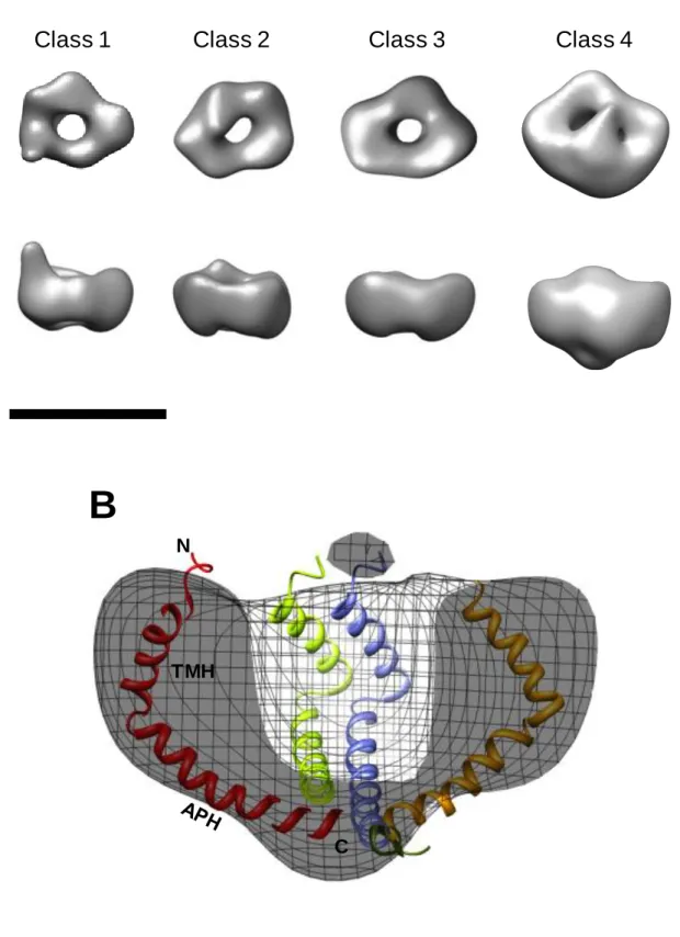
corresponding tilted particles, initial 3D models were generated. These density maps were then iteratively refined by matching the original tilted particles to projections of the initial models to improve their cross-correlation. In Fig. 3D the class averages are compared to reprojections of their corresponding final density map. In classes 1-3, the 3D density map appears as a small asymmetrical ring made up of 5 globular densities measuring ~2.5 nm across, and in class 4 there are 6 globular densities. No variation in ring thickness is present between the classes (Fig. 4A). Only the largest class (Class 4) shows significant occlusion of the central channel. This is more prominent on the face away from the grid. Pore size also does not appear to vary between the classes (measuring 2.5-3 nm across). The 3 smaller classes have a ring height of ~4 nm increasing to 5.5 nm for the larger class. The complexes are estimated to range from 70 kDa to 120 kDa based on the enclosed volume of the density maps at 3.8 σ. In order to aid interpretation,
11
seen to protrude from the circumference of the ring (Fig. 5, A and B), and such extensions are not seen in the larger classes or in any of the untilted particles. The total length of these extended regions is ~4 nm including the ring density. A possible candidate for forming these extensions could be the APH of the TatAd subunit, which could form a structure compatible with an aqueous channel. Such a conformational change has been suggested previously for E. coli TatA [43] and could be due to the flexibility of the hinge region [37]. The flexibility of the hinge region may permit alternative arrangements of the APH, explaining why the 'arm' structure is seen in only a subset of TatAd complexes. This conformational change could explain the slight variation in ring diameter across the TatAd complexes observed, with the TMH shifting from a tilted orientation to a straighter alignment, parallel to the APH. However, it is also conceivable that the 'arm' density is due to other structural rearrangements within the Class1 TatAd complexes, for example as proposed in the recent ‘charge zipper mechanism’ for TatA function [44].
3.3 B. subtilis TatAc forms small, homogeneous complexes and can restore export of TorA in a
ΔtatAE mutant.
12
we tested whether TatAc can functionally replace TatA and TatE and restore translocation activity in E. coliΔtatAE cells. The TMAO reductase activity assay was performed as described previously [12]. As shown in Fig. 6B, expression of TatAc is sufficient to restore translocation activity in the presence of native levels of E. coli TatB and TatC, with no activity detectable in the periplasmic fraction in the absence of a TatA-like component (ΔtatAE control). The periplasmic signal with TatAc complementation (ΔtatAE+TatAc cells) is lower than in wild type cells, raising the possibility that translocation is less efficient. Nonetheless, the ability of TatAc to substitute for TatA/E
provides strong evidence that the homogeneous TatAc complexes are physiologically relevant.
4.0 Discussion
Previous studies have focussed on the structures of Tat complexes from the Gram-negative bacterium E. Coli [12, 18]. Very recently, the crystal structure of TatC from the hyperthermophilic
bacterium Aquifex aeolicus was elucidated at 3.5 Å resolution [17]. These studies have prompted discussion concerning the possible mechanism of the Tat system with particular emphasis on the roles of separate TatA and TatBC complexes during the actual translocation event. In contrast, Tat complexes from Gram-positive bacteria have not been subjected to structural analysis and, to date, only the structure of the B. subtilis TatAd monomer has been solved by solution NMR [37]. Furthermore, bioinformatic structural predictions of the TatCd monomer have been confirmed by circular dichroism [45]. Important biochemical differences have been noted between Tat
complexes from Gram-positive and Gram-negative bacteria. Therefore, we have set out to investigate the structure of B. subtilis TatAd complexes through the generation of 2D and 3D structure models using EM techniques.
We show that TatAd forms ring-shaped structures of approximately 7.5 – 9 nm diameter, containing a potential pore of 2.5 – 3 nm, which is occluded at one end. Using electron microscopy
13
significance of such a 'lid' structure remains to be elucidated. A subset of the identified TatA complexes contain potential pores of diameters that could allow translocation of small Tat substrates and in this scenario the lid could seal a defined translocation channel at one end. On the other hand, our TatAd data argue against a 'simple' channel role for TatA-type complexes, because the diameters are too small to allow the passage of larger substrates, such as E. coli TorA which is 90 kDa in size. Notably, in the absence of clear data on how the small TatAd complexes could contribute to translocation, the role of the potential lid is unclear. Further experimentation is also required to accurately assess the protein contribution to the TatAd 3D density maps. The correlation of the presence of central density with complex size suggests a mixed population of conformational states. In such a case the arm-like extensions seen extruding from the smallest class could likely include the C-terminus of the amphipathic helix of the TatAd subunit.
Previous analysis of E. coli TatA and TatE complexes revealed a similar structure with a prominent potential pore in the centre of the complexes [12,18]. Nevertheless, the two types of complex exhibit major differences in other respects; TatA complexes are found in a large size range (9 - 13 nm diameter), whereas TatE complexes are much smaller and more homogeneous (6 – 8 nm diameter). The diameters of the potential pores are correspondingly different, and this
has major implications for models of the translocation mechanism. The diameters of the potential pores in TatA complexes vary from 3-7 nm [12] and this range of pores would be capable of translocating the wide range of known Tat substrates in a folded state. Intriguingly, the largest pores would be just capable of accommodating larger Tat substrates, such as TorA. However, TatE is capable of substituting for TatA [46], although the structures of TatE complexes are very different to those of TatA complexes [12]. Importantly, the much smaller potential pores in TatE complexes are too small to accommodate many of the known Tat substrates. It was therefore suggested that the Tat mechanism may instead involve the coalescence of relatively
14
complex. In fact, the TatAd complexes resemble TatE complexes in that TatAd complexes do not present a large size range, and the average TatAd particle diameter (8 nm) is smaller than that of the smallest observed class of TatA complexes. Importantly, we do not observe TatAd complexes with an internal channel large enough for the translocation of fully folded E. coli Tat substrates.
Finally, the small and uniform size of the TatAd complex appears to be a strictly conserved feature of TatA complexes from B. subtilis as is underscored by our present investigation of the TatAc complex and our previous investigations of the TatAy complex [25, 26]. We have therefore shown that all three TatA-type complexes of this Gram-positive bacterium form small homogenous complexes after purification. This challenges previous models on the role of TatA in protein
translocation, which are based, in the main, on the biochemical and structural properties of E. coli TatA. Altogether, our observations emphasise the need for further functional studies to understand the nature and role of TatA-type complexes in the overall Tat mechanism for protein traffic.
5.0 Acknowledgements
15 6.0 References
[1] T. Palmer, B.C. Berks, The twin-arginine translocation (Tat) protein export pathway, Nature reviews. Microbiology, 10 (2012) 483-496.
[2] C. Robinson, C.F.R.O. Matos, D. Beck, C. Ren, J. Lawrence, N. Vasisht, S. Mendel, Transport and proofreading of proteins by the twin-arginine translocation (Tat) system in bacteria, Biochimica et Biophysica Acta (BBA) - Biomembranes, 1808 (2011) 876-884.
[3] M. Müller, R.B. Klösgen, The Tat pathway in bacteria and chloroplasts (Review), Mol Membr Biol, 22 (2005) 113-121.
[4] K. Cline, W.F. Ettinger, S.M. Theg, Protein-specific energy requirements for protein transport across or into thylakoid membranes. Two lumenal proteins are transported in the absence of ATP, Journal of Biological Chemistry, 267 (1992) 2688-2696.
[5] C. Robinson, A. Bolhuis, Tat-dependent protein targeting in prokaryotes and chloroplasts, Biochimica et Biophysica Acta (BBA) - Molecular Cell Research, 1694 (2004) 135-147.
[6] C.-L. Santini, B. Ize, A. Chanal, M. Müller, G. Giordano, L.-F. Wu, A novel Sec-independent periplasmic protein translocation pathway in Escherichia coli, EMBO journal, 17 (1998) 101-112. [7] A.M. Chaddock, A. Mant, I. Karnauchov, S. Brink, R.G. Herrmann, R.B. Klösgen, C. Robinson, A new type of signal peptide: central role of a twin-arginine motif in transfer signals for the delta pH-dependent thylakoidal protein translocase., EMBO journal, 14 (1995) 2715-2722.
[8] N.R. Stanley, T. Palmer, B.C. Berks, The Twin Arginine Consensus Motif of Tat Signal Peptides Is Involved in Sec-independent Protein Targeting in Escherichia coli, Journal of Biological
Chemistry, 275 (2000) 11591-11596.
[9] F. Sargent, E.G. Bogsch, N.R. Stanley, M. Wexler, C. Robinson, B.C. Berks, T. Palmer, Overlapping functions of components of a bacterial Sec-independent protein export pathway, EMBO journal, 17 (1998) 3640-3650.
16
[11] J.H. Weiner, P.T. Bilous, G.M. Shaw, S.P. Lubitz, L. Frost, G.H. Thomas, J.A. Cole, R.J. Turner, A Novel and Ubiquitous System for Membrane Targeting and Secretion of Cofactor-Containing Proteins, Cell, 93 (1998) 93-101.
[12] J. Baglieri, D. Beck, N. Vasisht, C.J. Smith, C. Robinson, Structure of TatA Paralog, TatE, Suggests a Structurally Homogeneous Form of Tat Protein Translocase That Transports Folded Proteins of Differing Diameter, J. Biol. Chem., 287 (2012) 7335-7344.
[13] A. Bolhuis, J.E. Mathers, J.D. Thomas, C.M.L. Barrett, C. Robinson, TatB and TatC Form a Functional and Structural Unit of the Twin-arginine Translocase from Escherichia coli, Journal of Biological Chemistry, 276 (2001) 20213-20219.
[14] J. Oates, J. Mathers, D. Mangels, W. Kühlbrandt, C. Robinson, K. Model, Consensus Structural Features of Purified Bacterial TatABC Complexes, Journal of Molecular Biology, 330 (2003) 277-286.
[15] J. Oates, C.M.L. Barrett, J.P. Barnett, K.G. Byrne, A. Bolhuis, C. Robinson, The Escherichia coli Twin-arginine Translocation Apparatus Incorporates a Distinct Form of TatABC Complex, Spectrum of Modular TatA Complexes and Minor TatAB Complex, Journal of Molecular Biology, 346 (2005) 295-305.
[16] J. Frobel, P. Rose, F. Lausberg, A.S. Blummel, R. Freudl, M. Muller, Transmembrane insertion of twin-arginine signal peptides is driven by TatC and regulated by TatB, Nature communications, 3 (2012) 1311.
[17] S.E. Rollauer, M.J. Tarry, J.E. Graham, M. Jaaskelainen, F. Jager, S. Johnson, M.
Krehenbrink, S.M. Liu, M.J. Lukey, J. Marcoux, M.A. McDowell, F. Rodriguez, P. Roversi, P.J. Stansfeld, C.V. Robinson, M.S. Sansom, T. Palmer, M. Hogbom, B.C. Berks, S.M. Lea, Structure of the TatC core of the twin-arginine protein transport system, Nature, 492 (2012) 210-214. [18] U. Gohlke, L. Pullan, C. McDevitt, I. Porcelli, E. de Leeuw, T. Palmer, H. Saibil, B. Berks, The TatA component of the twin-arginine protein transport system forms channel complexes of variable diameter., Proc Natl Acad Sci U S A, 102 (2005) 10482-10486.
17
[20] C.G. Monteferrante, J. Baglieri, C. Robinson, J.M. van Dijl, TatAc, the third TatA subunit of Bacillus subtilis, can form active twin-arginine translocases with the TatCd and TatCy subunits., Appl Environ Microbiol, 78 (2012) 4999-5001.
[21] J.D.H. Jongbloed, U. Martin, H. Antelmann, M. Hecker, H. Tjalsma, G. Venema, S. Bron, J.M. van Dijl, J. Muller, TatC Is a Specificity Determinant for Protein Secretion via the Twin-arginine Translocation Pathway, Journal of Biological Chemistry, 275 (2000) 41350-41357.
[22] O. Pop, U. Martin, C. Abel, J.P. Muller, The Twin-arginine Signal Peptide of PhoD and the TatAd/Cd Proteins of Bacillus subtilis Form an Autonomous Tat Translocation System, Journal of Biological Chemistry, 277 (2002) 3268-3273.
[23] J.D.H. Jongbloed, U. Grieger, H. Antelmann, M. Hecker, R. Nijland, S. Bron, J.M. Van Dijl, Two minimal Tat translocases in Bacillus, Molecular Microbiology, 54 (2004) 1319-1325.
[24] J.P. Barnett, R.T. Eijlander, O.P. Kuipers, C. Robinson, A Minimal Tat System from a Gram-positive Organism: A BIFUNCTIONAL TatA SUBUNIT PARTICIPATES IN DISCRETE TatAC AND TatA COMPLEXES, J. Biol. Chem., 283 (2008) 2534-2542.
[25] J.P. Barnett, R. van der Ploeg, R.T. Eijlander, A. Nenninger, S. Mendel, R. Rozeboom, O.P. Kuipers, J.M. van Dijl, C. Robinson, The twin-arginine translocation (Tat) systems from
Bacillus subtilis display a conserved mode of complex organization and similar substrate recognition requirements, FEBS Journal, 276 (2009) 232-243.
[26] R. van der Ploeg, J.P. Barnett, N. Vasisht, V.J. Goosens, D.C. Pother, C. Robinson, J.M. van Dijl, Salt sensitivity of minimal twin arginine translocases, in: The Journal of biological chemistry, vol. 286, United States, 2011, pp. 43759-43770.
[27] T. Brüser, C. Sanders, An alternative model of the twin arginine translocation system, Microbiological Research, 158 (2003) 7-17.
[28] J. Sambrook, E.F. Fritsch, T. Maniatis, Molecular cloning: a laboratory manual, New York: Cold Spring Harbor Laboratory Press, 1989.
18
[30] J.L. Rubinstein, Structural analysis of membrane protein complexes by single particle electron microscopy, Methods, 41 (2007) 409-416.
[31] J. Frank, M. Radermacher, P. Penczek, J. Zhu, Y. Li, M. Ladjadj, A. Leith, SPIDER and WEB: Processing and Visualization of Images in 3D Electron Microscopy and Related Fields, Journal of Structural Biology, 116 (1996) 190-199.
[32] T.R. Shaikh, H. Gao, W.T. Baxter, F.J. Asturias, N. Boisset, A. Leith, J. Frank, SPIDER image processing for single-particle reconstruction of biological macromolecules from electron
micrographs, Nat. Protocols, 3 (2008) 1941-1974.
[33] J. Mindell, N. Grigorieff, Accurate determination of local defocus and specimen tilt in electron microscopy., J Struct Biol, 142 (2003) 334-347.
[34] H.E. White, H.R. Saibil, A. Ignatiou, E.V. Orlova, Recognition and Separation of Single Particles with Size Variation by Statistical Analysis of their Images, Journal of Molecular Biology, 336 (2004) 453-460.
[35] P. Penczek, M. Radermacher, J. Frank, Three-dimensional reconstruction of single particles embedded in ice, Ultramicroscopy, 40 (1992) 33-53.
[36] M. Radermacher, T. Wagenknecht, A. Verschoor, J. Frank, Three-dimensional reconstruction from a single-exposure, random conical tilt series applied to the 50S ribosomal subunit of
Escherichia coli., J Microsc, 146 (1987) 113-136.
[37] Y. Hu, E. Zhao, H. Li, B. Xia, C. Jin, Solution NMR Structure of the TatA Component of the Twin-Arginine Protein Transport System from Gram-Positive Bacterium Bacillus subtilis, Journal of the American Chemical Society, 132 (2010) 15942-15944.
[38] M.C. Leake, N.P. Greene, R.M. Godun, T. Granjon, G. Buchanan, S. Chen, R.M. Berry, T. Palmer, and B.C. Berks. Variable stoichiometry of the TatA component of the twin-arginine protein transport system observed by in vivo single-molecule imaging. Proc. Natl. Acad. Sci. USA. 105 (2008) 15376–15381.
[39] C. Dabney-Smith and K. Cline. Clustering of C-terminal stromal domains of Tha4 homo-oligomers during translocation by the Tat protein transport system. Mol. Biol. Cell. 20 (2009) 2060–
19
[40] S.D. Müller, A.A. De Angelis, T.H. Walther, S.L. Grage, C. Lange, S.J. Opella, A.S. Ulrich, Structural characterization of the pore forming protein TatAd of the twin-arginine translocase in membranes by solid-state 15N-NMR, Biochimica et Biophysica Acta (BBA) - Biomembranes, 1768 (2007) 3071-3079.
[41] C. Lange, S.D. Müller, T.H. Walther, J. Bürck, A.S. Ulrich, Structure analysis of the protein translocating channel TatA in membranes using a multi-construct approach, Biochimica et Biophysica Acta (BBA) - Biomembranes, 1768 (2007) 2627-2634.
[42] T.H. Walther, S.L. Grage, N. Roth, A.S. Ulrich, Membrane Alignment of the Pore-Forming Component TatAd of the Twin-Arginine Translocase from Bacillus subtilis Resolved by Solid-State NMR Spectroscopy, Journal of the American Chemical Society, 132 (2010) 15945-15956.
[43] K. Gouffi, F. Gérard, C.-L. Santini, L.-F. Wu, Dual Topology of the Escherichia coli TatA Protein, Journal of Biological Chemistry, 279 (2004) 11608-11615.
[44] T.H. Walther, C. Gottselig, S.L. Grage, M. Wolf, A.V. Vargiu, M.J. Klein, S. Vollmer, S. Prock, M. Hartmann, S. Afonin, E. Stockwald, H. Heinzmann, O.V. Nolandt, W. Wenzel, P. Ruggerone, A.S. Ulrich, Folding and Self-Assembly of the TatA Translocation Pore Based on a Charge Zipper Mechanism, Cell, 152 (2013) 316-326.
[45] O.V. Nolandt, T.H. Walther, S. Roth, J. Bürck, A.S. Ulrich, Structure analysis of the membrane protein TatCd from the Tat system of B. subtilis by circular dichroism, Biochimica et Biophysica Acta (BBA) - Biomembranes, 1788 (2009) 2238-2244.
[46] F Sargent, NR Stanley, BC Berks, T Palmer, Sec-independent protein translocation in
Escherichia coli. A distinct and pivotal role for the TatB protein. J Biol Chem. 274 (1999) 36073-82.
Figure legends
20
bracket indicates the position of TatAd which runs as a diffuse band. (B). A sample of the TatAd concentrate was applied to a Superdex 200 GL 30/100 gel filtration column. The run (240 µl sample, 0.5 ml/min flow, 0.02% DDM in buffer) shows a major peak at ~270 kDa with a shoulder towards lower molecular weights. The corresponding Western blot shows that TatAd elutes across fractions 19-26. Vo = Void volume. (C) Silver-stained gel of elution fractions which contain TatAd for different stages in column purification. T = Pooled Talon column elution fractions C = Pooled Talon fractions after concentration GF = Gel filtration elution fraction selected for electron microscopy studies. Positions of TatAd are indicated on the right of the figure and molecular weight markers are shown on the left.
Figure 2. Micrographs of TatAd tilt pair and example particles. The area of micrographs is shown untilted (A) and tilted to 50° (B). Corresponding particle pairs in both images are circled. The scale bar measures 100 nm. Images were acquired at ~57,000x magnification under low-dose conditions. (C) Particles picked from the untilted images are small and roughly circular measuring ~7.5 nm in diameter. Each contains a clear central pool of stain indicating a cavity or channel. The scale bar is set to 7.5nm to facilitate size comparison with the actual particles.
Figure 3. Assessment of size variations in TatAd complexes. (A) Initial centred sum image of
1990 particles. (B) Size class averages of TatAd reveal a slight size variation that correlates with internal density (see largest class, top right). The numbers of particles per class are as follows: class 1, 547; class 2, 477; class 3, 440; class 4, 352. (C) Size classes after reference-free rotational and translational alignment of the untilted particles. (D) Reprojections of TatAd 3D density maps for comparison to the class averages. Scale bar = 10 nm.
Figure 4. 3D density maps of TatAd complexes. (A) Shown from left to right are classes 1-4. The maps are filtered to 30 Å and contoured at ~ 4 σ (standard deviations above the mean
21
monomers are coloured in red, yellow, green and blue. Side views in (A) and (B) are shown with the potential cytoplasmic side facing down as suggested by the fitted TatAd NMR structure. TMH, transmembrane helix; APH, amphipathic helix.
Figure 5. Small TatAd complexes show extensions from the ring. (A) TatAd particles were identified from the Class 1 tilted images showing one or two extensions (white arrows indicate examples) from the circumference of the ring measuring 1-2 nm in length. (B) Closer view of two such particles with and without a custom mask to highlight the extensions (white arrows).
Figure 6. B. subtilis TatAc forms small homogeneous complexes, capable of complementing a ΔtatAE E. coli mutant. (A). Membranes were prepared from E. coli ΔtatAE, Δtat, and ΔtatB cells expressing B. subtilis TatAc, or from E. coli ΔtatAE cells expressing E. coli
7 - 17 - 23 - 30 - 46 - 58 - 80 - 175 -
FT W1 W2 W3 E1 E2 E3 E4 E5 E6 E7 E8 E9 E10 E11 E12 E13 E14 E15 E16 E17 E18 E19 E20
α- His
7 -
A
- 50
0 50 100 150 200
mAU
α -His
19 20 21 22 23 24 25 26 -270 kDa
V
oB
Fraction
Volume (ml)
2 4 6 8 10 12 14 16 18 20
7 - 17 - 23 - 30 - 46 - 58 - 80 - 175 -
T
C
GF
Tat Ad
Silver stain
C
A
Class 1
Class 2
Class 3
Class 4
Figure
4
TMH N
C
B
A
B
10 nm
10 nm
Figure 5
440
669
232
140
66
-anti-Strep
TatAc
TatA
A
Figure 6
B
1 Table 1. Plasmids and strains used in this work
Plasmids/Cells Relevant properties Reference/ source
pBAd-his pBAD24 derivative containing the [21] B. subtilis tatAd-his gene; Ampr
MC4100 F- ΔlacU169 araD139 rpsL150 relA ptsF rbs flbB5301; [36] MC4100 ΔtatABCDE tat deletion strain [8]
MC4100 ΔtatAE tatA and tatE deletion strain [8]
MC4100 ΔtatB tatB deletion strain [8]
MC4100 ΔtatC tatC deletion strain [8]
pBAD-A-Strep pBAD24 derivative containing the E. coli tatA gene with a
C-terminal Strep-IITM tag; Ampr; [37]
pBAD-Ac-Strep: pBAD24 derivative containing the B. subtilis tatAc-strepII gene;
Ampr ; This study
Ampr = ampicillin resistant