Quinolin-3-amine
Arun M. Islor,aB. Chandrakantha,bPrakash Shetty,b Thomas Gerber,cEric Hostencand Richard Betzc*
a
National Institute of Technology-Karnataka, Department of Chemistry, Organic Chemistry Laboratory, Surathkal, Mangalore 575 025, India,bManipal Institute of Technology, Department of Chemistry, Manipal 576 104, India, andcNelson
Mandela Metropolitan University, Summerstrand Campus, Department of Chemistry, University Way, Summerstrand, PO Box 77000, Port Elizabeth, 6031, South Africa Correspondence e-mail: [email protected]
Received 6 October 2012; accepted 11 October 2012
Key indicators: single-crystal X-ray study;T= 200 K; mean(C–C) = 0.002 A˚; Rfactor = 0.032;wRfactor = 0.091; data-to-parameter ratio = 10.0.
In the crystal structur of the achiral title compound, C9H8N2,
N—H N hydrogen bonds connect the molecules into zigzag chains in [100]. Weak intermolecular N–H interactions further consolidate the crystal packing.
Related literature
For novel applications of quinolin-3-amine and its derivatives, see: Rohmer et al. (2010); Kaneshiro et al. (2011). For the crystal structure of a rhodium coordination compound featuring the title compound as a ligand, see: Garraldaet al. (1999). For graph-set analysis of hydrogen bonds, see: Etteret al.(1990); Bernsteinet al.(1995).
Experimental
Crystal data
C9H8N2
Mr= 144.17
Orthorhombic,P212121 a= 7.6223 (3) A˚ b= 7.6289 (3) A˚ c= 12.6967 (4) A˚
V= 738.31 (5) A˚3
Z= 4
MoKradiation
= 0.08 mm1
T= 200 K
0.550.520.15 mm
Data collection
Bruker APEXII CCD diffractometer
Absorption correction: multi-scan (SADABS; Bruker, 2008) Tmin= 0.950,Tmax= 0.988
6898 measured reflections 1077 independent reflections 1015 reflections withI> 2(I) Rint= 0.013
R[F2> 2(F2)] = 0.032 wR(F2) = 0.091
S= 1.03 1077 reflections 108 parameters
H atoms treated by a mixture of independent and constrained refinement
max= 0.23 e A˚3
min=0.20 e A˚3
Table 1
Hydrogen-bond geometry (A˚ ,).
Cgis the centroid of the C1/C5–C9 ring.
D—H A D—H H A D A D—H A
N2—H2B N1i
0.90 (2) 2.22 (2) 3.0761 (17) 158.2 (18) N2—H2A Cgii 0.85 (2) 2.60 (2) 3.3101 (15) 142.3 (19)
Symmetry codes: (i)x1
2;yþ12;zþ1; (ii)x;y12;zþ12.
Data collection:APEX2(Bruker, 2010); cell refinement:SAINT (Bruker, 2010); data reduction:SAINT; program(s) used to solve structure:SHELXS97(Sheldrick, 2008); program(s) used to refine structure: SHELXL97 (Sheldrick, 2008); molecular graphics: ORTEPIII(Farrugia, 1997) andMercury(Macraeet al., 2008); soft-ware used to prepare material for publication: SHELXL97 and PLATON(Spek, 2009).
AMI is thankful to the Director of the National Institute of Technology for providing research facilities and also thanks the Board for Research in Nuclear Sciences, the Department of Atomic Energy and the Government of India for a Young Scientist award.
Supplementary data and figures for this paper are available from the IUCr electronic archives (Reference: CV5347).
References
Bernstein, J., Davis, R. E., Shimoni, L. & Chang, N.-L. (1995).Angew. Chem. Int. Ed. Engl.34, 1555–1573.
Bruker (2008).SADABS.Bruker Inc., Madison, Wisconsin, USA. Bruker (2010).APEX2andSAINT. Bruker AXS Inc., Madison, USA. Etter, M. C., MacDonald, J. C. & Bernstein, J. (1990).Acta Cryst.B46, 256–262. Farrugia, L. J. (1997).J. Appl. Cryst.30, 565.
Garralda, M. A., Hernandez, R., Pinilla, E. & Rosario Torres, M. (1999).J. Organomet. Chem.586, 150–158.
Kaneshiro, K., Fukuyama, Y., Iwamoto, S., Sekiya, S. & Tanaka, K. (2011). Anal. Chem.83, 3663–3667.
Macrae, C. F., Bruno, I. J., Chisholm, J. A., Edgington, P. R., McCabe, P., Pidcock, E., Rodriguez-Monge, L., Taylor, R., van de Streek, J. & Wood, P. A. (2008).J. Appl. Cryst.41, 466–470.
Rohmer, M., Meyer, B., Mank, M., Stahl, B., Bahr, U. & Karas, M. (2010). Anal. Chem.82, 3719–3726.
Sheldrick, G. M. (2008).Acta Cryst.A64, 112–122. Spek, A. L. (2009).Acta Cryst.D65, 148–155. Structure Reports
Online
supporting information
Acta Cryst. (2012). E68, o3155 [doi:10.1107/S1600536812042626]
Quinolin-3-amine
Arun M. Islor, B. Chandrakantha, Prakash Shetty, Thomas Gerber, Eric Hosten and Richard Betz
S1. Comment
3-Aminoquinoline and its derivatives have found applications in matrix-assisted laser desorption ionization (MALDI)
mass-spectrometry of oligosaccharides (Rohmer et al., 2010) and glycans (Kaneshiro et al., 2011). Herewith we present
the crystal structure of 3-aminoquinoline, (I).
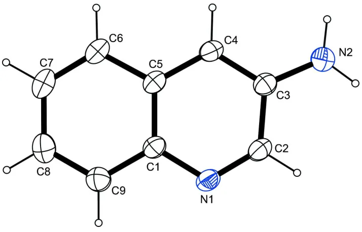
In (I) (Fig. 1), the molecule bears an amino group in meta position to the intracyclic nitrogen atom. Intracyclic angles in
the six-membered ring containing the nitrogen atom cover a range of 117.42 (12)–125.27 (11) ° with the smallest angle
found on the carbon atom bearing the amino group and the biggest angle present on the hydrogen-bearing carbon atom in
ortho position to the intracyclic nitrogen atom. The molecule is essentially planar (r.m.s. deviation of of all fitted
non-hydrogen atoms = 0.0091 Å). The least-squares planes defined by the non-non-hydrogen atoms of the heterocycle on the one
hand and the atoms of the amino group on the other hand intersect at an angle of 11.97(2.58) °.
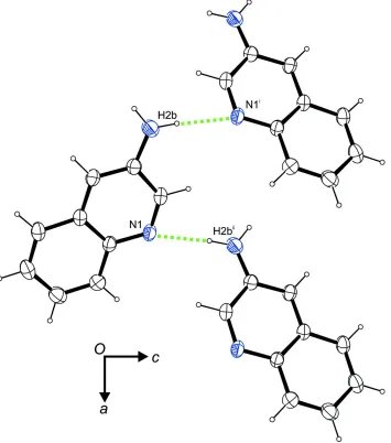
In the crystal, N–H···N hydrogen bonds (Table 1) are observed between the amino group and the intracyclic nitrogen
atom that connect the molecules to zigzag chains along the crystallographic a axis (Fig. 2). In terms of graph-set analysis
(Etter et al., 1990; Bernstein et al., 1995), the descriptor for these contacts is C1
1(5) on the unary level. In addition, a N–
H···π interaction (Table 1) involving the non-heterocyclic moiety of the quinoline core as acceptor contribute to the
crystal packing stability.
S2. Experimental
To a solution of 3-nitroquinoline (1 g, 0.0057 mol) in methanol (20 ml) 10% palladium on carbon (0.10 g) was added.
The batch was hydrogenated at a pressure of 10 bar for 12 h. Subsequently, the reaction mixture was filtered and
concentrated under reduced pressure to afford the title compound as a pale yellow solid. The solid was dissolved in
absolute ethanol and allowed to stand and evaporate at room temperature overnight. The crystalline solid that developed
was filtered and dried under high vacuum (yield: 0.8 g, 97.5%).
S3. Refinement
C-bound H atoms were placed in calculated positions (C—H 0.95 Å) and were included in the refinement in the riding
model approximation, with Uiso(H) set to 1.2Ueq(C). Both amino H atoms were located on a difference Fourier map and
Figure 1
The molecular structure of the title compound, with atom labels and anisotropic displacement ellipsoids (drawn at the
Figure 2
A portion of the crystal packing viewed along [010]. Dashed lines indicate N–H···N hydrogen bonds. Symmetry codes: (i)
x - 1/2, -y + 1/2, -z + 1; (ii) x + 1/2, -y + 1/2, -z + 1.
Quinolin-3-amine
Crystal data
C9H8N2 Mr = 144.17
Orthorhombic, P212121 Hall symbol: P 2ac 2ab a = 7.6223 (3) Å b = 7.6289 (3) Å c = 12.6967 (4) Å V = 738.31 (5) Å3 Z = 4
Dx = 1.297 Mg m−3 Melting point = 366–368 K Mo Kα radiation, λ = 0.71073 Å Cell parameters from 5242 reflections θ = 2.7–28.3°
Bruker APEXII CCD diffractometer
Radiation source: fine-focus sealed tube Graphite monochromator
φ and ω scans
Absorption correction: multi-scan (SADABS; Bruker, 2008) Tmin = 0.950, Tmax = 0.988
6898 measured reflections 1077 independent reflections 1015 reflections with I > 2σ(I) Rint = 0.013
θmax = 28.3°, θmin = 3.1° h = −6→10
k = −9→10 l = −16→16
Refinement
Refinement on F2 Least-squares matrix: full R[F2 > 2σ(F2)] = 0.032 wR(F2) = 0.091 S = 1.03 1077 reflections 108 parameters 0 restraints
Primary atom site location: structure-invariant direct methods
Secondary atom site location: difference Fourier map
Hydrogen site location: inferred from neighbouring sites
H atoms treated by a mixture of independent and constrained refinement
w = 1/[σ2(F
o2) + (0.0591P)2 + 0.1011P] where P = (Fo2 + 2Fc2)/3
(Δ/σ)max < 0.001 Δρmax = 0.23 e Å−3 Δρmin = −0.20 e Å−3
Special details
Refinement. Due to the absence of a strong anomalous scatterer, the Flack parameter is meaningless. Thus, Friedel opposites (737 pairs) have been merged and the item was removed from the CIF.
Fractional atomic coordinates and isotropic or equivalent isotropic displacement parameters (Å2)
x y z Uiso*/Ueq
Atomic displacement parameters (Å2)
U11 U22 U33 U12 U13 U23
N1 0.0323 (5) 0.0323 (6) 0.0193 (5) −0.0026 (5) −0.0030 (4) −0.0003 (4) N2 0.0318 (6) 0.0443 (7) 0.0316 (6) −0.0081 (5) 0.0052 (5) −0.0086 (6) C1 0.0277 (6) 0.0263 (6) 0.0207 (5) 0.0017 (5) −0.0018 (5) 0.0009 (5) C2 0.0340 (6) 0.0299 (6) 0.0191 (5) −0.0004 (5) −0.0012 (5) −0.0007 (5) C3 0.0279 (6) 0.0238 (5) 0.0243 (6) 0.0009 (5) −0.0004 (5) −0.0006 (5) C4 0.0306 (6) 0.0264 (6) 0.0226 (5) −0.0013 (5) −0.0021 (5) −0.0045 (5) C5 0.0303 (6) 0.0228 (6) 0.0202 (5) 0.0027 (5) −0.0006 (5) −0.0003 (5) C6 0.0382 (7) 0.0298 (6) 0.0218 (6) 0.0032 (5) 0.0015 (5) −0.0037 (5) C7 0.0430 (7) 0.0348 (7) 0.0254 (5) 0.0059 (6) 0.0084 (6) −0.0008 (5) C8 0.0320 (6) 0.0389 (7) 0.0350 (7) 0.0010 (6) 0.0070 (6) 0.0052 (6) C9 0.0294 (7) 0.0347 (7) 0.0298 (6) −0.0008 (5) −0.0009 (5) 0.0008 (6)
Geometric parameters (Å, º)
N1—C2 1.3091 (17) C4—C5 1.4133 (18) N1—C1 1.3740 (16) C4—H4 0.9500 N2—C3 1.3701 (17) C5—C6 1.4205 (17) N2—H2A 0.85 (2) C6—C7 1.369 (2) N2—H2B 0.90 (2) C6—H6 0.9500 C1—C9 1.4122 (18) C7—C8 1.410 (2) C1—C5 1.4167 (17) C7—H7 0.9500 C2—C3 1.4231 (17) C8—C9 1.375 (2) C2—H2 0.9500 C8—H8 0.9500 C3—C4 1.3792 (17) C9—H9 0.9500
C2—N1—C1 117.72 (11) C4—C5—C1 118.88 (11) C3—N2—H2A 119.4 (14) C4—C5—C6 122.66 (12) C3—N2—H2B 116.8 (12) C1—C5—C6 118.45 (12) H2A—N2—H2B 122.1 (18) C7—C6—C5 120.52 (13) N1—C1—C9 118.50 (12) C7—C6—H6 119.7 N1—C1—C5 121.52 (12) C5—C6—H6 119.7 C9—C1—C5 119.98 (11) C6—C7—C8 120.78 (12) N1—C2—C3 125.27 (11) C6—C7—H7 119.6 N1—C2—H2 117.4 C8—C7—H7 119.6 C3—C2—H2 117.4 C9—C8—C7 120.04 (13) N2—C3—C4 124.50 (12) C9—C8—H8 120.0 N2—C3—C2 118.07 (11) C7—C8—H8 120.0 C4—C3—C2 117.42 (12) C8—C9—C1 120.22 (13) C3—C4—C5 119.19 (12) C8—C9—H9 119.9 C3—C4—H4 120.4 C1—C9—H9 119.9 C5—C4—H4 120.4
N1—C2—C3—C4 0.3 (2) C1—C5—C6—C7 −0.5 (2) N2—C3—C4—C5 178.95 (12) C5—C6—C7—C8 0.3 (2) C2—C3—C4—C5 0.15 (18) C6—C7—C8—C9 0.1 (2) C3—C4—C5—C1 −0.61 (18) C7—C8—C9—C1 −0.4 (2) C3—C4—C5—C6 179.80 (12) N1—C1—C9—C8 −179.82 (13) N1—C1—C5—C4 0.66 (18) C5—C1—C9—C8 0.2 (2)
Hydrogen-bond geometry (Å, º)
Cg is the centroid of the C1/C5–C9 ring.
D—H···A D—H H···A D···A D—H···A
N2—H2B···N1i 0.90 (2) 2.22 (2) 3.0761 (17) 158.2 (18) N2—H2A···Cgii 0.85 (2) 2.60 (2) 3.3101 (15) 142.3 (19)
![Crystal structure of 2 [2 (pyridin 3 yl)diazen 1 yl]aniline](data:image/gif;base64,R0lGODlhAQABAIAAAP///wAAACH5BAEAAAAALAAAAAABAAEAAAICRAEAOw==)