ISSN Online: 2165-3410 ISSN Print: 2165-3402
DOI: 10.4236/aim.2019.93018 Mar. 27, 2019 248 Advances in Microbiology
Impact of Manure Storage Time and
Temperature on Microbial Composition and
Stable Fly (Diptera: Muscidae) Development
Janice L. Speshock
1*, Jeff A. Brady
2,3, Jessica Eastman
1,
Travis Roach
1, Samantha Hays
4, David Kattes
21Department of Biology, Tarleton State University, Stephenville, USA
2Department of Wildlife, Sustainability & Ecosystem Sciences, Tarleton State University, Stephenville, USA 3Texas A&M AgriLife Research & Extension Center, Stephenville, USA
4Department of Agriculture & Environmental Science, Tarleton State University, Stephenville, USA
Abstract
Samples are often frozen for preservation until needed for use. It has been a common practice to store fresh dairy manure in the freezer until needed for fly development studies. However, conflicting data have suggested that freez-er tempfreez-erature and duration of manure may impact fly development studies, and it is likely due to the change in microbial comminutes due to the freezer conditions. In this study manure storage conditions were assessed to ascertain how temperatures impact stable fly, Stomoxys calcitrans L., survival to pupa-tion and determine which bacterial populapupa-tions impacted fly development using massively-parallel sequencing and 16S metagenomic analysis. Stable fly survival to pupation was greater in manure that was stored warm (27˚C) or frozen (−20˚C or −80˚C) for 24 days as compared to fresh manure samples. Refrigeration (4˚C) of the manure for 24 days did not affect fly development and slightly decreased the pupal weights. Over 80 bacterial families were de-tected by sequencing allowing for a more thorough assessment of changes in bacterial populations. Only minor shifts were observed in bacterial family composition in the manure when refrigerated or frozen for 24 days, but sig-nificant population changes were observed when the manure was incubated for 24 days at 27˚C. Since it is the temperature and incubation time that yielded the greatest pupation rate, it is hypothesized that the manure micro-bial community impacts the growth and development of stable flies. This study has determined suggested freezer conditions for the best storage of manure samples to maintain bacterial diversity and retain the closest bacterial populations to freshly collected manure. Although untouched, aged (20 days) manure is best to use to assess fly development, it is not always feasible in la-How to cite this paper: Speshock, J.L.,
Brady, J.A., Eastman, J., Roach, T., Hays, S. and Kattes, D. (2019) Impact of Manure Storage Time and Temperature on Micro-bial Composition and Stable Fly (Diptera: Muscidae) Development. Advances in Microbiology, 9, 248-265.
https://doi.org/10.4236/aim.2019.93018
Received: February 12, 2019 Accepted: March 24, 2019 Published: March 27, 2019
Copyright © 2019 by author(s) and Scientific Research Publishing Inc. This work is licensed under the Creative Commons Attribution International License (CC BY 4.0).
DOI: 10.4236/aim.2019.93018 249 Advances in Microbiology
boratory experimentations. This study demonstrates the importance of pre-servation techniques on manure samples, which could also confer to storage of other biological specimens that contain resident microbes.
Keywords
Stomoxys calcitrans L., Manure Microbiota, 16S Metagenomics
1. Introduction
Stable flies, Stomoxys calcitrans L., are blood-sucking arthropods that can cause serious health issues for cattle and economic loss for cattle ranchers [1] [2]. They reduce productivity, lead to defense behaviors and bunching, and carry patho-gens of cattle, which make stable flies a serious pest [2]. It is estimated that 139 kg of milk is lost per annum due to stable fly infestations resulting in an estimated economic loss of $360 million each year for United States dairy cattle farmers [2]. It was originally thought that stable flies were only a pest problem for con-fined cattle, but more recent evidence indicates that stable flies can also impact pastured cattle [3]. The impact on pastured cattle is attributed to the use of hay bale feeders during the winter months, and more importantly, the manure ac-cumulation around those feeders [4]. Due to insecticide costs and concern over insecticide-resistance, adequate pasture control of pest flies has been unattaina-ble [1], which makes their development and control an important avenue of re-search.
DOI: 10.4236/aim.2019.93018 250 Advances in Microbiology
we demonstrated why freezing manure may complicate the consensus in fly de-velopment, and how studies performed on “ambient temperature” manure in various climates (warm or cold) may have drastic differences in results.
This study utilized a comparison of freshly collected dairy cattle manure to manure aged in a variety of temperatures at 1, 7, and 24 d post-collection, to de-termine the optimal storage conditions for stable fly development. The abun-dance of eggs developing to the pupal stage was compared to the bacterial com-position, both using live culture-based assays and 16S metagenomics, in order to determine factors that are important for the development of stable flies. The dif-ferences in storage conditions impact the bacteria microflora of the manure, which in turn will affect the development of stable flies. This data suggests that research on fly development and prevention needs to take storage into consider-ation and use ambient methods as much as possible to preserve the natural mi-crobiome.
2. Materials and Methods
2.1. Manure Collection
Fresh bovine manure was collected from five random dairy cattle at the South-west Regional Dairy Center in Stephenville, TX in October (average temperature 25˚C/14˚C (high/low)). The manure was placed into a 19-L plastic bucket, tho-roughly mixed and then separated into seventy-five 100 gram aliquots for the fly development studies and fifteen 10 g aliquots for the bacterial survey. Fifteen of the 100 g samples and three of the 10 g samples were placed into each of the fol-lowing storage methods: outdoors (27˚C) covered with fly screen to prevent oviposition but allow for air movement, refrigeration (4˚C), freezer (−20˚C), and deep freeze (−80˚C) for a time period of 1, 7, or 24 d. These storage conditions and time points were compared to manure that was processed the day of collec-tion (fresh).
2.2. Bacterial Population Survey
DOI: 10.4236/aim.2019.93018 251 Advances in Microbiology
extracted from 200 μl of the manure solution as previously described [12] for massively parallel sequencing and 16S metagenomic analysis of bacterial popula-tions. DNA concentration was determined using a Qubit® 2.0 fluorometer with a Qubit® dsDNA HS Assay Kit according to manufacturer’s instructions (Life Technologies, Carlsbad, CA). The DNA from each sample was normalized to a concentration of 5 ng/μl before subsequent analysis.
2.3. Massively-Parallel Sequencing
Bacterial 16S amplicons were generated using primers 341F (5’-CCTACGGGNGGCWGCAG-3’) and 785R
(5’-GACTACHVGGGTATCTAATCC-3’) that amplify the V3 and V4 regions [13] [14]. PCR amplification was accomplished through denaturation at 95 de-grees Celsius for 3 minutes, followed by 35 cycles of 95 dede-grees Celsius for 10 seconds, 55 degrees Celsius for 30 seconds, and 72 degrees Celsius for 30 seconds. Barcodes were constructed with the same PCR protocol. Paired-end sequence data were generated on an Illumina MiSeq instrument using v3 600 cycle kits (Illumina, San Diego, CA), with dual 6 basepair index sequences at-tached to each amplicon during indexing PCR as described in the Illumina 16S Metagenomic Sequencing Library Preparation protocol [15].
The raw sequencing reads were processed with a combination of QIIME [16], USEARCH [17] and FASTX-Toolkit [18] software packages, as well as a series of custom python scripts. Individual sequence tags were compared to the Green-genes 13.8 reference sequence database [19] using UCLUST [17] in order to pick referenced-based Operational Taxonomic Units (OTUs) at 97% similarity. The sequencing dataset was rarified to an equal sequencing depth of 860 sequences per sample by randomly subsampling sequences from each sample without replace-ment in order to provide even measures of bacterial alpha- and beta-diversity and to have equal sequencing depth for the production of all figures, tables, and statis-tical analyses.
2.4. Larvae Development
Fly eggs were obtained from the Knipling-Bushland U. S. Livestock Insect Re-search Lab (Kerrville, TX). Manure was removed from each storage condition, or used fresh, and 100 g of the substrate was placed into 510-g plastic cups (Hef-ty®, Pactiv Corporation, Lake Forest, IL) (n = 5). The manure was allowed to ac-climate to room temperature and 50 stable fly eggs were added to each cup. The cups were placed into an incubator set at 27˚C, 86 % RH at a photoperiod length of 14:10 L:D and the larvae were allowed to develop for eight days until eclosion. Following this incubation period, the pupae were enumerated and weighed.
2.5. Statistical Analysis
mi-DOI: 10.4236/aim.2019.93018 252 Advances in Microbiology
crobial community composition was significantly different between samples, PERMANOVA and bootstrapped Mann-Whitney U tests were conducted using the QIIME package [21] [22]. Differences in microbial taxa between different storage times and temperatures were determined with NCSS statistical package (NCSS, Kaysville UT). Analysis of variance (ANOVA) tests were used to deter-mine significant differences between the treatment groups for the fly and bac-terial assays.
Stable fly percentage data was normalized using arcsine transformation before analysis. Percent stable fly development to the pupal stage and pupal weights were analyzed using Statistix 10 and a Least Significant Difference test (LSD) was used to spate multiple means where appropriate. Analysis of results was consi-dered significant at P <0.05so specific P values are not presented.
3. Results
3.1. Viable Cell Counts
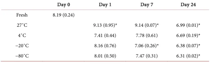
[image:5.595.205.540.628.734.2]To determine the impact of various storage temperatures and times on the number of viable prokaryotic cells, cells were microscopically counted using trypan blue, which does not permeate the membranes of live cells, and compared to the fresh extraction of manure on day 0. A significant decline in viable cell populations was evident following 20 days of storage using any of the storage conditions (Table 1). Growth at 27˚C demonstrated a significant increase in vi-able cell numbers over the first week, which was predominantly prokaryotic when the storage vial was left uncovered (Table 1), but predominantly eukaryo-tic (fungi) when the storage vial was sealed (data not shown). For this reason, and the noticeable impact on fly development (Figure 4), the covered 27˚C treatment group was omitted. Following refrigeration, the total viable prokaryo-tic cell counts decreased, but not significantly from control, except after 24 days of storage (Table 1). The freezer temperatures did not seem to impact viable cells much in the first week of storage, and did not seem to vary much from each other, although the −20˚C freezer did decline more rapidly, but they both did retain viable cells for the entire 24 day trial (Table 1). Due to the high prevalence of E. coli in manure samples, it was hypothesized that E. coli levels may impact the hatching of pupae and development of adult flies, thus E. coli viable cell
Table 1. Total Viable Prokaryotic Cell Count (log10 cells/ml) ± SEM. Microscopic cell
counts using trypan blue stain to differentiate between live and dead cells, with only via-ble cells included. *Statistically significant (P < 0.05) from control (fresh manure).
Day 0 Day 1 Day 7 Day 24
Fresh 8.19 (0.24)
27˚C 9.13 (0.95)* 9.14 (0.07)* 6.99 (0.01)*
4˚C 7.41 (0.44) 7.78 (0.61) 6.69 (0.19)*
−20˚C 8.16 (0.76) 7.06 (0.26)* 6.38 (0.07)*
DOI: 10.4236/aim.2019.93018 253 Advances in Microbiology
counts were also measured using EMB agar. At 27˚C (uncovered) the E. coli le-vels significantly increased by day 7, and although a decline was observed by day 24, there was still a significantly higher level of viable E. coli cells in this treat-ment group as compared to control manure (Table 2). Refrigeration in manure appears to have little effect on the viability of E. coli as it remains unchanged throughout the entire time course (Table 2). A slight decline in E. coli popula-tions was observed following deep freeze (−80˚C) and a significant decline was observed using a standard freezer (−20˚C), but neither appeared to be impacted by duration of freezing (Table 2).
3.2. Sequencing Metrics
Following quality filtering and joining of the paired-end reads, there were 538,249 16S DNA sequence tags for the 39 samples with an average length of 447 bp and an average Q score of 37.94. The lowest number of sequences per sample was 863. For all figures and statistical analyses of the sequencing data, the se-quence dataset was randomly subsampled to an even depth of 863 sese-quences per sample.
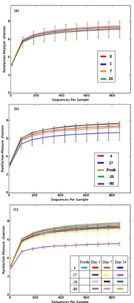
3.3. Bacterial Richness
Shannon diversity metrics were used to analyze alpha diversity within the mi-crobial populations comparing duration of storage (Figure 1(a)), temperature of storage (Figure 1(b)), or a combination of the parameters (Figure 1(c)). The rarefaction measure of species richness indicates that temperature (Figure 1(b)) has a greater impact than time (Figure 1(a)) on the number of species present. A comparison of the collective temperatures at each of the time points demon-strates very little change in species number indicating that the duration of time may not be a critical parameter of manure storage and prokaryotic species rich-ness (Figure 1(a)). Temperature did however appear to impact species richness, especially at 27 degrees Celsius (Figure 1(b)). The 27 degree temperature storage decreased the species richness (Figure 1(b)) with a significant impact observed after 24 days of storage (purple line; Figure 1(c)).
3.4. Bacterial Sample Variability
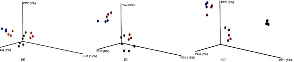
Beta diversity analysis was used to compare the amount of variability within the
Table 2. E. coli Cell Count (log10 cfu/ml) ± SEM. E. coli counts from EMB plates.
*Statistically significant (P < 0.05) from control (fresh manure).
Day 0 Day 1 Day 7 Day 24
Fresh 3.29 (0.44)
27˚C 3.65 (0.38) 5.12 (0.91)* 4.73 (0.32)*
4˚C 3.59 (0.70) 3.56 (0.17) 3.68 (0.15)
−20˚C 2.52 (0.3.6)* 2.34 (0.43)* 2.30 (0.28)*
DOI: 10.4236/aim.2019.93018 254 Advances in Microbiology
DOI: 10.4236/aim.2019.93018 255 Advances in Microbiology
sample set and to examine the similarity between treatment groups. Principle coordinate analysis plots were used to demonstrate the variation present within the dataset to confirm consistent replicates and demonstrate effects of experi-mental parameters. For all storage temperatures and durations analyzed, there are no observable outliers from any of the three replicates, which is indicated by the close clustering of spheres of similar colors. With only one day of incubation (Figure 2(a)), there is still close clustering of the samples from various storage temperatures to the microbial DNA amplified from the freshly-obtained manure samples (red), suggesting that this short of an incubation time may not have a dramatic effect on bacterial species diversity. However, the frozen samples (blue −20 and orange −80) are a greater distance from the 27˚C (purple) and refrige-rated (green) samples, suggesting that even a short freeze may influence bacterial populations (Figure 2(a)). By day 7 of storage, most storage conditions have se-parated from the fresh manure samples (Figure 2(b)) indicating that the effects of temperature are starting to cause variability in the bacterial populations in the manure samples. The refrigerated samples still remain the most closely asso-ciated with the fresh manure after one week (Figure 2(b)). Following 24 days of storage, only the refrigerated samples remain in close proximity to the fresh manure (Figure 2(c)), suggesting that refrigeration of manure will not dramati-cally change the prokaryotic cell population. The bacterial populations in the 27˚C stored manure exhibited much more variation from the fresh manure samples following 24 days of storage, confirming the change in species richness observed in the alpha diversity plots (Figure 1(c) and Figure 2(c)). The frozen samples remained dissimilar from the fresh manure after 24 days of storage, but there appears to be little variation between freezing at −20 degrees Celsius or −80 degrees Celsius (Figure 2(c)).
3.5. Bacterial Taxa Summary
[image:8.595.63.541.582.683.2]Although the primers used in this study are designed to selectively amplify bac-terial 16S sequences, a small number of 16S sequence tags were generated from archaea. The archaea sequence tags were treated as contaminants for the purpose of this work, and are not discussed. Taxonomic changes of bacteria at the family level were examined between freshly obtained manure and manure stored at
DOI: 10.4236/aim.2019.93018 256 Advances in Microbiology
[image:9.595.212.537.337.561.2]−20˚C, −80˚C, 4˚C, and 27˚C (Figure S1). The only consistent change observed following 24 days in all storage conditions was in the Succinivibrionaceae family (Figure 3), which declined 9.8%, 10.3%, 5%, and 8%, respectively (Figure S1). Besides the Succinivibrionaceae family, there were no other changes between the fresh manure and the manure stored in the refrigerator for 24 days, and the Suc-cinivibrionaceae were not quite statistically significant (Figure 3). More changes in population composition of various families were observed between the frozen manure (−20˚C and −80˚C) with little difference between the two temperatures, but the only large changes were observable increases in the Clostridiales order, although they were not statistically significant, and decreases in the family Bac-teroidaceae, also not statistically significant (Figure 3). Decreases were also ob-served in percentage of Campylobacteraceae and Desulfovibrionaceae that were minor, but statistically significant. In the Clostridiales order, families “other” and “undefined” doubling in percent of composition, Clostridiaceae increasing from 1.4% in fresh manure to 3.3% (−20˚C) or 4.0% (−80˚C), Lachnospiraceae increasing from 7.1% in fresh manure to 8.8% (−20˚C) or 10.6% (−80˚C), and Ruminococcaceae increasing from 1.7% in fresh manure to 26.1% (−20˚C) or
Figure 3. Taxa summary bar graphs at the level of family. The percentage of prokaryote OTU representation of each family is compared at 24 days incubation between the vari-ous storage temperatures and a fresh manure control. The pink sections in the box la-beled “A” depicts changes in Moraxellaceae (brown) and Xanthomonadaceae (green).
The section labeled “B” depict Succinivibrionaceae. The box around sections labeled “C” includes the Campylobacterales (light blue), Desulfovibrionaceae (yellow), Comamona-daceae (green), Alcaligenaceae (orange), and Erysipelotrichaceae (dark blue) families.The box labeled “D” includes the families in Clostridiales order (families Ruminococca-cae—purple, Lachnospiraceae (blue), Peptococcaceae—peach, undefined—grey, Chris-tensenellaceae—green, other—brown).The box labeled “E” includes the Paraprevotella-ceae (tan), Bacteroidales p-2534-18B5 (brown), Fibrobacteraceae (red),and Planococca-ceae (green) families. The designations “F” and “G” are for the Porphyromonadaceae and
DOI: 10.4236/aim.2019.93018 257 Advances in Microbiology
23.3%2 (−80˚C) (Figure 3; Figure S1). The most significant prokaryotic family shifts from the freshly obtained manure were observed in the manure stored at 27˚C for 24 days (Figure 3). The percentage of OTU sequences from the family Paraprevotellaceae (P = 0.055) decreased from 4.9% to 1.2% following a 24 day incubation at 27 degrees (Figure S1), and large increases were observed in the families Bacteroidales P-2534-18B5 (P = 0.055), Porphyromonadaceae (P = 0.054), Fibrobacteraceae (P < 0.05), Planococcaceae (P = 0.051), Comamonada-ceae (P < 0.05), Campylobacteraceae (P = 0.050), and Moraxellaceae (P < 0.05) (Figure 3; Figure S1).
3.6. Fly Development
Fresh manure extracted from dairy cattle and then inoculated with stable fly eggs resulted in an average of 9.5 percent developing to the pupal stage (Figure 4(a)). The average weight from these control pupae was 0.036 g (Figure 4(b)). Manure stored at −80˚C for 24 days had an increase of about double the number pupae produced (20.1) and nearly double the average pupal size (0.068 g) (Figure 4(a) & Figure 4(b)). There was little difference in pupation rate or average pupae size between manure frozen at −20˚C (data not shown) or −80˚C, so the latter was used for all subsequent studies. When the manure was stored at 27˚C uncovered for 24 days, there was a significant increase of over 3.5-fold for the number of eggs hatched (36.7 eggs) and a 2.7-fold significant increase for pupae weight (0.098 grams) (Figure 4(a) & Figure 4(b)). Manure stored at 4˚C appeared to have no impact on the number of eggs that hatched (Figure 4(a)), but the pupae that hatched were on average smaller (Figure 4(b)). The manure that was sealed and stored at 27˚C had decreases in pupae number (Figure 4(a)) and weight (Figure 4(b)), but these manure samples had an overgrowth of fungi, which
DOI: 10.4236/aim.2019.93018 258 Advances in Microbiology
likely impacted the development of stable flies.
4. Discussion
Due to the economic and medical importance of stable flies, much effort has been ongoing to attempt to better understand their breeding and development habits in order to reduce the fly populations around cattle and other livestock. As with many research objectives, there are many parameters that must be as-sessed to develop a full understanding of adequate methodology to obtain labor-atory goals. The major question addressed in this research was does manure sto-rage method and age impact bacterial growth, and in turn, stable fly larvae de-velopment? Often it is not feasible or practical to work with fresh manure sam-ples, and thus researchers rely on storage of the manure until the protocols can proceed. Therefore we utilized common storage conditions, outdoors (27˚C), typical refrigerator setting (4˚C), a standard freezer (−20˚C), and a deep freeze (−80˚C), and compared manure samples stored in these conditions for 1 day, 1 week, and 24 days to determine how storage impacted fly larvae development and the bacterial populations in the manure.
The data from the 1 day storage was largely omitted from this manuscript, because it appeared to only be long-term storage that affected manure composi-tion and quality. Although an overall viable cell number decrease was observed following refrigeration for 24 hours (Table 1), the overall percent composition of the microbes did not significantly change in this time period as compared with fresh manure (Figure 2). Conversely, an increase in total viable cell counts was measured following 24 hours at 27˚C (Table 1), but again the overall species diversity was not significantly affected. There was an increase in species diversity and richness following 7 days of incubation, but since the data trended towards what was observed following 20 days of incubation, the data presented here fo-cused mainly on the 24 day results.
DOI: 10.4236/aim.2019.93018 259 Advances in Microbiology
research began with examining E. coli levels since it was the largest constituent grown from the fresh manure cultures obtained, with 32% of all colonies on the agar plate representing E. coli (data not shown). However, further genomic ana-lyses indicated that the prevalence of E. coli using sequence-based determination was less than 0.0002% (data not shown) of the bacterial population of the ma-nure, confirming that standard plate techniques can be misleading. Metagenom-ic analysis provides a more thorough examination of the gut mMetagenom-icrobiota, and was used here to determine how changes in bacterial populations can impact stable fly larvae development.
The average pupation rate using fresh manure was 20%, and this rate doubled in frozen manure, and was greater than 70% with manure stored at 27˚C for 24 days (Figure 4(a)). Pupae size followed a similar trend with 27˚C incubation for 24 days resulting in the heaviest pupae, which were slightly heavier than from manure that was frozen for 24 days, and both of these stored samples produced heavier pupae than fresh manure (Figure 4(b)). Refrigeration of the manure had little effect on pupation rate compared to fresh, but did result in pupae of small-er size. Aftsmall-er detsmall-ermination of how tempsmall-eratures and durations impact stable fly development, the next question addressed was what changed in the 27˚C or fro-zen manure to allow for increased pupation rate and pupae size?
re-DOI: 10.4236/aim.2019.93018 260 Advances in Microbiology
sulted in comparable fly pupation, and frozen manure had significantly less E. coli, but had greater pupation than fresh manure (Table 2; Figure 4).
Previous studies have attempted to elucidate bacteria that are important for stable fly development, but they too relied on culture methods, which can be misleading [1] [6]. Both of these previous studies cultured bacteria from manure in aerobic conditions, but analysis of the bacterial phyla in ruminant animals in-dicates that a majority of the fecal microbiome consists of species from the phyla Bacteroidetes, Firmicutes,and Proteobacteria, all three of which contain mostly anaerobic species [27]. All families that had significant changes resulting from the various storage conditions tested here belonged to one of these three phyla, indicating that aerobic plating methods may not have allowed for easy depiction of the importance of these bacterial families to stable fly development. It is hy-pothesized that changes in population diversity among the phyla Bacteroidetes, Firmicutes,and Proteobacteria had the greatest contribution to increased stable fly pupation.
An approximate doubling of average eggs hatched and pupae size was ob-served when the manure was stored for 24 days at −80˚C compared to fresh manure, although due to sample variation it was not a significant increase (Figure 4). The frozen samples (−20˚C or −80˚C) had an increase in prokaryotic species richness (Figure 2(b)) and species variation (Figure 2(c)) over the fresh manure control (Figure 1(b)), suggesting a good preservation of the microbial population. The manure stored at 27˚C for 24 days allowed for a nearly 4-fold increase in hatch number and 2.7-fold increase in pupae size, both of which were statistically significant, over fresh manure pupation rates and growth (Figure 4). However, this manure actually demonstrated a dramatic decrease in bacterial species diversity at 24 days with even a slight, but noticeable decline at 7 days incubation (Figure 1(c)). The beta diversity analysis also indicated an increase in species variation, suggesting that the incubation at this temperature promoted the growth of certain bacterial species, while inhibiting the growth (or killing off) others (Figure 2 and Figure 3). All observed changes in bacterial families originated in the phyla Bacteroidetes, Firmicutes, and Proteobacteria, but no discernable trend can be established to suggest that one of these phyla contri-buted greatly to the abundance of stable flies.
DOI: 10.4236/aim.2019.93018 261 Advances in Microbiology
be attracted to a combination of acetic acid and ethanol, thus this glucose dissi-milation by Clostridiaceae may be producing a chemoattractant for the flies [30]. Lachnospiraceae have been implicated in the ability to stimulate host epithelial cell growth due to the production of butyric acid [31].
The manure stored at 27˚C for 24 days had many observable changes in bac-terial families, all from the phyla Bacteroidetes, Firmicutes,and Proteobacteria. No information was found regarding the unclassified Bacteroidetes family Bac-teroidales p2534-18B5, but the other family with increases in species composi-tion, Porphyromonadaceae and a closely-related family, Fibrobacteraceae, that used to be linked to the phylum, are both beneficial bacteria that aid in digestion of complex carbohydrates [32] [33]. Porphyromonadaceae digest fiber and other carbohydrates into short-chain fatty acids, such as butyrate, which is more di-gestible and has been shown to aid in healthy digestion and colon health [33]. Fibrobacteraceae are highly fibrolytic and cellulytic bacteria that aid in digestion and produce storage carbohydrates glycogen and cellodextrins [32]. Although the Firmicutes displayed large variation in manure stored at −20˚C and −80˚C, when stored at 27˚C only one family had any change, which were the Planococ-caceae which have been found in the rumen by other analyses [34], but whose function in digestion remains unknown. The remaining family diversity changes were observed in the phylum Proteobacteria, the largest and most diverse phy-lum of the bacteria. Significant increases were observed in the families Com-amonadaceae, denitrifying bacteria, the Campylobacteraceae, chemoorgano-trophs that reduce nitrate, and Moraxellaceae, which have been implicated in infectious bovine keratoconjunctivitis (IBK), but have not been reported in the rumen or fecal material [35] [36] [37].
DOI: 10.4236/aim.2019.93018 262 Advances in Microbiology
these parameters could drastically impact the results obtained. This knowledge is not only important for manure studies, but for any culture preservation that in-volves preserving a microbiome.
Acknowledgements
We would like to thank Dr. Kim Lohmeyer and Mr. Matt Waldon from the Agricultural Research Services of the United States Department of Agriculture in Kerrville, Texas for the stable fly eggs. We would also like to thank the Tarleton State University Office of Student Research and Creative Activities for funding the project.
Conflicts of Interest
The authors declare no conflicts of interest regarding the publication of this pa-per.
References
[1] Romero, A., Broce, A. and Zurek, L. (2006) Role of Bacteria in the Ovipostion Be-havior and Larval Development of Stable Flies. Medical and Veterinary Entomolo-gy, 20, 115-121. https://doi.org/10.1111/j.1365-2915.2006.00602.x
[2] Taylor, D.B., Moon, R.D. and Mark, D.R. (2012) Economic Impact of Stable Flies (Diptera: Muscidae) on Dairy and Beef Cattle Production. Journal of Medical En-tomology, 49, 198-209. https://doi.org/10.1603/ME10050
[3] Campbell, J.B., Skoda, S.R., Berkebile, D.R., Boxler, D.J., Thomas, G.D., Adams, D.C. and Davis, R. (2001) Effects of Stable Flies (Diptera: Muscidae) on Weight Gains of Grazing Yearling Cattle. Journal of Economic Entomology, 94, 780-783.
https://doi.org/10.1603/0022-0493-94.3.780
[4] Talley, J.A., Broce, B. and Zurek, L. (2009) Characterization of Stable Fly (Diptera: Muscidae) Larval Development Habitat at Round Hay Bale Feeding Sites. Journal of Medical Entomology, 46, 1310-1319. https://doi.org/10.1603/033.046.0609
[5] Broce, A.B. and Haas, M. (1999) Relation of Cattle Manure Age to Colonization by Stable Fly and House Fly (Diptera: Muscidae). Journal of the Kansas Entomological Society, 72, 60-72.
[6] Zurek, L. and Albuquerque, T. (2008) Microbial Ecology of Stables Flies: Effect of Bacterial Community of Aging Horse Manure on Stable Fly Fitness. 17th Annual K-State Research Forum, Manhattan, KS, 8 March 2012.
[7] Lysyk, T.J., Kalischuk-Tymensen, L., Selinger, L.B., Lancaster, R.C., Wever, L. and Cheng, K.J. (1999) Rearing Stable Fly Larvae (Diptera: Muscidae) on an Egg Yolk Medium. Journal of Medical Entomology, 36, 382-388.
https://doi.org/10.1093/jmedent/36.3.382
[8] Caporaso, J.G., Lauber, C.L., Walters, W.A., et al. (2012) Ultra-High-Throughput Microbial Community Analysis on the Illumina HiSeq and MiSeq Platforms. The ISME Journal, 6, 1621-1624. https://doi.org/10.1038/ismej.2012.8
[9] Myers, H.M., Tomberlin, J.K., Lambert, B.D. and Kattes, D. (2008) Development of Black Soldier Fly (Diptera: Stratiomyidae) Larvae Fed Dairy Manure. Environmen-tal Entomology, 37, 11-15. https://doi.org/10.1093/ee/37.1.11
DOI: 10.4236/aim.2019.93018 263 Advances in Microbiology
Value of Fresh and Composted Poultry Manure for House Fly (Diptera: Muscidae) Larvae. Journal of Economic Entomology, 94, 1308-1317.
https://doi.org/10.1603/0022-0493-94.5.1308
[11] Geden, C.J. (1999) Host Location by House Fly (Diptera: Muscidae) Parasitoids in Poultry Manure at Different Moisture Levels and Host Densities. Environmental Entomology, 28, 755-760. https://doi.org/10.1093/ee/28.4.755
[12] Brady, J.A., Faske, J.B., Castañeda-Gill, J.B., King, J.L. and Mitchell, F.L. (2011) High-Throughput DNA Isolation Method for Detection of Xylella fastidiosa in Plant and Insect Samples. Journal of Microbiological Methods, 86, 310-312.
https://doi.org/10.1016/j.mimet.2011.06.007
[13] Herlemann, D.P.R., Labrenz, M., Jurgens, K., Bertilsson, S., Waniek, J.J. and An-dersson, A.F. (2011) Transitions in Bacterial Communities along the 2000 km Salin-ity Gradient of the Baltic Sea. The ISME Journal, 5, 1571-1579.
https://doi.org/10.1038/ismej.2011.41
[14] Klindworth, A., Pruesse, E., Schweer, T., Peplies, J., Quast, C., Horn, M. and Glöckner, F.O. (2012) Evaluation of General 16S Ribosomal RNA Gene PCR Pri-mers for Classical and Next-Generation Sequencing-Based Diversity Studies.
Nucleic Acids Research, 41, e1. https://doi.org/10.1093/nar/gks808
[15] Illumina (2013) 16S Metagenomic Sequencing Library Preparation.
[16] Caporaso, J.G., Kuczynski, J., Stombaugh, J., et al. (2010) QIIME Allows Analysis of High-Throughput Community Sequencing Data. Nature Methods, 7, 335-336.
https://doi.org/10.1038/nmeth.f.303
[17] Edgar, R.C. (2010) Search and Clustering Orders of Magnitude Faster than BLAST.
Bioinformatics, 26, 2460-2461. https://doi.org/10.1093/bioinformatics/btq461
[18] Hannon. FASTX Toolkit. http://hannonlab.cshl.edu/fastx_toolkit/
[19] DeSantis, T.Z., Hugenholtz, P., Larsen, N., et al. (2006) Greengenes, a Chime-ra-Checked 16S rRNA Gene Database and Workbench Compatible with ARB. Ap-plied and Environmental Microbiology, 72, 5069-5072.
https://doi.org/10.1128/AEM.03006-05
[20] Lozupone, C. and Knight, R. (2005) UniFrac: A New Phylogenetic Method for Comparing Microbial Communities. Applied and Environmental Microbiology, 71, 8228-8235. https://doi.org/10.1128/AEM.71.12.8228-8235.2005
[21] Anderson, M.J. (2001) A New Method for Non-Parametric Multivariate Analysis of Variance. Austral Ecology, 26, 32-46.
[22] Anderson, M.J., Crist, T.O., Chase, J.M., et al. (2011) Navigating the Multiple Meanings of Beta Diversity: A Roadmap for the Practicing Ecologist. Ecology Let-ters, 14, 19-28. https://doi.org/10.1111/j.1461-0248.2010.01552.x
[23] Schmidtmann, E.T. and Martin, P.A.W. (1992) Relationship between Selected Bac-teria and the Growth of Immature House Flies, Musca domestica, in an Axenic Test System. Journal of Medical Entomology, 29, 232-235.
https://doi.org/10.1093/jmedent/29.2.232
[24] Zurek, L., Schal, C. and Watson, D.W. (2000) Diversity and Contribution of the In-testinal Bacterial Community to the Development of Musca domestica (Diptera: Muscidae) Larvae. Journal of Medical Entomology, 37, 924-928.
https://doi.org/10.1603/0022-2585-37.6.924
[25] Wade, W. (2002) Unculturable Bacteria—The Uncharacterized Organisms That Cause Oral Infections. Journal of the Royal Society of Medicine, 95, 81-83.
Mul-DOI: 10.4236/aim.2019.93018 264 Advances in Microbiology
tiplex Phenotyping of Human Gut Microbiota Allows Targeted Recovery of Pre-viously Uncultured Bacteria. Nature Communications, 5, Article No. 4714.
https://doi.org/10.1038/ncomms5714
[27] Jami, E. and Mizrahi, I. (2012) Composition and Similarity of Bovine Rumen Mi-crobiota across Individual Animals. PLoS ONE, 7, e33306.
https://doi.org/10.1371/journal.pone.0033306
[28] Wust, P.K., Horn, M.A. and Drake, H.L. (2011) Clostridiaceae and Enterobacteria-ceae as Active Fermentaers in Earthworm Gut Content. The ISME Journal, 5, 92-106. https://doi.org/10.1038/ismej.2010.99
[29] Tian, Z., Cabrol, L., Ruiz-Filippi, G. and Pullammanappallil, P. (2014) Microbial Ecology in Anaerobic Digestion at Agitated and Non-Agitated Conditions. PLoS ONE, 9, e109769. https://doi.org/10.1371/journal.pone.0109769
[30] Ladnolt, P.J., Cha, D.H. and Zack, R.S. (2015) Synergistic Trap Response of the False Stable Fly and Little House Fly (Diptera: Muscidae) to Acetic Acid and Etha-nol, Two Principle Sugar Fermentation Volatiles. Environmental Entomology, 44, 1441-1448. https://doi.org/10.1093/ee/nvv119
[31] Meehan, C.J. and Beiko, R.G. (2014) A Phylogenomic View of Ecological Specializa-tion in the Lachnospiraceae, a Family of Digestive Tract-Associated Bacteria. Ge-nome Biology and Evolution, 6, 703-713. https://doi.org/10.1093/gbe/evu050
[32] Qi, M., Nelson, K.E., Daugherty, S.C., Nelson, W.C., Hance, I.R., Morrison, M. and Forsberg, C.W. (2005) Novel Molecular Features of the Fibrolytic Intestinal Bacte-rium Fibrobacter intestinalis Not Shared with Fibrobacter succinogenes as Deter-mined by Suppressive Subtractive Hybridization. Journal of Bacteriology, 187, 3739-3751. https://doi.org/10.1128/JB.187.11.3739-3751.2005
[33] Zackular, J.P., Baxter, N.T., Iverson, K.D., Sadler, W.D., Petrosino, J.F., Chen, G.Y. and Schloss, P.D. (2013) The Gut Microbiome Modulates Colon Tumorigenesis.
mBio, 4, e00692-13. https://doi.org/10.1128/mBio.00692-13
[34] Kim, M., Morrison, M. and Yu, Z. (2011) Status of the Phylogenetic Diversity Cen-sus of Ruminal Microbiomes. FEMS Microbiology Ecology, 76, 49-63.
https://doi.org/10.1111/j.1574-6941.2010.01029.x
[35] Vandamme, P. and De Ley, J. (1991) Proposal for a New Family, Campylobactera-ceae. International Journal of Systematic and Evolutionary Microbiology, 41, 451-455. https://doi.org/10.1099/00207713-41-3-451
[36] Khan, S.T., Horiba, Y., Yamamoto, M. and Hiraishi, A. (2002) Members of the Family
Comamonadaceae as Primary Poly(3-Hydroxybutyrate-Co-3-Hydroxyvalerate)- Degrading Denitrifiers in Activated Sludge as Revealed by a Polyphasic Approach.
Journal of Applied & Environmental Microbiology, 68, 3206-3214.
https://doi.org/10.1128/AEM.68.7.3206-3214.2002
DOI: 10.4236/aim.2019.93018 265 Advances in Microbiology