ORIGINAL RESEARCH
PATIENT SAFETY
Artifact Reduction of Different Metallic Implants in
Flat Detector C-Arm CT
S.-C. Hung, C.-C. Wu, C.-J. Lin, W.-Y. Guo, C.-B. Luo, F.-C. Chang, and C.-Y. Chang
ABSTRACT
BACKGROUND AND PURPOSE: Flat detector CT has been increasingly used as a follow-up examination after endovascular intervention. Metal artifact reduction has been successfully demonstrated in coil mass cases, but only in a small series. We attempted to objectively and subjectively evaluate the feasibility of metal artifact reduction with various metallic objects and coil lengths.
MATERIALS AND METHODS: We retrospectively reprocessed the flat detector CT data of 28 patients (15 men, 13 women; mean age, 55.6 years) after they underwent endovascular treatment (20 coiling⫾stent placement, 6 liquid embolizers) or shunt drainage (n⫽2) between January 2009 and November 2011 by using a metal artifact reduction correction algorithm. We measured CT value ranges and noise by using region-of-interest methods, and 2 experienced neuroradiologists rated the degrees of improved imaging quality and artifact reduction by comparing uncorrected and corrected images.
RESULTS: After we applied the metal artifact reduction algorithm, the CT value ranges and the noise were substantially reduced (1815.3⫾ 793.7 versus 231.7⫾95.9 and 319.9⫾136.6 versus 45.9⫾14.0; bothP⬍.001) regardless of the types of metallic objects and various sizes of coil masses. The rater study achieved an overall improvement of imaging quality and artifact reduction (85.7% and 78.6% of cases by 2 raters, respectively), with the greatest improvement in the coiling group, moderate improvement in the liquid embolizers, and the smallest improvement in ventricular shunting (overall agreement, 0.857).
CONCLUSIONS: The metal artifact reduction algorithm substantially reduced artifacts and improved the objective image quality in every studied case. It also allowed improved diagnostic confidence in most cases.
ABBREVIATIONS:CCF⫽carotid cavernous fistula; FDCT⫽flat detector CT; MAR⫽metal artifact reduction; HU⫽Hounsfield unit
F
lat panel detectors have been used in radiography and fluoros-copy with the advantages of offering a higher dynamic range, dose reduction, direct digital readout, and a higher frame rate of dynamic image acquisition.1Earlier reports about projection databy C-arm-based flat detector– equipped angiography were
pub-lished in the 2000s.2,3With the advances of flat detector
technol-ogy, the angiographic suites equipped with C-arm flat detectors can generate 3D angiography and high-resolution volume images with soft-tissue contrast resolution. Today, the term “flat detector CT” (FDCT) refers to CT images with a series of projection data using a C-arm-based flat detector system, known as angiographic CT.4This technology has gained popularity and provided a
num-ber of important clinical applications.5
As a crucial tool for follow-up examinations in the angio-graphic suite, FDCT detects most intraparenchymal hemorrhages in emergency situations6,7and can recognize intracranial
compli-cations early during endovascular surgery, such as coiling of an intracranial aneurysm, stent placement, or other interventional procedures.7However, severe artifacts stemming from metallic
objects remarkably degrade the image quality of FDCT and pre-vent visualization of the adjacent brain parenchyma or hemor-rhage. Prell et al8 described a metal artifact reduction (MAR)
algorithm that significantly improved imaging quality in the FDCT of 7 patients. Further studies based on a previous MAR
Received September 17, 2013; accepted after revision November 18.
From the Department of Radiology (S.-C.H., C.-C.W., C.-J.L., W.-Y.G., C.-B.L., F.-C.C., C.-Y.C.), Taipei Veterans General Hospital, Taipei, Taiwan; and Department of Bio-medical Imaging and Radiological Sciences (S.-C.H.) and School of Medicine (S.-C.H., C.-C.W., C.-J.L., W.-Y.G., C.-B.L., F.-C.C., C.-Y.C.), National Yang-Ming Uni-versity, Taipei, Taiwan.
This work was supported, in part, by a collaborative project (T1100200) of Taipei Veterans General Hospital and Siemens. The MAR prototype was provided by Siemens.
Paper previously presented in part at: Annual Meeting of the Japanese Society of Neuroradiology, February 15–16, 2013; Kitakyushu, Japan.
Please address correspondence to WanYuo Guo, MD, PhD, No. 201, Sec 2, Shipai Rd, Beitou District, Taipei, Taiwan 11217; e-mail: [email protected]
Indicates article with supplemental on-line figures.
algorithm8have demonstrated the success in coiling and
stent-assisted coiling FDCT and CT angiography.9,10However, the
comparison of the efficacy of artifact reduction for various sizes and types of metal objects was seldom discussed. In this study, we investigated whether a MAR prototype software could reduce ar-tifacts stemming from different high-attenuation implants and improve the diagnostic confidence of radiologists.
MATERIALS AND METHODS
Our institutional review board approved this study. We retro-spectively reprocessed consecutive postinterventional FDCT im-ages of 28 patients (15 men, 13 women; mean age, 55.6 years; range, 13– 85 years) between January 2009 and November 2011. Twenty patients had aneurysms or carotid cavernous fistulas (CCFs) and were treated with endovascular coiling and stent placement. One patient had a left CCF, treated with 35%n-BCA. Five patients had cerebral AVMs or dural arteriovenous fistulas and were treated with Onyx (Covidien, Irvine, California). Two had hydrocephalus and were treated with ventricular shunt drainage.
All images were obtained from a flat panel angiographic C-arm system (Axiom Artis; Siemens, Erlangen, Germany) with the same FDCT protocol. The high-quality FDCT scan protocol in-cluded an acquisition range of 200° (20 seconds for acquisition, 1240⫻960 projection matrix, 0.4° angular increment) and 496 projections. The uncorrected FDCT images were reconstructed by using the dedicated software syngo InSpace 3D (Siemens).
All images were then postprocessed by using an MAR proto-type software on an off-line workstation (MAR_VB21, Version 10; Siemens). The MAR algorithm investigated in this work is a modification and, additionally, an extension of a recently pub-lished MAR algorithm.8The algorithm consists of several steps.
Initially, an uncorrected volume image is reconstructed from the measured data. By segmenting the metal objects in this volume, one can obtain a binary metal volume image. For each projection, this binary volume is forward-projected to yield a binary projec-tion image of metal regions on the detector in the respective po-sition. The projection data contained in these metal regions are generated by rays through metal objects, and thus these data are
responsible for the artifacts. The data along the metal region boundaries are used to replace these data by a nonlinear interpolation procedure. The so-far cor-rected volume is used for a second nor-malized MAR correction step.11This step
includes, additionally, iterative improve-ments of the metal region boundaries to enhance the consistency of the corrected data as a whole. Finally, a procedure min-imizing the total variation is applied to reduce residual streaks.
The uncorrected and corrected image volumes were reformatted to stacks of transverse images with section thick-nesses of 2.5 mm for the following analysis.
Image Analysis and Statistical Analysis
Objective Assessment. An experienced neuroradiologist (S.-C.H.) placed the first region of interest (ROI1) of 1.5⫻1.5 cm2in exactly the same location of the brain parenchyma closest
to the metal objects in both uncorrected and corrected images. A second same-size region of interest (ROI0) was placed in the paren-chyma in the section above or below the metal artifacts, which was used for the brain parenchyma reference without metal artifact inter-ference (Fig 1).
The range of Hounsfield unit (HU) values in the region of interest was defined as the difference of maximal (max) and min-imal (min) values,
HU range⫽HUmax⫺HUmin,
and the noise was defined as the SD of HU values in the region of interest.
Subjective Assessment. Two neuroradiologists (S.-C.H. and C.-C.W.) evaluated each examination independently on the com-mercialized PACS viewer of our hospital. All images were anony-mized; the raters were blinded to patient information and types of metal objects. The raters compared uncorrected and corrected images according to the artifacts around the metal objects and rated improved diagnostic confidence by using a 5-score system: (⫺1⫽worse metal artifacts, 0⫽no substantial change of metal artifacts, 1⫽mild reduction of artifacts but not helpful for the diagnosis, 2⫽marked reduction of metal artifacts and improved diagnostic confidence, 3⫽no residual artifacts).
Statistical Analysis
We divided the patients into 3 groups according to the metal objects, including coiling⫾stent placement, liquid embolizers (n-BCA or Onyx), and shunts. The coiling group was further divided into 3 subgroups according to the coil lengths (⬍25 cm,⭌25 cm but⬍100 cm,⭌100 cm). We used pairedttests to compare the range and noise of ROI1 between uncorrected and corrected images and between ROI0 and ROI1 in the corrected images. We calculated the overall interrater agreement by raw statistics and analyzed the reliability of the agreement by usingstatistics. We used the2and Fisher exact
FIG 1. Hounsfield unit value range and noise were measured by two 1.5⫻1.5 cm2square ROIs.
[image:2.594.53.370.48.205.2]tests to calculate the significance of the results. A 2-tailed level of
P⬍.05 was considered significant.
RESULTS
The images of all patients demonstrated streak artifacts and were processed by the MAR algorithm successfully. The results are summarized in Table 1.
Comparison among Metal Objects
The application of the MAR algorithm did not compromise the im-age quality in the section without metal artifact contamination
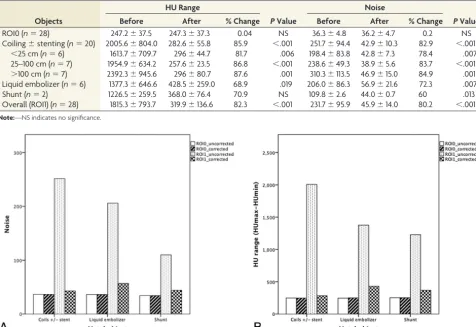
(ROI0) in all cases. The Hounsfield unit range and noise of ROI1 were higher in the coiling group compared with the liquid em-bolizer group (n-BCA or Onyx) or the ven-tricular shunt group in the uncorrected im-ages, but no significant difference existed (Fig 2). A significant reduction of metal ar-tifacts was achieved in all volume images of various metal objects, which showed a lower Hounsfield unit range and noise (P⬍
.001) (Fig 3). The Hounsfield unit range and noise of ROI1 in the corrected images remained significantly higher than those of ROI0 in the coiling group, but no signifi-cant difference existed in the other groups (Fig 2).
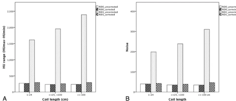
Comparison among Various Coil Lengths
In the coiling group, the Hounsfield unit range and noise of ROI1 were related to the coil length before the MAR algorithm correc-tion; however, no significant difference existed (Fig 4). A signifi-cant reduction of metal artifacts was achieved in all patients with different metal objects (P⬍.05). Except for the noise in the sub-group length between 25 and 100 cm, the Hounsfield unit range and noise of ROI1 in the corrected images showed no significant difference compared with those of ROI0 in the other subgroups (Fig 3).
FIG 2. A, Comparison of Hounsfield unit range among different objects.B, Comparison of noise among different metal objects.
[image:3.594.56.533.56.383.2]FIG 3. Illustrative case 1. FDCT without MAR (A) and with MAR (B) of a left-side ICA aneurysm post– coil embolization. The metal artifacts are significantly reduced, and the postcorrected images enable clearer evaluation of adjacent parenchyma.
Table 1: Quantitative assessment of metal artifacts before and after reduction
Objects
HU Range
PValue
Noise
PValue
Before After % Change Before After % Change
ROI0 (n⫽28) 247.2⫾37.5 247.3⫾37.3 0.04 NS 36.3⫾4.8 36.2⫾4.7 0.2 NS
Coiling⫾stenting (n⫽20) 2005.6⫾804.0 282.6⫾55.8 85.9 ⬍.001 251.7⫾94.4 42.9⫾10.3 82.9 ⬍.001 ⬍25 cm (n⫽6) 1613.7⫾709.7 296⫾44.7 81.7 .006 198.4⫾83.8 42.8⫾7.3 78.4 .007 25–100 cm (n⫽7) 1954.9⫾634.2 257.6⫾23.5 86.8 ⬍.001 238.6⫾49.3 38.9⫾5.6 83.7 ⬍.001 ⬎100 cm (n⫽7) 2392.3⫾945.6 296⫾80.7 87.6 .001 310.3⫾113.5 46.9⫾15.0 84.9 .001 Liquid embolizer (n⫽6) 1377.3⫾646.6 428.5⫾259.0 68.9 .019 206.0⫾86.3 56.9⫾21.6 72.3 .007
Shunt (n⫽2) 1226.5⫾259.5 368.0⫾76.4 70.9 NS 109.8⫾2.6 44.0⫾0.7 60 .013
[image:3.594.52.371.412.571.2]Subjective Assessment of Image-Quality Improvement
The results of the observer study are summarized in Tables 2 and 3. The overall agreement of the 2 raters was 85.7% (⫽0.517). No case had more severe artifacts or worse imaging quality after MAR correction. The number of cases in which the raters agreed on significant artifact reduction and improved image quality (score ofⱖ2) was⬎95% in the coiling group, 50% in the liquid embo-lizer group, and none in the shunt group.
DISCUSSION
An angiographic suite equipped with flat panel detectors can pro-vide in-room CT-like images before or after neurointerventional procedures without transferring patients to another conventional CT scanner. These images can identify the relationship of the metal implants and provide early recognition of rebleeding or intracranial procedure-related complications, such as incomplete stent deployment, stent migration, stent fracture, or coil disloca-tion.12-14However, the metallic artifacts stemming from metal
objects hinder clear visualization of a hematoma or parenchyma surrounding metal objects.14
Prell et al15demonstrated successfully reducing metal artifacts
by using their version of a MAR algorithm in a small series of 7 patients who had cerebral aneurysms and were treated with coil-ing or clippcoil-ing. Other studies, based on their own versions of a
MAR algorithm, have further demonstrated the visualization use-fulness of the stent strut and parent vessels in stent-assisted coil-ing.9,10However, the efficacy of the MAR algorithm, as described
above in managing different metal objects, has not been reported, to our knowledge.
The patients in our study did not undergo FDCT before the operation. Therefore, we chose the brain parenchyma in a higher or lower section, which was free of metallic artifacts, as the inter-nal reference of imaging quality. We demonstrated that the Sie-mens MAR software remarkably reduces artifacts and corrects image quality to a level close to the imaging quality of the native image by using adjacent parenchyma as the internal reference. Although the Hounsfield unit range and noise of the corrected images remained slightly higher than those of native images, no significant differences existed. We noticed that in most cases, cer-tain darker areas surrounding the implants remained after MAR correction. These artifacts may be the secondary artifacts intro-duced by the correction scheme and may be the reason the imag-ing quality remained inferior to that of native images.15,16In our
results, the types of metal objects and the coil lengths did not affect the results of the investigated MAR algorithm.
In assessing subjective image quality, we demonstrated that the MAR software substantially improved the visibility of the metal object vicinity, in which image qualities were most severely degraded before correction. Consequently, the overall imaging quality of surrounding regions improved in⬎78% of the cases following MAR. Nearly all coiling cases had increased diagnostic confidence following MAR. In contrast, the rater score was lower in the liquid embolizer group, though the Hounsfield unit range and noise in the corrected images showed no difference from
[image:4.594.50.530.46.252.2]FIG 4. A, Comparison of Hounsfield unit range among different coil lengths.B, Comparison of noise among different coil lengths.
Table 2: Results of the observer study rating the imaging quality with MAR
Objects
Rater 1 Score
PValue
Rater 2 Score
PValue
−1 0 1 2 3 −1 0 1 2 3
Coiling⫾stenting 0 0 0 18 2 0 0 1 13 6
Liquid embolizer 0 0 2 4 0 0 0 3 3 0
Shunt 0 0 2 0 0 0 2 0 0 0
Overall 0 0 4 22 2 .002 0 2 4 16 6 ⬍.001
Table 3: Inter-rater agreement of the observer study Overall
Agreement
Rater 1, Score ≤ 2 (%)
Rater 2, Score ≤ 2 (%) Overall imaging
quality
[image:4.594.52.531.288.357.2]those of the coiling group. Our explanation is that liquid emboli-zer shape and distribution were typically irregular and scattered in these cases. Despite significant artifacts reduction, the blurred re-gions and the secondary artifacts introduced by the replaced im-aging content of the original x-ray data accumulated in the central regions and significantly hindered the image interpretation (On-line Fig 1). Therefore, only half of the cases achieved higher diag-nostic confidence following MAR. None of the ventricular shunt-ing group provided additional diagnostic information followshunt-ing MAR because the shunt artifacts were typically limited to the tip of the shunt and were minor, which explains why most cases could be interpreted easily without requiring MAR correction (On-line Fig 2).
The main limitation of our study was the relatively small num-ber of cases. We only analyzed 6 patients receiving liquid emboli-zers and 2 patients undergoing ventricular drainage. Although the sample size was small, the MAR application resulted in promising results for correcting metal artifacts and improving image quality, indicating the need for further research. In the coiling group, we chose coil length rather than aneurysm packing attenuation as the coil attenuation parameter. Because the coil masses are irregular and bilateral in CCF cases, it is difficult to apply the formula of the aneurysm packing attenuation in these cases. Our results indicate the correlation of coil length with artifact severity and achieving reduced metal artifacts in all subgroups of different coil lengths. These findings may provide indirect evidence that the MAR soft-ware successfully reduces metal artifacts, regardless of various coiling densities.
CONCLUSIONS
We demonstrated using the MAR prototype to reduce streak ar-tifacts substantially around different metallic implants on FDCT and to improve objective and subjective imaging quality.
Disclosures: Sheng-Che Hung—RELATED:Provision of Writing Assistance, Medi-cines, Equipment, or Administrative Support: MAR prototype software,* Com-ments: collaborative project (T1100200) of the Taipei Veterans General Hospital and Siemens, the MAR. Wan-You Guo—RELATED:Other: supported in part by a collab-oration research contract between Taipei Veterans General Hospital and Siemens. *Money paid to the institution.
REFERENCES
1. Kalender WA, Kyriakou Y.Flat-detector computed tomography (FD-CT).Eur Radiol2007;17:2767–79
2. Akpek S, Brunner T, Benndorf G, et al.Three-dimensional imaging and cone beam volume CT in C-arm angiography with flat panel detector.Diagn Interv Radiol2005;11:10 –13
3. Kalender WA.The use of flat-panel detectors for CT imaging
[in German].Radiologe2003;43:379 – 87
4. Gupta R, Cheung AC, Bartling SH, et al.Flat-panel volume CT: fun-damental principles, technology, and applications.Radiographics
2008;28:2009 –22
5. Hung SC, Lin CJ, Guo WY, et al.Toward the era of a one-stop imag-ing service usimag-ing an angiography suite for neurovascular disorders.
Biomed Res Int2013;2013:873614
6. Struffert T, Richter G, Engelhorn T, et al.Visualisation of intracere-bral haemorrhage with flat-detector CT compared to multislice CT: results in 44 cases.Eur Radiol2009;19:619 –25
7. Do¨rfler A, Struffert T, Engelhorn T, et al.Rotational flat-panel com-puted tomography in diagnostic and interventional neuroradiol-ogy.Rofo2008;180:891–98
8. Prell D, Kalender WA, Kyriakou Y.Development, implementation and evaluation of a dedicated metal artefact reduction method for interventional flat-detector CT.Br J Radiol2010;83:1052– 62 9. van der Bom IM, Hou SY, Puri AS, et al.Reduction of coil mass
artifacts in high-resolution flat detector conebeam CT of cerebral stent-assisted coiling.AJNR Am J Neuroradiol2013;34:2163–70 10. Psychogios MN, Scholz B, Rohkohl C, et al.Impact of a new metal
artefact reduction algorithm in the noninvasive follow-up of intra-cranial clips, coils, and stents with flat-panel angiographic CTA: initial results.Neuroradiology2013;55:813–18
11. Meyer E, Raupach R, Lell M, et al.Normalized metal artifact reduc-tion (NMAR) in computed tomography.Med Phys2010;37:5482–93 12. Mordasini P, Al-Senani F, Gralla J, et al.The use of flat panel angioCT (DynaCT) for navigation through a deformed and fractured carotid stent.Neuroradiology2010;52:629 –32
13. Buhk JH, Kallenberg K, Mohr A, et al.Evaluation of angiographic computed tomography in the follow-up after endovascular treat-ment of cerebral aneurysms–a comparative study with DSA and TOF-MRA.Eur Radiol2009;19:430 –36
14. Clarenc¸on F, Piotin M, Pistocchi S, et al.Evaluation of stent visibility by flat panel detector CT in patients treated for intracranial aneu-rysms.Neuroradiology2012;54:1121–25
15. Prell D, Kyriakou Y, Struffert T, et al.Metal artifact reduction for clipping and coiling in interventional C-arm CT.AJNR Am J Neu-roradiol2010;31:634 –39