ORIGINAL RESEARCH
Basal Forebrain Involvement in Low-Functioning
Autistic Children: A Voxel-Based Morphometry
Study
D. Riva S. Bulgheroni D. Aquino F. Di Salle M. Savoiardo A. Erbetta
BACKGROUND AND PURPOSE:Imaging studies have revealed brain abnormalities in the regions in-volved in functions impaired in ASD (social relations, verbal and nonverbal communication, and adaptive behavior). We performed a VBM whole-brain analysis to assess the areas involved in autistic children with DD.
MATERIALS AND METHODS: Twenty-one developmentally delayed children with ASD (aged 3–10 years) were compared with 21 controls matched for age, sex, and sociocultural background. All ASD cases had been diagnosed according toDiagnostic and Statistical Manual of Mental Disorders, Fourth Editioncriteria, with the Autism Diagnostic Observation Schedule-Generic, and the Autism Diagnostic Interview-Revised. The VBM data, covaried with intelligence quotient, age, and brain volume, were analyzed.
RESULTS: ASD patients showed a pattern of regional GM reduction symmetrically affecting the basal forebrain, accumbens nucleus, cerebellar hemispheres, and perisylvian regions, including insula and putamen. Asymmetric involvement of GM was observed in other brain regions functionally connected to the basal forebrain, ie, an area located close to the medial and ventral surface of the frontal lobe. No regional WM differences were observed between the 2 groups. No significant differences between patients and controls were found regarding total brain volume, GM, and WM.
CONCLUSIONS:In children with ASD and DD, the novel finding of our VBM study was the demon-stration of reduced GM volume in the basal forebrain and the areas connected with it. This system is involved in social behavior, communication, and cognitive skills. Whether the involvement of the basal forebrain is characteristic of ASD or is related to the DD present in our patients needs further investigation.
ABBREVIATIONS:ASD⫽autistic spectrum disorders; BA⫽Brodmann area; BF⫽basal forebrain; DD⫽developmental delay; DLPFC⫽dorsolateral prefrontal cortex; FDR⫽false discovery rate; fMRI⫽functional MR imaging; GM⫽gray matter; IFG⫽inferior frontal gyrus; IQ⫽intelligence quotient; LF⫽low functioning; NACC⫽accumbens nucleus; PET⫽positron-emission tomogra-phy; rCBF⫽relative cerebral blood flow; SMA⫽supplementary motor area; VBM⫽voxel-based morphometry; WM⫽white matter
T
he ASD are currently seen as genetic neurodevelopmental disorders, though their underlying biologic causes remain to be established. The general behavioral phenotype defined as ASD includes diverse endophenotypes sharing 3 core features: impaired social interaction; verbal and nonverbal communi-cation anomalies; and restricted patterns of behavior, stereo-typed patterns of behavior, or both. Furthermore, frequent comorbidities (eg, epilepsy, mental retardation, and psychiat-ric disorders) make it difficult to isolate the neuropathologic picture of autism.1In patients with ASD, conventional and advanced imaging studies, such as VBM, demonstrated abnormalities in frontal lobe, temporal and parietal cortices, language areas, amygdala, caudate nucleus, and cerebellum, even though some findings were controversial.1Furthermore, most studies were carried out in high-functioning patients or patients with Asperger
syndrome. However, most of the autistic population is intel-lectually impaired, and⬇80% of autistic children are LF, ie, they have an IQ⬍70.2Despite this prevalence of LF children with ASD, these patients are rarely investigated,3-5probably because of the difficulties in cooperating for neuroradiologic investigations.
The aim of our study was to investigate LF children with ASD younger than age 10 with VBM, which is an unbiased, operator-independent method for detecting statistically sig-nificant differences between groups.6
Materials and Methods
Subjects
From 93 children with ASD, referred to our Developmental Neurol-ogy Unit from January 2006 to August 2008, a group of 21 LF children with idiopathic ASD without associated seizures or other neurologic diseases was selected. Patients with known infectious, metabolic, or genetic diseases, and chromosomal abnormalities were excluded. Children with disintegrative disorder also were excluded. In addition, we excluded patients in whom conventional MR imaging demon-strated structural abnormalities. Four patients were excluded because of motion artifacts.
Of the 21 patients (8 females and 13 males; age range, 3 years and 2 months–10 years and 10 months; mean, 6 years and 6 months; SD, Received October 12, 2010; accepted after revision December 17.
From the Division of Developmental Neurology (D.R., S.B.) and Department of Neuroradi-ology (D.A., M.S., A.E.), Fondazione IRCCS Istituto Neurologico C. Besta, Milan, Italy; and Department of Cognitive Neuroscience (F.D.S.), University of Maastricht, Maastricht, the Netherlands.
Please address correspondence to Daria Riva, MD, Developmental Neurology Division, Fondazione IRCCS Istituto Neurologico C. Besta, Via Celoria, 11-0133 Milan, Italy; e-mail: [email protected]
2 years and 5 months), 12 had a diagnosis of autism, and 9 of Pervasive Developmental Disorder-Not Otherwise Specified. In all cases, the diagnosis was established according to theDiagnostic and Statistical Manual of Mental Disorders, Fourth Editiondiagnostic criteria and confirmed by semistructured assessment by using Autism Diagnostic Observation Schedule-Generic and by interviewing the parents by using Autism Diagnostic Interview-Revised.
The cognitive functioning was assessed by using the Wechsler In-telligence Scale according to their respective age, the Leiter Interna-tional Performance Scale Revised for nonverbal children, and the Griffiths Mental Developmental Scale for those with chronologic or mental age⬍4 years. The IQ ranged from 32 to 66 (mean, 52.5⫾9.8). The control group consisted of 21 normally developing children (8 females and 13 males; age range, 3 years and 9 months–10 years and 3 months; mean, 6 years and 10 months; SD, 2 years and 1 month), with normal IQ, no signs of autistic behavior or other psychiatric or neurologic disorders, or family history of neurologic or psychiatric illness among their first-degree relatives. The study was approved by our local ethics committee, and all examinations were performed with the written informed consent of the patients’ parents.
MR Imaging Acquisition
All the autistic patients were examined under propofol sedation (1 mg/kg). The controls⬍6 years old also were examined under propo-fol sedation. They were recruited among inpatients with suspected spinal cord abnormalities and were included in the study if the brain and spine examination was normal. The controls⬎6 years old were examined without sedation; they were recruited among the children of the medical and technical staff involved in the study. Volumetric T1-weighted images were acquired on a 1.5T MR imaging system (Siemens, Erlangen, Germany) by using a magnetization-prepared gradient-echo sequence (TR⫽1640 ms, TE⫽2.48 ms, TI⫽552 ms, FOV⫽256⫻256 mm, matrix⫽256⫻256, 160 sagittal sections, voxel size⫽1⫻1⫻1 mm).
Structural imaging included axial proton-density/T2-weighted images (TR⫽3500 ms, TE⫽17 ms/84 ms, FOV⫽208⫻256 mm, matrix⫽208⫻256, section thickness⫽5 mm) and coronal turbo spin-echo T2-weighted images (TR⫽4100 ms, TE⫽143 ms, FOV⫽ 324⫻384 mm, matrix⫽324⫻384, section thickness⫽5 mm). Structural imaging was assessed by a senior neuroradiologist for the presence or absence of supratentorial and infratentorial abnormali-ties and signal intensity changes, based on visual inspection. The MR imaging obtained for the controls was similarly reviewed.
VBM Analysis
Images were processed according to the optimized VBM method im-plemented in SPM5 (http://www.fil.ion.ucl.ac.uk/spm/software/ spm5/) as described previously.7Original T1 images were first
nor-malized to a pediatric (5–9.5 years) brain template (Cincinnati Children’s Hospital Medical Center: CCHMC2_y; available at https://
irc.cchmc.org) by using a 12-parameter linear affine transformation and a nonlinear transformation through 7⫻8⫻7 nonlinear basis functions and then segmented into GM, WM, and CSF partitions. A custom template was then obtained as suggested by Good et al,7
nor-malizing, segmenting, smoothing, and averaging the images of the 42 subjects of our study to account for potential differences between our MR scanner and the scanner used to obtain the pediatric template and to account for differences between our sample and the sample used for the template. The original, native T1 images were then normalized and segmented again by using the new template and priors. To pre-serve the amount of a particular tissue (GM) within a voxel, a further modulation step was incorporated.
Finally, the images were smoothed with a Gaussian kernel filter of 8⫻8⫻8 mm full width half maximum to have data more normally distributed and to compensate for inexact spatial normalization.
Differences in GM and WM were assessed statistically using the general linear model based on the Gaussian field theory. An analysis of covariance, with total intracranial volume, age, IQ, and sex as covari-ates, was used. The significance levels were set atP⬍.05 corrected for multiple comparisons by using the FDR.
Localization of areas of significant tissue loss was determined by superimposing the regions of significant atrophy on the averaged T1-weighted image used to create the template. Regions of interest were reported in Montreal Neurologic Institute coordinates.
Total Volumes Analysis
The intracranial volumes were estimated by integrating the voxel val-ues across the modulated (unsmoothed) GM, WM, and CSF parti-tions, and results were converted from voxels into milliliters. Group differences in total intracranial volume as well as group differences in total GM, WM, and CSF volumes were assessed by using a 2-tailedt test.
Results
Quantitative Structural MR Imaging
No significant differences were found between cases and con-trols regarding the total volumes of brain (P⫽.49), GM (P⫽
.992), WM (P⫽.227), and CSF (P⫽.813). These results are summarized in Table 1.
Regional WM Differences
No regional differences in the WM were observed between the 2 groups.
Regional GM Differences
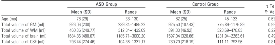
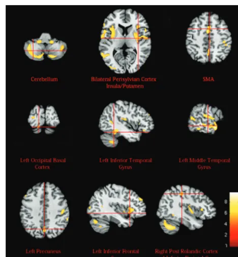
[image:2.594.51.532.59.139.2]The autistic patients had a reduced volume of GM in 13 3D clusters (significance threshold, P ⬍ .05, FDR corrected). Basal forebrain, nucleus accumbens, SMA, cerebellum, and perisylvian region (including the insula and putamen) showed Table 1: Total intracranial volumes in ASD and control groups
ASD Group Control Group tTest,
PValue
Mean (SD) Range Mean (SD) Range
Age (mo) 78 (29) 38–130 82 (25) 45–123 0.623
Total volume of GM (ml) 926.06 (230) 239.34–1485.22 925.50 (107.43) 775.89–1176.89 0.992 Total volume of WM (ml) 460.35 (249.77) 312.34–1439.69 391.33 (46.92) 323.69–478.83 0.227 Total volume of brain (ml) 1684.86 (480.07) 1185.71–3000.20 1597.04 (320.66) 1231.94–2263.61 0.490 Total volume of CSF (ml) 298.44 (274.46) 104.36–1321.17 280.20 (218.19) 111.11–793.96 0.813
PEDIATRICS
ORIGINAL
a reduced GM bilaterally. In the left hemisphere, a reduction in GM was recorded in the occipital basal cortex; inferior, middle, and superior temporal gyri; IFG; DLPFC; and precu-neus. In the right hemisphere, a GM reduction was visible in the postcentral cortex and inferior parietal lobe. These results are shown in Table 2 and Figs 1 and 2.
Discussion
In this study we applied unbiased whole brain VBM to assess neuroanatomic changes in LF autistic children compared with controls paired for age, sex, and social status. The choice of normal IQ for the control group was dictated by the fact that a group of DD children with IQ matching with that of the pa-tients would probably be a heterogeneous sample of various pathologies that would create even greater difficulties in the comparison.
Several VBM investigations in the autistic population have been reported in the literature. These studies demonstrated a volume loss in cortical and subcortical areas as well as in the cerebellum. The affected areas were the IFG,8,9insula,9 cere-bellum,5,10 precuneus,11 and basal ganglia.11,12 These areas also have been found affected in our study. Higher than nor-mal density, not observed by us, has been reported in inferior
frontal cortex10,13and cerebellum.3,8,14Other investigations identified areas of higher or lower GM density that were un-affected in our patients, such as the amygdala,8 cingulate gyrus,8,11fusiform gyrus,10,14and thalamus.5,12,13GM reduc-tion in the basal forebrain, which is the most remarkable find-ing of our study, was not observed previously.
In our series of ASD patients, volume loss was found to affect the basal forebrain, nucleus accumbens, perisylvian re-gions, and other cortical brain regions functionally connected to the abovementioned areas as well as the putamen and cerebellum.
The basal forebrain comprises a group of structures located in the medial and ventral surface of the frontal lobe. The basal forebrain consists of 3 functionally distinct compartments,15 ie, the mainly cholinergic corticopetal system, the extended amygdalae, and the ventral striatopallidal system. The cholin-Table 2: GM regions significantly reduced (P<.05 FDR corrected) in the autistic group by comparison with the controls
Anatomic Location
Talairach Coordinates Center of Cluster, x y z
Peak of Significance (tscore)
Cluster size (No. of voxels)
Basal forebrain, nucleus accumbens 0 1 ⫺2 4.05 1282
Supplementary motor area (BA6) 2 ⫺1 47 4.12 699
Left insula, left putamen, left perisylvian cortex (BA48) ⫺43⫺2 13 5.37 6945 Right insula, right putamen, right perisylvian cortex (BA48) 34⫺2 14 5.42 8266
Left cerebellum ⫺26⫺58⫺37 5.56 6452
Right cerebellum 23⫺55⫺39 5.29 10,441
Left DLPFC (BA47) ⫺44 15 34 4.93 1513
Left inferior temporal gyrus (BA20) ⫺46⫺40⫺14 5.40 1708
Left middle temporal gyrus (BA21) and left superior temporal gyrus (BA22) ⫺56⫺19⫺2 5.96 2844
Left precuneus (BA7) ⫺1⫺68 34 5.78 1225
Left occipito-basal cortex (BA18) ⫺14⫺78⫺18 5.48 359
Left inferior frontal gyrus (BA44) ⫺39 22 2 5.39 276
Right postcentral cortex (BA1,2,3), right inferior parietal lobe (BA40) 41⫺33 40 5.35 1382
Note:—All labels are derived from the Anatomical Automatic Atlas. The x, y, z coordinates are the Talairach coordinates of the center of each 3D cluster.43BAs also are provided.
Fig 1.Basal forebrain and accumbens nucleus volume differences between groups. GM volume differences between groups, overlaid on T1 images (consecutive coronal sections, rostral to caudal) showing the involvement of accumbens nucleus and basal forebrain. The relative decrease in GM attenuation observed in autistic subjects compared with controls is represented in a red-orange color scale. The statistical threshold isP⬍.05 (FDR corrected). L⫽left; R⫽right.
[image:3.594.52.533.59.214.2] [image:3.594.300.534.234.487.2] [image:3.594.52.285.235.393.2]ergic cells are concentrated in the septum, the diagonal band, and the nucleus basalis of Meynert16; their medial component projects to the medial frontal and cingulate cortex, whereas the more lateral subdivisions of this system project to the lat-eral neocortex, including the orbitoinsular, prefrontal, tempo-ral, parietal, and occipital cortices.16,17
Therefore, the term basal forebrain commonly refers to an extended continuum of subcortical neurons providing projec-tions to a variety of neocortical fields and limbic structures implicated in many cognitive functions and social behavior patterns.18The basal forebrain activates areas of the cerebral cortex and facilitates learning, by using acetylcholine as a prominent neurotransmitter. Functionally, the cholinergic system has been referred to as an “action system” that helps individuals to develop the ability to focus on the environment and provide a coherent behavioral response.19
Volume loss in the basal forebrain is consistent with the hypothesis proposed by Perry et al,20according to which, in autism, the cholinergic system of the basal forebrain is im-paired. In fact, in autistic subjects, they demonstrated a lower level of cholinergic receptors than in normal controls. Cholin-ergic afferents innervate the cerebral cortex during the early postnatal development, which is the most dynamic period of neuronal differentiation and synapse formation.21A disrup-tion of the cholinergic innervadisrup-tion results in a delayed cortical neuronal development and permanent changes in cortical cy-toarchitecture and cognitive functions.21
Involvement of the basal forebrain has been demonstrated by imaging studies in attention, learning, memory, and re-ward. In an auditory vigilance test, PET showed rCBF changes in the basal forebrain.22During classic conditioning tasks with auditory discrimination, PET showed that rCBF of the audi-tory cortex positively correlated with activity in the basal fore-brain, amygdala, and orbitofrontal cortex.23These results sup-port the notion that the basal forebrain’s cholinergic projection to the cortex is important in modulating the syn-aptic activity. Furthermore, PET studies suggested an activity in the nucleus of the diagonal band of Broca in episodic mem-ory recall.24The basal forebrain and DLPFC were activated in paradigms sensitive to working memory.25
The extended amygdalae stretch medially and rostrally from the centromedial nuclei of the amygdalae through the substantia innominata into the medial portion of the nucleus accumbens, thus joining the ventral striatopallidal system. In our study, the nucleus accumbens was found to be reduced in volume. This nucleus has a central role in the functional inter-play between basal forebrain components, and between the basal forebrain and other brain regions. It receives projections from the hippocampus; basolateral amygdalae; and the pre-frontal, insular, and temporal association cortex,15and it has efferent connections to the corticopetal cholinergic system, thus modulating the cholinergic output.15,16The nucleus ac-cumbens is therefore involved in memory; it is engaged both in incentive-reward processes and in adaptive responses to conditioned and unconditioned aversive stimuli. A reduced volume of the nucleus accumbens also was reported by Ball-maier et al26in children with schizophrenia. In their study, the smaller left nucleus accumbens was associated with linguistic errors and the smaller right nucleus accumbens with difficul-ties in planning and organized thinking.
A reduced volume of GM was found in the SMA (BA6) and putamen, structures involved in the motor and cognitive cir-cuits. SMA is functionally subdivided in the SMA proper and pre-SMA. Functional imaging studies in humans have shown that the pre-SMA has an important role in supramotor activ-ities.27Similarly, the putamen is activated both when a subject is preparing to move and when the movement is performed. Motor symptoms, such as postural abnormalities in the head and trunk, and stereotypical movements are prominent fea-tures of autism; they are probably due to involvement of both the fronto-striatal basal ganglia circuits and the cerebellum.28 A reduced volume of GM was identified bilaterally in the insula and the perisylvian region. The insular cortex works as a relay area for multiple neurocognitive systems and plays a part in many functions related to emotions or regulating the body’s homeostasis. This region may therefore be involved in the complex clinical features of autistic disorders.9The anterior cortical perisylvian areas are important for speech and lan-guage functions.
Our patients showed GM reduction in both cerebellar hemispheres. The cholinergic basal forebrain is connected with the cerebellum29and the cholinergic cerebellar neurons contribute to mental functions related to cognition, behavior and emotions. In autism, there are numerous reports of vol-ume changes in the cerebellar vermis. Individuals with early infantile autism show fourth ventricle enlargement, loss of Purkinje cells in the lateral and inferior cerebellar cortex, and abnormal or reduced numbers of neurons in the deep cerebel-lar nuclei.1
Unilateral involvement was found in different areas of the cerebral hemispheres and was more numerous on the left side. GM loss was found in the left DLPFC. This area is part of the neuronal network underlying social cognition in the frame of the theory of mind, and executive functions. GM loss in the frontal lobe of autistic children was confirmed by imaging and postmortem studies.1 Decreased connections between the frontal cortex and regions implicated in social cognition and in the empathic processes were demonstrated by diffusion ten-sor imaging.30
Reduced GM was prominent in the left middle temporal gyrus, which is functionally connected to the cholinergic sys-tem and nucleus accumbens.17The middle temporal cortex and superior temporal gyrus are important for perceiving and decoding other people’s gaze, a function deficient in autistic children. An impairment of this region also could contribute to the language difficulties and consequent communication problems of autistic subjects.31
GM loss in our ASD patients involved the precuneus that is connected with multiple areas of the interhemispheric cortex, convexity, basal ganglia, and thalami.32Changes occurring in this pivotal area are consistent with recent claims that autism is a connectivity disorder.33The precuneus is activated when we make judgments that demand empathy and when we attribute emotions to ourselves and to other people.34
tem-poral gyrus seems to be involved in common object percep-tion. In autistic subjects, Schultz et al35found fMRI abnormal-ities in the inferior temporal and fusiform gyri in face discrimination tasks.
Involvement of the left IFG deserves a specific comment. This region, the inferior parietal lobe and BA1, 2, 3 are part of the mirror neuron system implicated in understanding other people’s actions, intentions, and emotions.36In autistic pa-tients, a limited activation of the area containing the mirror neurons was demonstrated by fMRI.37In our study, these 2 last areas, inferior parietal lobe and BA1, 2, 3 were found af-fected only on the right side.
In contrast with the studies collected by Radua et al,38in agreement with other studies,9,10,13we did not find any vol-ume abnormalities in the WM. These different results can be attributed to the heterogeneity of the disorder and the differ-ent age of the population studied. Diffusion tensor imaging studies might be more sensitive to WM abnormalities.
Bilateral GM reduction could be consistent with more prominent behavioral abnormalities than unilateral GM in-volvement, as already shown in bilateral versus unilateral focal brain lesions.39 However, only a clinical correlation could confirm this hypothesis.
Abnormalities in the basal forebrain were reported in other syndromes with DD, such as Down syndrome,40 Rett syn-drome,41and fragile X syndrome,42but not in children with idiopathic DD.42
The autistic phenotype we examined cannot be merely summarized in the term autistic behavior plus intellectual dis-ability, because it comprises a syndromic constellation of symptoms, in which there are both signs of autism and of cognitive malfunctioning. Involvement of the basal forebrain, however, might be an indicator of DD rather than autism itself.
The limitations of our study are the small sample size and the lack of a control group with idiopathic DD. The analysis of such a group could answer the question whether basal fore-brain involvement is related to the mental retardation rather than autism itself.
Conclusions
The main finding of this study was the volume reduction in the basal forebrain, an area not previously found affected in VBM studies of autistic children. This area and its extensive, multi-ple cortical connections that also present volume reduction, are involved in social, communicative, and cognitive behavior. The basal forebrain involvement might be related to DD, which was always present in our autistic children.
Acknowledgments
We thank all the children who participated in the study and those who acted as the healthy controls and their families. We also thank Dagmar Di Fiore, RT, for helpfulness and accuracy in performing the MR examinations as well as the reviewers for many helpful suggestions.
References
1. Amaral DG, Schumann CM, Nordahl CW.Neuroanatomy of autism.Trends Neurosci2008;31:137– 45
2. Fombonne E.Epidemiology of pervasive developmental disorders.Pediatr Res
2009;65:591–98
3. Bonilha L, Cendes F, Rorden C, et al.Gray and white matter imbalance-typical structural abnormality underlying classic autism? Brain Dev
2008;30:396 – 401
4. Boddaert N, Chabane N, Gervais H, et al.Superior temporal sulcus: anatomical abnormalities in childhood autism: a voxel-based morphometry MRI study.
Neuroimage2004;23:364 – 69
5. Spencer MD, Moorhead TW, Lymer GK, et al.Structural correlates of intellec-tual impairment and autistic features in adolescents. Neuroimage
2006;33:1136 – 44
6. Ashburner J, Friston KJ.Voxel-based morphometry: the methods.Neuroimage
2000;11:805–21
7. Good CD, Johnsrude IS, Ashburner J, et al.A voxel-based morphometric study of ageing in 465 normal adult human brains.Neuroimage2001;14:21–36 8. Abell F, Krams M, Ashburmer J, et al.The neuroanatomy of autism: a voxel
based whole brain analysis of structural scans.Neuroreport1999;10:1647–51 9. Kosaka H, Omori M, Munesue T, et al.Smaller insula and inferior frontal
volumes in young adults with pervasive developmental disorders.Neuroimage
2010;50:1357– 63
10. Rojas DC, Peterson E, Winterrowd E, et al.Regional gray matter volumetric changes in autism associated with social and repetitive behavior symptoms.
BMC Psychiatry2006;6:56 – 68
11. McAlonan GM, Cheung V, Cheung C, et al.Mapping the brain in autism. A voxel-based MRI study of volumetric differences and intercorrelations in au-tism.Brain2005;128:268 –76
12. McAlonan GM, Suckling J, Wong N, et al.Distinct patterns of grey matter abnormality in high-functioning autism and Asperger’s syndrome.J Child Psychol Psychiatry2008;49:1287–95
13. Waiter GD, Williams JH, Murray AD, et al.A voxel-based investigation of brain structure in male adolescents with autistic spectrum disorder. Neuro-image2004;22:619 –25
14. Ke X, Hong S, Tang T, et al.Voxel-based morphometry study on brain struc-ture in children with high-functioning autism.Neuroreport 2008;19:921–25 15. Zahm DS.The evolving theory of basal forebrain functional-anatomical
mac-rosystems.Neurosci Biobehav Rev2006;30:148 –72
16. Selden NR, Gitelman DR, Salamon-Murayama N, et al.Trajectories of cholin-ergic pathways within the cerebral hemispheres of the human brain.Brain
2008;121:2249 –57
17. Zaborszky L, Hoemke L, Mohlberg H, et al.Stereotaxic probabilistic maps of the magnocellular cell groups in human basal forebrain. Neuroimage
2008;42:1127– 41
18. Baxter MG, Chiba AA.Cognitive functions of the basal forebrain.Curr Opin Neurobiol1999;9:178 – 83
19. Lam KS, Aman MG, Arnold LE.Neurochemical correlates of autistic disorder: a review of the literature.Res Dev Disabil2006;27:254 – 89
20. Perry EK, Lee ML, Martin-Ruiz CM, et al.Cholinergic activity in autism: ab-normalities in the cerebral cortex and basal forebrain.Am J Psychiatry
2001;158:1058 – 66
21. Hohmann CF, Berger-Sweeney J.Cholinergic regulation of cortical develop-ment and plasticity. New twists to an old story.Perspect Dev Neurobiol
1998;5:401–25
22. Paus T, Perry DW, Zatorre RJ, et al.Modulation of cerebral blood flow in the human auditory cortex during speech: role of motor-to-sensory discharges.
Eur J Neurosci1996;8:2236 – 46
23. Morris JS, Friston KJ, Dolan RJ.Experience-dependent modulation of tono-topic neural responses in human auditory cortex. Proc Biol Sci
1998;265:649 –57
24. Fujii T, Okuda J, Tsukiura T, et al.The role of the basal forebrain in episodic memory retrieval: a positron emission tomography study. Neuroimage
2002;15:501– 08
25. Swartz BE, Halgren E, Fuster JM, et al.Cortical metabolic activation in humans during a visual memory task.Cereb Cortex1995;5:205–14
26. Ballmaier M, Toga AW, Siddarth P, et al.Thought disorder and nucleus accum-bens in childhood: a structural MRI study.Psychiatry Res2004;130:43–55 27. Sakai K, Hikosaka O, Miyauchi S, et al.Presupplementary motor area
activa-tion during sequence learning reflects visuo-motor associaactiva-tion.J Neurosci
1999;19:RC1:1– 6
28. Rinehart NJ, Tonge BJ, Iansek R, et al.Gait function in newly diagnosed chil-dren with autism: cerebellar and basal ganglia related motor disorder.Dev Med Child Neurol2006;48:819 –24
29. Walker BR, Diefenbach KS, Parikh TN.Inhibition within the nucleus tractus solitarius (NTS) ameliorates environmental exploration deficits due to cere-bellum lesions in an animal model for autism. Behav Brain Res
2007;176:109 –20
30. Noriuchi M, Kikuchi Y, Yoshiura T, et al.Altered white matter fractional an-isotropy and social impairment in children with autism spectrum disorder.
Brain Res2010;1362:141– 49
32. Cavanna AE, Trimble MR.The precuneus: a review of its functional anatomy and behavioural correlates.Brain2006;129:564 – 83
33. Kennedy DP, Courchesne E.The intrinsic functional organization of the brain is altered in autism.Neuroimage2008;39:1877– 85
34. Siegal M, Varley R.Neural systems in theory of mind.Nat Rev Neurosci
2002;3:463–71
35. Schultz RT, Gauthier I, Klin A, et al.Abnormal ventral temporal cortical activ-ity during face discrimination among individuals with autism and Asperger syndrome.Arch Gen Psychiatry2000;57:331– 40
36. Rizzolatti G, Sinigaglia C.The functional role of the parieto-frontal mirror circuit: interpretations and misinterpretations. Nat Rev Neurosci
2010;11:264 –74
37. Dapretto M, Davies MS, Pfeifer JH, et al.Understanding emotions in others: mirror neuron dysfunction in children with autism spectrum disorders.Nat Neurosci2006;9:28 –30
38. Radua J, Via E, Catani M, et al.Voxel-based meta-analysis of regional white-matter volume differences in autism spectrum disorder versus healthy con-trols.Psychol Med2011;41:1539 –50
39. Ballantyne AO, Spilkin AM, Hesselink J, et al.Plasticity in the developing brain: intellectual, language and academic functions in children with ischaemic perinatal stroke.Brain2008;131:2975– 85
40. Chen Y, Dyakin VV, Branch CA, et al.In vivo MRI identifies cholinergic cir-cuitry deficits in a Down syndrome model.Neurobiol Aging2009;30:1453– 65 41. Riikonen R.Neurotrophic factors in the pathogenesis of Rett syndrome.
J Child Neurol2003;18:693–97
42. Hoeft F, Carter JC, Lightbody AA, et al.Region-specific alterations in brain development in one- to three-year-old boys with fragile X syndrome.Proc Natl Acad Sci U S A2010;107:9335–39