Multiple Organ Dysfunction Syndrome (MODS): Is It
Preventable or Inevitable?
*
Ayman El-Menyar1,2#, Hassan Al Thani3, El Rasheid Zakaria4, Ahmad Zarour3, Mazin Tuma3,
Husham AbdulRahman3, Ashok Parchani3, Ruben Peralta3, Rifat Latifi2,3,5
1The Clinical Research, Trauma Surgery, Hamad Medical Corporation (HMC), Doha, Qatar; 2Clinical Medicine, Weill Cornell
Medical School, Doha, Qatar; 3Trauma Surgery, Hamad Medical Corporation (HMC), Doha, Qatar; 4Medical Research Center, Hamad Medical Corporation (HMC), Doha, Qatar; 5Department of Surgery, University of Arizona, Tucson, Arizona, USA.
Email: #[email protected]
Received September 26th, 2012; revised November 11th, 2012; accepted November 24th, 2012
ABSTRACT
Multiple organ dysfunction syndrome (MODS) is a systemic, dysfunctional inflammatory response that requires long intensive care unit (ICU) stay. It is characterized with high mortality rate depending on the number of organs involved. It has been recognized that organ failure does not occur as an all-or-none rule, but rather a range of organ dysfunction exists resulting in clinical organ failure. In the absence of a gold standard scoring or tool for early diagnosis or predic-tion of MODS, a novel bio-clinical scoring is mandatory. Moreover, understanding the pathophysiology of MODS in medical, surgical and trauma, ICUs should take a priority to achieve a favorable outcome. Herein we reviewed the lit-eratures published in English language through the research engines (MEDLINE, Scopus, and EBASE) from 1982 to 2011 using key words: “multiorgan dysfunction”, “organ failure”, “intensive care units” to highlight the definition, me- chanism, diagnosis and prediction of MODS particularly at its earliest stages. Bring up new bio-clinical scoring to a stage where it is ready for field trials will pave the way for implementing new risk-stratification strategy in the intensive care to reduce the morbidity and mortality and save resources. Prospective studies are needed to answer our question and to shift MODS from an inevitable to a preventable disorder.
Keywords: Multiorgan Dysfunction; Failure; Intensive Care Units
1. Introduction
Multiple organ dysfunction syndrome (MODS) is a sys- temic, dysfunctional inflammatory response that requires long intensive care unit (ICU) stay and has high mortality rate of 27% - 100% depending on the number of organs involved [1-3]. However, there is no gold standard scor- ing system for MODS and also, the available registries and studies are not enough to understand, diagnose or predict the occurrence of MODS across the different types of ICUs. Herein we reviewed the literatures pub- lished in English language through the research engines (MEDLINE, Scopus, and EBASE) from 1982 to 2011 using key words: “multiorgan dysfunction”, “organ fail- ure”, “intensive care units” to highlight the definition, mechanism, diagnosis and prediction of MODS particu- larly at its earliest stages. Non-English articles, case re- ports, outdated abstracts, and unpublished data were ex- cluded.
2. Mechanism of Multiple Organ
Dysfunction Syndrome
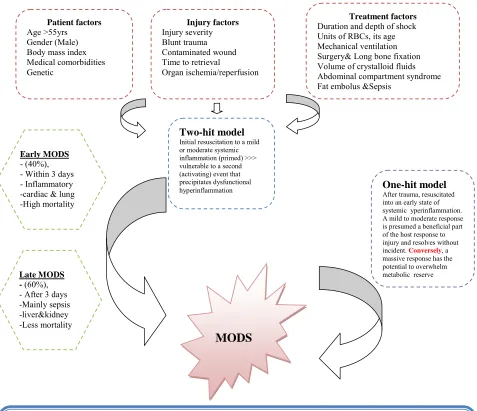
MODS is the leading cause of mortality in patients who survived the initial hours after trauma [2]. Moreover, MODS represents the most common cause of utilization of hospital resources. Initially MODS was thought to be an overwhelming, uncontrolled sepsis response, this was modified with the realization of the bimodal model of MODS (Figure 1) and the recognition that early MODS was unrelated to sepsis. In general, the cause of post in-jury MODS involves a mixed layering of patient, inin-jury and treatment factors (Figure 1). The dysregulated im- munological response is the crucial factor in the patho- physiology of post injury MODS [3].
3. Cycle and Stages of MODS
Patients with a heterogeneous trauma load and clinical picture are resuscitated into a similar state of systemic hyperinflammation, termed systemic inflammatory re- sponse syndrome (SIRS). This might be both beneficial *All authors read and approved the manuscript; there is no conflict of
interest and no financial issues to declare. #Corresponding author.
One-hit model
After trauma, resuscitated into an early state of systemic yperinflammation. A mild to moderate response is presumed a beneficial part of the host response to injury and resolves without incident. Conversely, a massive response has the potential to overwhelm metabolic reserve
Two-hit model
Initial resuscitation to a mild or moderate systemic inflammation (primed) >>> vulnerable to a second (activating) event that precipitates dysfunctional hyperinflammation
Late MODS - (60%), - After 3 days -Mainly sepsis -liver&kidney -Less mortality
Early MODS - (40%), - Within 3 days - Inflammatory -cardiac & lung -High mortality
MODS
Treatment factors Duration and depth of shock Units of RBCs, its age Mechanical ventilation Surgery& Long bone fixation Volume of crystalloid fluids Abdominal compartment syndrome Fat embolus &Sepsis
Patient factors Age >55yrs Gender (Male) Body mass index Medical comorbidities Genetic
Injury factors Injury severity Blunt trauma Contaminated wound Time to retrieval
Organ ischemia/reperfusion
[image:2.595.61.538.86.497.2]Injury>> shock >> whole body hypoperfusion >> resuscitation >> reperfusion of ischaemic gut >> release of cytokines (IL6, IL8 and IL10) >> proinflammatory lipids and proteins from the reperfused splanchnic bed >> these mediators return to vascular circulation via lymphatics >> prime PMNs >> PMNs mobilise into vascular circulation >> a significant neutrophillia at 3 h postinjury >> ‘‘vulnerable window’’ >> A second hit during this period??: cipitate MOF, eutrophillia normally resolves without endorgan damage. Yes >> pre No >>
Figure 1. Causes, mechanism and types of multiorgan dysfunction syndrome.
and compensatory mechanism in the early stage of the disease, resolving in the majority of patients as they re- cover. However, organ failure may occur if this inflame- matory response is exaggerated or sustained, eventually resulting in MODS. The initial magnitude of postinjury inflammatory response is depend on the amount of tissue injury, the degree of shock and the presence of host fac- tors [4]. Patients who develop MODS frequently have early respiratory dysfunction, which is the major contribu- tor to early MODS, occurring in 99% of postinjury MODS. Lung dysfunction precedes cardiac, hepatic and renal dysfunction [1,5]. The other dysfunctional organ systems
can be associated with or without sepsis, and occur gene- rally after 72 hrs of the primary insult (Late MODS). Late MODS patients require a second hit to progress to organ failure, and this hit is often sepsis. Nosocomial pneumonia, a common ICU complication, is the major in- fection associated with or precipitate late MODS [1,6].
4. MODS-Related Studies
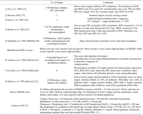
none phenomenon, but rather that a range of organ dys- function exists leading up to clinical failure [7]. It has been suggested that this continuous process of varying levels of organ function is designated “MODS” [8]. This change in the understanding of organ failure as a con- tinuous process has led to the development of a number of scoring systems that attempt to quantitate the degree of organ failure [9,10]. These variations in the definition of organ failure and the study of heterogeneous patient populations have made it difficult to establish the accu- rate incidence of organ failure in a given homogeneous population. Figure 2 shows the incidence of single and multiorgan failure (SOF and MOF) in various studies [11- 15].
5. Independent Predictors for MOF
For early risk stratification (12 hrs postinjury), several modifiable and non-modifiable predictors have been re- ported: high injury severity, amount of red blood cell transfusion, age of transfused products, age greater than 55 years old, high base deficit, uncorrected lactate at 12 - 24 hrs postinjury, obesity, male gender and abdominal compartment syndrome [1]. However, even during this early period, MOF already has been triggered and may
be inevitable [1,16].
6. Scoring and Criteria
[image:3.595.123.482.392.720.2]The pathophysiology of MODS is varying according to the mechanism of injury in the medical versus non-me- dical (trauma and surgical) ICUs. Certain criteria should be taken into consideration when assessing the value of any scoring system in clinical practice [17,18]. These criteria should include reliability, validity and the ability of a scoring system to unmask temporal changes in organ dysfunction if measured sequentially [18,19]. Studies evaluated the comparative prognostic value of the com- monly used organ dysfunction scoring systems concluded that some standardization of the included variables is needed before introducing a scoring tool in everyday prac- tice [20,21]. In the absence of a gold standard scoring or tool for diagnosis or prediction of MODS, validation is required to evaluate the association of different scores with objective, adverse outcomes, clinical status and re- source utilization. Table 1shows the diver sity of scor-ing systems for MOF [10,12,13,15-20]. Sauaia et al. [21] reported that both Denver and Marshall MOF scores perform reasonably well as indicators of unfavorable outcomes in critically ill patients; with the Denver MOF
Table 1. Different scoring systems for multiorgan failure.
N. of Organs Comments
1) Fry et al. 1980 [15] 4 (Pulmonary, hepatic, GIT, and renal)
One or more organs failed in 15% of patients. The incidence of SOF and MOF were 8.3% and 6.9% and Mortality rates were 30% for SOF, 60% for 2 organ, 85% for 3-system organ, and 100% for 4-OF. 2) Stevens 1983 [17] 7 Numeric Sepsis severity scoring system
3) Goris et al. 1985 [18] 7 Organ dysfunction/failure had 3 categories: (0 = normal,1 = organ dysfunction, 2 = OF)
4) Knaus et al. 1985 [13]
5 (CVS, respiratory, renal, hematologic, and neurological)
One or more OFs occurred in 49% of patients: SOF occurred in ≈1/3 of patients at some time during their ICU stay. MOF occurred in 15%. SOF lasting more than 3 days had mortality of 40%. Mortality was 60% for 2-OF and 100% for 3-OF.
5) Marshall et al. 1995 (MODS) [10]
6 (Pulmonary, renal, hepatic, cardiac, hematological and
neurological system)
Data collected from systematic review and clinical database
Marshall and SOFA scores Both scores are very sensitive but not specific: More sensitive score causes high incidence of MODS, while a more specific score causes high mortality.
6) Vincent et al. 1996 (SOFA) [19]
6 (Graded from 0 to 4 according to the degree of
dysfunction failure)
The score individualizes the degree
of dysfunction of each organ obtained daily(not to predict outcome but to describe a sequence of
complications).
7) Le Gall et al. 1996 (LODS) [20] 6
Physiological variables of 6 organ systems by using logistic regression: GCS, PaO2:FiO2 ratio, heart rate, BP, serum urea, creatinine, urine output, white blood cell, bilirubin, platelet count, and prothrombin. One or more organ systems failed in 25% of patients and 2 or more organ systems failed in 15% of patients. Mortality was 11% in patients with 1 organ system failure, 24% with 2 organ systems, 60% in patients with 3 failures, and 62% in patients with 4 OFs. 8) Moore et al. 1996 (Denver) [12] 4 (Pulmonary, renal, hepatic and cardiac)
Denver MODS score To define and monitor the severity of MODS in trauma with ISS > 15 who survived >48 hrs and who are 16 yrs or older. Must be collected daily (day 2 to discharge). It rates 4 organ systems; pulmonary, renal, hepatic and cardiac. It is more specific and less sensitive in trauma population.
Criteria of OF [7]
Cardiac: Heart rate ≤ 54 beats/min, Mean arterial pressure ≤ 49 mm Hg ventricular tachycardia, fibrillation, or both, Serum pH ≤ 7.24 with a PaCO2≤ 49 mm Hg.
Pulmonary : Respiratory rate ≤ 5 breaths/min or≥49 breaths/min, PaCO2≥ 50 mm Hg AaDO2 ≥ 350 mm Hg, Dependent on ventilator on the fourth day of organ failure. Renal: Urine ≤ 479 mL/24 h or ≤149 mL/8 h, BUN ≥ 100 mg/dL, Creatinine ≥ 3.5 mg/dL; Hepatic: Bilirubin ≥ 3 mg/dL Serum glutamic oxaloacetic transaminase greater than two times normal Protime INR >1.5.
GIT = gastrointestinal tract; SOF = single organ failure; MOF = multiorgan failure; CVS = cardiovascular system; ICU = intensive care unit; GCS = Glasgow coma scale; ≈ = approximately; OF = organ failure
scores performing slightly better due to greater specificity. Moreover, Denver MOF score performs better as a con- tinuous scale to monitor individual patient’s response to treatment. The analysis of individual organ dysfunction scores suggests that concepts of the two scores can be com- bined to develop more valid score [21].
However, what is validity and accuracy if the clinical scoring system is integrated into the biomarkers of organ dysfunction? This bio-clinical scoring has not tested yet.
7. MODS at Cellular Level
Numerous studies revealed that cells of the immune sys- tem (Polymorphic mononuclear [PMN], lymphocytes, mo- nocytes/macrophages, dendritic cells and endothelial cells) and the release of pro- and anti-inflammatory cytokines, chemokines, adhesion molecules, complement, protease, eicosanoids, reactive oxygen species (ROS) and nitric
oxide (NO) play a significant role in the pathogenesis of MOF. As these cells and molecules are released for pri- marily host defense, their release can be harmful to the host depending on the type and degree of injury, post- trauma surgery, intervention for diagnosis and therapy for trauma [22].
What is Systemic inflammatory response syndrome (SIRS)
Two or more of the following criteria are met:
—Temperature < 36.8˚C or temperature > 38.8˚C. – Heart rate > 90/min.
—Respiratory rate > 20 breaths/min or PCO2 < 32 mm
Hg.
—WBC < 4000/mL or WBC>12,000 mL or > 10% im- mature forms [1].
stimulus following prior exposure to a different stimulus. Clinically, priming is manifested by SIRS, characterized by alterations in body temperature, white blood cell count, respiratory dysfunction, and a hyperdynamic state. Primed PMNs cause a significant neutrophillia at 3 h postinjury. This neutrophillia represents the “vulnerable window” and a second hit during this period may precipitate MOF. In MOF there is a rapid neutropenia between 6 and 12 h postinjury (end organ sequestration). While in non-MOF, neutrophil priming and neutrophillia are not followed by neutropenia, and resolve over the next 36 h without end organ damage [1].
7. MODS at the Molecular Levels
Molecular events underlie the pathogenesis of MODS is not well-established, therefore, the temporal course of pathophysiological changes leading to the development of MODS are of great clinical and research interest. Keeping in mind “There is no gold standard diagnostic tool for MODS”, however, several studies have utilized one or two biomarkers for detection of single rather than multiple organ dysfunctions. General markers of inflam- mation including cytokines are correlated with posttrau- matic complications with a low sensitivity and specificity and are, therefore, of little utilization as prognostic mark- ers [23]. To date, all therapeutic strategies focused on a single mediator or receptor has failed to improve the clini- cal outcome associated with MODS [2]. However, a re- cent small study showed that cytokine expression du- ring shock may enable earlier identification of patients who are at risk for development of MODS [24].
There are only few registries and prospective studies that reported the prevalence of MODS worldwide [11,25]. Furthermore, there is no consensus to support the sensi- tivity and specificity, predictive values of one or more serum markers in the diagnosis or prediction of organ dysfunction before the overt clinical failure. Therapeutic strategies to combat the post injury MODS have focused on control of the post injury inflammatory response [25]. Early detection of MODS need extensive prospective studies as this will be reflected on the morbidity and mor- tality in all ICUs. For that purpose, ICUs should have prospective data collection on MODS in its database re- gistry. Subsequently, registries will pave ways for the ap- propriate evaluation and management.
8. Animal Models
Trauma, shock or sepsis, researchers studied the basic mechanisms that drive the pathogenesis of end-organ in- jury and MODS at the cellular, tissue and whole organ- ism levels. Previous studies demonstrated that endorgan injury and subsequent MODS result from a cause-effect relationship between three pathophysiologic events, which
likely interact in a time-dependent, tissue-specific fash- ion. First, a persistent and progressive splanchnic vaso- constriction and hypoperfusion leading to relative ische- mia/hypoxia [26,27]; Second, a gut-derived systemic in- flamematory response generated by the ischemic gut [28]; and third, inevitable fluid shifts at both the cellular level due to ionic-disequilibrium and at the capillary level due to alteration of the trans-capillary Starling forces that gov- ern fluid exchange [29,30].
9. Gut Hypothesis
Preclinical experimental studies showed that shock or trauma cause gut barrier failure and bacteria translocating to distant organs. Also, the subsequent gut released pro- inflammatory and tissue injurious factors could result in acute lung injury, bone marrow failure, myocardial dys- function, neutrophil activation, red blood cells injury and endothelial cell activation and injury. Interestingly, these factors are carried in the mesenteric lymphatics, but not in the portal blood flow and are sufficient to cause MODS [31].
10. The Trigger and Targeted Organ
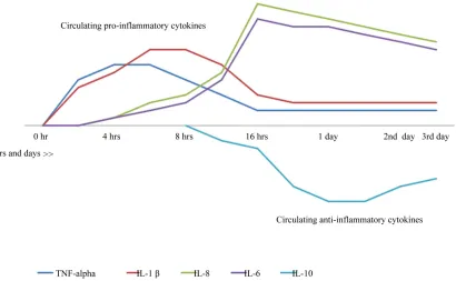
It has been shown that certain organs are more vulnerable and presage the chain or occurrence of MOD. There is no solid data showing whether the dysfunction will affect single organ or multiple organs. Post-traumatic lung dys- function precedes cardiac dysfunction by 0.6 days, he- patic dysfunction by 4.8 days and renal dysfunction by 5.5 days on average [1,4,5]. This early involvement of the lungs supports the contention of the time dependency and tissue (organ) specificity of SIRS. The mechanism of post-traumatic lung injury and dysfunction occur at least in part through mesenteric lymph-induced activation of neutrophils and activation/injury of endothelial cells. Sub- sequent infiltration of the tissue with activated neutron- phils is time dependent and organ specific [28]. In the shock lung, resuscitation from hemorrhagic shock sec- ondary to trauma increases the myeloperoxidase (MPO) level, an index of neutrophil tissue infiltration, in a near- linear fashion during the first 4 h following resuscitation. However, lung MPO level returned to baseline at 24 h following resuscitation from hemorrhagic shock [28]. This data suggest that a vulnerable window for neutron- phil- mediated lung damage exists during the first 4 h follow- ing resuscitation from hemorrhagic shock in rats [28]. Thus, demonstration of a time-dependency and organ-spe- cificity of the proposed composite biomarkers in a fash- ion similar to SIRS, adds more specificity and sensitivity to the composite biomarkers, which helps the develop- ment of specific intervention before the initiation of the pathogenesis of multi-system organ failure. Figure 3 shows the inflammatory response after trauma in hrs and days
Figure 3. Cytokines response after trauma in hrs and days.
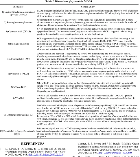
11. Role of Biomarkers
Different biomarkers [35-47] play various ways on the occurrence of organ dysfunction. Detailed information for the clinical utility of biomarkers is given in brief in Ta- ble 2. Certain markers have been studied frequently and prove important clinical impact on certain organs {i.e., lung (pentraxin-3 and IL-8 & 18), heart (NT-pro BNP, Adrenomedullin and pentraxin), kidney (NGAL) and liver (glutamate/glutamine and cytokeratin-18)} in addition to Vascular cell adhesion molecule (VCAM-1) and Endo- thelial cell-specific molecule 1 (ESM-1) [48]. Biomarkers for early detection of sepsis (i.e., procalcitonin) is also important. Several studies tackling proinflammtory (i.e., IL-8 &18) and anti-inflammatory (i.e., IL-6 &10) cyto- kines have shown that there is no single cytokine turned out to be legible enough to predict the outcome [35].
The cytokine response is an important factor in the development of SIRS as a response to trauma. Pro-and anti-inflammatory cytokines are released excessively du- ring the initial phase of trauma. The role of the anti-in- flammatory cytokines is to down-regulate the production of pro-inflammatory cytokines. Normally, the bala nce of pro- and anti-inflammatory cytokines is in equilibrium, however, when this natural balance is unbalanced with the release of predominantly pro-inflammatory cytokines, this leads to SIRS, while predominance of anti-inflam- matory cytokines causes immunosuppression, which lead to infection, sepsis and subsequently result in MOF [22]. The role of acute phase proteins: Levels of CRP rapidly
increase within 2 hours of acute insult, reaching a peak at 48 hours. With resolution of the acute phase response, CRP declines with a relatively short half-life of 18 hours. PCT is proposed to be a better and more specific marker for inflammation than CRP, as the kinetics of PCT more closely resembles the kinetics of inflammation [37,38]. Serum levels of PCT more rapidly increase after the on- set of inflammation and decline faster as inflammation di- minishes.
12. Recommendation and Conclusion
Bring up new bio-clinical scoring to a stage where it is ready for field trials will pave the way for implementing new risk-stratification strategy in the intensive care to re- duce the morbidity and mortality and save resources.
Further trials will be based on that to establish preven- tive measures and goal-targeted therapy in the ICUs. Un- derstanding the different pathphysiology of early MODS will discriminate the signs of intense inflammatory re- sponse from early signs of sepsis. This will reduce the inappropriate use of antibiotics or to correctly start anti- biotics at the early stages of sepsis and subsequently re- duce hospital stay and mortality. As there are no guide- lines or consensus for management of high-risk patients, further studies utilizing the integrated bio-clinical hy- pothesis will be of great interest. Further prospective stud- ies are needed to answer our question and convert MODS
Table 2. Biomarkers play a role in MODS.
Biomarker Clinical utility
1) Neutrophil gelatinase-associated lipocalin (NGAL)
NGAL is ideal biomarker for acute kodney injury (AKI); its concentration rapidly decreases with attenuation of renal injury; and it is readily and easily measured in plasma and urine. NGAL typically detected AKI 36 to 48 h earlier than creatinine.
2) Serum glutamate/glutamine
Glutamine itself may act as a key precursor for nucleic acids in glutamine consuming cells, but in many circumstances acts to provide glutamate, however, glutamate also serves as a precursor for the formation of glutamine.. Glutamate could be a marker of liver dysfunction during sepsis.
3) Cytokeratin-18 (CK-18)
CK-18 presents in most simple epithelial and parenchymal cells. Fragments of CK-18 are more specific for apoptotic cell death. The measurement of caspase-cleaved and uncleaved CK-18 appears to be an early predictor for survival in severe septic patients with hepatic dysfunction
4) Procalcitonin (PCT)
PCT supports early diagnosis and clinical decision making which could direct an effective therapy at the right time and avoid unnecessary spending for critically ill patients. Unlike CRP, PCT values typically declined rapidly after trauma. The rapid decline of the trauma-induced response of PCT towards its normal range compared with the long-lasting increase of CRP promises an earlier diagnostic use of PCT as a marker of sepsis and infection than of CRP. The PCT half life of about 22 hours
5) Adrenomedullin (AM)
AM production and secretion is augmented by several pro-inflammatory and pro-atherogenic factors. Circulating adrenomedullin levels are elevated in acute cardiac injury. plasma levels are markedly increased in early septic shock. Plasma AM and IL-8 levels correlated positively with (APACHE) II score, peak MODS score during the first month and prognosis in patients with septic shock, as did plasma IL-6 levels in patients with traumatic shock. Adrenomedullin has a circulating half life of 22 ± 1.6 min
6) Pentraxin-3 (PTX3)
PTX3 is a rapid marker for primary local activation of innate immunity and inflammation It is associated with acute lung and heart injury. PTX3 behaves as an acute phase response protein, as the blood levels of PTX3, low in normal conditions (<2 ng/mL in humans), increase rapidly (peaking at 6 - 8 h after induction) and dramatically (200 - 800 ng/mL) during endotoxic shock, sepsis and correlating with the severity of the disease.
7) NT-pro-BNP
NT-proBNP, is supposed to be a better marker of myocardial dysfunction and prognosis in patients with severe sepsis and septic shock. Its levels correlate with the severity of organ dysfunction as assessed by the SOFA score in septic patients. The half-life of human NT-proBNP is consideredto be 60 - 120 min (depending on renal function).
8) VCAM-1
Vascular cell adhesion protein 1 is also known as vascular cell adhesion molecule 1 .VCAM-1 protein mediates the adhesion of lymphocytes, monocytes, eosinophils, and basophils to vascular endothelium. It also functions in leukocyte-endothelial cell signal transduction.
9) Cytokines (interleukine; IL)
MODS is associated with higher levels of systemic proinflammatory cytokines(IL6, IL8 and IL10). Patients who develop late MODS show a second peak of IL6 at day 7, while in early MODS, IL6 returns to baseline levels by day 4. IL8 can activate PMNs via two different receptors. IL-8 is a chemokine and its production following trauma leads to leucocyte recruitment and activation at the site of injury.
In contrast to NT-proBNP and PCT,initial IL-6 are highly predictor of mortality after myocardial infarction with shock. Increased IL-6 is associated with narrowed macro-and microcirculation,a center pathomechanism for development of MODS. After trauma, the levels of IL-8 correlate with the subsequent development of ARDS and MODS.
10) Endothelial cell-specific molecule 1 (ESM-1)
Inflammatory cytokines such as TNF-α strongly stimulate human endothelial cell to increase the secretion, synthesis and expression of endocan. Studies agreed on the endocan’s prognostic value and how it could be of huge help to predict the outcome of sepsis. As its increase at ICU admission is indicative of poor prognosis.
REFERENCES
[1] D. Dewar, F. A. Moore, E. E. Moore and Z. Balogh, “Postinjury Multiple Organ Failure,” Injury, Vol. 40, No. 9, 2009, pp. 912-918. doi:10.1016/j.injury.2009.05.024 [2] B. Maier, R. Lefering, M. Lehnert, H. L. Laurer, W. I.
Steudel, E. A. Neugebauer and I. Marzi, “Early versus Late Onset of Multiple Organ Failure Is Associated with Dif- fering Patterns of Plasma Cytokine Biomarker Expression and Outcome after Severe Trauma,” Shock, Vol. 28, No. 6, 2007, pp. 668-674.
[3] D. Dewar, N. Butcher, K. King and Z. Balogh, “Post Injury Multiple Organ Failure,” Trauma, Vol. 13, No. 1, 2011, pp. 81-91.
[4] D. J. Ciesla, E. E. Moore, J. L. Johnson, A. Sauaia, C. C.
Cothren, J. B. Moore and J. M. Burch, “Multiple Organ Dysfunction during Resuscitation Is Not Postinjury Mul- tiple Organ Failure,” Archives of Surgery, Vol. 139, No. 6, 2004, pp. 590-594. doi:10.1001/archsurg.139.6.590 [5] D. J. Ciesla, E. E. Moore, J. L. Johnson, et al., “The Role
of the Lung in Postinjury Multiple Organ Failure,” Sur-gery, Vol. 138, No. 4, 2005, pp. 749-757.
doi:10.1016/j.surg.2005.07.020
[6] F. A. Moore, A. Sauaia, E. E. Moore, et al., “Postinjury Multiple Organ Failure: A Bimodal Phenomenon,” Jour- nal of Trauma, Vol. 40, No. 4, 1996, pp. 501-502. doi:10.1097/00005373-199604000-00001
Trauma Patients,” Journal of Trauma, Vol. 55, No. 4, 2003, pp. 608-616.
doi:10.1097/01.TA.0000092378.10660.D1
[8] R. Bone, R. Balk, F. Cerra, et al., “Definitions for Sepsis and Organ Failure and Guidelines for the Use of Innova-tive Therapies in Sepsis: The ACCP/SCCM Consensus Conference Committee—American College of Chest Phy- sicians/Society of Critical Care Medicine,” Chest, Vol. 101, No. 6, 1992, pp. 1644-1655.
[9] J. Marshall, “A Scoring System for the Multiple Organ Dysfunction Syndrome (MODS),” In: K. Rheinhart, K. Ey- rich and C. Sprung, Eds., Sepsis: Current Perspectives in Pathophysiology and Therapy, Springer-Verlag, Berlin, 1994, pp. 38-49.
[10] J. Marshall, D. Cook, N. Christou, G. Bernard, C. Sprung and W. Sibbald, “Multiple Organ Dysfunction Score: A Reliable Descriptor of a Complex Clinical Outcome,” Cri- tical Care Medicine, Vol. 23, No. 10, 1995, pp. 1638-1652. [11] D. J. Ciesla, E. E. Moore, J. L. Johnson, J. M. Burch, C. C.
Cothren and A. Sauaia, “A 12-Year Prospective Study of Postinjury Multiple Organ Failure: Has Anything Changed?” Archives of Surgery, Vol. 140, No. 5, 2005, pp. 432-438. doi:10.1001/archsurg.140.5.432
[12] F. A. Moore, A. Sauaia, E. E. Moore, et al., “Postinjury Multiple Organ Failure: A Bimodal Phenomenon,” Jour- nal of Trauma, Vol. 40, No. 4, 1996, pp. 501-502. doi:10.1097/00005373-199604000-00001
[13] W. A. Knaus, E. A. Draper, D. P. Wagner and J. E. Zimmer- man, “Prognosis in Acute Organ System Failure,” Annals of Surgery, Vol. 202, No. 6, 1985, pp. 685-693.
doi:10.1097/00000658-198512000-00004
[14] E. Faist, A. E. Baue, H. Dittmer, G. Heberer, “Multiple Organ Failure in Polytrauma Patients,” Journal of Trauma, Vol. 23, No. 9, 1983, pp. 775-787.
doi:10.1097/00005373-198309000-00002
[15] D. E. Fry, L. Pearlstein, R. L. Fulton and H. C. Polk, “Multi- ple System Organ Failure: The Role of Uncontrolled In- fection,” Archives of Surgery, Vol. 115, No. 2, 1980, pp. 136-140.
[16] H. G. Cryer, K. Leong, D. L. McArthur, et al., “Multiple Organ Failure: By the Time You Predict It, It’s Already There,” Journal of Trauma, Vol. 46, No. 4, 1999, pp. 597- 604. doi:10.1097/00005373-199904000-00007
[17] L. E. Stevens, “Gauging the Severity of Surgical Sepsis,” Archives of Surgery, Vol. 118, No. 10, 1983, pp. 1190- 1192. doi:10.1001/archsurg.1983.01390100060015 [18] R. J. Goris, T. P. te Boekhorst, J. K. Nuytinck and J. S. Gim-
bree, “Multiple-Organ Failure: Generalized Autodestruc- tive Inflammation?” Archives of Surgery, Vol. 120, No. 10, 1985, pp. 1109-1115.
doi:10.1001/archsurg.1985.01390340007001
[19] J. L. Vincent, R. Moreno, J. Takala, et al., “The SOFA (Sepsis-related Organ Failure Assessment) Score to De- scribe Organ Dysfunction/Failure,” Intensive Care Medi- cine, Vol. 22, No. 7, 1996, pp. 707-710.
doi:10.1007/BF01709751
[20] J. R. Le Gall, J. Klar, S. Lemeshow, et al., “ICU Scoring Group. The Logistic Organ Dysfunction System: A New Way to Assess Organ Dysfunction in the Intensive Care
Unit,” Journal of the American Medical Association, Vol. 276, No. 10, 1996, pp. 802-810.
doi:10.1001/jama.1996.03540100046027
[21] A. Sauaia, E. E. Moore, J. L. Johnson, D. J. Ciesla, W. L. Biffl and A. Banerjee, “Validation of Postinjury Multiple Organ Failure Scores,” Shock, Vol. 31, No. 5, 2009, pp. 438-447. doi:10.1097/SHK.0b013e31818ba4c6
[22] T. Tsukamoto, R. S. Chanthaphavong and H. C. Pape, “Cur- rent Theories on the Pathophysiology of Multiple Organ Failure after Trauma,” Injury, Vol. 41, No. 1, 2010, pp. 21-26. doi:10.1016/j.injury.2009.07.010
[23] S. Oda, H. Hirasawa, T. Sugai, et al., “Comparison of Sepsis Related Organ Failure Assessment (SOFA) Score and CIS (Cellular Injury Score) for Scoring of Severity for Patients with Multiple Organ Dysfunction Syndrome (MODS),” Intensive Care Medicine, Vol. 26, No. 12, 2000, pp. 1786-1793. doi:10.1007/s001340000710
[24] V. Pettila, M. Pettila, S. Sarna, P. Voutilainen and O. Tak- kunen, “Comparison of Multiple Organ Dysfunction Scores in the Prediction of Hospital Mortality in the Critically Ill,” Critical Care Medicine, Vol. 30, No. 8, 2002, pp. 1705-1711. doi:10.1097/00003246-200208000-00005 [25] D. C. Dewar, P. Mackay and Z. Balogh, “Epidemiology
of Post-Injury Multiple Organ Failure in an Australian Trauma System,” ANZ Journal of Surgery, Vol. 79, No. 6, 2009, pp. 431-436.
doi:10.1111/j.1445-2197.2009.04968.x
[26] E. R. Zakaria, R. N. Garrison, D. A. Spain, P. J. Mathe-son, P. D. Harris and D. J. RichardMathe-son, “Intraperitoneal Resuscitation Improves Intestinal Blood Flow Following Hemorrhagic Shock,” Annals of Surgery, Vol. 237, No. 5, 2003, pp. 704-713.
doi:10.1097/01.SLA.0000064660.10461.9D
[27] E. R. Zakaria, R. T. Hurt, P. J. Matheson and R. N. Gar- rison, “A Novel Method of Peritoneal Resuscitation Im- proves Organ Perfusion after Hemorrhagic Shock,” Amer- ican Journal of Surgery, Vol. 186, No. 5, 2003, pp. 443- 448. doi:10.1016/j.amjsurg.2003.07.006
[28] E. R. Zakaria, J. E. Campbell, J. C. Peyton and R. N. Garrison, “Post-Resuscitation Tissue Neutrophil Infiltra- tion Is Time-Dependent and Organ-Specific,” Journal of Surgical Research, Vol. 143, No. 1, 2007, pp. 119-125. doi:10.1016/j.jss.2007.04.008
[29] E. R. Zakaria, P. J. Matheson, M. F. Flessner and R. N. Garrison, “Hemorrhagic Shock and Resuscitation-Medi- ated Tissue Water Distribution Is Normalized by Adjunc-tive Peritoneal Resuscitation,” Journal of the American College of Surgeons, Vol. 206, No. 5, 2008, pp. 970-980. doi:10.1016/j.jamcollsurg.2007.12.035
[30] E. R. Zakaria, N. Li, P. J. Matheson and R. N. Garrison, “Cellular Edema Regulates Tissue Capillary Perfusion Following Hemorrhage Resuscitation,” Surgery, Vol. 142, No. 4, 2007, pp. 487-496. doi:10.1016/j.surg.2007.08.007 [31] E. A. Deitch, “Gut-Origin Sepsis: Evolution of a Concept,”
The Surgeon, Vol. 10, No. 6, 2012, pp. 350-356. doi:10.1016/j.surge.2012.03.003
pp. 97-102.
[33] R. M. Roumen, H. Redl and G. Schlag, “Inflammatory Mediators in Relation to the Development of Multiple Organ Failure in Patients after Severe Blunt Trauma,” Cri- tical Care Medicine, Vol. 23, No. 3, 1995, pp. 474-480. [34] P. V. Giannoudis, “Current Concepts of the Inflammatory
Response after Major Trauma: An Update,” Injury, Vol. 34, No. 6, 2003, pp. 397-404.
[35] T. Visser, J. Pillay, L. Koenderman and L. P. Leenen, “Postinjury Immune Monitoring: Can Multiple Organ Fail- ure Be Predicted?” Current Opinion in Critical Care, Vol. 14, No. 6, 2008, pp. 666-672.
doi:10.1097/MCC.0b013e3283196522
[36] A. M. Ferreira and Y. Sakr, “Organ Dysfunction: General Approach, Epidemiology, and Organ Failure Scores,” Se- minars in Respiratory and Critical Care Medicine, Vol. 32, No. 5, 2011, pp. 543-551.
doi:10.1055/s-0031-1287862
[37] R. M. Dorizzi, E. Polati and P. Sette, “Procalcitonin in the Diagnosis of Inflammation in Intensive Care Units,” Clini- cal Biochemistry, Vol. 39, No. 12, 2006, pp. 1138-1143. [38] A. Luzzani, E. Polati and R. Dorizzi, “Comparison of Pro-
calcitonin and C-Reactive Protein as Markers of Sepsis,” Critical Care Medicine, Vol. 31, No. 6, 2003, pp. 1737- 1741. doi:10.1097/01.CCM.0000063440.19188.ED [39] M. Krueger, A. Heinzmann and M. Nauck, “Adhesion
Molecules in Pediatric Intensive Care Patients with Organ Dysfunction Syndrome,” Intensive Care Medicine, Vol. 33, No. 2, 2007, pp. 359-363.
doi:10.1007/s00134-006-0453-6
[40] S. Hofer, T. Brenner, C. Bopp, J. Steppan, C. Lichten-stern, J. Weitz, T. Bruckner, E. Martin, U. Hoffmann and M. A. Weigand, “Cell Death Serum Biomarkers Are Ear-ly Predictors for Survival in Severe Septic Patients with Hepatic Dysfunction,” Critical Care, Vol. 13, No. 3, 2009, pp. R93. doi:10.1186/cc7923
[41] N. Kume, H. Mitsuoka, K. Hayashida and M. Tanaka, “Pen- traxin 3 as a Biomarker for Acute Coronary Syndrome: Com- parison with Biomarkers for Cardiac Damage,”Journal of
Cardiology, Vol. 58, No. 1, 2011, pp. 38-45. doi:10.1016/j.jjcc.2011.03.006
[42] X. He, B. Han, X. Bai, Y. Zhang, M. Cypel, M. Mura, S. Keshavjee and M. Liu, “PTX3 as a Potential Biomarker of Acute Lung Injury: Supporting Evidence from Animal Experimentation,” Intensive Care Medicine, Vol. 36, No. 2, 2010, pp. 356-364. doi:10.1007/s00134-009-1720-0 [43] M. Haase, P. Devarajan, A. Haase-Fielitz, et al., “The
Outcome of Neutrophil Gelatinase-Associated Lipocalin- Positive Subclinical Acute Kidney Injury: A Multicenter Pooled Analysis of Prospective Studies,” Journal of the American College of Cardiology, Vol. 57, No. 17, 2011, pp. 1752-1761. doi:10.1016/j.jacc.2010.11.051
[44] I. T. Klip, A. A. Voors, S. D. Anker, H. L. Hillege, et al., “OPTIMAAL Investigators Prognostic Value of Mid-Re- gional Pro-Adrenomedullin in Patients with Heart Failure after an Acute Myocardial Infarction,” Heart, Vol. 97, No. 11, 2011, pp. 892-898.
[45] C. Kirchhoff, B. A. Leidel, S. Kirchhoff, V. Braunstein, V. Bogner, U. Kreimeier, W. Mutschler and P. Biber- thaler, “Analysis of N-Terminal Pro-B-Type Natriuretic Peptide and Cardiac Index in Multiple Injured Patients: A Prospective Cohort Study,” Critical Care, Vol. 12, No. 5, 2008, p. R118. doi:10.1186/cc7013
[46] M. Poeze, Y. C. Luiking, P. Breedveld, S. Manders and N. E. Deutz, “Decreased Plasma Glutamate in Early Phases of Septic Shock with Acute Liver Dysfunction Is an In- dependent Predictor of Survival,” Clinical Nutrition, Vol. 27, No. 4, 2008, pp. 523-530.
doi:10.1016/j.clnu.2008.04.006
[47] S. Hofer, T. Brenner, C. Bopp, et al., “Cell Death Serum Biomarkers Are Early Predictors for Survival in Severe Septic Patients with Hepatic Dysfunction,” Critical Care, Vol. 13, No. 3, 2009, p. R93. doi:10.1186/cc7923
[48] P. Paulus, C. Jennewein and K. Zacharowski, “Biomarkers of Endothelial Dysfunction: Can They Help Us Deci- phering Systemic Inflammation and Sepsis?” Biomarkers, Vol. 16, Suppl. 1, 2011, pp. S11-S21.