EFFECTIVENESS OF LOCKING VERSUS DYNAMIC COMPRESSION
PLATES FOR DIAPHYSEAL FOREARM FRACTURES
*Imad Hasan Qasim
[M.B.Ch.B, F.I.C.M.S], Head of Orthopaedic Department at Al Wasity Teaching hospital
Baghdad, Iraq.
ABSTRACT
This study compares the results of the locking compression plate
(LCP) and the dynamic compression plate (DCP) in the treatment of
diaphyseal forearm fractures in adults and defines the indications for
the use of the LCP. Forty-two patients with diaphyseal forearm
fractures were retrospectively analyzed. Of those, 22 had been treated
with the LCP (LCP group) and 20 had been treated with the DCP
(DCP group). The AO/ASIF classification was used to classify the
fractures. Patients were assessed using the Grace-Eversmann criteria
and the Disabilities of the Arm and Shoulder and Hand questionnaire
during the final follow-up. Mean follow-up was 21 months (range,
20-24 months) in the LCP group and 23 months (range, 19-26 months) in
the DCP group. Union was achieved in all patients. Mean time to union
was 15 weeks (range, 12-25 weeks) in the LCP group and 17 weeks (range, 13-24 weeks) in
the DCP group. In each group, 1 patient experienced delayed union, which required no
additional surgical intervention. No significant difference was found regarding the time to
union between the groups (P > .05). No significant difference existed between the 2 groups in
terms of Grace-Eversmann criteria and Disabilities of the Arm and Shoulder and Hand scores
(both P > .05). The results of these different fixation methods for forearm fractures in adults are similar. As such, the correct selection and application of surgical technique is more
important than the type of plate used.
Volume 6, Issue 5, 64-80. Research Article ISSN 2277– 7105
*Corresponding Author
Dr. Imad Hasan Qasim
[M.B.Ch.B, F.I.C.M.S],
Head of Orthopaedic
Department at Al Wasity
Teaching hospital Baghdad,
Iraq.
Article Received on 01 March 2017,
Revised on 21 March 2017, Accepted on 11 April 2017
A
B
Figure: Preoperative lateral radiograph of the forearm showing fracture of both bones
(A). Lateral radiographs taken 10 months after locking compression plate fixation
showing fracture union (B).
In the treatment of adult forearm fractures, the main goals are to reestablish forearm length,
restore axial and rotational alignment and maintain them until union is achieved.[1] Open
reduction and internal fixation with the use of a dynamic compression plate (DCP) is a
Cortical porosis and refractures were considered secondary to excessive plate– bone contact
in DCPs that interfered with cortical perfusion.[4] According to this theory, the limited contact
DCP (LC-DCP) was developed to reduce the plate’s interference with cortical perfusion, thus
decreasing cortical porosis.[5] The subsequent development of the point contact fixator
reduced plate–bone contact to the point that it was essentially negligible.[6]
Recently developed locking compression plates (LCPs) combine the properties of both
locking plates and DCPs.[7] With their combined hole, an unlocked compression screw and a
locking screw can be used.[7] Locking compression plates have been shown to provide a
stronger fixation compared with DCPs in biomechanical studies.[8] In addition, LCPs can be
placed using a bridging plate technique, allowing biological fixation for the treatment of
comminuted fractures.[7] These advantages of the LCP have been considered to accelerate
fracture healing and reduce the problems of delayed union and nonunion.[8]
However, LCPs have some disadvantages, including difficulties during removal and a higher
cost.[9] A limited number of studies compare LCPs with conventional plates and have
reported similar results with both implants in the treatment of diaphyseal forearm
fractures.[10,11] Although LCPs have some theoretical advantages, the superiority of the LCP
over conventional plates remains to be proven.
The goal of the current study was to compare the results of LCPs and DCPs in the treatment
of diaphyseal forearm fractures in adults and to define the indications for the use of LCPs.
MATERIALS AND METHODS
A total of 51 patients treated with the DCP or LCP due to forearm fractures between 2008
and 2010 and 42 patients who had adequate follow-up were included in the study. Of those,
22 patients (17 men and 5 women; mean age, 28 years [range, 16-74 years]) were treated with
the LCP (LCP group) and 20 patients (14 men and 6 women; mean age, 32 years [range,
16-69 years]) were treated with the DCP (DCP group). The AO/ASIF classification was used for
fracture classification (Table 1).[12] The distribution of patients according to the fractured
bone is shown in Table 2. In the LCP group, 2 patients had Gustilo-Anderson grade 1
fractures and in the DCP group, 1 patient had a Gustilo-Anderson grade 2 open fracture.[13]
Patients with pathologic fractures, rheumatoid arthritis treated with corticosteroids for a long
period of time, functional loss prior to the fracture, and inadequate follow-up were excluded
In the LCP group, 16 (73%).
Table 1: Distribution of Fractures According to AO/ASIF Classification
AO/ASIF Classification LCP Group DCP Group
A1 4 3
A2 2 2
A3 8 6
B1 1 1
B2 1 1
B3 3 5
C1 2 2
C2 1 0
C3 0 0
Total 22 20
Abbreviations: DCP, dynamic compression plate; LCP, locking compression plate.
Table 2: Distribution of Patients According to the Fractured Bone
Fractured Bone LCP Group DCP Group
Radius 4 2
Ulna 5 6
Radius & ulna 13 12
Total 22 20
Abbreviations: DCP, dynamic compression plate; LCP, locking compression plate
In the LCP group, 16 (73%) patients experienced low-energy trauma and 6 (27%)
experienced high-energy trauma. In the DCP group, 13 (65%) patients experienced
low-energy trauma and 7 (35%) experienced high-low-energy trauma. In the LCP group, 19 (86%)
patients had an isolated forearm fracture and 3 (14%) had multiple fractures. In the DCP
group, 16 (80%) patients had an isolated forearm fracture and 4 (20%) had multiple fractures.
All patients were administered 1 g of a first-generation cephalosporin 30 minutes
preoperatively as a prophylactic measure (continued for 24 hours). Average time from injury
to surgery was 3 days (range, 1-8 days) in the LCP group and 4 days (range, 1-6 days) in the
DCP group.
Surgical Technique
A volar Henry incision was used for radius fractures of the third middle and distal region and
a dorsal Thompson incision was used for the proximal third region. The ulna fractures were
approached through an incision of the subcutaneous border. Care was taken to not damage
To determine the length and alignment of both bone forearm fractures, the simple fractures
were stabilized first. In the absence of specific references to the rotation, the fracture was
reduced temporarily and the rotation was controlled after reduction of the other fracture. In
the simple transverse fractures treated with the LCP, the locking screw was placed in the
other holes after the application of compression with at least 1 unlocked screw proximal or
distal to the fracture line. In the oblique fractures in which the LCP was used, the locking
screw was placed in the other holes after obtaining compression on the fracture line with the
interfragmenter lag screw technique using the hole on the plate. In the comminuted fractures
in which the LCP was used, fixation was provided by locking screws after obtaining the
length and alignment by selecting the appropriate length of plate using the bridging
technique. In patients who underwent fracture fixation using the DCP, 2 compression screws
were inserted proximal and distal to the fracture line after fracture reduction, followed by the
insertion of other screws.
In all patients, 3.5-mm LCP or DCP plates were used. The authors attempted to provide
fixation with a total of 3 screws (6 cortex) on both sides of the fracture line. After the
tourniquet was removed and hemostasis was obtained, a hemovac drain was placed and the
wound was closed. A splint was applied for 2 weeks. Elbow and wrist exercises were started
immediately while the arm was still in the splint.
The patients were followed up with radiographic studies once per month until fracture union
was achieved. The quality of fracture reduction was evaluated according to the criteria
proposed by Leung and Chow.[14] According to this definition, anatomic reduction refers to
full reduction in which the complete length and alignment are achieved and the compression
of the butterfly fragments is provided by means of the lag screws. Nonanatomic reduction
refers to an incomplete anatomic reduction in which the length and alignment of the fracture
fragments are achieved.
Bone union was defined as the presence of bridging the periosteal callus in 3 or 4 cortexes or
the primary closure fracture line on anteroposterior and lateral radiographs.[15] According to
the criteria proposed by Anderson et al,[1] fracture healing in less than 6 months was
considered union, fracture healing lasting longer than 6 months without the need for
additional surgical intervention was considered delayed union and the absence of fracture
healing requiring additional surgery was considered nonunion. Callus formation on the
For functional evaluation, the system described by Grace and Eversmann[2] was used.
Accordingly, complete fracture union with at least 90% rotation of the forearm was
considered a perfect result, complete fracture union with at least 80% forearm rotation was
considered a good result, complete fracture union with at least 60% forearm rotation was
considered an acceptable result, and nonunion of the fracture with a forearm rotation less than
60% was considered a poor result.
Patient satisfaction was assessed using the Disabilities of the Arm, Shoulder and Hand
(DASH) questionnaire.[16] In this scoring system, which is used to indicate the functional
status of the upper extremity, 0 reflects an excellent extremity and 100 reflects a
nonfunctional extremity.
Data were analyzed using SPSS version 16 statistical software (SPSS Inc, Chicago, Illinois).
Mann-Whitney U test was performed. A P value less than .05 was considered statistically significant.
RESULTS
Union was achieved in all patients in both groups. Mean follow-up was 21 months (range,
20-24 months) in the LCP group and 23 months (range, 19-26 months) in the DCP group.
Mean operative time was 76 minutes (range, 62-98 minutes) in the LCP group and 88
min-utes (range, 58-102 minmin-utes) in the DCP group. Mean time to union was 15 weeks (range,
12-25 weeks) in the LCP group (Figure 1) and 17 weeks (range, 13-24 weeks) in the DCP group
(Figure 2). No difference existed between the 2 groups in terms of operative time and time to
union (both P > .05).
1B
1D
Figure 1: Preoperative anteroposterior (A) and lateral (B) radiographs of the forearm
showing fracture of both bones. Anteroposterior (C) and lateral (D) radiographs taken
10 months after locking compression plate fixation showing fracture union.
[image:8.595.199.398.70.372.2]2B
2C
2D
[image:9.595.115.473.68.709.2]Anatomical fracture reduction was achieved in 22 (63%) patients in the LCP group and 26
(79%) patients in the DCP group. In the remaining cases, fracture fixation was obtained with
nonanatomic reduction. No significant difference existed in terms of time to union between
the 2 groups (P > .05).
In the LCP group, of the 19 fractures without an anatomical reduction, callus formation was
seen in 15 (79%) patients. In the DCP group, of the 6 fractures without an anatomical
reduction, callus formation was seen in 4 (67%) patients. In the LCP group, of the 16
fractures with an anatomical reduction, callus formation was seen in 2 (13%) patients. In the
DCP group, of the 26 fractures without an anatomical reduction, callus formation was seen in
2 (8%) patients. In both groups, a significant difference existed between the subgroups with
and without anatomic reduction in terms of callus formation (P5.003 and .004, respectively).
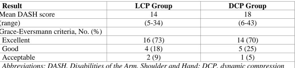
According to the Grace-Eversmann criteria, in the LCP group, 16 patients had excellent
results, 4 patients had good results, and 2 patients had acceptable results. In the DCP group,
14 patients had excellent results, 5 patients had good results and 1 patient had acceptable
re-sults (Table 3). Mean DASH score was 14 (range, 5-34) in the LCP group and 18 (range,
6-43) in the DCP group. No significant difference existed between the 2 groups in terms of
[image:10.595.58.540.484.595.2]Grace-Eversmann criteria and DASH scores (both P > .05).
Table 3: Functional Results
Result LCP Group DCP Group
Mean DASH score 14 18
(range) (5-34) (6-43)
Grace-Eversmann criteria, No. (%)
Excellent 16 (73) 14 (70)
Good 4 (18) 5 (25)
Acceptable 2 (9) 1 (5)
Abbreviations: DASH, Disabilities of the Arm, Shoulder and Hand; DCP, dynamic compression plate; LCP, locking compression plate
The plates that caused discomfort under the skin were removed 18 months postoperatively. In
the LCP group, plates and screws were removed in 6 patients. During removal in 2 patients,
cold fusion was observed between the locked screw head and the plate. Because the screws
could not be separated from the plate, the plate was left in situ. In the DCP group, the
implants were removed in 10 patients with no difficulties. No patient in either group
No patient had deep infection, synostosis, plate fracture, compartment syndrome, or
iatrogenic neurological or vascular damage. One patient in each group had delayed union;
both had open fractures. However, the fractures healed with no additional surgical
procedures. Superficial infection occurred in 2 patients in the LCP group and 1 patient in the
DCP group; all recovered with local wound care and antibiotic treatment.
DISCUSSION
This study compared LCPs and DCPs for the treatment of diaphyseal forearm fractures in
adults. Similar results were obtained in terms of operative time, time to union, complications,
and range of motion.
Anatomic reduction and stable internal fixation are effective methods for achieving optimal
functional results for diaphyseal forearm fractures in adults.[1-3] Union rates ranging from
92% to 98% after treatment with a DCP have been reported in many studies.[1,2,15] However,
cortical porosis and refracture observed in the DCP design were attributed to decreased blood
flow due to increased friction and intimate contact between the plate and bone[4]
Development of the LC-DCP and point contact fixator was based on the theory that, if
friction and intimate contact between plate and bone reduces, vascular damage, osteonecrosis
and cortical porosis beneath the plate will be reduced.[5,6] Jain et al[17] measured cortical blood
flow of canine tibias fixed with the LC-DCP or DCP and found no significant difference in
cortical blood flow between the groups. Uthoff et al[18] found no positive correlation between
necrosis and porosis, and they explained porosis by stress shielding. Moreover, the reduced
contact area (LC-DCP) and limited unicortical fixation without compression (point contact
fixator) failed to improve the clinical outcome in diaphyseal forearm fractures.[19]
The recently developed LCP has properties of the DCP and point contact fixator, which
provide locking and compression via its combined hole.[7] The locking compression plate acts
as a fixed-angle device and provides stronger fixation than conventional plates.[20] In
addition, the LCP can be used as a bridging plate, allowing biological fracture fixation in
comminuted fractures.[20] In this way, it provides relative fixation, facilitating callus
forma-tion and secondary fracture healing.[21] Due to these advantages, the LCP has recently began
to replace conventional plates in the treatment of patients with osteoporosis, fractures close to
Diaphyseal fractures of the forearm are commonly encountered in the daily practice of
orthopedic surgeons. With the theoretical advantages and the expansion of indications for
locked plate systems, the question remains whether LCPs are more effective than
conventional plates in the treatment of forearm fractures.[10,11] Although LCPs are
increasingly preferred in the treatment of forearm fractures, a limited number of studies
compare the effectiveness of LCPs and conventional plates.[10,11]
In a series of 36 patients with forearm fractures, Saikia et al[11] compared LCPs and LC-DCPs
(18 patients in each group). All patients achieved union, which occurred at an average of 16
weeks in the LCP group and 14 weeks in the LC-DCP group. One case of delayed union
oc-curred in the LC-DCP group, and 1 case of synostosis and 1 case of osteomyelitis ococ-curred in
the LCP group. The authors concluded that the LCP and LC-DCP provided similar results.[11]
In 32 patients (45 fractures) with forearm fractures who were treated with LCPs, Leung and
Chow[14] reported that union was achieved at an average of 20 weeks. However, they
encountered delayed union in 2 (6.3%) cases. Anatomic reduction was obtained in 33% of
pa-tients. No or minimal callus formation occurred in 56% of patients, and moderate callus
formation occurred in 44% of patients. The authors reported that LCP was effective as a
bridging device in the treatment of comminuted diaphyseal forearm fractures.[14]
In the current study, mean time to union was 15 weeks in the LCP group and 17 weeks in the
DCP group. These results are comparable with those in the literature.[11,14] Callus formation
was significantly higher in the LCP group than in the DCP group. This finding supports
previous studies.[11,14] Furthermore, in both groups, callus formation was more prominent
when anatomic reduction was not achieved. Nonanatomic reduction (especially in
comminuted fractures) was obtained using the bridging technique, in which the fracture was
not completely separated from the soft tissues to keep the blood supply intact. This technique
provided callus formation in both groups.
Stevens and ten Duis[10] compared DCPs and LCPs in the treatment of type A diaphyseal
forearm fractures. Union was obtained in all cases. However, time to union was an average of
10 weeks shorter when compression was applied in both groups. The authors concluded that
time to union is determined by the axial compression applied to the fracture line rather than
by the type of plate.[10]
Simple transverse or short oblique fractures require maximum compression for optimal
healing. Because of the combined hole in the LCP, one can apply compression at the fracture
surfaces using screws through the plate eccentrically. For this reason, the current authors
applied compression using a combined hole in the LCP group when possible. After
achieve-ment of compression at the fracture surface, locking screws were added to the fixation with
the purpose of neutralization. However, the number of locking screws necessary for
additional fixation remains to be evaluated.
The risk of refracture after plate removal has been reported to range from 4% and 22%.[1,11,23]
Leung and Chow[14] reported 2 (9%) cases of refracture in patients treated with the LCP
during removal at approximately 12 months. These were both simple transverse fractures and
healed primarily without callus formation. The authors recommended the removal of the plate
after 18 months to reduce the risk of refracture.[14]
Problems encountered during locked plate removal have been increasingly reported. In a
series of 43 patients with forearm fractures, Henle et al[9] reported cold fusion of the plates
with at least 1 screw head in 6 of 10 patients in whom material removal was attempted. They
also reported an ulnar fracture during removal. It was necessary for the authors to use special
devices to remove these screws. The authors proposed manual tightening of screws to avoid
cold fusion.[9]
In the current study, the authors did not remove the plates routinely due to the risk of
refracture. However, the plates that caused discomfort under the skin were removed 18
months postoperatively. In the LCP group, of the 8 patients with an indication for removal of
the plates, 2 patients experienced cold fusion of the locked screw head and the plate.
There-fore, the plate was left in place in these 2 patients. However, in the DCP group, the implants
were removed in 10 patients (14 fractures) with no difficulties. The authors concur with
Henle et al[9] with regard to hand-tightening the locked screws. Furthermore, patients who
want to have the locking plate removed should be informed preoperatively that not every
plate can be removed. However, during the removal procedure, a special screw removal kit
should be available.
In a radius cadaver model, Gardner et al[24] reported that the biomechanical properties of
LCPs and DCPs are similar. However, in a biomechanical study of an osteoporotic bone
axial compression test compared with conventional plates. In addition, a biomechanical study
of an osteoporotic diaphyseal fracture model by Doornink et al[26] demonstrated that hybrid
plates delivered higher torsional strength, similar bending strength, and a minimal decrease in
axial strength than all locked plates.
Experimental studies have reported that LCPs are superior to DCPs and all locked plates in
osteoporotic fracture models.[25,26] However, clinical trials have failed to demonstrate the
superiority of LCPs over conventional plates in forearm fractures.[10,11] The series reported by
Stevens and ten Duis[10] consisted of only A-type simple fractures. Similarly, in a study by
Saikia et al,[11] most fractures were type A and B, and only 5% were type C. Although the
type C fracture rate in the current study was slightly higher (9% and 6% in the LCP and DCP
groups, respectively) than in the study by Saikia at al,[11] it was not sufficient to compare the
efficacy of the LCP and DCP in type C fractures. Therefore, future studies are needed to
compare the LCP and DCP to determine their efficacy in comminuted or osteoporotic
fractures.
Increasing medical care costs is an important problem affecting patients and the health care
budgets of many countries. Locking compression plates are 3 times more expensive than
DCPs. The findings of the current study and others in the literature demonstrate that the
DCPs provide similar results to the LCPs in type A and B diaphyseal forearm
fractures.[10,11,14] Therefore, using DCPs in these fracture types seems to be more reasonable.
This study has several limitations. First, it is retrospective and comprises a relatively small
number of patients. Second, a comparison could not be made between subgroups of the
diaphyseal forearm fractures (eg, comminuted or osteoporotic fractures). Future studies
con-sisting of homogeneous subgroup types with similar degrees of osteoporotic bone may reveal
more accurate results for the indications and effectiveness of LCPs and DCPs.
CONCLUSION
Locking compression plates and DCPs have similar functional and radiological results in the
treatment of adult diaphyseal forearm fractures. Therefore, the authors believe that the correct
surgical technique is more important than the plate type in such fractures. Future studies with
REFERENCES
1. Anderson LD, Sisk D, Tooms RE, Park WI III. Compression-plate fixation in acute
di-aphyseal fractures of the radius and the ulna. J Bone Joint Surg Am. 1975; 57(3): 287-297.
2. Grace TG, Eversmann WW Jr. Forearm fractures: treatment by rigid fixation with early
motion. J Bone Joint Surg Am. 1980; 62(3): 433-438.
3. Chapman MW, Gorden JE, Zissimos AG. Compression-plate fixation of acute fractures
of the diaphyses of the radius and ulna. J Bone Joint Surg Am. 1989; 71(2): 159-169.
4. Perren SM, Cordey J, Rahn BA, Gautier E, Schneider E. Early temporary porosis of bone
induced by internal fixation implants: a reaction to necrosis, not to stress protection? Clin
Orthop Relat Res. 1988; (232): 139-151.
5. Perren SM, Klaue K, Pohler O, Predieri M, Steinemann S, Gautier E. The limited contact
dynamic compression plate (LC-DCP). Arch Orthop Trauma Surg. 1990; 109(6):
304-310.
6. Tepic S, Perren SM. The biomechanics of the PC-Fix internal fixator. Injury. 1995;
26(suppl 2): 5-10.
7. Frigg R. Locking compression plate (LCP): an osteosynthesis plate based on the dynamic
compression plate and the Point Contact Fixator (PC-Fix). Injury. 2001; 32(suppl 2): 63-66.
8. Fulkerson E, Egol KA, Kubiak EN, Liporace F, Kummer FJ, Koval KJ. Fixation of
diaphyseal fractures with a segmental defect: a biomechanical comparison of locked and
conventional plating techniques. J Trauma. 2006; 60(4): 830-835.
9. Henle P, Ortlieb K, Kuminack K, Mueller CA, Suedkamp NP. Problems of bridging plate
fixation for the treatment of forearm shaft fractures with the locking compression plate.
Arch Orthop Trauma Surg. 2011; 131(1): 85-91.
10.Stevens CT, ten Duis HJ. Plate osteosynthesis of simple forearm fractures: LCP versus
DC plates. Acta Orthop Belg. 2008; 74(2): 180- 183.
11.Saikia KC, Bhuyan SK, Bhattacharya TD, Borgohain M, Jitesh P, Ahmed F. Internal
fixation of fractures of both bones forearm: comparison of locked compression and
lim-ited contact dynamic compression plate. Indian J Orthop. 2011; 45(5): 417-421.
12.Mueller ME, Nazarian S, Koch P, Schatzker J, eds. The Comprehensive Classification of
13.Gustilo RB, Anderson JT. Prevention of infection in the treatment of one thousand and
twenty-five open fractures of long bones. retrospective and prospective analyses. J Bone
Joint Surg Am. 1976; 58(4): 453-458.
14.Leung F, Chow SP. Locking compression plate in the treatment of forearm fractures: a
prospective study. J Orthop Surg. 2006; 14(3): 291-294.
15.Hertel R, Pisan M, Lambert S, Ballmer FT. Plate osteosynthesis of diaphyseal fractures of
the radius and ulna. Injury. 1996; 27(8): 545-548.
16.Hudak PL, Amadio PC, Bombardier C. Development of an upper extremity outcome
measure: the DASH (disabilities of the arm, shoulder and hand) [corrected]. The Upper
Extremity Collaborative Group (UECG). Am J Ind Med. 1996; 29(6): 602-608.
17.Jain R, Podworny N, Hupel TM, Weinberg J, Schemitsch EH. Influence of plate design
on cortical bone perfusion and fracture healing in canine segmental tibial fractures. J Orthop Trauma. 1999; 13: 178-186.
18.Uhthoff HK, Boisvert D, Finnegan M. Cortical porosis under plates. Reaction to
unload-ing or to necrosis? J Bone Joint Surg Am. 1994; 76(10): 1507-1512.
19.Leung F, Chow SP. A prospective, randomized trial comparing the limited contact
dynamic compression plate with the point contact fixator for forearm fractures. J Bone Joint Surg Am. 2003; 85: 2343-2348.
20.Egol KA, Kubiak EN, Fulkerson E, Kummer FJ, Koval KJ. Biomechanics of locked
plates and screws. J Orthop Trauma. 2004; 18(8): 488-493.
21.Perren SM. Evolution of the internal fixation of long bone fractures. The scientific basis
of biological internal fixation: choosing a new balance between stability and biology. J Bone Joint Surg Br. 2002; 84(8): 1093-1110.
22.Sommer C, Gautier E, Müller M, Helfet DL, Wagner M. First clinical results of the
lock-ing compression plate. Injury. 2003; 34(suppl 2): B43-B54.
23.Hidaka S, Gustilo RB. Refracture of bones of the forearm after plate removal. J Bone Joint Surg Am. 1984; 66(8): 1241-1243.
24.Gardner MJ, Brophy RH, Campbell D, et al. The mechanical behavior of locking
com-pression plates compared with dynamic comcom-pression plates in a cadaver radius model. J
Orthop Trauma. 2005; 19(9): 597-603.
25.Snow M, Thompson G, Turner PG. A mechanical comparison of the locking compression
plate (LCP) and the low contact-dynamic compression plate (DCP) in an osteoporotic
26.Doornink J, Fitzpatrick DC, Boldhaus S, Madey SM, Bottlang M. Effects of hybrid
plating with locked and nonlocked screws on the strength of locked plating constructs in