391
© 2017 by the Serbian Biological Society How to cite this article: Valenzuela PL, de Melo Aroeira AE, Torrella JR, de la Villa P. The effect of high-frequency neuromuscular electrical stimulation training on skeletal muscle properties in mice. Arc Biol Sci. 2017;69(3):391-7.
The effect of high-frequency neuromuscular electrical stimulation training on skeletal
muscle properties in mice
Pedro L. Valenzuela1,*, Andresa E. de Melo Aroeira1, Joan R. Torrella2 and Pedro de la Villa1
1Physiology Unit, Department of Systems Biology, School of Medicine, University of Alcalá, Spain
2Department of Physiology and Immunology, Faculty of Biology, University of Barcelona, Spain
*Corresponding author: [email protected]
Received: September 25, 2016; Revised: October 15, 2016; Accepted: October 18, 2016; Published online: November 11, 2016
Abstract: The aim of this study was to analyze the effects of high-frequency neuromuscular electrical stimulation training (NMES) on the structure, function and oxidative capacity of the skeletal muscle using a mice model (C57BL/6J strain, n=8). The left tibialis anterior muscle in mice was electro-stimulated (ST) whereas the right muscle was maintained as an internal control (CT). The ST limb was submitted to eight surface (100 Hz) NMES sessions in two weeks, with a minimum gap of 24 h between sessions. NMES training increased muscle mass (42.0±3.3 vs. 36.1±5.4 mg, p<0.05, effect size [ES] r=0.55), the mean fiber cross-sectional area (FCSA) (3318±333 vs. 2577±405 µ2, p<0.001, ES=0.71), maximal force (224.7±13.8 vs. 184.5±30.9
mN, p<0.01, ES=0.64), and the rate of force development (1.63±0.14 vs. 1.34±0.20 mN/ms, p<0.05, ES=0.64), with no effects on the muscle oxidative profile. These results demonstrate that surface NMES induced muscle hypertrophy and instigated an improvement in the contractile properties of the TA muscle in mice. Therefore, this animal model appears to be suitable for the study of hypertrophic processes as it enables better control of the stimulus properties (intensity, duration, frequency, etc.) than other traditionally used animal models and does not require negative reinforcements or surgical procedures.
Key words: skeletal muscle; neuromuscular electrical stimulation; muscle mass; hypertrophy; muscle force
INTRODUCTION
One of the main characteristics of skeletal muscle is its plasticity, as it can adapt to several stimuli, such as training or injuries, but also atrophy with aging or during prolonged immobilization periods [1]. Muscle mass plays a central role in the prevention of metabol-ic diseases by increasing the resting metabolmetabol-ic rate [2] and improving carbohydrate metabolism and insulin sensitivity [3]. In addition, recent evidence indicates that contracting muscle works as an endocrine organ that releases into the bloodstream certain molecules (myokines) with antiinflammatory effects, which ex-ert several relevant effects in the organism [4], such as slowing cancer cell growth or protecting against chronic health problems caused by cardiovascular diseases and type II diabetes [5,6].
The role of high-frequency NMES as a “resist-ance exercise-like simulator” has been widely studied in recent years, as it activates the same hypertrophic signaling pathways (mammalian target of rapamycin,
mTOR) as resistance training [7], and enables the ac-tivation of type II fibers without the necessity of high intensity exercise through a non-selective and tempo-rally synchronic activation of muscle fibers [8]. This tool has been shown to be effective in the prevention of muscle atrophy in populations with difficulties in performing volitional exercise, such as the elderly [9], during immobilization periods [10-12], and in indi-viduals recovering from injuries [13,14].
The aim of our study was to establish whether a pro-tocol of surface high-frequency NMES could be a valid tool to increase skeletal muscle mass and force develop-ment in mice, and would therefore be a suitable method for inducing resistance training-like adaptations, and if it could be used to study hypertrophic mechanisms.
MATERIALS AND METHODS
A total of 8 adult and middle-aged male mice of the C57BL/6J strain (6.4±3.8 months, 25.4±3.3 g) were in-cluded in the present study. The animals were housed in ventilated racks with independent jails and a light/ dark cycle of 12:12 h, temperature of 21±1ºC and rela-tive air humidity of 55±10% (means±standard error). Food (Panlab A04 rat/mouse feed) and water were
provided ad libitum. All the processes carried out in
this study were accepted by the University of Alcalá Animal Research and Experimentation Ethics Com-mittee and complied with the European Guidelines on Laboratory Animal Care.
NMES training
Mice were unilaterally submitted to 8 sessions of high-frequency NMES training during two weeks. The left limb was electro-stimulated (ST, n=8) and the right limb served as an internal control (CT, n=8). The animals were anesthetized with inhaled isoflurane and the ST limb was shaved and placed in a supine position with a knee and hip flexion of 90º. For the
stimulation of the tibialis anterior (TA), two surface
electrodes were placed over the animal’s deep fibular nerve, located anterior to the fibular head, after the application of a conductance gel to improve the con-tact of the electrodes. The correct placement of the electrodes was confirmed when the stimulation elic-ited full ankle dorsiflexion and extension of the digits. The high-frequency NMES training protocol used in this study was like that used by previous authors [17-19]. The ST limb was trained evoking five sets of five contractions of 5 s (pulse width 3 ms, voltage 20 V, frequency 70 Hz). A rest of 5 s was left between con-tractions and a 5-min rest was left between sets. The CT limb was maintained inactive during the whole protocol. The animals were submitted to four sessions per week with a minimum gap of 24 h between them.
Analysis of contractile properties
Forty-eight h after the last NMES session the animals were anesthetized by an intraperitoneal injection of a mixture of ketamine (100 mg/kg) and xylazine (10 mg/kg), and placed in a supine position with a knee and hip flexion of 90º. The foot of the animal was tied to a force transducer (TRI201, Letica Scientific Instruments, Spain) with surgical thread. The optimal initial length of the muscle, defined as the tension that enabled a higher twitch contraction, was assessed (~40-60 mN) and kept steady for all the measure-ments of that muscle. Torque signals were collected continuously using a 16-bit analog-to-digital con-verter (PowerLab/16SP; AD Instruments, UK) and analyzed using Power Lab Chart 5 Software (AD In-struments, UK).
The contraction of the TA was evoked by percu-taneously electro-stimulating the common peroneal nerve with two needle electrodes. Again, to ensure that the common peroneal nerve was being
stimu-lated, the activation of both the TA and the extensor
digitorum longus (EDL) was checked. The maximal force evoking a tetanic contraction with a train of ten high-frequency stimuli (pulse width 3 ms, voltage 20 V, frequency 70 Hz) was measured. This protocol was repeated three times with a 3-min gap between them and the contraction that provoked the greater peak tetanic force (termed as maximal force) was analyzed. The time to peak force and the slope of the force/ time curve at 50% of the maximal tetanic force (rate of force development, RFD) were also evaluated.
Structural and histological analysis
Once muscle force testing was complete, the animals were killed by cervical dislocation and both TA were dissected out and weighed with a precision of 0.001 g. The muscles were submerged in pre-cooled 3-me-thilbutane as a cryoprotective substance and finally frozen in liquid nitrogen and kept at -80ºC until their posterior histochemical analysis.
stain-ing and immunohistochemistry [20,21]. The immun-ofluorescence was performed with primary anti-slow myosin heavy chain (MHC) antibodies (Abcam, Unit-ed Kingdom), followUnit-ed by secondary Cy3-couplUnit-ed goat anti-mouse antibody (Jackson InmunoResearch Laboratories, USA). Individual images of the slides were taken with a 20x optical zoom across the entire cross section with a camera (Olympus DP71, Japan) attached to a microscope (Olympus BX61, Japan). The images were then assembled into a composite pano-ramic image (Fig. 1A and 1B) with Adobe Photoshop CC Software (Adobe Systems, California, USA).
Microphotographs (Fig. 1C and 1D) including an
area of 0.59 mm2 from the anterior part of the TA
were taken and the FCSA of each SDH microphoto-graph (213±47 fibers per muscle) were measured with ImageJ Software (1.46r ImageJ, National Institutes of Health, USA). This area was chosen due to the pre-dominance of type II muscle fibers [21], which have been reported to be more responsive to NMES train-ing [22]. The muscle fibers were divided accordtrain-ing to their SDH activity into aerobic (strong purple), inter-mediate (pale purple) and anaerobic (not stained); the oxidative capacity of the muscle was determined by counting the number of aerobic muscle fibers versus the total number of fibers.
Statistical analysis
The normal distribution (Shapiro-Wilk test) and ho-moscedasticity (Levene test) of the data were checked prior to the statistical treatment. Paired t-tests were used make a comparison between the CT and the ST limbs. An alpha level of p<0.05 was established as the minimal level of significance. Effects sizes (Pearson’s correlation coefficient, r) were calculated to determine the magnitude of the differences [23]. All data are expressed as means±SD. The Statgraphics Plus 5.1 software (Statgraphics, Spain) was used for statistical analysis.
RESULTS
A representative histological section in which the slow/fast and aerobic/anaerobic phenotype of the muscle fibers can be differentiated is shown in Fig.
1A and 1B, respectively. A predominance of fast fibers was found in the analyzed area (Fig. 1C), in which we also detected a majority of anaerobic fibers (53.5±8.1%) in comparison with aerobic (21.2±6.4 %) and intermediate (25.3±8.0 %) ones (Fig. 1D).
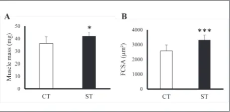
Eight sessions of high-frequency NMES training elicited a marked hypertrophy of the TA muscle in all analyzed mice (Fig. 2), with a significant increment of the muscle mass (p<0.05, r=0.55, Fig. 2A) and FCSA (p<0.001, r=0.71, Fig. 2B) in the ST limbs.
An improvement in contractile properties was also clearly observed after NMES training (Fig. 3A), with a significant increment in maximal force (p<0.01,
Fig. 1. Representative transversal cross sections of the Tibialis Anterior and microphotographs of the analyzed area (marked with a yellow rectangle). Serial cross sections were treated by im-munohistochemistry (A) and enzyme histochemistry (B) so as to differentiate fibers into slow (red) or fast (not labeled) with regard to their myosin heavy chain composition (C), as well as into anaerobic (white), aerobic (dark purple) or intermediate (pale purple) fibers with regard to their oxidative capacity (D). Symbols (*, +, •) represent the same muscle fibers. The scale bar represents 1000 µm in A and B and 200 µm in C and D.
r=0.64, Fig. 3B) and RFD (p<0.05, r=0.64, Fig. 3C). No differences were found between the CT and ST in the time to peak (115±10 and 117±8 ms, respectively). Regarding the oxidative capacity of the muscle fibers, there were no differences in the percentage of aerobic, intermediate and anaerobic fibers between the CT and ST limbs (Fig. 4).
DISCUSSION
Neuromuscular electrical stimulation (NMES) has been proposed as an interesting tool for the improve-ment of skeletal muscle properties [24]. Our results show that 8 sessions of high-frequency NMES training induced hypertrophy of the TA muscle of mice, with a significant increase in the muscle mass and the FCSA.
An increase of the quadriceps cross-sectional area after NMES training in humans has been previously described [22,25,26]. In the case of rodents, increments in muscle mass and FCSA of a fast-twitch muscle, such as the rat’s gastrocnemius, have also been reported after the application of intermittent high-frequency NMES [17,18,27-29], similarly to those observed in our study in a mixed muscle such as the TA. As muscle plasticity is dependent on the muscle fiber type, being for exam-ple type II fibers more prone to atrophy during aging [30] or to hypertrophy with NMES [22], more studies analyzing the effects of different stimulation patterns in purely slow or fast-twitch muscles are required to draw conclusions about the fiber type-dependent ef-fects of NMES training [31].
The hypertrophy found in our study after NMES training could be both sarcoplasmic and/or sarcomer-ic. An increase in the cell volume (cell swelling) could be produced because of the inflammatory response to muscle damage [32] and due to the accumulation of intracellular liquid because of the increase in the cell glycogen content [29]. Sarcomeric hypertrophy could result after an increase in protein synthesis, since the NMES protocol applied in our study has been pre-viously reported to increase anabolism through the phosphorylation of p70 ribosomal protein S6 kinase beta-1 (p70s6k) [17,18], downstream of the mTOR pathway. In addition, similarly to resistance training [33,34], NMES increases growth hormone levels as a consequence of the metabolic stress induced by the
ac-cumulation of H+ and lactate [35,36], which may also
enhance the anabolic processes through the activation of mTOR [37]; some authors have not found an as-Fig. 4. Effects of neuromuscular electrostimulation training on
tibialisanterior oxidative capacity. Percentage of aerobic, inter-mediate and anaerobic fibers of the control (CT) and stimulated (ST) limbs. No significant differences were found between control and stimulated limbs.
sociation between the exercise-related acute increase in anabolic hormones and muscle hypertrophy [38].
We also observed an improvement in the contrac-tile properties after NMES training, with an increase in the maximal tetanic force and the RFD. Benefits in the muscle contractile properties after NMES training have been previously reported in humans [22,25] and rodents. The increments observed in our study are similar to those found in mice [39] and rats [18,21]. Contractile properties are dependent on many param-eters, including neural mechanisms, energy disposal and muscle intrinsic properties, such as the muscle
fiber type composition, Ca2+ kinetics, the myofibrillar
protein content or the muscle size and architecture (pennation angle, musculotendinous stiffness, etc.) [40,41]. NMES has been shown to increase the mus-cle glycogen supply [29] and musmus-cle size [18], as well as to improve the neural drive [22,25,26]. Therefore, all these mechanisms could have played a role in the observed improvement of the contractile properties.
The high-frequency NMES protocol used in our study did not elicit any change in the proportion of oxidative muscle fibers (determined as oxidative ca-pacity). It is known that low-frequency NMES pro-motes endurance training-like adaptations, such as capillarization and mitochondrial biogenesis [42], through the activation of the AMP-activated protein kinase (AMPK)/peroxisome proliferator-activated receptor-γ coactivator (PGC-1α) signaling pathway [7]. In contrast, and according to our findings, it has been shown that moderate high-frequency NMES (40 Hz) does not improve the oxidative capacity in rats [43]. Resistance training and high-frequency NMES induce the activation of the mTOR hypertrophic pathway [7,17,18] and an interference between the AMPK and mTOR signaling pathways has been re-ported [44]. This could explain the absence of changes in the muscle oxidative profile after high-frequency NMES training.
CONCLUSIONS
Eight sessions of surface high-frequency NMES train-ing elicited muscle hypertrophy and an improvement
in the contractile properties of the tibialis anterior
muscle in mice. NMES appears to be a suitable
ani-mal model to study hypertrophic processes as it allows for better control of the training variables (intensity, duration, frequency) than other models, and does not require negative reinforcement or surgical procedures.
Acknowledgements: The authors gratefully acknowledge Javier Vicente-Tejedor, Laura Ramirez and David Rizo for their help, as well as the University of Alcalá Animal Research Center for their technical support. These experiments were financially supported by the funds of the Department of Systems Biology.
Authors’ contribution: P.L.V., J.R.T. and P.V. conceived and de-signed the research. P.L.V. and A.E.M.A. performed the experi-ments and analyzed the data. P.L.V., J.R.T. and P.V. interpreted the results of the experiments. P.L.V. prepared the figures and drafted the manuscript. P.L.V., A.E.M.A., J.R.T. and P.V. revised the manuscript and approved the final version of manuscript. Conflict of interest disclosure: No conflicts of interests, financial or otherwise, are declared by the authors.
REFERENCES
1. Matsakas A, Patel K. Skeletal muscle fibre plasticity in response to selected environmental and physiological stim-uli. Histol Histopathol. 2009;24:611-29.
2. Zurlo F, Larson K, Bogardus C, Ravussin E. Skeletal muscle metabolism is a major determinant of resting energy expen-diture. J Clin Invest. 1990;86(5):1423-7.
3. Wolfe RR. The underappreciated role of muscle in health and disease. Am J Clin Nutr. 2006;84:475-82.
4. Fiuza-Luces C, Garatachea N, Berger NA, Lucia A. Exercise is the real polypill. Physiology. 2013;28:330-58.
5. Pedersen BK. Exercise-induced myokines and their role in chronic diseases. Brain Behav Immun. 2011;25(5):811-6. 6. Pedersen BK. Muscles and their myokines. J Exp Biol.
2011;214(Pt 2):337-46.
7. Atherton PJ, Babraj J, Smith K, Singh J, Rennie MJ, Wack-erhage H. Selective activation of AMPK-PGC-1α or PKB-TSC2- mTOR signaling can explain specific adaptive responses to endurance or resistance training-like electrical muscle stimulation. FASEB J. 2005;19:786-8.
8. Bickel CS, Gregory CM, Dean JC. Motor unit recruitment during neuromuscular electrical stimulation: A critical appraisal. Eur J Appl Physiol. 2011;111:2399-407.
9. Caggiano E, Emrey T, Shirley S, Craik RL. Effects of electri-cal stimulation or voluntary contraction for strengthening the quadriceps femoris muscles in an aged male population. J Orthop Sport Phys Ther. 1994;20(1):22-8.
10. Dirks ML, Wall BT, Snijders T, Ottenbros CLP, Verdijk LB, Van Loon LJC. Neuromuscular electrical stimulation pre-vents muscle disuse atrophy during leg immobilization in humans. Acta Physiol. 2014;210(3):628-41.
12. Gerovasili V, Stefanidis K, Vitzilaios K, Karatzanos E, Poli-tis P, Koroneos A, Chatzimichail A, Routsi C, Roussos C, Nanas S. Electrical muscle stimulation preserves the muscle mass of critically ill patients: a randomized study. Crit Care. 2009;13(5):R161.
13. Snyder-Mackler L, Ladin Z, Schepsis AA, Young JC. Elec-trical stimulation of the thigh muscles after reconstruc-tion of the anterior cruciate ligament. J Bone Jt Surg. 1991;73(7):1025-36.
14. Taradaj J, Halski T, Kucharzewski M, Walewicz K, Smykla A, Ozon M, Slupska L, Dymarek R, Ptaszkowski K, Rajfur J, Pasternok M. The effect of neuromuscular electrical stimu-lation on quadriceps strength and knee function in profes-sional soccer players: Return to sport after ACL reconstruc-tion. Biomed Res Int. 2013;2013:1-9.
15. Cholewa J, Guimarães-Ferreira L, da Silva Teixeira T, Naimo MA, Zhi X, de Sá RBDP, Lodetti A, Cardozo MQ, Zanchi NE. Basic models modeling resistance training: An update for basic scientists interested in study skeletal muscle hyper-trophy. J Cell Physiol. 2014;229(9):1148-56.
16. Lowe DA, Alway SE. Animal models for inducing muscle hypertrophy: are they relevant for clinical applications in humans? J Orthop Sports Phys Ther. 2002;32(2):36-43. 17. Tsutaki A, Ogasawara R, Kobayashi K, Lee K, Kouzaki K,
Nakazato K. Effect of Intermittent Low-Frequency Electrical Stimulation on the Rat Gastrocnemius Muscle. Biomed Res Int. 2013;2013:480620.
18. Ogasawara R, Kobayashi K, Tsutaki A, Lee K, Abe T, Fujita S, Nakazato K, Ishii N. mTOR signaling response to resistance exercise is altered by chronic resistance training and detrain-ing in skeletal muscle. J Appl Physiol. 2013;114:934-40. 19. Kobayashi K, Ogasawara R, Tsutaki A, Lee K, Ochi E,
Naka-zato K. Genetic Strain-Dependent Protein Metabolism and Muscle Hypertrophy Under Chronic Isometric Training in Rat Gastrocnemius Muscle. Physiol Res. 2012;61:527-35. 20. Nachlas MM, Tsou KC, De Souza E, Cheng CS, Seligman
AM. Cytochemical demonstration of succinic dehydroge-nase by the use of a new p-nitrophenyl substituted ditetra-zole. J Histochem Cytochem. 1957;(5):420-36.
21. Bloemberg D, Quadrilatero J. Rapid determination of myo-sin heavy chain expression in rat, mouse, and human skeletal muscle using multicolor immunofluorescence analysis. PLoS One. 2012;7(4):e35273.
22. Gondin J, Brocca L, Bellinzona E, D’Antona G, Maffiuletti N a, Miotti D, Pellegrino M a, Bottinelli R. Neuromuscular electrical stimulation training induces atypical adaptations of the human skeletal muscle phenotype: a functional and proteomic analysis. J Appl Physiol. 2011;110:433-50. 23. Cohen J. Statistical power analysis for the behavioral
sci-ences. 2nd ed. Hillsdale, New Jersey: Lawrence Erlbaum Associates; 1988.
24. Maffiuletti NA, Minetto MA, Farina D, Bottinelli R. Elec-trical stimulation for neuromuscular testing and training: State-of-the art and unresolved issues. Eur J Appl Physiol. 2011;111:2391-7.
25. Gondin J, Guette M, Ballay Y, Martin A. Electromyostimula-tion training effects on neural drive and muscle architecture. Med Sci Sports Exerc. 2005;37(6):1291-9.
26. Maffiuletti NA, Zory R, Miotti D, Pellegrino MA, Jubeau M, Bottinelli R. Neuromuscular adaptations to electro-stimulation resistance training. Am J Phys Med Rehabil. 2006;85(2):167-75.
27. Haddad F, Qin AX, Zeng M, McCue SA, Baldwin KM. Effects of isometric training on skeletal myosin heavy chain expression. J Appl Physiol. 1998;84(6):2036-41.
28. Adams GR, Cheng DC, Haddad F, Baldwin KM. Skeletal muscle hypertrophy in response to isometric, lengthening, and shortening training bouts of equivalent duration. J Appl Physiol. 2004;96:1613-8.
29. Durigan JLQ, Cancelliero KM, Guirro RR, Da Silva CA, Ozores ML. Effects of neuromuscular electric stimulation on rats soleus muscle: A morphometric and metabolic analysis. Acta Ortop Bras. 2008;16(4):238-41.
30. Nilwik R, Snijders T, Leenders M, Groen BBL, van Kranen-burg J, Verdijk LB, Van Loon LJC. The decline in skeletal muscle mass with aging is mainly attributed to a reduction in type II muscle fiber size. Exp Gerontol. 2013;48(5):492-8. 31. Boonyarom O, Kozuka N, Matsuyama K, Murakami S.
Effect of electrical stimulation to prevent muscle atrophy on morphologic and histologic properties of hindlimb suspended rat hindlimb muscles. Am J Phys Med Rehabil. 2009;88(9):719-26.
32. Nosaka K, Aldayel A, Jubeau M, Chen TC. Muscle dam-age induced by electrical stimulation. Eur J Appl Physiol. 2011;111(10):2427-37.
33. Kraemer WJ, Ratamess NA. Hormonal responses and adaptations to resistance exercise and training. Sports Med. 2005;35(4):339-61.
34. Kraemer WJ, Marchitelli L, Gordon SE, Harman E, Dziados JE, Mello R, Frykman P, McCurry D, Fleck SJ. Hormonal and growth factor responses to heavy resistance exercise proto-cols. J Appl Physiol. 1990;69(4):1442-50.
35. Matsuse H, Nago T, Takano Y, Shiba N. Plasma growth hor-mone is elevated immediately after resistance exercise with electrical stimulation and voluntary muscle contraction. Tohoku J Exp Med. 2010;222:69-75.
36. Sartorio A, Jubeau M, Agosti F, Col A De, Marazzi N, Lafor-tuna CL, Maffiuletti NA. GH responses to two consecutive bouts of neuromuscular electrical stimulation in healthy adults. Eur J Endocrinol. 2008;158:311-6.
37. Hayashi A, Proud C. The rapid activation of protein synthe-sis by growth hormone requires signaling through mTOR. Am J Physiol Metab. 2007;292(6):1647-55.
38. West DWD, Burd NA, Tang JE, Moore DR, Staples AW, Hol-werda AM, Baker SK, Phillips SM. Elevations in ostensibly anabolic hormones with resistance exercise enhance neither training-induced muscle hypertrophy nor strength of the elbow flexors. J Appl Physiol. 2010;108(1):60-7.
39. Distefano G, Ferrari RJ, Weiss C, Deasy BM, Boninger ML, Fitzgerald GK, Huard J, Ambrosio F. Neuromuscular elec-trical stimulation as a method to maximize the beneficial effects of muscle stem cells transplanted into dystrophic skel-etal muscle. PLoS One. 2013;8(3):e54922.
41. Josephson RK. Extensive and intensive factors deter-mining the performance of striated muscle. J Exp Zool. 1975;194(1):135-53.
42. Reichmann H, Hoppeler H. Biochemical and ultrastructural changes of skeletal muscle mitochondria after chronic elec-trical stimulation in rabbits. Eur J Physiol. 1985;404(1):1-9.
43. Egginton S, Hudlická O. Early changes in performance, blood flow and capillary fine structure in rat fast muscles induced by electrical stimulation. J Physiol. 1999;515 (Pt 1):265-75.