Innovative and New Approaches to Laboratory Diagnosis
of Zika and Dengue: A Meeting Report
Adriana Goncalves,1 Rosanna W. Peeling,1 May C. Chu,2 Duane J. Gubler,6 Aravinda M. de Silva,3 Eva Harris,4 Maurine Murtagh,5 Arlene Chua,8 William Rodriguez,9 Cassandra Kelly,9 and Annelies Wilder-Smith7,10,11
1London School of Hygiene and Tropical Medicine, United Kingdom; 2Department of Epidemiology, Colorado School of Public Health, University of Colorado, Anschutz Medical Center, Aurora; 3Department of Microbiology and Immunology, University of North Carolina School of Medicine, Chapel Hill; 4Division of Infectious Diseases and Vaccinology, School of Public Health, University of California, Berkeley, and 5The Murtagh Group, Palo, Alto, California; 6Emerging Infectious Diseases Program, Duke-NUS Medical School, and 7Lee Kong Chian School of Medicine, Singapore; 8World Health Organization and 9Foundation for Innovative New Diagnostics, Geneva, Switzerland; 10Institute of Public Health, University of Heidelberg, Germany; and 11Department of Global Health and Epidemiology, University of Umea, Sweden.
Epidemics of dengue, Zika, and other arboviral diseases are increasing in frequency and severity. Current efforts to rapidly iden-tify and manage these epidemics are limited by the short diagnostic window in acute infection, the extensive serologic cross-reac-tivity among flaviviruses, and the lack of point-of-care diagnostic tools to detect these viral species in primary care settings. The Partnership for Dengue Control organized a workshop to review the current landscape of Flavivirus diagnostic tools, identified cur-rent gaps, and developed strategies to accelerate the adoption of promising novel technologies into national programs. The rate-lim-iting step to bringing new diagnostic tools to the market is access to reference materials and well-characterized clinical samples to facilitate performance evaluation. We suggest the creation of an international laboratory-response consortium for flaviviruses with a decentralized biobank of well-characterized samples to facilitate assay validation. Access to proficiency panels are needed to ensure quality control, in additional to in-country capacity building.
Keywords. Dengue; Zika; flavivirus; arbovirus; laboratory; diagnostics; serology; surveillance.
Zika virus (ZIKV) and the dengue viruses (DENVs) are arthro-pod-borne viruses (arboviruses) of the Flaviviridae family that cocirculate in tropics and subtropics along with other arbovi-ruses that share the same Aedes species mosquito vectors [1]. Several factors, including viral evolution, redistribution of vec-tors, ineffective vector control strategies, population growth, urbanization, and globalization have contributed to the global spread of DENV, ZIKV, and other arboviruses [2].
Up to 400 million DENV infections are estimated to occur every year [3], and infection with any of the 4 DENV serotypes (DENV1–4) can cause severe and sometimes fatal disease. The geographical expansion of dengue is increasingly associated with more-severe disease outcomes [2, 4]. ZIKV is following the global spread of DENV [2]. ZIKV infections were first thought to only cause sporadic and mild disease in parts of Africa and Asia [5]. A major Zika outbreak with a high attack rate occurred for the first time in 2007. During a subsequent outbreak in the Pacific (French Polynesia) in 2013, ZIKV was linked to severe neurological disease in humans [6]. The recent explosive
outbreak in the Americas unmasked the association between prenatal ZIKV infections and severe birth defects [2, 6].
No specific therapeutic options exist for DENV or ZIKV infections. For DENV, a vaccine was recently licensed but has not yet been implemented widely in any of the affected countries [7]; for ZIKV, at least 45 vaccine candidates are now in devel-opment, but a licensed vaccine will not be available for years to come [8]. There is an urgent need for highly specific diagnostic assays that can identify and discriminate between cocirculating DENV and ZIKV for efficient case management, surveillance, control, and vaccine trials. In May 2017, the Partnership for Dengue Control (PDC) [9] organized a workshop with approx-imately 80 key stakeholders and thought leaders to address crit-ical issues related to the diagnosis and surveillance of ZIKV and DENV. The workshop was organized around 3 questions: What is the status of Zika and dengue diagnostic tools? What tech-nological innovations might be available in the near, interme-diate, and long-term future? and What is needed to make these technologies readily available where they are most needed? The following is a summary of key outcomes that emerged from the meeting.
WHAT IS THE STATUS OF ZIKA AND DENGUE DIAGNOSTIC TOOLS?
Individuals infected with DENV and ZIKV may be asympto-matic or display a similar constellation of initial clinical symp-toms [10]. Hence, virus-specific assays are required for accurate diagnosis. Since the first isolation of DENV during World War STANDARD
Received 18 September 2017; editorial decision 19 December 2017; accepted 22 December 2017; published online December 25, 2017.
Correspondence: A. Wilder-Smith, MD, PhD, Institute of Public Health, University of Heidelberg, Germany, Lee Kong Chian School of Medicine, Singapore, Department of Global Health and Epidemiology, University of Umea, Umea, Sweden ([email protected]).
The Journal of Infectious Diseases® 2018;217:1060–8 © The Author(s) 2017. DOI: 10.1093/infdis/jix678 1
217
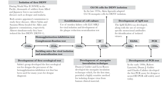
II [11, 12], a number of diagnostic methods commonly used for viral detection, such as viral isolation, plaque reduction neu-tralization test (PRNT), the immunoglobulin M (IgM) enzyme-linked immunosorbent assay (ELISA), and, in the 1990s, reverse transcription–polymerase chain reaction (RT-PCR) [13] were developed for DENV (Figure 1) and other medically relevant flaviviruses.
Assays to detect DENV and ZIKV can be divided into 2 main categories: (1) assays to detect the pathogen (viral isolation, viral nucleic acid testing, or viral antigen detection); and (2) assays to detect exposure to the pathogen (detection of virus-specific antibodies such as IgM, immunoglobulin G, and immunoglob-ulin A). Assay selection depends both on the timing of sample collection and the purpose of testing (Figure 2). The viremic period of flaviviral infections is transient and short-lived; the duration of viral shedding and the presence of ZIKV RNA can be variable across sample types (eg, serum, whole blood, urine, saliva, and amniotic fluid) [6, 14] and different hosts (eg, preg-nant women and other adults) [15]. A negative viral isolation and/or nucleic acid test result does not exclude the presence of a current infection.
In the convalescent phase of infection, serologic meth-ods are preferred, although paired acute and convalescent phase samples are required to distinguish current from past infections [16]. The major challenge of ZIKV and DENV diagnosis by serologic analysis is the extensive cross-reac-tivity of antibody responses resulting from prior flaviviral infections and/or vaccination [17–19]. Figure 3 details the applications, advantages, and limitations of the different
types of assays available for the detection of DENV and ZIKV infections.
LANDSCAPE OVERVIEW AND EXISTING GAPS Both in-house laboratory-developed tests (LDTs) and com-mercial kits are available to detect ZIKV and DENV infections (Figure 4). Most of the available technologies require laboratory facilities with appropriate diagnostic competence (Figure 4); point-of-care assays remain limited. Zika commercial kits in-clude nucleic acid tests and serologic assays. The current ZIKV nucleic acid tests have not yet gone through much rigorous evaluations [20], and the evidence is even scarcer for serologic assays. Antigen detection assays for the diagnosis of ZIKV infections are currently not available on the market.
Performance of commercial dengue diagnostic tools has improved over the last decade. These include 2 Food and Drug Administration (FDA)–approved assays (1 RT-PCR and 1 IgM-capture ELISA). Additionally, there are several rapid lateral flow assays (RDTs) for the detection of DENV NS1 antigen, DENV-specific IgM antibodies, or both (Figure 4), none of which are FDA approved. RDTs hold promise as future point-of-care applications; however, the clinical performance of these assays has been highly variable [21].
While dengue serologic assays have been clinically validated, their specificity has decreased by cross-reactivity in the con-text of the recent cocirculation of ZIKV [22]. To date, very few dengue and Zika diagnostic assays have been adequately and independently evaluated using clinical specimens from both ZIKV-infected and DENV-infected populations. Diagnostic
Isolation of first DENV
Establishment of cell culture
Hemagglutination-inhibition test Complement-fixation test
Suckling mice for viral isolation and neutralization test
Development of mosquito
inoculation technique Development of PCR test Development of first serological test
1940s 1950s 1960s 1970s 1980s 1990s
PCR ELISA
IF
C6/36 cells for DENV isolation
Development of IgM test During World War II (WWII) in the
Pacific, thousands of soldiers from Allied and Japanese forces succumbed to diseases such as dengue and malaria.
Use of monkey kidney cells LLC-MK2 for viral isolation and development of the plaque reduction neutralization test
In the late 1970s, Akira Igarashi adapted C6/36 mosquito cells for DENV isolation.
The IgM ELISA was developed, along with the use of serotype-specific monocional antibodies for identification of isolated viruses.
Both armies appointed commissions to study these diseases. Albert Sabin and Susumu Hotta headed the Allies and Japanese commissions, respectively. Almost simultaneously they have isolated the first DENV (DENV-1).
Sabin’s group developed the first serological test for dengue-the precursor of the hemagglutination-inhibition test that has been used for many years for dengue diagnosis
Duane J. Gubler and Leon Rosen developed the mosquito inculation technoque which, for the first time, provided a highly sensitive method for isolating dengue virus from human clinical material.
In the early 1990s, Robert Lanciotti, Duane J. Gubler and Charles Calisher developed the first PCR assay for dengue-a nested RT-PCR still widely used today.
assays that can accurately detect and distinguish cocirculating flaviviral infections and predict severe disease outcomes at or near the point-of-care are urgently needed.
WHAT TECHNOLOGICAL INNOVATIONS MIGHT BE AVAILABLE IN THE NEAR, INTERMEDIATE, AND LONG-TERM FUTURE?
Different companies and research groups were invited to pre-sent technologies to detect DENV and ZIKV. In the following, we discuss the technologies in the pipeline (Figure 4) and their potential to change the paradigm of flaviviral diagnosis.
Pipeline Assays to Detect the Pathogen
Nucleic-Acid Testing
Simpler and faster alternatives to traditional RT-PCR meth-ods have the potential to be used at or near the point-of-care. These include cartridge-based sample-in-answer-out multiplex real-time RT-PCR assays that can simultaneously detect ZIKV, DENV1–4, other arboviruses (eg, chikungunya virus, an alpha-virus), and other viruses (3-plex to 6-plex combinations) from a single specimen in <2 hours. Arboviral assays are being de-veloped for use on existing industry platforms that were previ-ously validated and implemented for other molecular tests. This strategy illustrates the usefulness of open-platform systems that can easily incorporate newly developed molecular amplification methods to suit an emergent medical need, such as the ZIKV
epidemic. Another advantage of these systems is the ability to transmit data wirelessly and monitor the results remotely. The disadvantages are that these platforms are costly and that some require technical expertise and laboratory infrastructure that are not widely available.
The development of more-portable molecular platforms linked to faster isothermal amplification methods independent of thermal cycling, such as recombinase polymerase amplifica-tion [23] and loop-mediated isothermal amplificaamplifica-tion, are also underway for singleplex and multiplex detection of ZIKV and other arboviruses. In prototype formats, results can be achieved in <1 hour, and assays can potentially be applied to settings without electricity or highly trained users. Proof-of-principle studies exist [23–30], but further simplification of sample prep-aration and more-robust clinical performance evaluation will be required. Innovative technologies, such as clustered regu-larly interspaced short palindromic repeats (CRISPR)–based RNA sensing, robotics, microfluidics, smartphones, and 3D printers, are being used to develop these assays. Other nucleic acid testing innovations include the use of paper-based strips for multiplex detection of ZIKV, DENV, and chikungunya virus end point RT-PCR products [31].
Antigen Detection Assays
High-affinity monoclonal antibodies that recognize specific epitopes on ZIKV antigens are required to develop antigen
Viral isolation
A) Assays to detect the pathogen:
B) Assays to detect exposure the pathogen:
Viremia
Acute phase Convalescent phase
IgG
IgM Antibody response C) Combination assays: Antigen + IgM/IgG detection
Viral NAT
IgM/IgG detection by ELISA/RDT Plaque reduction neutralization tests Viral antigen detection
Symptoms
Exposure 0 Days Months Years
detection assays and are either in development or were devel-oped for NS1 [32, 33], including the development of RDTs [34].
Pipeline Assays to Detect Past Exposure
Given our understanding of the cross-reactivity of current an-tibody detection methods for flaviviruses, there is a lack of reli-able reference diagnostic tools against which to compare newly developed specific serologic assays. Detection of virus-specific neutralizing antibodies by PRNT can be useful to discriminate viral species and serotypes in primary infections. However, the specificity of PRNT in sequential DENV infections or sequen-tial DENV and ZIKV infections and at early time points after
infection is limited [32, 35, 36]. Interestingly, little cross-neu-tralization is detected in samples collected during the late convalescent phase (ie, >2 months) after DENV and ZIKV in-fection [37]. These observations highlight the importance of the timing of sample collection and the history of exposure to past infections to inform the serodiagnosis of flaviviral infections. It is critical to evaluate multiple flaviviruses simultaneously in neutralization assays to interpret the results appropriately.
Strongly neutralizing human monoclonal antibodies target quaternary structure epitopes that typically bind across envelope proteins displayed on the surface of the viral particles [38–40]. Epitopes with high sequence homology among serotypes and
A) Assays to detect the pathogen: B) Assays to detect exposure to the pathogen:
C) Combination assays
Application
Advantages
Limitations
• Application
- IgM/IgG (paired serum specimens):
- IgM/IgG (single/paired serum specimens):
- IgM/IgG (single/paired serum specimens): - IgM (sigle serum specimen):
- IgG (sigle serum specimen):
- Plaque reduction neutralization test:
- Plaque reduction neutralization test:
- Plaque reduction neutralization test:
•
Application •
Application •
Application •
•
Advantages
• •Advantages
Advantages •
Advantages •
•
Limitations •
Limitations •
Limitations •
Limitations •
Viral isolation: Serology:
NAT:
Antigen detection (DENV NS1)
Antigen (DENV NS1) and IgM/IgG combination assays: Detection of active infection. Interpretation of positive result: confirmatory. Allows identification of viral species and DENV
serotypes. Detection of active infection upon seroconversion (negativeIgM/IgG in an acute specimen followed by a positive IGM/IgG in a convalescent specimen) or a >4-fold rise in IgG or total antibody titer. Interpretation of positive result: confirmatory.
Detection of recent past infection. Interpretation of positive result: probable. Useful for surveillance.
Detection of past infection. Classification of primary or secondary infections (IgG avidity, IgM/IgG ratio, titer)
Can be used as ELISA (high throughput) or as RDT at or near the point-of-care; easy to perform; affordable.
Limited specificity due to high cross-reactivity among flaviviruses;
Limited sensitivity during acute phase of infection; Testing single serum specimens is not confirmatory of current infection;
Confirmatory results can only be obtained after a second visit (convalescent phase samples are difficult to obtain); Variation among laboratories.
Time-and labor-intensive with variation among laboratories; Not always able to resolve cross-reactivity (particularly in secondary infections).
Detection of past infection. Identification of viral species and infecting DENV serotypes in primary infection and some secondary infections. Useful for research and vaccine studies.
Higher specificity than IgM/IgG assays;
Possible identification of infecting viral species and DENV serotypes during the convalescent phase;
Indicates the level of protection against an infecting virus. Detection of active infection. Interpretation of positive result:
confirmatory.a Allows identification of viral species and DENV serotypes.
Detection of active infection. Interpretation of positive result: confirmatory.
Detection of active infection. Interpretation of positive result: confirmatory. Most specific and conclusive diagnosis; only method that
allows the detection and isolation of living virus.
Highly accurate if performed correctle; faster and less laborious than viral isolation; multiplex and near point-of-care testing possible.
Diagnostic window of DENV NS1 up to day 8 post-onset of DENV infection with less sensitivity; ELISA (high throughput) or RDT; can be used at or near the point-of-care; easy to perform; less expensive.
Less accurate than viral isolation and NAT, requires acute sample, variable results according to serotype and immune status. No antigen antigen detection assay availbale for ZIKV at present.
Less accurate than viral isolation and NAT during acute phase. Variation among laboratories. No assays availbale for ZIKV at present. Entire temporal spectrum following infection (applicable in acute and convalescent samples); RDT; can be used at or near the point of care; easy to perform; less expensive; can use whole blood.
Mainly laboratory based; requires acute sample; complex; requires power supply; expensive; potential false positives.
Lab-based; requires acute sample; takes more than one week to complete; expensive; laborious and impractical in point-of-care and resource-limited settings.
Figure 3. Applications, advantages, and limitations of the main diagnostic tests in use for the detection of flaviviral infections. A, Assays to detect the pathogen directly: viral isolation, nucleic acid amplification tests (NATs), and antigen detection assays. B, Assays to detect exposure to infection. C, Combination assays to detect both the pathogen and exposure to infection. aRNA copy number is not an accurate measure of infectious virus (viral RNA can persist for longer periods than infectious virus). DENV,
viral species can trigger cross-reactive antibody responses, whereas unique epitopes can lead to type-specific antibody responses. This information is being used to rationally design ZIKV and DENV envelope and NS1 recombinant antigens for specific serologic assays.
Isolated human ZIKV type-specific anti-NS1 monoclonal antibodies were used to identify type-specific recognition sites on ZIKV NS1 protein by antibody competition assays [32]. One of these antibodies was adapted to a competition-based ELISA, in which serum antibodies are measured for their ability to block the binding of a ZIKV NS1-specific monoclonal antibody to sol-id-phase ZIKV NS1 [41]. This approach, named “ZIKV NS1 blockade-of-binding (BOB) ELISA,” was shown to be more spe-cific than traditional ELISAs. Clinical validation in large multi-center cohorts of patients stratified by exposure to DENV and
ZIKV infection, immune status, and timing of sample collection confirmed the high specificity and sensitivity of the assay [41]. The ZIKV NS1 blockade-of-binding ELISA has been imple-mented in laboratories of 6 different countries (Brazil, Italy, Nicaragua, Switzerland, United Kingdom, and United States).
Nanotechnology assays have also been developed, including simple-to-use readout platforms with data connectivity that use disposable microfluidic cartridges for rapid detection of ZIKV and DENV antibodies/antigen, and a multiplex serologic assay that uses near‐infrared fluorescence enhanced imaging on a nanoscale plasmonic gold microarray antigen platform (12-plex) for antibody detection on 2 different channels [42]. The latter was shown to detect and distinguish IgG antibodies from ZIKV- and DENV-infected patients, as well as determine the timing of exposure to infection by measuring IgG avidity.
R&D Evaluation
Proof of principle
Proof of principle Product
prototype TPP
A
In-house LDTs
Commercial kits Public health
emergency
Clinical validation
NATa (DENV, ZIKV, multiplex)
IgM capturea (DENV, ZIKV)
PRNTa (DENV, ZIKV)
ZIKV NS1 BOB ELISAa
NAT assaysa:
FDA cleared: 1 DENV assay (DENV-1-4 real-time RT-PCR; CDC)
FDA cleared: 1 DENV assay (DENV Detect IgM capture ELISA; InBios) EUA: 3 ZIKV assays
IgM/IgG antibody detection (DENV, ZIKV, multiplex)
Multiplex “sample-in-answer-out” RT-PCR platformsa
Multiplex nanotechnology-based microarray platforma
(antibody detection)
ZIKV/DENV automated ELISAa
(antigen and antibody detection) ZIKV antigen detection ELISAsa and RDTs
ZIKV/multiplex portable isothermal molecular platforms ZIKV/DENV nanotechnology-based portable platform (antigen and antibody detection)
NS1 antigen detection (DENV)
NS1 antigen detection (DENV) + IgM/IgG (DENV/multiplex) combination RDTs EUA: 11 ZIKV and 1 multiplex assay (ZIKV, DENV, CHIKV)
EUAL: 1 ZIKV and 1 multiplex assay (ZIKV, DENV, CHIKV) IgM-capturea:
RDTsb:
Clinical validation
Emergency use authorization
Commercial
product and distributionManufacturing Regulatoryapproval Ready foradoption Commercial kits adopted
for emergency use
Commercial kits adopted into use Ready for
adoption In-house assays developed and adoptedinto use by public health laboratories
Implementation
B
C
D
E
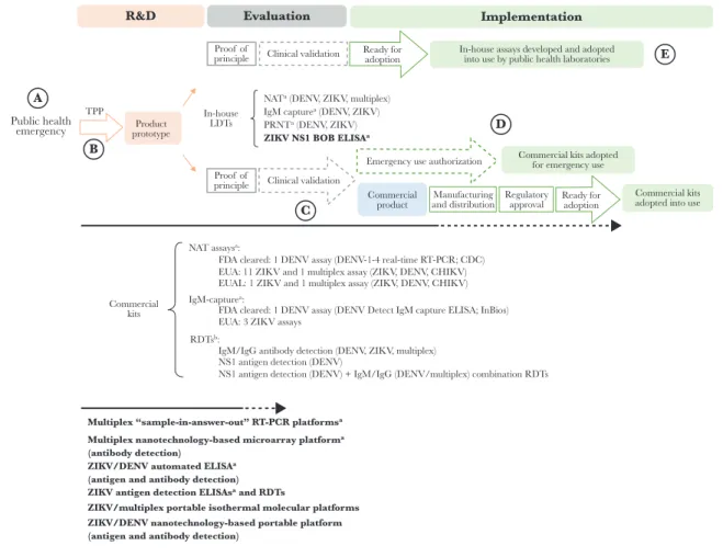
Figure 4. Diagnostic landscape to detect Zika virus (ZIKV) and dengue virus (DENV) infections and the pathway to adoption. The Zika public health emergency triggered significant efforts toward the development of new diagnostic assays. Commercial kits and in-house laboratory-developed tests (LDTs) are available to detect ZIKV and DENV infections. Multiplex assays that simultaneously detect ZIKV and other arboviral infections (DENV, CHIKV and others) are also available. Target product profiles (TPP) are used to define the desired technical and operational characteristics of a test. Quality-assured clinical laboratories can develop, validate, and then implement their in-house LDTs. Commercial kits require clinical validation, scale production, distribution, and regulatory approval to be adopted for wide use. Two DENV commercial kits (1 nucleic acid amplification test [NAT] and 1 immunoglobulin M [IgM]–capture enzyme-linked immunosorbent assay [ELISA]) have been cleared by the Food and Drug Administration (FDA). Emergency use authorization mechanisms by the FDA (the Emergency Use Authorization [EUA] authority) and the World Health Organization (the Emergency Use Assessment and Listing [EUAL] procedure) were put into place to accelerate adoption of ZIKV commercial kits in response to the Zika public health emergency. In bold are the types of assays that are currently in the pipeline (most assays are stuck at the evaluation stage because of the lack of access to well-characterized clinical specimens). BOB, blockade of binding; CDC, Centers for Disease Control and Prevention; IgG, immunoglobulin G; PRNT, plaque-reduction neutralization test; RT-PCR, reverse transcription–polymerase chain reaction; R&D, research and development. aAssays that require laboratory infrastructure. bNo rapid diagnostic test (RDT) has been cleared by the FDA nor received
WHAT IS NEEDED TO MAKE THESE TECHNOLOGIES AVAILABLE IN THE FIELD?
For the last 25 years, routine diagnostic approaches have mainly included laboratory-based RT-PCR, IgM detection, and PRNT. Recognition of Zika as a public health emergency of interna-tional concern has galvanized the development of new diag-nostic assays to detect flaviviral infections. While these efforts must be encouraged, it is equally important to look down-stream and identify the issues around translating research into a product that is available for use in the field, robust, easy to use, reasonably inexpensive, and accurate and has demonstrable clinical impact. Previous research and development experience has shown that the path from diagnostic development to adop-tion is long and fragmented [16]. There is a massive loss be-tween the number of tests that undergo initial development and the ones that are taken into adoption for routine use.
HOW CAN WE ACCELERATE TRAVEL ON THE PATHWAY FROM DISCOVERY TO ADOPTION?
The 5 major steps identified for optimizing the time to adoption (Figure 4A–E) are discussed below and summarized in Table 1.
Market Uncertainty
For diagnostic countermeasures to be readily available, research and development must happen before rather than in response to an outbreak [43]. The unpredictable and episodic nature of outbreaks brings uncertainty to the market, and diagnostic companies are left unable to adequately forecast demand and establish business models that allow a return on investment. Even when a product is brought into the market, there is no guarantee that it will be adopted by national health authorities. Once a public health emergency of international concern has ended, sustained manufacturing support of the product may be at stake. Sustainable markets are required to ensure that vali-dated, approved, high-quality diagnostic tools remain available for use in the next outbreak event. As such, innovative financial
incentives are needed to achieve sustainable emergency prepar-edness for diagnostic tools. From investments in product devel-opment to the establishment of partnerships and the creation of models to support scalable adoption into national programs, a variety of mechanisms have been proposed or established to overcome some of the challenges.
The WHO R&D Blueprint for Actions to Prevent Epidemics has initiated a call for open-platform technologies to improve research and development preparedness against global health emergencies, so that in the event of an epidemic diagnostic kits can be made available in a short time frame [44]. Furthermore, there was a call for a coalition between diagnostic preparedness efforts and programs that finance and manage the development of vaccines [43]. As a result, CEPI∙dx, a new partnering model between the Coalition for Epidemic Preparedness Innovations, the Foundation for Innovative New Diagnostics, and other di-agnostic partners, has been created.
Target Product Profiles (TPPs)
TPPs are used to define the desired minimum technical and oper-ational characteristics of diagnostic tests, to ensure the develop-ment of the most-impactful products. TPPs are aspirational in nature; however, excessively stringent requirements may deter industry partners from developing new products and lead to a lack of diagnostic tests meeting those requirements. Strategies on how to best define the desired characteristics of TPPs and/or inform the use of diagnostic tests when those requirements are not met have been proposed. For example, a slightly less accu-rate test might provide a higher public health impact in terms of increased access to testing, compared with a more accurate but expensive or complex test (this was the approach used to approve the use of HIV self-testing and malaria RDTs in the past). As such, it is important to consider how the assay will be used, in which setting, and for what purpose (eg, surveillance, early warning, or clinical management at the point-of-care), as dif-ferent applications will have difdif-ferent technical and operational
Table 1. Summary Table of the Challenges and Drivers Along the Pathway to Adoption
Stepa Challenge(s) Drivers
A Market failure due to uncertainty and lack of demand of public health emergencies
Research and development models for diagnostic preparedness, including product development; product development partnerships, such as CEPI.dx; other innovative financing models
B TPP: the performance characteristics that are set in the TPP are aspirational in nature; TPPs are often deemed to be too stringent
Risk and benefit models to set accuracy targets may help inform use of diagnostic tests when they do not meet the minimum or ideal characteristics set in the TPP
C Lack of clinical samples and resources for clinical validations Development of international reference standard for assay comparability; improved access to qualified field laboratory networks; access to proficiency panels; develop-ment of standardized protocols
D Regulatory approval that is region specific, nontransparent, com-plex, slow, and costly
Establishment of regulatory networks, common strategies, information sharing, and early partnerships
E Limited in-country capacity for wide adoption. Mechanisms for appropriate transfer of technology in a more streamlined fashion; reg-ulation of quality of local laboratories and in-house assays for national scale-up and sustainable implementation
Abbreviation: TPP, target product profile.
requirements. A weighted risk and benefits approach within dif-ferent use scenarios may be more appropriate not only to define but also to guide regulatory approval and adoption.
Assay Optimization and Clinical Validation
Internationally accepted reference preparations to compare and potentially standardize the different assays are crucially important [45]. The WHO has established numerous reference preparations, most of them as WHO international standards. For ZIKV RNA, the biological standard for molecular tests was characterized for the majority of nucleic acid tests available [46], and the complete sequence of the ZIKV of this reference preparation was published [47] and established as a WHO international standard. Lack of access to biobanks of well-characterized clinical specimens delays the process of test optimization, clinical validation, and product adoption. This lack was identified as the most significant bottle-neck along the pathway from development to adoption.
Of note, the pathway to adoption of in-house assays and of commercial kits differs substantially. Quality-assured clinical laboratories can develop, validate, and then implement their in-house assays. In contrast, commercial diagnostic kits go through regulatory approval processes that may require large clinical validation studies, manufacturing under a quality man-agement system, and some level of distribution capacity. The different streams of test development make it challenging to determine relative comparability of the accuracy of the differ-ent tests because very few of them share the same calibration controls (ie, internal positive controls used for measuring the reactivity of a diagnostic test) or screening panels (ie, a small set of coded samples that include high-positive, low-positive, cross-reactive, and negative samples, to measure the specificity and sensitivity of a diagnostic test). Obtaining irreplaceable clinical specimens is costly; the same test materials cannot be used throughout the development process. Access to clinical samples becomes even more challenging during an outbreak, with multiple demands to prioritize assay validation in a short time frame and the inability to do head-to-head comparisons.
A coordinated network of quality-assured laboratories with staff that are well trained in assay validation and performance evaluation could be leveraged a priori. Such an approach would alleviate pressure on the countries involved in outbreak response, yet provide access to clinical samples and data in a way that may be acceptable to the different parties. Of note, local restrictions on the export of clinical samples (as witnessed during the ZIKV outbreak) limits sample sharing for product validation outside the affected countries [43]. The involvement of a network of capable local laboratories would have the advantage to over-come the need for out-of-country sample transfer and facilitate country involvement and capacity development at an early stage of product development. A transparent and fair process of en-gagement needs to be put into place to minimize distrust and ensure access and equitable sharing of specimens and data. The
creation of a governance system to provide access to reference panels and protocols for test validation has been proposed.
Regulatory Approval
Regulation is essential to ensure the safety, quality, and effec-tiveness of diagnostic tests, yet >50% of countries do not inde-pendently regulate in vitro diagnostic tools [48]. The regulatory landscape for in vitro diagnostic tools is highly variable, and reg-ulatory approval mechanisms vary from country to country. This makes assay uptake processes slow, costly, and nontransparent. Regulatory harmonization between international and national regulatory agencies, coupled with coordinated information shar-ing among the different interest groups (industry, regulators, researchers, laboratories, health systems, and patients), is required.
During outbreak events, emergency use authorizations are generally used to provide regulatory oversight for diagnostic tools that have not previously been evaluated and yet are ur-gently needed for global response. Both the FDA and the WHO have implemented programs (the Emergency Use Authorization [EUA] authority and the Emergency Use Assessment and Listing [EUAL] procedure, respectively) to address the evaluation of new diagnostic tools in an emergency setting. It is important to note that in both cases, EUA and/or EUAL approval does not extend approval for use outside of an emergency setting. Once an emergency has ended, industry will need to seek approval for regular use of their products in the intended settings, using ei-ther FDA 510(k) or WHO prequalification procedures. The data obtained during EUA and/or EUAL approval may be included in the application package for regular approval; however, it is likely that additional data will be required for full approval. In these instances, it can be challenging for industry to provide sufficient data, as limited access to well-characterized samples can pre-vent evaluation of the products to the extent required for FDA and WHO approval. As of May 2017, several ZIKV diagnostic assays have received approval through the EUA (15 assays) and/ or EUAL (2 assays) [45]; however, no single ZIKV assay has been cleared by the FDA to date (Figure 4).
Sustainable In-Country Capacity
Sustainable in-country capacity is needed for diagnostic tools to respond to the intermediate and long-term infectious diseases threats. Higher-cost commercial kits are unlikely to solve this issue at a national level in many resource-constrained countries. Therefore, key reagents, protocols, and quality control standards (eg, proficiency panels) must be made available to national ref-erence laboratories and other such public-sector entities to en-sure wide and sustainable adoption.
CONCLUSIONS
flaviviral diseases. The rate-limiting bottleneck is early access to calibration controls and screening panels, as well as access to well-characterized samples for development, validation, and comparison of the performance of different assays. Proficiency testing for both serologic and molecular diagnostic tools should be developed for all regions of endemicity, paired with capacity building. We suggest that an international reference laboratory re-sponse for flaviviruses is needed, which would include networks of in-country laboratories and preparation of protocols for evalua-tion studies. This could be achieved through initiatives such as the Global Dengue and Aedes Transmitted Disease Consortium, the European Virus Archive, the future EVD-LabNet by the European Centre for Disease Prevention and Control, or the Zika research consortia funded by the European Commission [49]. The know-ledge obtained should be put into the public domain. Researchers and policymakers alike need to ensure mechanisms for greater re-agent availability and sharing of standard rere-agents, such as refer-ence materials, antigens, monoclonal antibodies, cell lines, control sera, and standardized protocols. While this workshop focused on challenges for arbovirus diagnostic development, the key outcomes highlighted here translate to all pathogens of epidemic potential.
Notes
Acknowledgments. We thank the Fondation Merieux, for
supporting the meeting; Richard Vaux, Benedicte Pansier, and Severine Comignani, for their assistance with logistics; and Raman Preet, for assistance for all ZikaPLAN consortium– related matters during this meeting.
Financial support. This work was supported by the
Gates Foundation; the European Union’s 2020 Research and Innovation Programme, through the ZikaPLAN consortium (grant 734584); the EPSRC IRC in Early-Warning Sensing Systems for Infectious Diseases (EP/K031953/1); bioMérieux (unrestricted grant); Sanofi Pasteur (unrestricted grant); Takeda (unrestricted grant); the Foundation for Innovative and New Diagnostics (in-kind support); the World Health Organization (WHO) Special Program for Research and Training in Tropical Diseases (in-kind support); and the WHO R&D Blueprint (in-kind support).
Potential conflicts of interest. All authors: No reported
conflicts of interest. All authors have submitted the ICMJE Form for Disclosure of Potential Conflicts of Interest. Conflicts that the editors consider relevant to the content of the manu-script have been disclosed.
References
1. Musso D, Gubler DJ. Zika virus. Clin Microbiol Rev 2016; 29:487–524.
2. Wilder-Smith A, Gubler DJ, Weaver SC, Monath TP, Heymann DL, Scott TW. Epidemic arboviral diseases: pri-orities for research and public health. Lancet Infect Dis 2017; 17:e101–6.
3. Bhatt S, Gething PW, Brady OJ, et al. The global distribution and burden of dengue. Nature 2013; 496:504–7.
4. Guzman MG, Gubler DJ, Izquierdo A, Martinez E, Halstead SB. Dengue infection. Nat Rev Dis Primers 2016; 2:16055. 5. Faye O, Freire CC, Iamarino A, et al. Molecular evolution of
Zika virus during its emergence in the 20(th) century. PLoS Negl Trop Dis 2014; 8:e2636.
6. Baud D, Gubler DJ, Schaub B, Lanteri MC, Musso D. An update on Zika virus infection. Lancet 2017; 390:2099–109. 7. Wilder-Smith A, Vannice KS, Hombach J, Farrar J, Nolan T. Population perspectives and World Health Organization recommendations for CYD-TDV Dengue vaccine. J Infect Dis 2016; 214:1796–9.
8. Durbin A, Wilder-Smith A. An update on Zika vaccine developments. Expert Rev Vaccines 2017; 16:781–7. 9. Gubler DJ. The partnership for dengue control—a new
global alliance for the prevention and control of dengue. Vaccine 2015; 33:1233.
10. Duffy MR, Chen TH, Hancock WT, et al. Zika virus out-break on Yap Island, Federated States of Micronesia. N Engl J Med 2009; 360:2536–43.
11. HOTTA S. Experimental studies on dengue. I. Isolation, identi-fication and modiidenti-fication of the virus. J Infect Dis 1952; 90:1–9. 12. SABIN AB. Research on dengue during World War II. Am J
Trop Med Hyg 1952; 1:30–50.
13. Lanciotti RS, Calisher CH, Gubler DJ, Chang GJ, Vorndam AV. Rapid detection and typing of dengue viruses from clinical samples by using reverse transcriptase-polymerase chain reaction. J Clin Microbiol 1992; 30:545–51.
14. Schaub B, Vouga M, Najioullah F, et al. Analysis of blood from Zika virus-infected fetuses: a prospective case series. Lancet Infect Dis 2017; 17:520–7.
15. Suy A, Sulleiro E, Rodó C, et al. Prolonged Zika virus vire-mia during pregnancy. N Engl J Med 2016; 375:2611–3. 16. Peeling RW, Artsob H, Pelegrino JL, et al. Evaluation of
diagnostic tests: dengue. Nat Rev Microbiol 2010; 8:S30–8. 17. Priyamvada L, Hudson W, Ahmed R, Wrammert J. Humoral
cross-reactivity between Zika and dengue viruses: implica-tions for protection and pathology. Emerg Microbes Infect 2017; 6:e33.
18. Keasey SL, Pugh CL, Jensen SM, et al. Antibody responses to Zika virus infections in environments of flavivirus ende-micity. Clin Vaccine Immunol 2017; 24:e00036–17. 19. Andrade DV, Harris E. Recent advances in understanding
the adaptive immune response to Zika virus and the effect of previous flavivirus exposure. Virus Res 2017; doi: 10.1016/j. virusres.2017.06.019.
22. van Meer MPA, Mögling R, Klaasse J, et al. Re-evaluation of routine dengue virus serology in travelers in the era of Zika virus emergence. J Clin Virol 2017; 92:25–31.
23. Teoh BT, Sam SS, Tan KK, et al. Early detection of dengue virus by use of reverse transcription-recombinase poly-merase amplification. J Clin Microbiol 2015; 53:830–7. 24. Gootenberg JS, Abudayyeh OO, Lee JW, et al. Nucleic acid
de-tection with CRISPR-Cas13a/C2c2. Science 2017; 356:438–42. 25. Pardee K, Green AA, Takahashi MK, et al. Rapid, low-cost
detection of Zika virus using programmable biomolecular components. Cell 2016; 165:1255–66.
26. Yaren O, Alto BW, Gangodkar PV, et al. Point of sampling de-tection of Zika virus within a multiplexed kit capable of detect-ing dengue and chikungunya. BMC Infect Dis 2017; 17:293. 27. Lee D, Shin Y, Chung S, Hwang KS, Yoon DS, Lee JH. Simple
and highly sensitive molecular diagnosis of Zika virus by lateral flow assays. Anal Chem 2016; 88:12272–8.
28. Chan K, Weaver SC, Wong PY, et al. Rapid, affordable and portable medium-throughput molecular device for Zika virus. Sci Rep 2016; 6:38223.
29. Abd El Wahed A, Sanabani SS, Faye O, et al. Rapid mo-lecular detection of Zika virus in acute-phase urine sam-ples using the recombinase polymerase amplification assay. PLoS Curr 2017; 9:doi: 10.1371/currents.outbreaks. a7f1db2c7d66c3fc0ea0a774305d319e.
30. Priye A, Bird SW, Light YK, Ball CS, Negrete OA, Meagher RJ. A smartphone-based diagnostic platform for rapid de-tection of Zika, chikungunya, and dengue viruses. Sci Rep 2017; 7:44778.
31. Niwa K, Oribe A, Okumura H, et al. Tag/hybridiza-tion-based sensitive detection of polymerase chain reaction products. Anal Biochem 2014; 464:12–6.
32. Stettler K, Beltramello M, Espinosa DA, et al. Specificity, cross-reactivity, and function of antibodies elicited by Zika virus infection. Science 2016; 353:823–6.
33. Rose N, Pinho-Nascimento CA, Ruggieri A, et al. Generation of monoclonal antibodies against native viral proteins using antigen-expressing mammalian cells for mouse immuniza-tion. BMC Biotechnol 2016; 16:83.
34. Bedin F, Boulet L, Voilin E, Theillet G, Rubens A, Rozand C. Paper-based point-of-care testing for cost-effective diagnosis of acute flavivirus infections. J Med Virol 2017; 89:1520–7.
35. Patel B, Longo P, Miley MJ, Montoya M, Harris E, de Silva AM. Dissecting the human serum antibody response to sec-ondary dengue virus infections. PLoS Negl Trop Dis 2017; 11:e0005554.
36. Beltramello M, Williams KL, Simmons CP, et al. The human immune response to Dengue virus is dominated by highly
cross-reactive antibodies endowed with neutralizing and enhancing activity. Cell Host Microbe 2010; 8:271–83. 37. Collins MH, McGowan E, Jadi R, et al. Lack of
dura-ble cross-neutralizing antibodies against Zika virus from Dengue virus infection. Emerg Infect Dis 2017; 23:773–81. 38. Rouvinski A, Guardado-Calvo P, Barba-Spaeth G, et al. Recognition determinants of broadly neutralizing human antibodies against dengue viruses. Nature 2015; 520:109–13. 39. Dejnirattisai W, Wongwiwat W, Supasa S, et al. A new class of highly potent, broadly neutralizing antibodies iso-lated from viremic patients infected with dengue virus. Nat Immunol 2015; 16:170–7.
40. Teoh EP, Kukkaro P, Teo EW, et al. The structural basis for serotype-specific neutralization of dengue virus by a human antibody. Sci Transl Med 2012; 4:139ra83.
41. Balmaseda A, Stettler K, Medialdea-Carrera R, et al. Antibody-based assay discriminates Zika virus infection from other flaviviruses. Proc Natl Acad Sci U S A 2017; 114:8384–9.
42. Zhang B, Pinsky BA, Ananta JS, et al. Diagnosis of Zika virus infection on a nanotechnology platform. Nat Med 2017; 23:548–50.
43. Perkins MD, Dye C, Balasegaram M, et al. Diagnostic pre-paredness for infectious disease outbreaks. Lancet 2017; 390:2211–4.
44. World Health Organization. About the R&D Blueprint. http://www.who.int/blueprint/about/en/. Accessed 10 January 2018.
45. Chua A, Prat I, Nuebling CM, Wood D, Moussy F. Update on Zika diagnostic tests and WHO’s related activities. PLoS Negl Trop Dis 2017; 11:e0005269.
46. Baylis SA, Hanschmann KO, Schnierle BS, Trösemeier JH, Blümel J; Zika Virus Collaborative Study Group. Harmonization of nucleic acid testing for Zika virus: devel-opment of the 1st World Health Organization International
Standard. Transfusion 2017; 57:748–61.
47. Trosemeier JH, Musso D, Blumel J, Theze J, Pybus OG, Baylis SA. Genome sequence of a candidate World Health Organization reference strain of Zika virus for nucleic acid testing. Genome Announc 2016; 4:doi: 10.1128/ genomeA.00917-16.
48. Special Program for Research and Training in Tropical Diseases. Diagnostics for tuberculosis: global demand and market potential. Geneva: World Health Organization, 2006. http://www.who.int/tdr/publications/documents/ tbdi.pdf. Accessed 10 January 2018.