MATERNAL UBE3A IS REQUIRED FOR NEUROTYPICAL MOTIVATIONAL DRIVE AND GABA CO-RELEASE
Janet Berrios
A dissertation submitted to the faculty at the University of North Carolina at Chapel Hill in partial fulfillment of the requirements for the degree of Doctor of Philosophy in
the Neurobiology Curriculum in the School of Medicine.
Chapel Hill 2015
Approved by: Thomas Kash Paul Manis
ii © 2015 Janet Berrios
iii ABSTRACT
Janet Berrios: Maternal UBE3A is required for neurotypical motivational drive and GABA co-release
(Under the direction of Benjamin D. Philpot)
Dopaminergic projections from the ventral tegmental area (VTA) to the nucleus accumbens (NAc) are known to be critical players in motivated, reward-seeking behaviors and are involved in learning processes during
positive-reinforcement. These neurons co-release several neurotransmitters in the NAc, however; their differential role in motivation and positive-reinforcement is unknown. Angelman syndrome (AS) is a neurodevelopmental disorder caused by the deletion or mutation of the E3-ubiquitin ligase, Ube3a. Ube3a is monoallelically expressed from the maternal allele and is paternally imprinted in a tissue specific manner. Mouse models of AS have been shown to have dopaminergic related phenotypes such as ataxia and decreased brain stimulation reward (BSR) threshold. This decrease in BSR is thought to be mediated, in part, by an increase in dopamine release within the NAc as evidenced within AS model mice using electrically mediated BSR. Because of this increase in dopamine release within the NAc, we hypothesized that VTA-to-NAc projections were hyperexcitable leading to
iv
Using circuit-specific manipulations, we found that there was not a change in dopamine release in AS model mice, but that there was a significant increase in motivational drive to self-stimulate these VTA axons in the NAc using a 1:1
fixed-ratio operant task. Next, we determined if Ube3a is required for neurotypical motivational drive using a novel conditional deletion mouse model (Ube3aFLOX/p+) in
which Ube3a was deleted from tyrosine hydroxylase (TH) positive neurons. We found that upon conditional Ube3a deletion, dopamine release remained at similar levels to wildtype mice however, motivational drive was significantly increased to a similar degree as in AS model mice.
We then hypothesized that the change in motivational drive was mediated by GABA or glutamate co-release. Previously, an inhibitory imbalance phenotype was found in AS model mice in which they have a decrease in inhibitory drive. We used in vitro optogenetics in conjunction with electrophysiology to determine if GABA and glutamate co-release was affected when Ube3a is specifically ablated in TH-positive neurons. We found that GABA co-release was severely compromised when Ube3a is ablated but glutamate co-release was unaffected. We also found that these phenotypes can be rescued upon exogenous expression of VGAT, a vesicular GABA transporter.
v
vi
ACKNOWLEDGEMENTS
First, I would like to thank my advisor, Ben Philpot. Ben has encouraged me to always follow my instinct, good or bad. Without such a supportive advisor, I would not have stumbled upon such a great project that has solidified my love for science. It has not been an easy road and I thank Ben for making every day coming into lab exciting and providing a great working environment that was not only intellectually enriching but also, a fun place to be that made work feel less like work. Ben has always encouraged me to persevere and follow my gut, and without this support I would not have believed that I was capable of achieving what I have done.
Second, I would like to thank Garret Stuber. He welcomed me into his lab and he, and his lab members, made me feel welcomed and a part of their team. They have taught me an incredible amount and the knowledge they shared was
invaluable. Without you all, the days would have been much longer, surgeries much less fun, and the experiments near impossible. Thank you Pranish Kantak, Alice Stamatakis Jennings, Joshua Jennings, Dennis Sparta, Shanna Resendez, Jim Otis, Zoe McElligott, and Jenna McHenry.
vii
In addition, I would like to thank my dissertation committee members Tom Kash, Donita Robinson, Garret Stuber, and Paul Manis. I always looked forward to committee meetings as they have always been extremely helpful and has guided my project in the best direction. I am so appreciative and thankful to have such a
supportive and constructive committee.
viii
TABLE OF CONTENTS
CHAPTER 1 ... 1
INTRODUCTION ... 1
1.1 Angelman Syndrome: identification and known pathogenic molecular players ... 2
1.1.1 The discovery and early characterization of Angelman syndrome ... 2
1.1.2 Genetic causes of AS ... 2
1.1.3 The pathogenesis of AS by a single gene, Ube3a ... 3
1.1.4 Neuronal phenotypes and proposed substrates of UBE3A ... 4
1.1.5 Dopaminergic phenotypes correlated with Ube3a loss ... 6
1.2 Circuitry underlying reward-seeking and motivation ... 9
1.2.1 The identification of “pleasure centers” and dopamine as a rewarding neuromodulator ... 9
1.2.2 The VTA: cell types, projections, and electrophysiological properties ... 10
1.2.3 Dopaminergic activity during reward and aversion ... 14
ix
1.2.5 Newly appreciated roles of dopamine and basal
ganglia circuitry ... 20
1.3 Dopaminergic phenotypes characterized neurodevelopmental models ... 22
1.3.1 Mouse models of 16p11.2 loss ... 23
1.3.2 Neuroligin-3 mutations associated with striatal dysfunction ... 24
1.3.3 Dopaminergic associated phenotypes in Rett syndrome... 24
CHAPTER 2 ... 26
LOSS OF UBE3A FROM TH-EXPRESSING NEURONS SUPPRESSES GABA CO-RELEASE AND ENHANCES REWARD-SEEKING BEHAVIOR ... 26
2.2 Introduction ... 26
2.3 Results ... 27
2.5 Materials and Methods ... 31
2.5.1 Experimental Subjects and Stereotaxic Surgeries ... 31
2.5.2 Slice preparation for whole-cell electrophysiology and voltammetry ... 32
2.5.3 Voltage-clamp Recordings ... 33
x
2.5.5 Fast-scan Cyclic Voltammetry ... 36
2.5.6 Immunohistochemistry... 36
2.5.7 In vivo optogenetic excitation ... 37
2.5.8 Real-time Place Preference ... 37
2.5.9 Positive reinforcement procedures and Optical Self-Stimulation ... 38
2.5.10 Statistics and Data Analysis ... 39
CHAPTER 3 ... 52
POTENTIAL MECHANISMS UNDERLYING HYPERMOTIVATION IN CELL TYPE-SPECIFIC LOSS OF UBE3A ... 52
3.1 Striatal mechanisms in AS model mice that may contribute to synaptic dysregulation ... 52
3.2 Mechanisms of GABA co-release and mechanisms that may mediate motivational drive ... 54
3.5 Materials and Methods ... 58
3.5.1 Experimental Subjects and Stereotaxic Surgeries ... 58
3.5.2 In vivo optogenetic excitation ... 59
3.5.3 Real-time Place Preference ... 59
3.5.4 Voltage-clamp Recordings ... 60
xi
CHAPTER 4 ... 66
DISCUSSION... 66
4.1 Upstream pathways that could contribute to circuit
dysfunction ... 66
4.2 Molecular constituents in AS model mice that may
contribute to disrupted GABA co-release... 69
xii
LIST OF FIGURES
Figure 2.1: Ube3am–/p+ mice are hyper-motivated to self-stimulate
TH-positive VTA-to-NAc terminals………41
Figure 2.2: THCRE-mediated recombination of the maternally inherited
Ube3aFLOX allele abolishes UBE3A within VTA but not striatal
neurons……….………...42
Figure 2.3: THCRE::Ube3am–/p+ mice exhibit greater preference for
stimulation of TH-positive VTA-to-NAc terminals than wild-type mice
but learn on similar timescales……….………..44
Figure 2.4: Optically evoked dopamine release is similar in TH-positive
VTA-to-NAc terminals in Ube3am–/p+ andUbe3am+/p+ mice……..………..46
Figure 2.5: Maternal deletion of Ube3a in TH-positive neurons has no apparent effect on intrinsic excitability and inhibitory input onto
VTA neurons………..………...47
Figure 2.6: Deleting Ube3a in TH-positive neurons decreases GABA
co-release and is sufficient to enhance motivational behavior………….……….49
Figure 3.1: Cell type-specific deletion of Ube3a in TH+ neurons may
lead to insensitivity to D2-antagonism.…………..……….……..62
Figure 3.2: Exogenous expression of VGAT in TH+ cells ameliorates
enhanced reward-seeking and normalizes GABA co-release in
THCRE::Ube3aFLOX/p+ mice………...63
Figure 3.3: Glutamate co-release is similar between THCRE::Ube3am+/p+
xiii
xiv
LIST OF ABBREVIATIONS
AMPA – α-amino-3-hyroxy-5-methyl-4-isoxazolepropionic acid
AMPAR – AMPA receptor
AP – Action Potential
AS – Angelman Syndrome
BLA – Basolateral Amygdala
BSR – Brain Stimulation Reward
CeA – Central Amygdala
ChR2 – Channel Rhodopsin-2
Cm – Capcitance
CPP – Conditioned Place Preference
DA – Dopamine
EYFP – Enhanced Yellow Fluorescent Protein
FSCV – Fast Scan Cyclic Voltammetry
GABA – gamma-aminobutyric acid
HPV – Human Papilloma Virus
Hz – Hertz
xv i.p. – intraperitoneal
L-DOPA – levodopa
LTP – Long Term Potentiation
MeCP2 – methyl CpG binding domain 2
mEPSC/IPSC – miniature Excitatory/Inhibitory Post-syanptic currents
ms – milliseconds
MSN – Medium Spiny Neuron
NAc – Nucleus Accumbens
NMDA – N-methyl-D-aspartic acid
NMDAR – NMDA receptor
oIPSC/oEPSC – optical E/IPSC
PFC – Prefrontal Cortex
PPR – Paired-Pulse Ratio
RTPP- real-time place preference
sEPSC/IPSC – spontaneous Excitatory/Inhibitory post-synaptic current
SNc – Substantia Nigra pars compacta
TDE – Temporal Difference Error
TH – Tyrosine Hydroxylase
TH+ - TH positive
xvi VMAT2 – vesicular monoamine transporter – 2
VTA – Ventral Tegmental Area
WT – wildtype
1
CHAPTER 1
INTRODUCTION
The aim of this dissertation project was to address the hypotheses that the gene, Ube3a, (i) is a potential molecular regulator of motivational drive, (ii) causes hyperexcitability in reward circuitry within Ube3a deficient mice, and (iii) is not directly involved in dopamine release from ventral tegmental area projections to the nucleus accumbens. To address these hypotheses I employed a combinatorial approach of in vivo and in vitro optogenetics along with in vitro electrophysiology, in vitro fast-scan cyclic voltammetry, and operant conditioning paradigms.
2
1.1 Angelman Syndrome: identification and known pathogenic molecular
players
1.1.1 The discovery and early characterization of Angelman syndrome Angelman syndrome (AS) was first described by Harry Angelman in 1965 in which he identified four key features in addition to severe developmental delay: flat occiput, tremulous movement, protruding tongues, and frequent laughter (Angelman, 1965). Because of these core features, he first described AS as “happy puppet syndrome.” Principal features associated with AS have now been expanded and include an exhaustive list of associated phenotypes including (but not limited to): severe developmental delay, ataxia, frequent laughter, lack of speech, seizures, abnormal EEG, abnormal sleep-wake cycles, and tremors in adulthood (Williams et al., 2006).
1.1.2Genetic causes of AS
3
imprinting deficits, and (iv) Ube3a mutation or deletion, a gene that is encoded by 15q11-13 (Clayton Smith and Laan., 2003).
1.1.3 The pathogenesis of AS by a single gene, Ube3a
UBE3A is a HECT (Homologous to E6-AP Carboxyl Terminus)-containing E3 ubiquitin ligase that is encoded by the 15q11-13 chromosome. UBE3A was first identified as E6-Associated Protein (E6-AP) as a regulator of p53 protein expression in HeLa cells, assigning it an oncogenic role in human papilloma virus (HPV) (Talis et al., 1998). E6, along with E6-AP (or UBE3A), transfers ubiquitin to the substrate, p53, and then targets this protein for proteasomal degradation. This role for UBE3A, within heterologous cell lines, provided some mechanistic insight into the
pathogenesis of AS which will be further described in section 1.1.4.
The chromosomal region containing the gene Ube3a (15q11-13) was a
known “hot-spot” for genomic imprinting or, silencing (Mabb et al., 2011; Matsuura et al., 1997; Nicholls and Knepper, 2001). This epigenetic phenomenon of imprinting results in alleles that are either paternally or maternally expressed making
parent-of-origin inheritance of each allele critical. The paternal allele of Ube3a is regulated by differential methylation of a particular imprinting center -- the Prader-Willi Syndrome Imprinting Center (PWS-IC) -- that is upstream of the Snurf and Snrpn promoter regions (Landers et. al., 2005). The result of this differential
4
al., 1997). The imprinting of the paternal allele renders the maternal Ube3a allele vulnerable to mutations that could result in a loss-of-function or dysregulation of UBE3A protein. Intriguingly, this imprinting is conserved in mice as in humans lending to the feasibility of making mouse models mimicking the human heredity of Ube3a. Using a mouse model in which the yellow-fluorescent protein (YFP) is knocked into the Ube3a locus (Ube3aYFP) it was discovered that Ube3a imprinting
only occurs within post-mitotic neurons in the brain but is biallecially expressed within glia leading to cell type-specific expression of UBE3A within the brain (Dindot et al., 2008).
1.1.4 Neuronal phenotypes and proposed substrates of UBE3A
The AS field, in regards to described neuronal phenotypes, began with the engineering of an AS model mouse by Yong-hui Jiang that contained a targeted deletion at exon 2 (Jiang et al., 1998). This targeted disruption of Ube3a resulted in a null allele. Since maternal Ube3a is the allele that is expressed in neural tissue, it is of utmost importance to note parent-of-origin inheritance for the null allele.
Therefore, all AS model mice are maternally deficient for Ube3a while the paternal allele remains intact. Because of the paternal imprinting, this renders the animal null for all neuronal UBE3A expression and AS model mice are denoted as: Ube3am–/p+.
The first phenotypes described within Ube3am–/p+ involved motor dysfunction,
5
inactivates UBE3A protein. This seminal paper and novel mouse line spawned a new opportunity for neuroscientists to elucidate circuit deficits in a rare
neurodevelopmental disorder. Neuronal phenotypes that have been described using this mouse line include (but are not limited to): decreased dendritic spine number and altered morphology (Dindot et al., 2008; Sato and Stryker, 2010; Yashiro et al., 2009), experience dependent plasticity deficits (Sato and Stryker, 2010; Yashiro et al., 2009), altered intrinsic membrane properties (Kaphzan et al., 2011), loss of inhibitory drive correlated to an increase in clathrin coated vesicles (Wallace et al., 2012), decreased locomotion (Huang et al., 2013; Riday et al., 2012), aberrant dopamine release (Riday et al., 2012), and dysregulation in circadian rhythm (Shi et al., 2015).
6
Interactions within the proteomic pathway are fairly quick (within minutes) and are composed of weak thioester covalent bonds (Cooper et al., 2004; Martinez-Noel et al., 2012). In addition, there are five subtypes of three different UBE3A isoforms, that differ within the N-terminus, leading to additional difficulty in identifying neuronal specific roles in anatomically distinct regions (Yamamoto et al., 1997). Because of the mechanism of action, the ubiquitin proteolysis pathway is, in itself, transient in that these complexes will be degraded within the proteasome. Biochemical
analyses of complexes have therefore, been performed under conditions inhibiting the proteasome with MG132 (Cooper et al., 2004; Martinez-Noel et al., 2012; Yamamoto et al., 1997). These factors as a whole have, therefore, made the identification of a true neuronal-specific substrate difficult.
1.1.5 Dopaminergic phenotypes correlated with Ube3a loss
There are phenotypes present within AS patients that can be ascribed to dopaminergic dysfunction. These symptoms include ataxia and tremors that have been characterized as early-onset Parkinson’s (Harbord, 2001; Riday et al., 2012). The presence of these symptoms have led to the hypothesis that dopaminergic dependent circuits are affected in AS patients and have led to the use of levodopa (L-DOPA) in clinical trials (Harbord, 2001); Tan W-H, in progress). While the use of L-DOPA in two elderly AS patients ameliorated tremors, it remained unclear as to how these symptoms arose (Harbord, 2001). In 2010, the Jana group suggested that the loss of Ube3a resulted in a loss of TH positive (TH+) neurons in the substantia
7
(Mulherkar and Jana, 2010). However, this work does not take into account that the loss of TH+ neurons does not necessarily coincide with a decrease in dopamine (DA)
release or directly relate to behavioral phenotypes. In drosophila, it was identified that UBE3A associates with GTP cyclohydrolase the rate limiting factor in
monoamine synthesis (Ferdousy et al., 2011). This work was further expanded upon in AS model mice by using microdialysis to measure the concentration of DA, serotonin, and norepinephrine within the striatum, midbrain and frontal cortex (Farook et al., 2012). Using this technique, an increase in catecholaminergic and serotoninergic concentration were found, however, microdialysis is only sufficient to measure extracellular concentration averaged across several minutes and does not take into account release mechanisms that occurs on a millisecond timescale.
The proposed dopaminergic phenotypes had only been correlated with changes in dopamine release and have not been shown to have a causal
8
electrically-evoked DA release in Ube3a-deficient mice and an increase within the mesolimbic pathway (VTA-to-NAc). This evidence corroborates previous
publications suggesting that there is a decrease in DA within AS model mice that leads to motor deficits however, it was unknown how an increase would affect behaviors associated with the mesolimbic pathway. Interestingly, Riday and colleagues also validated that there was not a difference in the number of TH+
neurons or dopamine tissue content in AS model mice when compared to controls which is in contrast to the previously described increase in catecholaminergic
concentration (Farook et al., 2012; Mulherkar and Jana, 2010; Riday et al., 2012). Intracranial self-stimulation (ICSS) is behavior paradigm that has been
classically used to determine reward-threshold via electrical stimulation and is often referred to as brain stimulation reward (BSR) (Olds and Milner, 1954). This method of stimulation causes DA release and is essential to elicit synthetic “rewarding” behaviors. Accumbal DA release is associated with the euphoric effects associated with drugs of abuse (e.g. opioids, cocaine) as well as food and copulation (Fields and Margolis, 2015; Wise and Rompre, 1989). By placing a stimulating electrode within the medial forebrain bundle (MFB), this ICSS paradigm was used in AS model mice to test the hypothesis if the increase in electrically-evoked DA release found with FSCV resulted in an expected behavioral consequence. Indeed, Ube3am–/p+
9
caveat that should be considered. The stimulating electrode used for both the ICSS paradigm and FSCV was placed within the MFB. The MFB contains axons that are heterogeneous and do not only project to the VTA but to other structures that will result in DA release within the NAc (Tsai et al., 2009). Therefore, observed changes in DA release using MFB stimulation are not necessarily due to DAergic
neuron-specific excitation but may occur through additional circuit mechanisms.
1.2 Circuitry underlying reward-seeking and motivation
1.2.1 The identification of “pleasure centers” and dopamine as a
rewarding neuromodulator
Neural circuits responsible for regulating motivation are necessary for
survival. An animal must identify what behaviors should be repeated in order to gain access to pleasurable “rewards” such as sex, food, and social interaction. Olds and Milner (1954) hypothesized that there are “pleasure centers” of the brain and used BSR in various brain regions to assay which areas supported self-stimulation in mice. Their research identified a circuit responsible for “reward-seeking” by identifying anatomical regions responsible for the learning process that lead to positive-reinforcement which is, in part, mediated by dopamine (Robinson and
10
(Schultz, 1998), helped in the identification of DA as a rewarding neuromodulator. The mesolimbic pathway, which is composed of projections from the VTA to the NAc, is the most well defined circuit involved in reward-seeking/motivational behavior.
1.2.2 The VTA: cell types, projections, and electrophysiological
properties
The VTA, along with the SNc, is a major source of DA to the cortex and a majority of subcortical regions (Bjorklund and Dunnett, 2007; Bromberg-Martin et al., 2010). Neurons in the VTA are heterogeneous in cell types; most neurons are dopaminergic (~70%), and there are also GABAergic (~30%) interneurons, and a small percentage of glutamatergic neurons (~2%) (Nair-Roberts et al., 2008; Walsh and Han, 2014; Yamaguchi et al., 2007). The VTA sends a majority of its
dopaminergic projections to the NAc but it is important to note that the VTA also sends dopaminergic projections to the prefrontal cortex (PFC), central amygdala (CeA), basolateral amygdala (BLA), and hippocampus (Russo and Nestler, 2013; Walsh and Han, 2014). In addition to the cell type heterogeneity within the VTA, dopaminergic neurons are now known to release several neurotransmitters in addition to dopamine, including GABA and glutamate (Stuber et al., 2010; Tritsch et al., 2012).
Dopaminergic neurons fire in two distinct patterns that have been
11
which results in a basal level of available dopamine in target structures that is
required for neurotransmission in those terminal fields (Bromberg-Martin et al., 2010; Grace, 1991; Grace et al., 2007; Grace and Onn, 1989; Lammel et al., 2008;
Schultz, 2007; Tsai et al., 2009; Yim and Mogenson, 1980). Phasic firing rates are increased to >15 Hz and are triggered by external stimuli that signal the presence of reward and cause a large increase in DA concentration in downstream targets (Schultz, 1998; Tsai et al., 2009).
Identifying dopaminergic neurons within the VTA was previously simplified by looking at two distinct electrophysiological properties: the presence of an Ih
(hyperpolarization-activated cyclic nucleotide-gated) current and intrinsic membrane properties that were first described within the SNc (Margolis et al., 2006). Because the SNc is adjacent to the VTA, and the neuronal composition is fairly homogenous (~90% dopaminergic) an assumption was made that the electrophysiological
properties defined for TH+ neurons within the SNc would apply to those in the VTA.
However, Margolis et. al. determined that selecting for neurons in this fashion, within the VTA, results in inaccurate selection of DA containing neurons. Past hypotheses proposed that selecting for neurons with the presence of a pronounced Ih current is
indicative of dopamine content. In contrast, thorough electrophysiological and molecular characterizations have shown that those cells with larger Ih currents are
likely TH-negative and that the size of the current differs based on the downstream targets of those TH+ projections (Ford et al., 2006; Margolis et al., 2006). In addition
12
between TH+ and TH-negative cells (Margolis et al., 2006). Another characteristic of
VTA dopaminergic neurons that increases the difficulty of accurately identifying them is that their electrophysiological properties will differ depending on where they send projections to (Lammel et al., 2008; Lammel et al., 2011; Lammel et al., 2014).
The challenge in identifying dopaminergic neurons within the VTA comes in the heterogeneity of responses. DA neurons that are within the medial VTA project to the mPFC, BLA, and NAc (core and shell). These neurons have little to no Ih, fire
APs at high frequencies averaging 20-30 Hz, have broad action potentials and small after hyperpolarization, and have synapses with a high AMPAR/NMDAR
conductance ratio (~0.6) (Lammel et al., 2014). DA neurons that have been shown to project to the NAc lateral shell are within the lateral VTA and have characteristic electrophysiological properties that were initially defined as: prominent Ih, AP firing
frequencies up to 10 Hz, short AP half-width with large after hyperpolarizations and a low AMPA/NMDA ratio (~0.4) (Lammel et al., 2014). Because of the heterogeneity present in DA neurons within the VTA, there are several tools that have become necessary when selecting DA neurons for electrophysiology. These tools include post-hoc immunohistochemistry, RetroBeads (LumaFluor), fluorescently labeled cholera toxin, fluorescently coupled transgenic lines (e.g. TH-EGFP), and CRE dependent reporters (e.g. Ai9, Ai6, Ai3, etc.).
Each of these tools, however, come with their own set of caveats. Post-hoc immunohistochemistry will reliably label TH+ cells however will result in
13
fluorescently labeled microsphere within a region of interest. These beads will then be transported in a retrograde fashion, which allows the user to select cell bodies of interest in the appropriate projection neurons. This trafficking takes time, however, and the user must wait weeks for the RetroBeads to travel to the cell bodies of interest. Fluorescently-labeled cholera toxin is another form of retrograde labeling that requires surgical intervention. Viral expression, however, is quick (on the order of days) but is toxic to cells over a long period of time. The advent of mouse
reporter lines have allowed for the engineering of promoters of interest (e.g. TH and DAT) to be fused with fluorescent reporters which are long lasting within the mouse in vivo. Transgenic reporters, along with other reporter lines that are CRE
dependent such as the Ai9 reporter (STOP-floxed-tdTomato), allow the user to
combine a CRE line of interest to fluorescently label those cells upon recombination. There are a few considerations when selecting a CRE line for experimental use, once being that once the CRE has performed the recombination event, those cells will express the fluorophore for the lifetime of the animal and thus the expression of the reporter may not be representative of protein expression at the time the animal is sacrificed. In addition, the THCRE lines have come under much scrutiny as of late
and it has been shown that THCRE lines exhibit low dopamine specificity whereas the
DATCRE lines exhibit high dopamine specificity within the ventral midbrain (Lammel
14
1.2.3 Dopaminergic activity during reward and aversion
A topic of increasing interest is how DA efferents originating in the VTA encode two distinct behaviors: motivational salience/value and avoidance. Dopaminergic neurons have been classically defined to have a role in “reward-prediction error” that aids in positive-reinforcement learning (Schultz, 2007). Wolfram Schultz found that if a reward is larger than predicted, then DAergic
neurons are strongly excited, but, if a reward is smaller than predicted then DAergic neurons are inhibited. In addition, if a reward is just as predicted, then this has no consequence on DA release. Moreover, DA neurons encode for “temporal
15
particular cues that triggers the cognitive process for reward-seeking or avoidance (Bromberg-Martin et al., 2010).
But how are these behaviors encoded on a neuronal level? With the advent of optogenetics, and the use of in vivo single unit recordings, circuit-level processing for these two behaviors is becoming elucidated. In vivo electrophysiological recordings have shown that DA neurons change their firing frequency from a low, tonic frequency pattern to a high, phasic firing pattern to encode reward-prediction and motivational salience (Tsai et al., 2009). In contrast, these neurons are inhibited upon the presence of an aversive stimulus. While the correlation between DAergic activity and reward valence is tantalizing, these studies did not provide a causative link between the two. Causal links were first established using optogenetic
approaches. For example, Tsai and colleagues performed experiments in which they injected a CRE dependent channel rhodopsin-2 (DIO-ChR2-EYFP) within the VTA of THCRE positive animals and tested them in a conditioned place preference
paradigm (CPP). ChR2 allows for the precise control of the neurons of interest (on the order of ms) and in this case, provides the additional precision of manipulating a selected cell-type of interest (e.g. TH+). This provided the first evidence that the
activation of dopaminergic cell bodies alone results in behavioral conditioning by firing TH+ neurons at a phasic firing frequency (50 Hz). In addition, this seminal
paper illustrated that only phasic activation of TH+ neurons results in robust DA
16
1.2.4 Targets of the VTA that promote reward-seeking: NAc, mPFC, and
dorsal striatum
The mesolimbic pathway has long been associated with reward-seeking behavior. As mentioned in section 1.2.2, phasic DA bursts from the VTA in several regions of the ventral and dorsal striatum encode reward-seeking behavior.
However, the VTA has also been implicated in encoding avoidance therefore, DAergic phasic bursts to particular targets play an important role in encoding
behavioral output. What are the differences within VTA target structures that encode these opposing behaviors? The striatum is a heterogeneous structure that is
17
suppression of movement via the indirect pathway (Bromberg-Martin et al., 2010; Frank, 2005). Further evidence for this is seen in optogenetic mediated CPP that is enhanced by cocaine. Optogenetic stimulation of D1 containing MSNs has shown to result in enhanced CPP when cocaine is on board whereas, stimulation of D2
containing MSNs has the opposite effect (Russo and Nestler, 2013). In addition, chronic administration of cocaine has been well described to have a role in structural and functional plasticity within MSNs, an effect that is illustrated by the increase in dendritic spine number and change in morphology (Russo and Nestler, 2013). There is a vast literature present on DAergic responses to reward from the VTA, however, there is still little evidence on how reward-seeking behavior is encoded to VTA accumbal targets. What has come to light is that DA is most important when target neurons are most excitable but, has little consequence otherwise (Floresco, 2015). Therefore, instead of directly promoting reward-seeking, DA may potentially be responsible for augmenting responses from the VTA that are being driven
additionally by upstream glutamatergic inputs such as the PFC, glutamatergic projections from the VTA, hippocampus, and BLA.
18
(Keeler et al., 2014). This PAS circuit suggests that D1-expressing MSNs prepare the circuit for the possible motor actions that can be taken while the D2-expressing MSNs further refine this to achieve a respective goal. D1 and D2 receptors have different binding affinities for DA. D1 receptors have low binding affinity for DA and are therefore more sensitive to phasic dopamine release whereas, D2 receptors have high binding affinity for DA and are sensitive to tonic dopamine release (Dreyer et al., 2010; Keeler et al., 2014; Surmeier et al., 2007). These MSNs accumulate inputs from multiple regions (excitatory and dopaminergic) to then dictate a
19
addition to D1Rs) via internalization mechanisms and is correlated by the presence of low D2R expression. In the presence of a motivating stimulus (e.g. hunger or sex) tonic DA concentration is increased and will therefore, activate more D2Rs and in turn cause their internalization, biasing reward-seeking via D1R activation upon the presentation of a reward (Dreyer et al., 2010; Rice and Cragg, 2008).
The mesocortical pathway is composed of projections from the VTA to the PFC. These dopaminergic projections also have a long standing association with the reward-seeking pathway. However, these DA projections are not intrinsically rewarding per se but rather regulate essential circuits that are imperative for
motivation such as higher-order motor control, motivation, and cognition (Seamans and Yang, 2004). DA release within the PFC is not related to reward-prediction error as in the NAc but rather plays an important role when the animal is performing the task. For instance, in a delayed response task, DA is released at the beginning of the task and is maintained during the performance of the task, suggesting that DA is required for the encoding and the use of working memory but is not necessary for retention of the task or to encode reward-value (Seamans and Yang, 2004). In primates, in vivo single-unit recordings in conjunction with microinontophoresis have provided empirical evidence that DA levels are the highest within the PFC during the delayed-period and that this effect of DA was mediated by D1 receptors specifically (Sawaguchi and Goldman-Rakic, 1994; Seamans and Yang, 2004). Lammel et. al. (2008) suggested that the VTA neurons which project to the PFC arise from a unique population within the medial posterior PFC and exhibit different
20
participating in reward-seeking by modulating working-memory, the PFC also plays an integral part in the extinction of a learned behavior associated with a reward (Sparta et al., 2014) that also consists of an active learning process. The mPFC is known to play a role in both appetitive and aversive conditioning and is the crucial circuit node for the expression and extinction of cue-mediated behaviors. Sparta and colleagues showed that this is a phenomenon that is likely mediated by parvalbumin positive fast-spiking interneurons (Sparta et al., 2014). The
mesolimbic, mesocortical, and striatal circuits are therefore integral nodes for the expression, execution, acquisition, and extinction for motivation and reward-seeking.
1.2.5 Newly appreciated roles of dopamine and basal ganglia circuitry Dopamine is the neuromodulator most strongly linked to reward prediction error (Schultz, 1998). However, basal ganglia / dopaminergic circuitry have also been strongly linked to reinforcement learning. Reinforcement learning, in the greater context of basal ganglia circuitry, is based on two different models of evaluation: model-based and model-free (Huys et al., 2015). Model-based
evaluation is built on an animal’s ability to create a probabilistic model of the world, which takes into account how predictive outcomes vary with the particular course of actions (Huys et al., 2015; Laurent and Balleine, 2015). On the other hand, model-free evaluation factors in how motivational states can introduce prediction errors that influence the ability to assess the long-range outcomes; chanegs in phasic
21
(1) evaluation (e.g. value of an action), (2) execution (e.g. choosing an action), (3) acquisition (e.g. learning to perform so an action can be taken), and (4) interruption (e.g. interrupting a previously acquired behavior when a better alternative is
available) (Laurent and Balleine, 2015). Three distinct basal ganglionic loops execute the four elements of goal directed behaviors. These loops are (i) motor loops, which initiates motor commands and occurs in both free and model-based evaluation. The (ii) cognitive loop, which plans actions and also occurs in both model-free and model-based evaluation. Lastly, the (iii) limbic loop which permits an action to be implemented given the motivational state of the animal. The pattern and amount of dopamine release influence the outcomes of these basal ganglionic loops, such that tonic dopamine release is thought to influence the utility of an action within these cases whereas, phasic dopamine release is thought to modulate
reinforcement learning on a model-free basis (Huys et al., 2015; Laurent and Balleine, 2015).
22
findings were the first to suggest an immediate role for phasic dopamine release in the bidirectional modulation of depressive symptoms caused by stressors, such as in a tail-suspension assay (Tye et al., 2013).
1.3 Dopaminergic phenotypes characterized neurodevelopmental
models
Impaired social interactions, language deficits, and repetitive stereotyped behaviors are core phenotypes associated with autism (Geschwind and Levitt,
2007). Within these ascribed phenotypes, the hormone oxytocin has been elucidated to be an important social reinforcement signal within the NAc core (Dolen et al., 2013) and that polymorphisms of oxytocin receptors are associated with autism. While these findings do not directly implicate dopamine within autism spectrum disorders, they do provide novel contextual evidence for a decrease in appetitive (social) behavior within autism models that are mediated by D1/D2 receptor containing MSNs. However, many of the dopaminergic pathway dysfunctions characterized have centered on those phenotypes that are related to repetitive
23 1.3.1 Mouse models of 16p11.2 loss
16p11.2 is a common chromosomal mutation that has been associated with autism spectrum disorders (Portmann et al., 2014). Patients presenting with this chromosomal mutation have phenotypes much like those described within AS such as: motor deficits, speech/language delay, and severe cognitive impairments. Interestingly a duplication in 16p11.2 has been associated with schizophrenia (McCarthy et al., 2009; Portmann et al., 2014). The Dolmetsch group engineered a novel mouse line exhibiting a mutation within the syntenic region to the human homolog (Portmann et al., 2014). Using this neurodevelopmental mouse model, the deletion in 16p11.2 caused anatomical (within striatum and cortex) and
neurophysiological abnormalities that could be correlated to motor dysfunction. These anatomical abnormalities included an increase in the number of D2 containing MSNs while exhibiting a decrease in D1-containing MSNs with no change in
24
affects motor dysfunction and is mediated by the loss of D1R containing MSNs and loss of Darpp-32, an integral DA signaling component.
1.3.2 Neuroligin-3 mutations associated with striatal dysfunction
Neuroligin is a post-synaptic cell-adhesion protein that modulates the
formation of synapses. Mutations in neuroligin-3 (NL3) are associated with autism in humans and synaptic deficits in mice (Rothwell et al., 2014). There are several mouse models with various NL3 mutations that have no overlap in synaptic
phenotypes but exhibit the same enhanced repetitive motor routines that are due to cerebellar or striatal deficits but not due to synaptic impairment within the NAc (Rothwell et al., 2014). Conditional deletion of NL3 in D1- and D2-containing MSNs increased rotorod learning by specifically impeding synaptic inhibition onto accumbal D1-containing MSNs but not D2-containing neurons. NL3 mutant mice had a
decrease in intrinsic excitability, and increase in rheobase, and a decrease in AMPA/NMDA ratio in D1-containing MSNs (Rothwell et al., 2014). While these phenotypes were characterized within basal ganglia circuitry that receive robust dopaminergic inputs, they are not indicative of a direct dopamine deficiency.
1.3.3 Dopaminergic associated phenotypes in Rett syndrome
25
conditional loss of MeCP2 within TH+ neurons that is known to reduce locomotion
26 CHAPTER 2
LOSS OF UBE3A FROM TH-EXPRESSING NEURONS SUPPRESSES GABA CO-RELEASE AND ENHANCES REWARD-SEEKING BEHAVIOR1
2.1 Overview
Motivated reward-seeking behaviors are governed by dopaminergic ventral tegmental area projections to the nucleus accumbens. In addition to dopamine, these mesoaccumbal terminals co-release other neurotransmitters including GABA, whose roles in regulating motivated behaviors are unknown. Here we demonstrate that specific loss of the Angelman syndrome-associated UBE3A protein in tyrosine hydroxylase-expressing neurons does not affect dopamine release, but impairs mesoaccumbal GABA co-release and enhances reward-seeking behavior.
2.2 Introduction
Loss-of-function of the maternal UBE3A allele causes Angelman syndrome (AS), a severe neurodevelopmental disorder characterized by cognitive impairments,
27
epilepsy, lack of speech, and motor deficits (Williams et al., 2006). Some AS patients exhibit symptoms consistent with dopaminergic dysfunction (Dichter et al., 2012; Harbord, 2001), of which there is evidence in the mesoaccumbal pathway of AS model mice (Ube3am–/p+) (Riday et al., 2012). Notably, Ube3am–/p+ mice exhibit
increased electrically-evoked dopamine release within the nucleus accumbens (NAc) and correlated enhancements in reward-seeking behavior in response to electrical stimulation of the medial forebrain bundle (Riday et al., 2012), suggesting that UBE3A may directly regulate dopamine release from mesoaccumbal terminals. Alternatively, extracellular activation of heterogeneous, non-dopaminergic
projections within the medial forebrain bundle might lead to differential dopamine release through complex circuit-level mechanisms (Robinson et al., 2003). Here, we optogenetically interrogated putative roles for UBE3A in regulating neurotransmitter release from tyrosine hydroxylase-positive (TH+) mesoaccumbal terminals and, in
turn, motivational drive.
2.3 Results
To target catecholaminergic neurons and the mesoaccumbal pathway, mice expressing CRE recombinase within TH+ neurons (THCRE) were crossed with
Ube3am–/p+ mice or their wildtype littermates (Ube3am+/p+). To specifically manipulate
the axon terminals of NAc-projecting TH+ neurons, we transduced CRE-dependent
AAV5-channelrhodopsin-2 fused to enhanced yellow fluorescent protein (ChR2-eYFP) into the VTA and implanted an optical fiber in the NAc (Fig. 2.1a1). We
28
2.1a2, 2.2), but no expression following viral treatment in wildtype control mice. To determine how the loss of UBE3A affects reward-related phenotypes through TH+
neurons projecting to the NAc, we tested mice in a real-time place preference task across a range of optical stimulation frequencies (20, 30, and 40 Hz) (Fig. 2.3a). When mice crossed into the designated stimulation side, light was constantly pulsed until they crossed back into the other side (Stamatakis and Stuber, 2012).
THCRE::Ube3am–/p+ mice showed a preference for receiving optical stimulation,
regardless of stimulation frequency (Fig. 2.3a2-4). This preference was statistically greater than that of both THCRE::Ube3am+/p+ and wildtype mice at 30 Hz stimulation
(Fig. 2.3a3), and furthermore, did not coincide with a change in movement velocity (Fig. 2.3b). These data indicate that maternal Ube3a loss enhances reward-seeking behavior that is elicited by the selective optogenetic activation of TH+ VTA-to-NAc
terminals.
To determine if there are differences in positive-reinforcement behavior, we trained THCRE::Ube3am–/p+ and THCRE::Ube3am+/p+ mice to nose-poke for 30 Hz
stimulation at a 1:1 fixed-ratio schedule. These mice similarly learned an appetitive nose-poke operant task (Fig. 2.3c). However, THCRE::Ube3am–/p+ mice nose-poked
to receive optical stimulation significantly more than THCRE::Ube3am+/p+ mice (Fig.
2.1b). These data suggest that the loss of UBE3A enhances motivation driven through TH+ terminals within the NAc, thus increasing reward-seeking.
29
voltammetry in brain slices from THCRE::Ube3am–/p+ and THCRE::Ube3am+/p+ mice
expressing ChR2-eYFP within VTA-to-NAc terminals. To probe for possible changes in dopamine release and quantal content, we optically stimulated either with a single pulse (Fig. 2.4a2) or with stimulation trains across a range of
frequencies (Fig. 2.4a3). Contrary to our initial hypothesis, the loss of UBE3A had no apparent effect on dopamine availability or release. Moreover, using in vitro whole-cell electrophysiology, we found that intrinsic excitability and inhibition onto VTA neurons were unchanged in THCRE::Ube3am–/p+ mice compared to controls (Fig.
2.5). Collectively, these findings suggest that enhanced reward seeking in Ube3am– /p+ mice is not due to changes in dopamine release from VTA-to-NAc dopaminergic
terminals or due to altered excitability of dopaminergic VTA neurons in Ube3am–/p+
mice.
Because VTA neurons exhibited typical excitability and retain a normal capacity to release dopamine in Ube3am–/p+ mice (Figs. 2.4-2.5), we questioned if
maternal Ube3a deletion selectively in catecholaminergic neurons would be
sufficient to alter motivational drive. To test this, we used a novel conditional Ube3a knockout mouse (Ube3aFLOX) to selectively delete maternal Ube3a in a THCRE
-dependent manner (Fig. 2.2). We injected THCRE::Ube3aFLOX/p+ and
THCRE::Ube3am+/p+ mice with CRE-dependent ChR2-eYFP into the VTA to determine
if deletion of Ube3a within TH+ neurons would be sufficient to phenocopy
reward-seeking phenotypes observed in AS model mice. Mice were trained to nose-poke (as described above) for optical stimulation of CRE+ terminals within the NAc.
30
THCRE::Ube3am+/p+ mice (Fig. 2.6a). This difference occurred in the absence of
observable changes in optically-evoked dopamine release (Fig. 2.6b1-2). These data demonstrate that the selective loss of UBE3A in TH+ neurons is sufficient to enhance
motivational drive despite the lack of a detectable deficit in NAc dopamine release. In the NAc, dopaminergic terminals are capable of also releasing glutamate and GABA (Stuber et al., 2010; Tritsch et al., 2012; Tritsch et al., 2014; Zhang et al., 2015). Thus, we tested whether Ube3a loss in TH+ neurons could alter transmitter
co-release. To assess this, we optogenetically activated VTA-to-NAc terminals and measured GABAergic currents in ventral striatal medium spiny neurons while
blocking glutamatergic responses with AMPA and NMDA receptor antagonists (Fig. 2.6c). We applied a single light-pulse (20 ms) to measure peak amplitude and
kinetics of the resulting current by averaging ≥ 6 consecutive traces (Fig. 2.6d1). We confirmed post hoc that these currents were GABAR-mediated by bath applying SR95531, a selective GABA receptor antagonist (Fig. 2.6d1). We found that
GABAR-mediated currents in THCRE::Ube3aFLOX/p+ mice showed a >50% reduction in
peak amplitude relative to controls but displayed normal decay kinetics (Fig. 2.6d2-3). Using an optical stimulation paradigm similar to that used in the behavioral
experiments, we also found that THCRE::Ube3aFLOX/p+ mice exhibit diminished
GABAergic currents at 30 Hz stimulation of VTA-to-NAc terminals compared to control mice (Fig. 2.6d4).
Collectively, our data indicate that UBE3A regulates circuits involved in reward-seeking behavior and, in particular, GABA co-release from putative
31
co-release and enhanced reward-seeking behavior occur in the absence of other measured cellular and synaptic deficits in the mesoaccumbal pathway. This
suggests that GABA co-release from VTA-to-NAc terminals may be causally linked to motivational drive in Ube3a-deficient mice.
2.5 Materials and Methods
2.5.1 Experimental Subjects and Stereotaxic Surgeries
Cg-Tg-TH:Cre mice (JAX #: 008601), Rosa26-stop-floxed-tdTomato (Ai9, JAX #: 007909), and Ube3a-deficient (JAX #: 016590) mice were obtained through
Jackson Laboratories. Ube3a-floxed mice were engineered in conjunction with the UNC Animal Models Core. These mice were on a C57BL/6 background except for the Ube3a-floxed mice which were mixed with a 129S6:C57BL/6 background but backcrossed eight generations before experimental use. Mice were housed on a 12:12 light-dark cycle. Mice for electrophysiological recordings were aged P60-P90 and were compared to wild-type age- and sex-matched controls. The experimenter was blind to genotype, and littermate controls were used when possible. Behavioral mice were group housed until surgery and were taken at 25-30g (approximately P60). Mice were anesthetized with ketamine (150mg/kg) and xylazine (50mg/kg), and then placed in a stereotaxic frame (Kopf Instruments) for bilateral injections (0.5µl) of purified adeno-associated virus (~1012 viral genomes/ ml, packaged and
32
neurons in THCRE positive Ube3am–/p+, Ube3aFLOX/p+, or Ube3a+/+ mice were
transduced with virus encoding ChR2-eYFP under the control of the EF1α promoter. Following surgery, mice were individually housed. For behavioral experiments, mice were implanted with bilateral chronic fibers directed above the NAc (coordinates from bregma: +1.2 A/P, ±1.6 M/L, -4.6 D/V at a 10°angle). We performed all
experiments 5-8 weeks post-surgery. All procedures were conducted in accordance with the Guide for the Care and Use of Laboratory Animals as adopted by the
National Institutes of Health, and with approval of the UNC Institutional Animal Care and Use committees.
2.5.2 Slice preparation for whole-cell electrophysiology and voltammetry
Mice were anesthetized with pentobarbital (40mg/kg) and intracardially
perfused with ice-cold dissection buffer (in mM: 87 NaCl, 2.5 KCl, 1.25 NaH2PO4, 26
NAHCO3, 75 sucrose, 10 dextrose, 1.3 ascorbic acid, 7 MgCl2 and 0.5 CaCl2)
bubbled with 95% O2-5% CO2 after disappearance of corneal reflexes. Brains were
then rapidly removed and immersed in ice-cold dissection buffer. VTA sections were dissected and 200µM horizontal slices were prepared using a vibrating microtome (Leica VT1200S). NAc sections were dissected and 250µM coronal slices were prepared as described within the VTA. Slices recovered for 20 min in a 35°C
submersion chamber filled with oxygenated artificial cerebrospinal fluid (ACSF) (in mM: 124 NaCl, 3 KCl, 1.25 NaH2PO4, 26 NAHCO3, 1 MgCl2, 2 CaCl2, and 20
33 2.5.3 Voltage-clamp Recordings
Spontaneous Inhibitory Postsynaptic Currents: To isolate spontaneous inhibitory postsynaptic currents (sIPSCs), slices were placed in a submersion chamber, maintained at 27°C and perfused at 2 ml/min with oxygenated ACSF (as described above) and held at the AMPAR reversal potential (+10mV). AMPAR reversal potential was empirically determined by applying a series of 10 pA current injections (-70 to +60 mV) in the presence of picrotoxin and D,L-APV. sIPSCs were confirmed post hoc by the addition of 10µM SR95531. Cells were visualized using a Zeiss Examiner microscope equipped with infrared differential interference contrast (IR-DIC) optics. Putative VTA-to-NAc dopaminergic neurons were identified by tdTomato fluorescence medial to the medial terminal nucleus of the accessory optic tract in THCRE::Ai9::Ube3am–/p+ or wildtype mice. Patch pipettes were pulled from
thick-walled borosilicate glass (P2000, Sutter Instruments Novato, CA). Open tip resistances were between 2.5-5 MΩ and were back-filled with an internal containing (in mM): 100 CsCH3SO3, 15 CsCl, 2.5 MgCl2, 10 Hepes, 5 QX-314, 5 BAPTA, 4
Mg-ATP, 0.3 Mg-GTP, and 0.025 Alexa-488 with pH adjusted to 7.25 with 1M CsOH and osmolarity adjusted to ~295 mOsm by the addition of sucrose. Voltage-clamp
34
by giving a test pulse every 30s and measuring the amplitude of the capacitive current. Cells were discarded if series resistance rose above 30 MΩ.
Optogenetic activation of THCRE::ChR2-eYFP fibers and measurement of
GABA co-release: Widefield ChR2 mediated photostimulation was provided through a 20X/0.8 NA objective using single-photon excitation through a 470 nm λ filter. Light power was provided by a Lambda DG-4 300 W Xenon bulb (Sutter
Instruments). This light was coupled to a Mosaic microelectro-mechanical-system digital micromirror device (Andor Technology) and was shuttered via pClamp mediated TTL pulse to the Lambda DG-4 as previously described (Larsen et al., 2014). GABA co-release from TH positive terminals originating in the VTA was measured in medium spiny neurons in response to a single 20 ms pulse as well as to a 1 s train of pulses at 30 Hz stimulation. Medium spiny neurons were identified by their shape and passive membrane properties (Cm, Rm, and decay constant)
immediately after break-in in the voltage-clamp configuration holding at -70mV.
Activation of ChR2-expressing fibers was performed by using square
35
pipette open-tip resistances were between 2.5-6 MΩ and were backfilled with (in mM): 125 CsCl, 10 TEA-Cl, 0.1 EGTA (Cs-OH), 10 Hepes, 3.3 QX314, 1.8 MgCl2, 4
ATP, 0.3 GTP, 8 Na2-Phosphocreatine with pH adjusted to 7.25 with 1M CsOH and
osmolarity adjusted to ~295 by the addition of sucrose. The high internal chloride concentration increased the chloride driving force and allowed for oIPSCs to be more easily resolved at -70 mV. Changes in series and input resistance were
monitored throughout the experiment and did not differ between genotypes (p>0.05). Recordings were discarded if series resistance rose above 30 MΩ.
2.5.4 Current-clamp Recordings
Intrinsic excitability experiments were performed at -50 to -60 mV in ACSF containing picrotoxin (50 µM), DNQX (20 µM), and DL-APV (100 µM) to block
excitatory and inhibitory transmission. Putative VTA-to-NAc dopaminergic cells were selected as described above and pipettes were backfilled with (in mM): 100
K-gluconate, 20 KCl, 10 Hepes, 0.2 EGTA, 4 ATP, 0.3 GTP, 10 Na2-Phosphocreatine,
and 0.015 Alexa-488 with pH adjusted to 7.25 with 1M KOH and osmolarity adjusted to ~295 by the addition of sucrose. For frequency-current plots, current was injected at 40 pA steps and average AP frequency was calculated to the same current
36
by giving a test pulse every 30s and measuring the amplitude of the capacitive current. Cells were discarded if series resistance rose above 30 MΩ.
2.5.5 Fast-scan Cyclic Voltammetry
Ventral striatum sections were prepared as described above. Electrochemical data were acquired using a custom-written software in LabVIEW (Tar Heel CV) and filtered at 1 kHz offline. Briefly, carbon fiber microelectrodes (50 µM in length) were scanned from -0.4 V to 1.3 V at a rate of 400 V/s. Samples were acquired at a rate of 10 Hz. Light pulses (5 ms, 473 nm, 1 mW) were delivered through a 40X objective via a high-powered LED (Thorlabs) to evoke dopamine release. A single pulse or 5 light pulses were delivered at 1, 5, 10, 20, 30, and 40 Hz in a randomized order. Immediately after optical stimulation of the slice, background-subtracted cyclic voltammograms were generated, which were characteristic of dopamine (peak oxidation potential of 600-700 mV). For complete methods please see (Stamatakis et al., 2013).
2.5.6 Immunohistochemistry
Mice were anesthetized with pentobarbital and then perfused with 4%
37
TH (1:650 Millipore, AB152), chicken EGFP (1:1000 Aves), and mouse anti-UBE3A (1:750 Sigma clone 3E5, SAB1404508). Transgenic fluorescent proteins expressed via CRE-mediated recombination (Ai9 mice) were not further antibody enhanced. Secondary detection was performed with Alexa Fluor 488, 568, or 633 conjugated goat anti-rabbit, anti-chicken, or anti-mouse antibodies (Invitrogen). Mounted sections were imaged on a Zeiss LSM 710 Confocal Microscope using 20X/0.8, or 40X/1.3 NA objectives. Immunohistochemistry was used to validate viral injection, fiber placement, and recombination efficiency post-hoc in experimental animals.
2.5.7 In vivo optogenetic excitation
For all behavioral experiments, mice were injected with AAV5-EF1α-DIO-ChR2-eYFP virus and implanted with bilateral custom-made optical fiber targeted to the NAc core (Sparta et al., 2012). Mice were connected to a ‘dummy’ optical patch cable 5 days before the experiment each day for 60 min to habituate them to the tether procedure. Following the tethering procedure, we ran the mice in several behavioral procedures (detailed below). We used a 10 mW, 473 nm laser with a stimulation frequency of 30 Hz and a 5 ms pulse width duration for all behavioral assays unless otherwise noted.
2.5.8 Real-time Place Preference
38
Mice were initially placed in the nonstimulation side at the onset of the experiment and were delivered a 5, 10, 20, 30, 40, or 60 Hz (randomized) constant laser
stimulation each time the mouse crossed to the stimulation side of the chamber until the mouse crossed back into the nonstimulation side. We recorded behavioral data via a CCD camera interfaced with Ethovision XT software (Noldus Information Technologies). Mice underwent one session per day during their respective dark cycle.
2.5.9 Positive reinforcement procedures and Optical Self-Stimulation
Behavioral training and testing occurred in mouse operant chambers (Med Associates) interfaced with optogenetic stimulation equipment. Behavioral
39 2.5.10 Statistics and Data Analysis
We plotted all data and performed all statistical analyses on GraphPad Prism software. All graphs are represented as the mean ± SEM. For statistical analyses, we used two-way ANOVA (Figs. 2.1b2, 2.6a-b2, 2.4a3), one-way ANOVA (Fig. 2.6a2-b), or two-tailed student’s t-test (Fig. 2.6d2-4, 2.3c2, 2.4a2, and 2.5). Statistical significance is represented as follows: *p<0.05, **p<0.02, and ***p<0.001. Minimum sample sizes were estimated from previously published data sets with similar
41
Figure 2.1: Ube3am–/p+ mice are hyper-motivated to self-stimulate TH-positive
VTA-to-NAc terminals.
(a1) Schematic representation of DIO-ChR2-eYFP viral transduction within the
VTA along with NAc chronic fiber placements into THCRE-positive mice. (a2)
Immunohistochemistry of ChR2-eYFP (green), and DAPI (blue) in a THCRE ::Ube3am– /p+ mouse. Scale bar = 500 µM. (b1) Cumulative response plots showing
cumulative nose pokes that trigger a 30 Hz, 473 nm stimulus (active nose pokes) in representative mice. (b2) Average number of nose pokes for triggering (active nose pokes) or not triggering (inactive) 30 Hz optical stimulation across a 60 minute session. THCRE::Ube3am–/p+ mice were significantly more motivated to trigger optical
stimulation than THCRE::Ube3am+/p+ mice (n=7 for each group,*p<0.05). All bars
43
Figure 2.2: THCRE-mediated recombination of the maternally inherited
Ube3aFLOX allele abolishes UBE3A within VTA but not striatal neurons.
(a1) Schematic of strategy used to generate C57BL/6 mice carrying the
Ube3aFLOX allele. Note that these mice have been fully anatomically and molecularly
validated (Judson et al., in review). (a2) Immunohistochemistry showing UBE3A-positive (white) and TH-UBE3A-positive (red) neurons in THCRE::Ube3aFLOX/p+ mice. Scale
bar = 500µM (top) or 50µM (bottom). High magnification views are of area outlined by white box. Arrowhead represents residual UBE3A expression within a TH-positive neuron. (a3) Quantification demonstrating a recombination efficiency of ~80% (n=2 mice). (b) Immunohistochemistry showing ChR2-eYFP (green), UBE3A (white), and DAPI (blue) in the striatum of THCRE::Ube3aFLOX/p+ mice following injection of
45
Figure 2.3: THCRE::Ube3am–/p+ mice exhibit greater preference for stimulation
of TH-positive VTA-to-NAc terminals than wild-type mice but learn on similar
timescales.
(a) Wire frame and heat plots representing a single mouse’s movements in real-time place preference arena during a 20 min session. (a2-4) Average data showing % time THCRE::Ube3am–/p+, THCRE::Ube3am+/p+, and THCRE negative control
mice spent in side triggering optical stimulation at 20, 30, or 40 Hz (p’s<0.05). (b) Mean velocity in THCRE::Ube3am–/p+, THCRE::Ube3am+/p+, and THCRE negative controls
(n=5/genotype). (c1) Plot showing active nose pokes for each mouse across sucrose training days. (c2) Average number of nose pokes performed on the last sucrose training day for THCRE::Ai9::Ube3am–/p+ and THCRE::Ai9::Ube3am+/p+ mice (p=0.84).
46
Figure 2.4: Optically evoked dopamine release is similar in TH-positive
VTA-to-NAc terminals in Ube3am–/p+ andUbe3am+/p+ mice.
(a1) Representative fast-scan voltammetric recordings from ventral striatal slices in both THCRE::Ube3am–/p+ and THCRE::Ube3am+/p+ mice. Insets represent
background subtracted electrochemical signal characteristic of oxidized dopamine. Right: Consecutive background subtracted voltammogram recorded over an 8-s interval. Applied electrode potential (Eapps vs. Ag/AgCl reference electrode) is shown
48
Figure 2.5: Maternal deletion of Ube3a in TH-positive neurons has no apparent
effect on intrinsic excitability and inhibitory input onto VTA neurons.
(a) Representative traces and average data showing action potential firing rates to increasing current injections in Ai9-positive VTA neurons in
THCRE::Ai9::Ube3am–/p+ and THCRE::Ai9::Ube3am+/p+ mice (n=18/ group, p=0.70). (b)
50
Figure 2.6: Deleting Ube3a in TH-positive neurons decreases GABA co-release and is sufficient to enhance motivational behavior.
(a) Average nose pokes for inactive and active ports triggering 30 Hz optical intracranial self-stimulation in a 60 minute behavioral session (n=5 and 7, *p<0.02). Experimental design was similar to that schematized in Figure 1a1, except that
THCRE::Ube3aFLOX/p+ and THCRE::Ube3am+/p+ mice were examined to selectively
delete Ube3a and optically stimulate TH+ VTA-to-NAc terminals. (b1) Representative
fast-scan cyclic voltammograms assessing dopamine release within the ventral striatum of THCRE::Ube3aFLOX/p+ and THCRE::Ube3am+/p+ mice. Dopamine release
was evoked by 30 Hz (5-pulses) optical stimulation. Insets represent background subtracted electrochemical signal characteristic of oxidized dopamine. (b2) Averaged optically-evoked dopamine release at a range of frequencies demonstrates that there are no statistical differences between genotypes (n=6 and 7, p>0.05). (c) Schematic representing protocol for whole-cell optical IPSC (oIPSC) recordings within the NAc of THCRE::Ube3aFLOX/p+ mice. IPSCs were pharmacologically isolated
(see Methods). (d1) Representative oIPSC traces measured in THCRE::Ube3aFLOX/p+
and THCRE::Ube3am+/p+ mice and evoked by single pulses (20ms) or 30 Hz trains of
stimulation. The pharmacological isolation of GABAergic currents was validated by application of the antagonist SR95531. (d2) Average peak amplitude of single oIPSCs demonstrate a significant reduction of GABAergic currents in Ube3aFLOX/p+
mice (n=13 and 15, **p<0.05). (d3) Average decay kinetics of oIPSCs analyzed in panel d2 (p=0.61). Representative traces are shown with normalized amplitude to
51
using a similar 30 Hz optical stimulation paradigm employed behaviorally (see panel “a”) and tested in vitro (bottom traces of “d1”). THCRE::Ube3aFLOX/p+ animals showed
52 CHAPTER 3
POTENTIAL MECHANISMS UNDERLYING HYPERMOTIVATION IN CELL TYPE-SPECIFIC LOSS OF UBE3A
The following chapter is subdivided into three sections to provide a
comprehensive overview of experimental topics that were expanded upon as future directions informed by the data presented in Chapter 2. These subdivisions are organized as follows: (i) striatal mechanisms in AS model mice that may contribute to synaptic dysregulation, (ii) mechanisms of GABA co-release that may mediate motivational drive, and (iii) differing mechanisms of glutamate co-release that are unaffected with the loss of Ube3a.
3.1 Striatal mechanisms in AS model mice that may contribute to
synaptic dysregulation
Ube3am–/p+ mice have been shown to be less affected by D2 antagonism
53
is a candidate within AS model mice, they respond to the same degree to D1-receptor antagonism as WT mice (Riday et al., 2012). D2-receptors are a Gi/o-coupled inhibitory auto-receptor that is associated with activation of the indirect
pathway and acts to inhibit dopaminergic neuron excitability and attenuates DA release (Section 1.2.3; (Ford et al., 2006)). Given that D1- and D2-receptors are located both pre- and post-synaptically, the hyper-motivation phenotype observed within Ube3aFLOX/p+ mice could potentially arise from a similar mechanism as
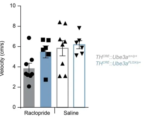
suggested in the AS model mice. Using a real-time place preference (RTPP) paradigm, we coupled i.p. administration of raclopride along with optogenetic
stimulation in THCRE::Ube3aFLOX/p+ to determine if D2-receptors are similarly affected
and in AS model mice when Ube3a is eliminated pre-synaptically in TH-expressing VTA neurons. Using 30 Hz stimulation, there was a trend for a decrease in
sensitivity to D2-antagonism mediated by raclopride in THCRE::Ube3aFLOX/p+