Introduction
T
he first cases of HIV-1 infection in USSR weredetected in late 1980s,1but the expansion of HIV-1
ep-idemic started only in 1996 when a subtype A virus strain was introduced into the population of people who inject drugs
(PWID).2Since then the HIV-1 epidemic in Russia has been
growing rapidly, resulting in a total of 1,114,815 cases of
infection, with over 100,000 new cases reported in 2016.3
The city of St. Petersburg is the second largest city in Russia and one of the regions with the highest prevalence of
HIV-1 infection that approached 1% of the city population in 2016. PWIDs historically have been at the highest risk of
HIV-1 acquisition in Russia and St. Petersburg4,5; however,
the virus has been also spreading into the general population.6
In 2016, 35% of new HIV-1 infections in St. Petersburg were attributed to injection drug use (data of St. Petersburg City AIDS Center, www.hiv-spb.ru, in Russian).
Acute HIV-1 infection (AHI) cases are characterized by extremely high viral loads and a proportionally high risk of further spread of infection, so monitoring and clinical man-agement of such cases have become a standard practice in
1
Lineberger Comprehensive Cancer Center, University of North Carolina at Chapel Hill, Chapel Hill, North Carolina. 2
Peter the Great St. Petersburg Polytechnic University, St. Petersburg, Russian Federation. 3Pavlov State Medical University, St. Petersburg, Russian Federation.
4
The Biomedical Center, St. Petersburg, Russian Federation.
5Laboratory for AIDS Vaccine Research and Development, Department of Surgery, Duke University, Durham, North Carolina. 6
School of Medicine, University of North Carolina at Chapel Hill, Chapel Hill, North Carolina. 7
College of Public Health, The Ohio State University, Columbus, Ohio. 8
Department of Biochemistry and Biophysics, and the UNC Center for AIDS Research, University of North Carolina at Chapel Hill, Chapel Hill, North Carolina.
Characterization
of
the
Transmitted
Virus
in
an
Ongoing
HIV-1
Epidemic
Driven
by
Injecting
Drug
Use
ElenaDukhovlinova,1AlexeyMasharsky,2 AleksandraVasileva,2AlessandroPorrello,1ShuntaiZhou,1 OlgaToussova,3SergeiVerevochkin,2EkaterinaAkulova,4DmitrijFrishman,2DavidMontefiori,5CeliaLabranche,5
IrvingHoffman,6WilliamMiller,7MyronS.Cohen,6AndreiP.Kozlov,2,4andRonaldSwanstrom1,8
Abstract
Understanding
features
of
the
HIV-1
transmission
process
has
the
potential
to
inform
biological
interventions
for
prevention.
We
have
examined
the
transmitted
virus
in
a
cohort
of
people
who
inject
drugs
and
who
are
at
risk
of
HIV-1
infection
through
blood
contamination
when
injecting
in
a
group.
This
study
focused
on
seven
newly
infected
participants
in
St.
Petersburg,
Russia,
who
were
in
acute
or
early
infection.
We
used
end-point
dilution
polymerase
chain
reaction
to
amplify
single
viral
genomes
to
assess
the
complexity
of
the
transmitted
virus.
We
also
used
deep
sequencing
to
further
assess
the
complexity
of
the
virus.
We
interpret
the
results
as
indicating
that
a
single
viral
variant
was
transmitted
in
each
case,
consistent
with
a
model
where
the
exposure
to
virus
during
transmission
was
limited.
We
also
looked
at
phenotypic
properties
of
the
viral
Env
protein
in
isolates
from
acute
and
chronic
infection.
Although
differences
were
noted,
there
was
no
consistent
pattern
that
distinguished
the
transmitted
variants.
Similarly,
despite
the
reduced
genetic
heterogeneity
of
the
more
recent
subtype
A
HIV-1
epidemic
in
St.
Petersburg,
we
did
not
see
reduced
variance
in
the
neutralization
proper-ties
compared
to
isolates
from
the
more
mature
subtype
C
HIV-1
epidemic.
Finally,
in
looking
at
members
of
injecting
groups
related
to
the
acute
HIV-1
infection/early
subjects,
we
found
examples
of
sequence
linkage
consistent
with
ongoing
and
rapid
spread
of
HIV-1
in
these
groups.
These
studies
emphasize
the
dynamic
nature
of
this
epidemic
and
reinforce
the
idea
that
improved
prevention
methods
are
needed.
many countries.7The studies of viral genetic and phenotypic diversity in AHI cases are aimed at finding the patterns in the transmitted isolates that can guide the design of a
region-specific vaccine.8,9A stringent genetic bottleneck in
hetero-sexual transmission has been observed and extensively
studied for the subtype B and C viruses.10–13
We previously reported a cross-sectional study of active PWID who were in acute or early stages of HIV-1 subtype A infection, and found the HIV-1 genetic bottleneck present upon parenteral transmission, with the majority of infection
cases attributed to a single viral variant.14In a similar study
of PWID in Bangkok, the parenteral transmission was also
associated with a similarly strong genetic bottleneck.15 In
contrast, the analysis of the transmitted HIV-1 in a PWID cohort of Montreal showed a less stringent bottleneck and a
higher percentage of infections with a complex viral population.16
In this article, we analyzed longitudinal plasma samples collected through a cohort study that monitored HIV-1 ac-quisition in real time among PWID in St. Petersburg, Russia. The study design and cohort description are published
else-where.17We used single genome amplification (SGA) and
Illumina next-generation sequencing to detect minor variants present near the time of transmission among acutely or recently infected PWID. We confirmed the strong genetic bottleneck of HIV-1 transmission in a majority of studied participants. We also followed early evolution in longitudinal samples and observed a superinfection event during acute infection. Molecular epidemiology allowed the identifica-tion of transmission clusters within injecting subgroups. We also tested the idea that the overall less diverse isolates in St. Petersburg, due to the more recent introduction of HIV-1 into this population, might be more homogeneous in their neutrali-zation properties than a more diverse and older epidemic.
Materials and Methods
Cohort description
A detailed description of the cohort is published
else-where.17Briefly, we recruited and followed up individuals
who were at high risk of HIV acquisition based on the criteria of frequent use of injectable drugs. The participants signed informed consent to donate the blood samples to be tested by real-time PCR, ELISA, and Western blot for HIV-1. They also agreed to answer a series of questions about their drug use behavior. Appropriate counseling was provided to each individual before and after the tests for HIV. The study was approved by Institutional Review Boards of UNC Chapel Hill and the Biomedical Center.
Single genome amplification
In SGA, the cDNA copies of the viral RNA are diluted to endpoint and nested polymerase chain reaction (PCR) am-plifications are done at a dilution where a majority of the reactions are negative, ensuring that a majority of the positive amplifications were initiated from a single template; this approach ensures that each amplicon represents an indepen-dent observation, and the total number of amplicons obtained defines the sample size for querying the genetic diversity of
the viral population.18Amplicons of individual copies of the
HIV-1envgene were obtained starting with viral RNA
ex-tracted from plasma and then amplifying cDNAs spanning
theenvgene using semi-nested PCR with the primers listed
below. For the first round of SGA, we used cEnvA and EnvN, and for second round of SGA, we used cEnvA and EnvM. Primer sequences used for SGA sequencing are listed in Supplementary Table S1 (Supplementary Data are available online at www.liebertpub.com/aid).
The SGA sequences were deposited to the Genbank, ID: MH603967-MH604583. Sequence analysis was performed using BioEdit and Mega 6.0 software. Sequence alignment and Poisson Fitter analysis (after correction for APOBEC3G/F mutations) were performed using LANL database tool kit (https://www.hiv.lanl.gov/content/sequence/HIV/HIVTools .html). Calculations of mean pairwise nucleotide distances per sample and phylogenetic analysis of SGA-derived amplicons were performed using Mega 6.0 and plotted by Prism 6.0.
Illumina MiSeq sequencing
We used a previously published Primer ID approach to
Illumina next-generation sequencing (NGS).19The NGS
li-braries were created with the env-specific primers A3F
(NGSAdapter1-CAGTCATGACCTGGATGCAATGGGACA) and A3R (NGSAdapter2-CCCTATCTGTCCACCCAGCTA CT) and sequenced using MiSeq at the UNC High-Throughput Sequencing Facility. We performed the bioinformatics data
analysis as previously reported.19
For the NGS sequence analysis, we used two types of consensus sequences. First, the template consensus sequence (TCS) was generated from the sequence reads that contained the same Primer ID since each sequence was generated from a single cDNA molecule (and RNA template). Due to the high number of TCS sequences, we used randomly generated sets of 200 TCS sequences to compare the pairwise genetic diversity between the subjects.
For the phylogenetic analysis, we used collapsed identical TCS to focus on the diversity within the viral population. Thus, we were able to assess the viral variants present in the subject’s viral population and the representation of these variants, which was done using 200 randomly sampled sequences from the collapsed population. The calculations of pairwise distances and phylogenetic analysis were performed using Mega 6.0.
Pseudovirus generation
The SGA-derivedenvamplicons were reamplified using
the Phusion Hot-start High-Fidelity DNA polymerase (Finn-zymes), gel-purified using the QIAquick Gel Extraction Kit (QIAGEN), and cloned using the pcDNA3.1 Directional TOPO Expression Kit (Thermo Fisher Scientific) according to the manufacturer’s protocol. Sequence-verified vectors were used for a transient transfection of 293T cells in equal ratio with the luciferase-containing backbone vector pNL4-3.LucR-E- plas-mid (AIDS Research and Reference Reagent Program, Divi-sion of AIDS, NIAID, NIH) with the FuGENE 6 transfection reagent (Roche). The viral supernatants were collected after
48 h, filtered through 0.45lm filters (Millipore), and stored in
small aliquots at-80C to minimize freeze–thaw cycles.
Neutralization assay
values toward 0 and make the kurtosis values nearer to 3, thus obtaining probability distributions closer to the normal before performing statistical inference on them using parametric methods. In particular, (1) the absolute values of the skew-ness of all the probability distributions of the analyzed Abs were reduced after log transforming the data and (2) from a more qualitative perspective and more complexly, also, the absolute values of the difference between the kurtosis of these distributions (originally, all leptokurtic) and the number 3 were reduced after the log transformation. Additionally, the statistical tests described below were utilized relying on their robustness.
Then, we tested the differences of variance and mean concerning four groups of viruses (group 1: acute infection, subtype A; group 2: chronic infection, subtype A; group 3: acute infection, subtype C; and group 4: chronic infection, subtype C). In particular, we compared group 1 versus 3 and
group 2 versus 4 for each Ab. First,F-tests (two sample tests
for equal variance) were performed for these two compari-sons. Later, for the similar two comparisons, we performed a
t-test (of equality of the means) that either assumes that the
samples come from independent random samples with the
same variance (when the correspondingF-test p value was
greater than .05) or that does not make this assumption
(Welch’st-test, when theF-testpvalue was less than .05).
Canonically, theF-tests were used to compare the spreading
of the values around the means, that is, the variances, between
groups, while thet-tests assessed the differences between the
means themselves. Since there was no significant difference in the mean and variance within subtype A and C of samples of acute and chronic viruses for four Abs (PG9, 4E10, sCD4, and SA-C72), we tested for statistical differences between the subtype A (acute and chronic cases) and C (acute and chronic
cases) using the F-test as well as the standard or Welch’s
t-test, based on the results of the correspondingF-test.
For the sake of completeness, also, the statistical results of the fifth Ab (HIVIG-C) were calculated and are shown with the others, despite the caveat that acute and chronic viruses of this Ab have a statistically significant difference in their variance for subtype C. All statistical tests were two tailed.
Tropism assay
We assessed the usage of CD4 and coreceptors in the in-ducible Affinofile cell line according to the protocol described
elsewhere.22 Pseudoviruses were incubated with Affinofile
cells that were induced to either CD4high/CCR5highor CD4low/
CCR5highexpression profiles, which approximate the level of
CD4 on T cells or macrophages, respectively. The pseudo-viruses were incubated with cells in the presence or absence of
AMD3100 and/or maraviroc (10lM) to determine the ability
to use CXCR4 or CCR5 as the coreceptor. Data were analyzed and plotted using Prism 6.0.
Results
Characterization of subjects with AHI with injection drug use as their major transmission risk factor
Briefly, we recruited a cohort of HIV-negative persons who were actively injecting drugs (PWID) in St. Petersburg,
Russia, described in greater detail elsewhere.17 The cohort
participants were frequently monitored for plasma HIV-1
usingastandardprotocol.20Thedataarereportedeitheras
theserumdilutionortheconcentrationofamonoclonal
an-tibodyneededtoinhibit50%ofthevirusinfectivity(ID50
or IC50, respectively), as interpolated using 5-parameter
curve-fitting.
Neutralizationdataanalysis
Ouranalysisofantibody(Ab)concentrationsobtainedina
neutralizationassayforsubtypeApseudovirusessplitintotwo
groups(acutevs.chronicinfection)wasperformedinthe
fol-lowingmanner:preliminarily,datawereorganizedinamatrix
withvirusesontherowsandAbsonthecolumns.Forachieving
abalanceddataanalysisthataccountsforthestepsdescribed
below, when an Ab concentration was above the range of
measurability,itwasrepresentedbyanumberthatequalstwice
themaximumofthatrange.Viceversa,whenanAb
concen-trationwasbelowtherangeofmeasurability,itwasrepresented
byanumberthatequalshalftheminimumofthatrange.Then,
concentrationvalueswerelog2-transformed,andsubsequently,
medians were calculated for each column and subtracted
from them. The log transformation exploits the fact that
when,forinstance,x=2·y, log2(x/y)=1;instead,ifx=0.5·y,
log(x/y)= -1.Consequently,proportionalchangesineither
direction(increasesandreductions)always become
sym-metricalwithrespecttothe0(ifthereisnovariation,i.e.,
whenx=y: log2(x/y)=log2(x/x)=0).
Thereafter,weperformedahierarchicalclusteringbothof
therowsand thecolumns usingthe uncenteredcorrelation
coefficientasoursimilaritymetricandthe averagelinkage
clusteringmethod.Clusteringresultsweredisplayedthrough
aheatmapandtwodendrograms(oneforthecolumns,shown
onthetop,andonefortherows,shownontheleftside).Dueto
thecolumn-by-columnmediancenteringthatweused,all
log-transformedvaluesarerecalculatedwithrespecttothemedians
(i.e.,bysubtractingthemedians),whichhavethewell-known
propertytohavethesamenumberof(column)valuesgreater
andlowerthanthemselves.Consequently,intheheatmapof
log-transformeddata,ageneralchromaticbalanceisachieved,
ineachcolumn,betweenbluesquares(log-transformedvalues
thatwereabovethemedianbeforebeingmediancentered)and
yellowsquares (log-transformedvalues thatwerebelow the
medianbeforebeingmediancentered),anddifferencesamong
virusesaremoreeasilydetectable.
Wecompared subtypeApseudovirus neutralizationdata
withapanelofsubtypeCvirusestestedinasimilarwayand
usingsharedAbs.21Inthisnewdataset,fivecasesofsubtype
Avirusesthatwererecombinantsbetweenfoundervariants
ofthe virusof subjectAHI_Dand acomplexrecombinant
virusSC1283wereremovedfromtheanalysis;also,seven
missingvaluesofsubtypeCcouldnotbeusedforthePG9
Ab.Asapreliminarystep,weadjustedneutralizationvalues
thatwere above orbelow the detectionthreshold, separate
andindependentofourclusteringanalysisofsubtypeA,for
the five Abs available. These extreme values, which were
recordedinourspreadsheetsas<Xor>Y,werechangedinto
X/2and2Y,respectively.Then,allthesevalueswere
log10-transformed, keeping into account that log2- and
log10-transformed valuesare directlyrelated by theapproximate
formula: 3.3219$log10(x)=log2(x). These log
transforma-tions,differentfromwhatwaspreviouslydescribedforthe
RNA to identify cases of AHI. Based on their drug use pat-terns and frequency of injection, these subjects were at high risk of HIV-1 acquisition through injection and this allowed analysis of the transmitted virus upon injecting drug use (IDU) transmission. A summary of the demographics of the seven subjects identified in acute infection, their risk factors, and the dates of plasma sampling after transmission are presented in Table 1.
Based on the provided behavioral data, that is, number of sex partners, number of sexual contacts before AHI detection, and frequency of drug injection, we assigned drug use (IDU), sexual transmission (Sex), or both as potential route of transmission for each AHI subject. Due to the low sample size and variations of responses, these assumptions cannot be verified statistically and should be treated as informational/ qualitative data.
Also, included in the table are the days of sampling since the last HIV-negative test as well as the Fiebig stage based on the assessment of their antibody response. The day of de-tection of HIV-1 was used as their entry day (day 0) into the
AHI cohort. In addition, eight members of their risk net-works, defined as sharing needles and syringes, were re-cruited into this study.
We were interested in assessing the genetic complexity of the virus transmitted by IDU and in characterizing the phe-notypic features of the viral Env protein encoded in these transmitted viruses. To accomplish these goals, we per-formed SGA of viral RNA isolated from longitudinal plasma samples of seven AHI subjects and the eight members of their
networks. The number ofenvgene amplicons obtained for
each plasma sample is presented in Table 1. All the viral strains obtained in this study belonged to the subtype A virus lineage specific to the Former Soviet Union countries.
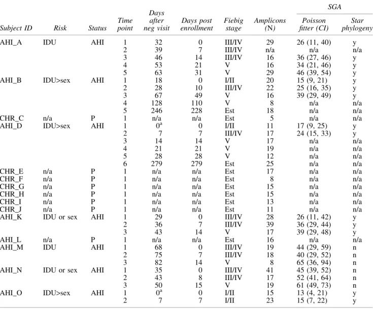
A phylogenetic analysis of a sampling of all of theenvgene
sequences is shown in Figure 1. This analysis revealed three transmission clusters: (1) AHI subject AHI_D with partners CHR_F and CHR_G, although the reported infected partners CHR_E, AHI_H, CHR_I, and CHR_J had viruses unrelated to this cluster (by phylogenetic analysis); (2) AHI subjects AHI_A and AHI_B with partner CHR_C; and (3) AHI
Table1. Study Subjects and Single Genome Amplification Data Summary
Subject ID Risk Status
Time point
Days after neg visit
Days post enrollment
Fiebig stage
Amplicons (N)
SGA Poisson fitter (CI)
Star phylogeny
AHI_A IDU AHI 1 32 0 III/IV 29 26 (11, 40) y
2 39 7 III/IV n/a n/a n/a
3 46 14 III/IV 16 36 (27, 46) y
4 53 21 V 16 34 (21, 46) y
5 63 31 V 29 46 (39, 54) y
AHI_B IDU>sex AHI 1 18 0 I/II 20 15 (9, 21) y
2 28 10 III/IV 22 25 (16, 35) y
3 67 49 V 16 39 (29, 49) y
4 128 110 V 8 n/a n/a
5 246 228 Est 18 n/a n/a
CHR_C n/a P 1 n/a n/a Est 5 n/a n/a
AHI_D IDU>sex AHI 1 0a 0 I/II 11 17 (9, 25) y
2 7 7 III/IV 17 24 (15, 33) y
3 14 14 V 17 n/a n/a
4 21 21 V 19 n/a n/a
5 28 28 V 12 n/a n/a
6 279 279 Est 25 n/a n/a
CHR_E n/a P 1 n/a n/a Est 17 n/a n/a
CHR_F n/a P 1 n/a n/a Est 8 n/a n/a
CHR_G n/a P 1 n/a n/a Est 15 n/a n/a
CHR_H n/a P 1 n/a n/a Est 15 n/a n/a
CHR_I n/a P 1 n/a n/a Est 13 n/a n/a
CHR_J n/a P 1 n/a n/a Est 11 n/a n/a
AHI_K IDU or sex AHI 1 29 0 III/IV 28 26 (11, 42) y
2 36 7 III/IV 39 36 (29, 44) y
3 43 14 V 17 39 (29, 48) y
AHI_L n/a P 1 n/a n/a Est 16 n/a n/a
AHI_M IDU AHI 1 68 0 III/IV 19 44 (29, 59) n
2 75 7 III/IV 18 40 (29, 52) n
3 82 14 V 8 65 (36, 94) n
AHI_N IDU or sex AHI 1 35 0 III/IV 41 45 (39, 52) n
2 43 8 III/IV 17 52 (41, 64) n
3 50 15 V 19 61 (49, 73) n
AHI_O IDU>sex AHI 1 0a 0 I/II 15 13 (4, 21) y
2 7 7 I/II 23 15 (7, 22) y
a
Identified as AHI at enrollment.
subject AHI_K and partner CHR_L. AHI subjects AHI_M, AHI_N, and AHI_O each appeared as a distinct phylogenetic lineage. The features of the viral sequences in the three transmission clusters are described below.
Network cluster 1contained sequences from AHI subject AHI_D and the sequences related to two of his PWID net-work members CHR_F and CHR_G, who had established HIV-1 infection as assessed by Western blot results. The viral population of AHI_D was homogeneous at the time of initial entry screening and 7 days after entry into the study, in each case giving rise to a star-like phylogeny. The presence of a homogeneous population is consistent with the transmis-sion of a single viral genome in the transmistransmis-sion event. The presence of a largely homogeneous population early after transmission allowed the use of Poisson Fitter to estimate the days postinfection, which, after elimination of sequences, likely to have been hypermutated by APOBEC3G/F, was estimated to be 17 days, consistent with the Fiebig Stage I/II designation based on antibody assessment (Table 1).
The phylogenetic analysis and highlighter plot of
full-length envsequences from the entry time point of AHI_D
indicated that the founder virus of AHI_D was most closely related to the viruses in CHR_G. Even though CHR_G was WB positive at enrollment and was classified as chronically infected, the viral population of CHR_G, as seen in the highlighter plot (Supplementary Fig. S1A), was relatively homogeneous. The viral population of CHR_F was also re-lated to AHI_D, although not as close as to CHR_G, and homogeneous, indicating these were likely recent infections within the AHI_D cluster from a common source.
Surprisingly, at 14 days after study entry, the viral popu-lation of AHI_D revealed a second largely homogeneous variant that was distinct from the entry founder virus and could not be attributed to contamination from any sample tested in this study. We interpret this pattern as a superin-fection event during the acute insuperin-fection period. The plasma samples from day 21 after study entry and later revealed extensive recombination between the initial founder virus and the superinfecting variant; however, we cannot exclude the possibility that a third variant was introduced during these later time periods, which appeared as recombinants with the initial two viruses.
Network cluster 2included 2 AHI subjects, AHI_A and his network and sexual partner AHI_B, as well as their PWID network member CHR_C, who had an established infection. Subject AHI_A was considered AHI, given a visit 30 days previously where he was assessed as HIV negative. We de-tected a largely homogeneous viral population (Supplemen-tary Fig. S1B), and analysis of the diversity with the Poisson Fitter program gave an estimate of 26 days postinfection for this subject, consistent with his Fiebig III/IV designation. The homogeneity of the population is consistent with the trans-mission of a single variant.
Subject AHI_B tested positive for HIV-1 at screening, and her virus was highly homogeneous, consistent with a recent transmission event of a single virus variant. Poisson Fitter analysis suggested a population age of just 15 days, consis-tent with the Fiebig I/II designation.
The viral population of network partner CHR_C was di-verse and related to both AHI_B and AHI_A, suggesting CHR_C as a possible source of infection for both AHI_B and AHI_A (or an equivalent partner with a similar virus with
FIG. 1. Phylogenetic analysis of full-length env genes
obtained by SGA from the transmitted (red) and chronic
(black) virus shows the transmission clusters and a genetic
bottleneck among AHI subjects. Brackets indicate the
transmission clusters. Triangles indicate the phylogenetic
clusters of diverse yet related sequences collapsed for visual
convenience; the length of thetriangles is proportional to
similar diversity). The placement of these sequences within the phylogenetic tree (Fig. 1) indicates that the virus was not passed directly between AHI_A and AHI_B.
Network cluster 3 included AHI subject AHI_K and the chronically infected network partner CHR_L (Fig. 1). The viral population of AHI_K was homogeneous at entry (Sup-plementary Fig. S2A) with the Poisson Fitter analysis sug-gesting days postinfection at 26, consistent with the Fiebig III/ IV designation from antibody analysis (Table 1). Again, the homogeneity of this population was consistent with a single variant being transmitted. In contrast, the viral population in CHR_L was much more diverse, indicating that this person had been infected for a much longer period of time.
We also had three individuals with AHI, which did not have any risk network members enrolled in the study: AHI_M, AHI_N, and AHI_O. The lineages for these viruses were distinct from the three network clusters and from each other (Fig. 1). Subject AHI_O was identified as being in acute infection at screening with a Fiebig I/II designation. The viral population was very homogeneous (Supplementary Fig. S2B), with the Poisson Fitter analysis suggesting an infection 15 days before screening, and the homogeneity of the viral population is again consistent with transmission of a single variant. The viral populations of AHI_M and AHI_N that appeared to be older in those single nucleotide variants (Supplementary Fig. S2C, D) were becoming fixed in the population, making the Poisson Fitter estimates of transmissions at 44 and 45 days before sampling, respectively, less accurate, but still consistent with the Fiebig stage designation of III/IV for each. How-ever, the lack of distinct lineages within either of these entry samples is still consistent with the transmission of a single variant in each case.
In this cohort of seven individuals with AHI, all seven subjects displayed a severe transmission bottleneck, which we interpret as a single variant being transmitted in each case. In addition, it was relatively easy to identify linked trans-missions within these self-identified partner networks. In one network, two of six partners had viruses that were phyloge-netically linked to the AHI subject (AHI_D), and all three of the viral populations within this network were relatively young, suggesting recent linked transmission events within the network. Similarly, the network of AHI subject AHI_A included a linked chronically infected partner and a partner in acute infection, which was even more recent. The other three AHI subjects were not linked to the other members of the cohort. However, the ability to find linked infections within these networks without extensive sampling indicates they represent foci of active HIV-1 transmission.
The low variability on pairwise nucleotide diversity among transmitted isolates, as analyzed using the
full-lengthenvgene sequences (SGA), was consistent with the
bottleneck hypothesis (Fig. 2A). The nucleotide diver-sity mean values with range for transmitted isolates were 0.0007 (0.0004–0.0013), while for chronic isolates, they
were much higher—0.01345 (0.0023–0.0326), p=.0003
by Mann–Whitney test.
Nucleotide substitution rate varies among transmitted isolates
We analyzed the nucleotide substitution rate of the trans-mitted virus using the BEAST software package, employing
tip-dating by incorporating into the presets the days postin-fection according to the Fiebig stage, or alternatively using a consensus sequence of the HIV-1 strain created for each subject based on their viral population present at the earliest available time point of infection. The viral population of AHI_D was excluded from the analysis due to the multiple variants present.
The results for the AHI subjects show some differences in the substitution rates (Fig. 2B). The estimated substitution rate
values fluctuate between 2·10-5to 7·10-5. There are
sev-eral reasons why there may be such a range in apparent sub-stitution rates. First, the different founder viruses may vary in the rate of misincorporation by reverse transcriptase. Second, very early immune responses may provide diversifying se-lective pressure; these early responses could be due to a higher state of immune activation in some individuals, reactivity to fortuitous cross-reactive antigens, or the presence of low-level cytotoxic T lymphocyte (CTL) activity due to repeated ex-posure to HIV-1 before establishing a systemic infection (these drivers of early diversity are not mutually exclusive). Finally, repeated superinfection from the same donor who is in an acute or early stage of HIV infection is another possi-bility for adding diversity to a population that would mimic an apparent high substitution rate.
Primer ID deep sequencing results reveal few minor variants in the pool of transmitted viruses
The samples previously analyzed by SGA were sequenced using the Primer ID NGS approach that corrects the error rate and quantifies the sampling depth for the detection of minor
variants. We chose to sequence a fragment of theenvgene that
spans the region from C1-V1-V2-C2, although the paired-end reads do not quite meet. The use of Primer ID allows each RNA template to be evaluated independently as a distinct se-quence even after the PCR step, avoiding the problem of PCR resampling. The goal was to determine if we could identify any minor viral variants not sampled by SGA, which might be present at a lower level than the founder virus.
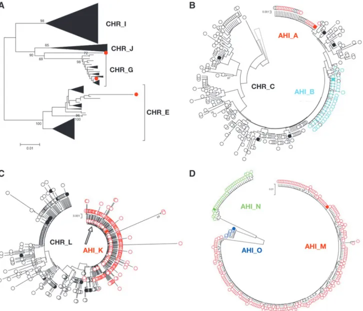
For the subject AHI_D, we were able to detect the second transmitted variant within 2 weeks postscreening, compared to 3 weeks by SGA. We also performed NGS analysis of his PWID network members CHR_E, CHR_G, CHR_I, and CHR_J (Fig. 3A). No sample was available for analysis from subject CHR_F. We detected the same founder variant that belonged to CHR_G group, and a superinfection with the variant that belonged to an unidentified subject, consistent with what was observed by SGA.
In the network cluster 2, the virus from both AHI subjects AHI_A and AHI_B cluster with different variants presents in CHR_C more than with each other, indicating that CHR_C, either directly or indirectly, was a donor for both of these subjects (Fig. 3B).
The NGS analysis of the other study subjects revealed that AHI_M had closely related viral variants that could be due to multiple variants or multiple events of superinfection, but from a person in early infection, or, more likely, the early diversification of a single transmitted variant in this some-what later AHI case (Fig. 3D). Similarly, the viral popula-tions of AHI_O and AHI_N showed the single viral variant for each (Fig. 3D).
Using the Primer ID NGS approach, we characterized viral populations of AHI subjects and their partners. We were able to detect an additional donor for AHI_D not sampled at an earlier time point by SGA. However, overall, the conclusions about population diversity drawn from the SGA analysis were confirmed using the deep sequencing approach, spe-cifically that transmission in these IDU cases is dominated by
a severe genetic bottleneck that most often results in the in-fection being established by a single variant.
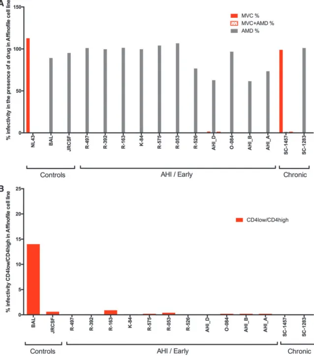
Phenotypic analysis of transmitted strains shows predominantly R5 T cell-tropic strains among AHI subjects
We used the full-lengthenvgenes of the isolates from the
subjects of this and our previously published study of PWID in
St. Petersburg14to create a panel ofenvgenes from selected
SGA amplicons cloned into an expression vector representing 11 different subjects in acute infection. The expression vectors were used to create pseudotyped viruses encoding a reporter gene (luciferase) that was used to test for infection of Affinofile cells. We first assessed coreceptor specificity by carrying out
FIG. 2. Assessment of nucleotide diversity and viral evolution among AHI and chronic (CHR) subjects. (A) Mean
pairwise nucleotide distance with standard deviation for either full-lengthenvgene (AHI) or its fragment (NGS). The values
are shown for members of the network cluster 1 (blue), network cluster 2 (red), and network cluster 3 (green), as well as for
the subjects with unrelated virus (gray). (B) Estimation of nucleotide substitution rates of AHI-derived full-length env
amplicons by BEAST. We used lognormal type of molecular clock for tip-dating of the viral variants by setting a consensus
sequence set as point 0. The order of the subjects onX-axis is determined by the increase in nucleotide substitution rate.
the infections with or without a CCR5 antagonist and/or a CXCR4 antagonist. As can be seen in Figure 4A, all of the
envgenes from viruses in acute infection encoded proteins
that used CCR5 for entry and not CXCR4, as is typically
seen in sexual transmission. One of the env genes cloned
from a participant in chronic infection (SC1457) used CXCR4 exclusively for entry.
We next tested theenv clones for entry phenotype with
respect to whether they required a high density of CD4 for efficient entry (T cell tropic) or if they were variants that could use a low density of CD4 on the cell surface for effi-cient entry (macrophage tropic). Shown in Figure 4B are the
results indicating that all theenvgenes from both the acute
and chronic infection samples encoded proteins that failed to
mediate efficient entry at a low density of CD4, thus making
all theenvgenes tested T cell tropic, again similar to that seen
in sexual transmission.
Neutralization sensitivity assays of chronic and acute subtype A isolates, and comparing subtype A and subtype C isolates
We tested the neutralization sensitivity of the pseudotyped viruses derived from transmitted and chronic isolates to a panel of HIV-specific antibodies. The panel included 4 polyclonal serum samples from HIV-infected individuals of subtype C, HIVIG-C from pooled sera, soluble CD4, and the monoclonal antibodies 4E10, PG9, and CH01-31.
FIG. 3. Phylogenetic analysis of the deep sequencing data (TCS) obtained for the clusters and unrelated AHI cases
reaffirm a single-variant transmission in all the AHI cases. Collapsed TCS are labeled withopen circlesand SGA-derived
variants placed on the same tree for the reference are labeled withclosed circles.Different colorsindicate the AHI subjects,
blackindicates chronic subjects.(A)Network cluster 1 and other PWID related to AHI_D by risk behavior, with AHI_D
viral variantshighlightedinred closed circles.(B)Network Cluster 2.(C)Network Cluster 3, with thearrowpointing to the
identical sequences of AHI_K (red) and CHR_L (black).(D)Three unrelated AHI subjects that were placed on the same
A hierarchical clustering analysis revealed three groups of subtype A viruses that differed in their neutralization pattern (Supplementary Fig. S3). Group 1 consisted of seven viruses predominantly derived from acutely or recently infected subjects (top of the heat map); Group 3 contained three isolates from chronic and two from acute subjects (bottom of the heat map, five viruses); and finally, Group 2 was equally split between both chronic and acute isolates (central portion of the heat map, six viruses). At present, we do not know the basis for this clustering, but it is clear that the AHI viruses are distributed throughout the three groups, arguing there is no specific phenotype for the transmitted variants, at least as assessed with these reagents.
Because the introduction of a subtype A virus into the IDU populations of the former Soviet Union countries happened more recently than the establishment of the subtype C
epi-demic in southern Africa11and the subtype B epidemic in the
United States,10 the viral population in the subtype A
epi-demic is less diverse. This is apparent comparing the
se-quences of the full-lengthenvgenes collected from subjects
in acute infection of these three geographical regions ap-proximately in the same period (Supplementary Fig. S4). We were interested in testing the hypothesis that the neutraliza-tion properties would be more homogeneous in the more genetically homogeneous viral population, that is, subtype A.
FIG. 4. Affinofile cell culture assay of HIV-1 subtype A pseudoviruses shows little variability between acute and chronic
isolates.(A)CXCR4 virus detected in a chronically infected subject.(B)All the viruses tested in the assay had a T
We compared previously published data21 that focused on subtype C isolates from individuals with a predominantly heterosexual risk of acquisition versus our data set of subtype A isolates.
The two main variables of these probability distributions (mean and variance) were statistically compared and the
correspondingpvalues are listed in Supplementary Table S2.
Specifically, we were interested in comparing isolates from acute infection between the two groups, and isolates from chronic infection between the two groups. Chronic subjects of subtype A and C differed in both variance and mean of the neutralization sensitivity to 4E10, with the subtype A prob-ability distribution being less compact and with a lower mean. For HIVIG-C, subtype A pseudoviruses had a lower mean compared with subtype C for both transmitted and chronic isolates; for sCD4, the mean value was higher for acute virus of subtype A versus subtype C pseudoviruses. However, there was no consistent pattern of difference in variance between subtype A and C viruses, despite the dif-ference in genetic heterogeneity, and the difdif-ferences that were observed would not be significant after accounting for multiple comparisons.
To increase the power of our analyses, we compared sub-type A and subsub-type C clones, pooling AHI and chronic iso-lates, assuming that the similarities within subtypes would be larger than the putative differences between subtypes. Most of the trends (of the means) in the AHI and chronic comparisons remained, but we did not see less variance for the subtype A isolates compared to the genetically more diverse subtype C isolates. We conclude that the bottleneck of introduction of the subtype A virus into eastern Europe, and specifically into St. Petersburg, happened sufficiently long ago to obscure phenotypic similarities in neutralization properties.
Discussion
We analyzed the cell-free HIV-1 subtype A viral popula-tions of PWID that had a very high risk of injection-related HIV-1 infection. A viral population that originates from a single viral variant, as an extreme genetic bottleneck, in the absence of selective pressure should evolve in a so-called ‘‘star phylogeny’’ pattern. The viral sequence data obtained by SGA and analyzed with the Poisson Fitter model confirmed a star phylogeny pattern for five out of seven AHI subjects and indicated the possibility of multiple consecutive infections within a PWID network, specifically in the case of the subject AHI_D. In the failed Poisson Fitter model (subjects AHI_M and AHI_N), the viral population contained minor variants that were genetically very similar to the founder virus, likely coming from the same donor (if they were in acute infection) or representing the initial fixation of a few mutations from a single transmission event; given this interpretation, this would indicate that all seven AHI subjects identified in this study were each infected with a single variant.
There are several scenarios that could increase early di-versity in a newly infected person. In the population with high HIV-1 incidence like in St. Petersburg, and PWID networks where multiple members are infected, frequent exposure to the virus before establishing a systemic infec-tion could result to early CTL escape mutainfec-tions in the re-cipients. Alternatively, superinfection events from the same donor due to frequent contacts through drug use or sex may
also lead to additional early diversity, but given the high similarity of the variants, this would only be the case if the donor was also in a very early stage of infection; multiple variants from a donor in chronic infection would appear much more distinct.
We performed Primer ID deep sequencing to test the hy-pothesis that at the earliest stages of AHI, there are multiple viruses that later are outgrown by the most fit variant. Our data reject this hypothesis and confirm the presence of a very strong bottleneck in AHI subjects. In a previously published cross-sectional study using SGA, we observed a genetic bottleneck in 9 out of 13 subjects with acute and recent
HIV-1 infection.14In this longitudinal study combining the data
obtained by SGA and NGS, we interpret the sum of the data as being most consistent with transmission of a single variant in each case. Combining these data with the previously de-scribed findings, we see that 16 out of 20 (80%) PWID in-dividuals infected with HIV-1 subtype A had a severe genetic bottleneck during the HIV-1 transmission event.
This phenomenon is similar to that seen in heterosexual transmission and provides a framework for considering transmission under these circumstances. It appears to dis-tinguish a model where there is a high probability of trans-mission because of virus-containing blood where multiple variants would be expected to be transmitted from a model where the amount of exposure to infectious material is much less, such that when infection does occur, it does so with a lower probability as evidenced by the presence, most often, of a single variant.
If the genetic bottleneck is due to biological selection of the fittest virus, we might expect to see differences in the pheno-typic properties of Env protein derived from AHI compared to chronically infected subjects. The results did not reveal phe-notypic differences in transmitted versus chronically circulat-ing viruses of IDU subtype A, nor any macrophage-tropic virus present in our study group, so all the transmitted isolates and all but one chronic isolate belonged to CCR5-dependent T cell-tropic class of viruses. Thus parenteral transmission does not select strongly for a specific phenotype, at least by the measures used, and the transmitted variants appear similar to those that are typically found in the blood of chronically in-fected people. However, the sample size in this study was relatively small and therefore not able to detect differences of smaller magnitude, if they exist.
We assessed the neutralization sensitivity of the pseudo-viruses to a panel of anti-HIV-1 sera and antibodies and compared the data within the panel and with the published
HIV-1 subtype C data.21The hierarchical clustering
analy-sis identified three clusters of the clones: one derived from mostly acute/recent infection cases, and two others derived from a mix of both chronic and acute cases. Although the nature of the higher sensitivity of viruses in the group (1) is unclear, they may be useful to further study as the candidate strains for the vaccine design against the transmitted isolates of subtype A. However, we were unable to generate evidence that the more recent nature of this subtype A epidemic retains less diversity in its neutralization properties, despite the fact that there is less diversity in the genetic population relative to older portions of the HIV-1 epidemic.
References
1. Kozlov AP, Volkova GV, Malykh AG, Stepanova GS, Gle-bov AV: Epidemiology of HIV infection in St. Petersburg, Russia. J Acquir Immune Defic Syndr 1993;6:208–212.
2. Bobkov A, Cheingsong-Popov R, Selimova L, et al.:
An HIV type 1 epidemic among injecting drug users in the former Soviet Union caused by a homogeneous subtype A strain. AIDS Res Hum Retroviruses 1997;13:1195–1201. 3. Beyrer C, Wirtz AL, O’Hara G, Le´on N, Kazatchkine M: The expanding epidemic of HIV-1 in the Russian Federa-tion. PLoS Med 2017;14:e1002462.
4. Kozlov AP, Shaboltas AV, Toussova OV, et al.: HIV
in-cidence and factors associated with HIV acquisition among injection drug users in St Petersburg, Russia. AIDS 2006; 20:901–906.
5. Dukhovlinova EN, Masharsky A, Toussova O, et al.: Two
independent HIV epidemics in Saint Petersburg, Russia revealed by molecular epidemiology. AIDS Res Hum Ret-roviruses 2014:150127063121001.
6. Toussova O, Shcherbakova I, Volkova G, Niccolai L, Heimer R, Kozlov A: Potential bridges of heterosexual HIV transmission from drug users to the general population in St. Petersburg, Russia: Is it easy to be a young female? J Urban Health 2009;86:121–130.
7. Rutstein SE, Sellers CJ, Ananworanich J, Cohen MS: The HIV treatment cascade in acutely infected people: In-forming global guidelines. Curr Opin HIV AIDS 2015;10: 395–402.
8. Ronen K, Sharma A, Overbaugh J: HIV transmission bi-ology: Translation for HIV prevention. AIDS 2015;29:2219– 2227.
9. Rademeyer C, Korber B, Seaman MS, et al.: Features of
recently transmitted HIV-1 Clade C viruses that impact antibody recognition: Implications for active and passive immunization. PLOS Pathog 2016;12:e1005742.
10. Keele BF, Giorgi EE, Salazar-Gonzalez JF,et al.: Identifi-cation and characterization of transmitted and early founder virus envelopes in primary HIV-1 infection. Proc Natl Acad Sci U S A 2008;105:7552–7557.
11. Abrahams MR, Anderson JA, Giorgi EE,et al.: Quantitating the multiplicity of infection with human immunodeficiency virus type 1 subtype C reveals a non-poisson distribution of transmitted variants. J Virol 2009;83:3556–3567.
12. Carlson JM, Schaefer M, Monaco DC,et al.Selection bias at the heterosexual HIV-1 transmission bottleneck. Science 2014;345:1254031–1254031.
13. Carlson JM, Du VY, Pfeifer N, et al.: Impact of
pre-adapted HIV transmission. Nat Med 2016;22:606–613. 14. Masharsky AE, Dukhovlinova EN, Verevochkin SV,et al.:
A substantial transmission bottleneck among newly and recently HIV-1-infected injection drug users in St Peters-burg, Russia. J Infect Dis 2010;201:1697–1702.
15. Sterrett S, Learn GH, Edlefsen PT,et al.: Low multiplicity of HIV-1 infection and no vaccine enhancement in VAX003 injection drug users. Open Forum Infect Dis 2014;1:ofu056. 16. Bar KJ, Li H, Chamberland A,et al.: Wide variation in the multiplicity of HIV-1 infection among injection drug users. J Virol 2010;84:6241–6247.
17. Toussova O, Kozlov A, Verevochkin S, et al.: A Cohort approach to real time detection of acute HIV infections among people who inject drugs in St. Petersburg, Russia. AIDS Res Human Retroviruses 2017;AID.2017.0076. 18. Salazar-Gonzalez JF, Bailes E, Pham KT, et al.:
Deci-phering human immunodeficiency virus type 1 transmission
drugsandmanifesteddifferentriskbehavior.Themembers
ofSt. Petersburg and Bangkokcohort reported heroinuse,
whereastheparticipants ofMontrealcohortreported
injec-tionof cocaine.Injection of cocaine mayimply more
fre-quentdrugadministration, thusincreasingthe concomitant
infectionrisksofneedlesharingandsexualactivity.23Sothe
high multiplicity of the infection reported inthe Montreal
cohortmaybeassociatedwiththemultipleviraltransmission
eventsfromthesameordifferentdonors.
An epidemiological study of Russian PWID networks
publishedearliersuggeststhatthespreadofHIV-1
infec-tion among PWID in Russia happens most often within
the small clusters or networks of drug users who share
unsafe injection practices.24 In our study, we detected
three transmission clusters, and our analysis ofthe viral
diversityinPWIDnetworks alongwith the
epidemiolog-ical data gathered in the previous studies suggests that
the HIV-1 prevention measures focused on PWID
net-workswouldbe oneofthemostefficientwaystocontrol
theepidemic.
Russia has seen an unprecedented growth of HIV-1
transmissionamongPWIDsince theonsetoftheepidemic,
with extremely high incidence rates inSt. Petersburg and
othercitiesthathavealargepopulationofPWID.Itisknown
that primary HIV-1 infection is characterized by an
ex-tremelyhighbloodviralloadandproportionallyhigh
prob-abilityoftransmission.Thegeneticbottleneckandanoverall
lowheterogeneityofsubtypeAHIV-1 inRussia maybea
direct consequence of a scenario when a low-complexity
viralpopulationisgettingtransmitted,andtheviralspreadin
alimitedspaceofadruguseclusterwithfrequentinjections
isfast andefficient.Inthiscase,themultiple-variant
trans-missioncouldhappenbeforeselectivepressureinthedonor,
whichcould leadtoanunderestimate of theaverage
com-plexityofthetransmittedvirus.
Sofar,therearelittletonopreventivemeasureinRussia
thatfocusonreducingtheriskofspreadfromacutelyinfected
individuals, and we show that such individuals are
wide-spreadwithinthenetworksofactivedrugusers.Theclinical
testing requirementsinthehigh-risk groups,hospitals, and
bloodbanksshould beexpandedtoincludeHIV-1RNAor
p24assessmentthatwouldallowamoreaccurateandtimely
diagnosis of HIV-1 infection and subsequent preventive
measuresfortheat-risknetworkmembers.
Acknowledgments
This study was supported by NIH grant P30 A150410
UNC Center for AIDS Research, and agreement between
the UNC and the Biomedical Center; E.D. was partially
supported through IAS-NIDA Postdoctoral Research
Fel-lowshipsinHIVandDrugUse;thedataanalysisand
publi-cationwerepartiallysupportedbytheGrantoftheRussian
ScienceFoundation#15-14-00026.
AuthorDisclosureStatement
UNCispursuingIPprotectionforPrimerID,andR.S.is
listedas aco-inventor and has receivednominal royalties.
Presently, E.D. is employed with Janssen Pharmaceutical
CompaniesofJohnsonandJohnson.Nocompetingfinancial
and early envelope diversification by single-genome am-plification and sequencing. J Virol 2008;82:3952–3970. 19. Zhou S, Jones C, Mieczkowski P, Swanstrom R: Primer ID
validates template sampling depth and greatly reduces the error rate of next generation sequencing of HIV-1 genomic RNA populations. J Virol 2015;JVI.00522-15-52.
20. Montefiori DC: Measuring HIV neutralization in a lucif-erase reporter gene assay. Methods Mol Biol 2009;485: 395–405.
21. Ping L-H, Joseph SB, Anderson JA,et al.: Comparison of viral Env proteins from acute and chronic infections with subtype C human immunodeficiency virus type 1 identifies differences in glycosylation and CCR5 utilization and sug-gests a new strategy for immunogen design. J Virol 2013;87: 7218–7233.
22. Joseph SB, Lee B, Swanstrom R: Affinofile Assay for Identifying Macrophage-Tropic HIV-1—BIO-PROTOCOL. Available at bio-protocolorg, accessed July 10, 2018.
23. Bruneau J, Daniel M, Abrahamowicz M,et al.: Trends in human immunodeficiency virus incidence and risk be-havior among injection drug users in Montreal, Canada: A 16-year longitudinal study. Am J Epidemiol 2011;173: 1049–1058.
24. Hoffman IF, Latkin CA, Kukhareva PV, et al.: A
peer-educator network HIV prevention intervention among in-jection drug users: Results of a randomized controlled trial in St. Petersburg, Russia. AIDS Behav 2013;17:2510–2520.
Address correspondence to: