Original Research Article
A study of age as a risk factor in ischemic stroke of elderly
Hari Priya Reddy
1, Jaganath A.
2*, Nagaraj N.
1, Visweswara Reddy Y. J.
1INTRODUCTION
Stroke mostly occurs in elderly people and outcome after stroke are highly influenced by age. The WHO clinically defines stroke as “the rapid development of clinical signs and symptoms of a focal neurological disturbance lasting more than 24 hours or leading to death with no apparent cause other than vascular origin.”1 In the global burden
of disease report of 1997, cerebrovascular disease is the second leading cause of death worldwide.2 It is the
leading cause of disability among elderly worldwide.3
Treatment options are limited to thrombolysis, but only few patients receive this treatment owing to restrictions in application time and indication.4 Thus, primary
prevention remains the most important general strategy for reducing stroke and its impact.5
Several well-established modifiable risk factors for stroke are hypertension (HTN), smoking, cardiac disease, diabetes, etc. Stroke appears to be a preventable disease to a large extent.6,7 Lifestyle changes are likely to
influence risk factor prevalence, which in turn may modify the stroke risk.8 There are certain non-modifiable
risk factors-age is one of them.
The brain changes during aging.9,10 Human post-mortem
studies indicate that brain weight decreases between the ages of 20 and 60 years, with more-rapid loss thereafter.11
There is a good correlation between MRI studies and
1Department ofMedicine,2Department ofAnaesthesia, PESIMSR, Kuppam, Chittoor, Andhra Pradesh, India Received: 29 October 2018
Revised: 20 March 2019
Accepted: 03 April 2019
*Correspondence:
Dr. Jaganath A.,
E-mail: [email protected]
Copyright: © the author(s), publisher and licensee Medip Academy. This is an open-access article distributed under the terms of the Creative Commons Attribution Non-Commercial License, which permits unrestricted non-commercial use, distribution, and reproduction in any medium, provided the original work is properly cited.
ABSTRACT
Background: The aim of the study was to determine the effect of age as a risk factor and a determinant of outcome in elderly ischemic stroke patients.
Methods: This is an observational study. One hundred, successive elderly patients aged 60 years and above, admitted with acute ischemic stroke in PESIMSR over a period of 18 months were prospectively studied. Patients with hemorrhagic stroke, neurological deficits following trauma or following infection were excluded. Demographics, risk factors, stroke severity at admission were estimated by NIHSS. Risk factors and clinical profile were noted and compared among male and female patients. Outcome at discharge was measured by-mRS-modified ranking score.
Results: Patients in age group 60-75 years presented with less severe stroke and better mRS when compared to >75 years age group. Complications were significantly higher among the older age group.
Conclusions: The risk factors identified for ischemic stroke in the present study are diabetes, hypertension, dyslipidaemia, obesity, smoking, and alcohol. Severity of stroke at presentation, clinical outcome and complication rate during the in-hospital stay were all significantly affected by the age, more so in ischemic stroke. Age specific factors of stroke prevention are crucial for successful prevention and implementation of well-organized stroke care.
Keywords: Complications, Elderly, Ischemic stroke, Severity
brain weight studies.12 Moderate to severe changes in
white matter occur in up to one-third of people aged 65-84 years and are termed leukoaraiosis.13,14 In elderly
people, leukoaraiosis has been shown to predict decline in motor performance, the onset of dementia, and rapid global functional decline.15,16 Leukoaraiosis has been
seen in up to 44% of patients with stroke or transient ischemic attack (TIA), and the degree of leukoaraiosis correlates with the risk of recurrent stroke.17-19
The brain microvasculature that forms the blood-brain barrier (BBB) changes during aging, the capillary surface area decreases, while capillary diameter, volume and total length increase.20-22 Such age-related degeneration of
brain vasculature structure and function might lead to the disruption of local perfusion.23-24 Aging-related
alterations in cerebral vessels might eventually reduce cerebrovascular reserves and increase the susceptibility of the brain to vascular insufficiency and ischemic injury.25
METHODS
This is an observational study. One hundred, successive elderly patients aged 60 years and above, admitted with acute ischemic stroke (An episode of neurological dysfunction caused by focal cerebral, spinal, or retinal infarction) in PESIMSR (People’s Education Society Institute of Medical Sciences and Research Kuppam- 517425, Chittoor District, Andhra Pradesh) over a period of 18 months from October 2013 to March 2015 were prospectively studied. The inclusion criteria for the study are patients with above age limit (60 years and above) and those fulfilling the definition of ischemic stroke (an episode of neurological dysfunction caused by focal cerebral, spinal or retinal infarction). Those patients with hemorrhagic stroke, neurological deficits following trauma or due to infection, inflammation and demyelination were excluded. Demographics, presenting complaints such as headache, vomiting, speech defects, convulsions and associated limb weakness, bowel and bladder symptoms were documented. Risk factors, co-morbidities such as diabetes mellitus, hypertension, smoking-tobacco chewing habits, and alcohol consumption were documented. A through physical examination was done including complete neurological examination. Stroke severity at admission was documented by using NIHSS scale (NIHSS-National Institute of Health Stroke score). NIHSS is a 15-element scoring system used to estimate severity of stroke with minimum score of zero and maximum of 32.
Relevant investigations were requested such as echocardiography to rule out cryptic strokes, Doppler-carotid and vertebral to identify any stenosis. Fasting lipid profile along with other organ functions were assessed. Outcome at discharge was measured by modified Rankin's score which is a functional dependency score ranging from 0 to 6, 0 being completely independent and 6 being dead. Stroke severity, complications rate and discharge outcome were
compared among patients aged 60-75 years and those aged >75 years. Risk factors and clinical profile were noted and compared among male and female patients. Data was entered in Microsoft excel and analysis was done using SPSS version 20. Descriptive statistical analysis was done. Results on continuous measurements are presented as mean and standard deviation. Results on categorical measurements are presented as percentages. Significance is assessed at 5 % level of significance. Student’s’ test (independent, two tailed) is used to find out the significance of study parameters on a continuous scale between two groups. Chi square test is used to find out the significance of study parameters on a categorical scale between two groups.
RESULTS
Statistical analysis (Table 1) depicts NIHSS score.
Table 1: Comparison of stroke severity at admission.
NIHSS
Age group N Mean SD P
60-75 87 8.46 4.546 0.001 >75 13 15.59 4.576
Age group 60-75 years presented with less severe stroke (NIHSS-8.46) when compared to those in age group >75 years (NIHSS-15.54) (Table 2) are indicating the ‘Modified Rankin’s score’ at the time of discharge.
Table 2: Modified Rankin’s Score(mRS), at the time of discharge.
mRS
Age N mean SD P
60-75 87 2.52 0.645
0.001 >75 13 4.23 0.725
Clinical outcome at discharge in 60-75 years age group was better (mRS-2.52) when compared to >75 years age group (mRS-4.23). Complications were significantly higher among the older group (92%), as compared to the younger group (21%). UTI was 42.10% Vs 4.59%, pneumonia was 36.84% Vs 6.89%, bed-sore was 36.84% Vs 11.49% and DVT was 10.52% Vs 2.29%. The same has been shown in (Table 3). Complications were significantly higher among the older group (92%), as compared to the younger group (21%).
Table 3: Comparison of complications among 60-75years age group and >60-75years age group.
Age group Total number
of cases
Number of cases with complications
60-75 years 87 19 >75 years 13 12
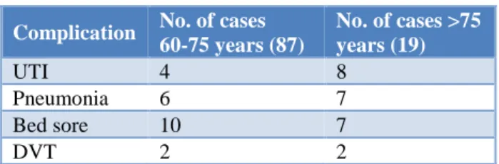
Table 4: Type of complications.
Complication No. of cases 60-75 years (87)
No. of cases >75 years (19)
UTI 4 8
Pneumonia 6 7
Bed sore 10 7
DVT 2 2
It was found that patients aged 75 years and above had overall a higher rate of complications and almost all the patient had complications. Risk factor profiles are displayed in (Table 5).
Table 5: Risk factor profile.
Risk factors Female (%) Male (%) P
Diabetes 38 (71.7) 25 (52.2) 0.056 Hypertension 35 (66.0) 26 (55.3) 0.27 Dyslipidaemia 24 (45.3) 20 (42.6) 0.784 CAD 14 (26.4) 17 (36.2) 0.292 Obesity 13 (24.5) 8 (17.0) 0.358 Tobacco 6 (11.3) 29 (61.7) <0.001 Alcohol 6 (11.3) 16 (34.0) 0.006 There is statistically significant difference of stroke severity between the age groups analyzed. In patients aged above 75 years almost every patient had complications.
DISCUSSION
Stroke mostly occurs in elderly people and outcome after stroke are highly influenced by age. Cerebrovascular disease is the second leading cause of death and of disability among elderly worldwide.2,3 Treatment options
are limited.4 Thus, primary prevention remains the most
important general strategy for reducing stroke and its impact.5
Several well-established modifiable risk factors for stroke are reported. There are certain non-modifiable risk factors-age is one of them. The brain changes during aging.9,10 Human post-mortem studies indicate that brain
weight decreases by ≈ 0.1% per year between the ages of 20 and 60 years, with more-rapid loss thereafter.11 In
agreement with brain weight studies, MRI shows that brain volume decreases by 0.1-0.2% per year from 30 to 50 years of age, and by 0.3-0.5% per year in people aged >70 years.12 Moderate to severe changes in white matter
occur in up to one-third of people aged 65-84 years13 and are termed leukoaraiosis.14 In elderly people,
leukoaraiosis has been shown to predict decline in motor performance, the onset of dementia, and rapid global functional decline.15,16 Leukoaraiosis has been seen in up
to 44% of patients with stroke or transient ischemic attack (TIA), and the degree of leukoaraiosis correlates with the risk of recurrent stroke.17-19
Studies in healthy individuals have reported that age-related degeneration of brain vasculature structure and function might lead to the disruption of local perfusion.23,24 Aging-related alterations in cerebral
vessels might eventually reduce cerebrovascular reserves and increase the susceptibility of the brain to vascular insufficiency and ischemic injury. Such changes could underlie the increase in morbidity and mortality rates following ischemic stroke in older.25
The mean NIHSS score in our cohort of patients in younger group is 8.46 and that of the older group is 15.54 which is statistically significant (p -value<0.05). Younger age group patients presented with less severe stroke when compared to the older age group patients. Similar findings are reported by Deng XY et al study.26 Their
study revealed statistically significant (p<0.05) difference of NIHSS score at admission and at discharge among the age groups of 18-45 years, 46-64 years, 66-80 years and those >80 years. Gur AY et al, studied characteristics and outcome in patients aged<85 years and those with≥85-year-old.27 The very elderly presented with more severe
strokes; 36.3% of the≥85-year-old patients had an NIHSS score ≥11 compared with 22.0% in the younger age group (p<0.05).
The mean modified Rankin scale score at discharge also varied significantly among both the groups, the younger group having a score of 2.52 and the older 4.23 respectively. Similar findings are reported by Knoflach M et al, they studied 43,163 ischemic stroke patients prospectively enrolled in the Austrian stroke unit Registry.28 In multivariable analysis, age emerged as a
significant predictor of outcome independent of stroke severity, etiology, and intervention of thrombolysis, sex, risk factors, and stroke complications. When the age stratum 56-65 years was used as a reference, odds ratios (95% confidence interval (95% CI) of good outcome were 3.4 (1.9-6.4), 2.2 (1.6-3.2), and 1.5 (1.2-1.9) for patients aged 18-35, 36-45, and 46-55 years and 0.70 (0.60-0.81), 0.32 (0.28-0.37), and 0.18 (0.14-0.22) for those aged 66-75,76-85, and>85 years (p<0.001). Findings applied equally to sexes and patients with and without IV thrombolysis.
Complications related to acute ischemic stroke were studied in all patients during the hospital stay. Pneumonia, urinary tract infection, bedsore, deep vein thrombosis were the main complications and rate of complications increased with age and there was more than one complication in most of the patients aged >75 years. Of the 87 patients aged between 60-75years, 19 patients had complications and of the 13 patients aged >75years 12 patients had complications. This difference is statistically significant p-value <0.05.
Chen CM et al, studied the complications in stroke patients.29 The incidence of symptomatic UTI was higher
stay in the rehabilitation ward (p<0.001). Similar findings are reported by Pandian JD et al, in their study of 449 patients.30
CONCLUSION
There is significant difference in both the groups that is between 60 to 75 years and above 75 years in terms of presentation, outcome and complications. Age plays an important determinant role in determining them specifically so in ischemic stroke. Age specific factors of stroke are crucial for successful prevention and implementation well-organized stroke care for this population. The risk factors identified for ischemic stroke in the present study are diabetes, hypertension, dyslipidemia, obesity, smoking, and alcohol
The occurrence of stroke steeply rises with age peaking around 65 years. Hence it is of paramount importance to consider it with much greater vigor. A holistic approach encompassing stroke awareness, establishment of stroke units, dispensing timely care to these victims, are the need of the hour to contain the tough challenge of stroke in our country which is now poised to become a giant threat in near future. The government health planners and research personnel should focus on this problem to improve life span and quality of life after the event.
Funding: No funding sources Conflict of interest: None declared Ethical approval: Not required
REFERENCES
1. Hatano S. Experience from a multicentre stroke register: a preliminary report. Bulletin World Health Org. 1976;54(5):541.
2. Murray CJ, Lopez AD. Mortality by cause for eight regions of the world: global burden of disease study. Lancet. 1997;349(9061):1269-76.
3. Lloyd-Jones. Heart disease and Stroke statistics-2009 update: A Report from the American heart association statistics committee and stroke statistics subcommittee. Circul. 2009;119(3):E182
4. Dávalos A. Thrombolysis in acute ischemic stroke: successes, failures, and new hopes. Cerebrovascular Dis. 2005;20(2):135-9.
5. Leys D, Deplanque D, Mackowiak-Cordoliani MA, Lucas C, Bordet R, Mounier-Vehier C. Stroke prevention: management of modifiable vascular risk factors. J Neurol. 2003;250(9):1125-6.
6. Kuller LH. Epidemiology and prevention of stroke, now and in the future. Epidemiol Reviews. 2000;22(1):14-7.
7. Sacco RL. American heart association prevention conference: IV. prevention and rehabilitation of stroke-risk factors. Stroke. 1997;28:1507-17. 8. Hu G, Sarti C, Jousilahti P, Peltonen M, Qiao Q,
Antikainen R, et al. The impact of history of hypertension and type 2 diabetes at baseline on the
incidence of stroke and stroke mortality. Stroke 2005;36: 25.
9. Raz N. In: New Frontiers in Cognitive Ageing. Dixon RA, Backman L, Nilsson LG, eds. Oxford University Press: Oxford. 2004: 115-134.
10. Esiri MM. Ageing and the brain. J Pathol Society Great Britain Ireland. 2007;211(2):181-7.
11. Anderson JM, Hubbard BM, Coghill GR, Slidders W. The effect of advanced old age on the neurone content of the cerebral cortex: Observations with an automatic image analyser point counting method. J Neurol Sci. 1983;58(2):235-46.
12. Pfefferbaum A, Mathalon DH, Sullivan EV, Rawles JM, Zipursky RB, Lim KO. A quantitative magnetic resonance imaging study of changes in brain morphology from infancy to late adulthood. Archives Neurol. 1994;51(9):874-87.
13. Breteler MM, Van Swieten JC, Bots ML, Grobbee DE, Claus JJ, Van Den Hout JH, et al. Cerebral white matter lesions, vascular risk factors, and cognitive function in a population‐based study: the Rotterdam study. Neurol. 1994;44(7):1246.
14. Fernando MS, Simpson JE, Matthews F, Brayne C, Lewis CE, Barber R, et al. White matter lesions in an unselected cohort of the elderly: molecular pathology suggests origin from chronic hypoperfusion injury. Stroke. 2006;37(6):1391-8. 15. Prins ND, van Dijk EJ, den Heijer T, Vermeer SE,
Koudstaal PJ, Oudkerk M, et al. Cerebral white matter lesions and the risk of dementia. Archives Neurol. 2004;61(10):1531-4.
16. Inzitari D, Pracucci G, Poggesi A, Carlucci G, Barkhof F, Chabriat H, et al. Changes in white matter as determinant of global functional decline in older independent outpatients: three-year follow-up of LADIS (leukoaraiosis and disability) study cohort. BMJ. 2009;339:b2477.
17. Pantoni L, Garcia JH. Pathogenesis of leukoaraiosis: a review. Stroke. 1997;28(3):652-9.
18. Koton S, Schwammenthal Y, Merzeliak O, Philips T, Tsabari R, Orion D, et al. Cerebral leukoaraiosis in patients with stroke or TIA: clinical correlates and 1‐year outcome. European J Neurol. 2009;16(2):218-25.
19. Fu JH, Lu CZ, Hong Z, Dong Q, Luo Y, Wong KS. Extent of white matter lesions is related to acute subcortical infarcts and predicts further stroke risk in patients with first ever ischaemic stroke. J Neurol Neurosurg Psychiatry. 2005;76(6):793-6.
22. Bertsch K, Hagemann D, Hermes M, Walter C, Khan R, Naumann E. Resting cerebral blood flow, attention, and aging. Brain Res. 2009;1267:77-88. 23. Mitchell GF. Effects of central arterial aging on the
structure and function of the peripheral vasculature: implications for end-organ damage. J Applied Physiol. 2008;105(5):1652-60.
24. Qin CC, Hui RT, Liu ZH. Aging-related cerebral microvascular degeneration is an important cause of essential hypertension. Med Hypotheses. 2008;70(3):643-5.
25. Ueno M, Tomimoto H, Akiguchi I, Wakita H, Sakamoto H. Blood brain barrier disruption in white matter lesions in a rat model of chronic cerebral hypoperfusion. J Cerebral Blood Flow Metabol. 2002;22(1):97-104.
26. Deng YX, Wang YL, Gao BQ, Wang CX, Zhao XQ, Liu LP, et al. Age differences in clinical characteristics, health care, and outcomes after ischemic stroke in China. CNS Neurosci Therapeutics. 2012;18(10):819-26.
27. Gur AY, Tanne D, Bornstein NM, Milo R, Auriel E, Shopin L, et al. Stroke in the very elderly: characteristics and outcome in patients aged≥ 85
years with a first-ever ischemic stroke. Neuroepidemiol. 2012;39(1):57-62.
28. Knoflach M, Matosevic B, Rücker M, Furtner M, Mair A, Wille G, et al. Functional recovery after ischemic stroke-a matter of age: data from the Austrian stroke unit registry. Neurol. 2012;78(4):279-85.
29. Chen CM, Hsu HC, Chang CH, Lin CH, Chen KH, Hsieh WC, et al. Age-based prediction of incidence of complications during inpatient stroke rehabilitation: a retrospective longitudinal cohort study. BMC Geriat. 2014;14(1):41.
30. Pandian JD, Kaur A, Jyotsna R, Sylaja PN, Vijaya P, Padma MV, et al. Complications in acute stroke in India (CAST-I): a multicenter study. J Stroke Cerebrovascular Dis. 2012;21(8):695-703.