Immediate placement and loading of dental
implants into infected sites with and without
antibiotic prophylaxis: An exploratory study
Edward Givens Jr, DDS
A thesis submitted to the faculty of the University of North Carolina at Chapel Hill in partial fulfillment of the requirements for the degree of Masters of Science in the Department of
Prosthodontics.
Chapel Hill 2012
Approved by:
Sompop Bencharit, DDS, MS, PhD
Carlos Barrero, DDS, MS
Ceib Phillips, MS, PhD
ii
ABSTRACT
EDWARD GIVENS JR, DDS: Immediate placement and loading of dental implants into infected sites with and without antibiotic prophylaxis: An exploratory study.
(Under the direction of Sompop Bencharit, DDS, MS, PhD, Carlos Barrero, DDS, MS, Ceib Phillips, MS, PhD, and Donald Tyndall, DDS, PhD)
The objective of this prospective clinical trial was to evaluate the influence of pre- and post-operative antibiotic therapy on the survival rate of implants immediately placed and loaded into sites with infection. Fifteen subjects were enrolled in the study. All subjects underwent extraction of an infected tooth. All but two received an implant, abutment, and provisional crown at the same visit. Follow-up visits at week 1, 4, and months 6 and 12 were completed. Of the thirteen implants placed, two failed to integrate. Of the two failed
iii
iv
ACKNOWLEDGEMENTS
v
TABLE OF CONTENTS
LIST OF TABLES………..vii
LIST OF FIGURES………...viii
Chapter I. Introduction...1
II. Review of the literature...3
2.1 Animal Studies...4
2.2 Human Studies……….6
2.3 Antibiotics and Implant Therapy...8
2.4 Discussion………...12
2.5 Conclusion………...13
III. Prospective Clinical Trial...14
3.1 Introduction………...14
3.2 Materials and methods...16
3.3 Results...25
3.4 Discussion.………...27
3.5 Conclusion………...30
vi
LIST OF TABLES
2.1. Animal Studies………...4
2.2 Human Studies………...7
2.3 Studies Comparing Antibiotic Efficacy………...10
3.1 Inclusion/Exclusion Criteria……….16
3.2 Implant Success Criteria………...25
3.3 Implant Distribution and Survival by Gender………..26
3.4 Implant Survival by Antibiotic or Placebo………...26
3.5 Implant Survival by Anterior-Posterior Position………..27
3.6 Implant Survival by Jaw Location………27
vii
LIST OF FIGURES
3.1 Pre-extraction CBCT of Tooth #7………18
3.2 Pre-extraction Clinical Photo………...18
3.3 Extraction Socket……….18
3.4 4.1mm x 11.5mm Zimmer TSV Implant……….19
3.5 Implant Placement………...19
3.6 Occlusal View of Implant Placement………..20
3.7 Provisional Restoration in Place….……….20
3.8 Definitive All-Zirconia Abutment………...21
3.9 All-Ceramic Definitive Restoration……….21
3.10 6-Month Follow-Up Photograph..………...22
3.11 12-Month Follow-Up Photograph………...22
3.12 Baseline Radiograph………23
I. INTRODUCTION:
The practice of placing dental implants, and immediately loading them after
placement has been studied extensively, and has become a common procedure under certain clinical situations1-5. Advantages to placing and immediately loading dental implants include immediate restoration of function and appearance, decreased morbidity as a result of reduced surgical visits, as well as a reduction in the amount of resorption of soft and hard tissues adjacent to the implant2. Several clinical studies have demonstrated survival rates
comparable to those of implants placed in a conventional manner; that is after osseous and gingival tissues have undergone an appropriate period of healing4.
There is a concern by some practitioners that implants should not be placed immediately within sites that demonstrate periradicular pathology7-9. While no evidence exists to support this claim, there is limited data to suggest that the immediate placement of implants into such sites is possible, and very limited data to suggest that immediate loading of implants placed into such sites is possible as well10-23.
II. REVIEW OF THE LITERATURE:
Endosseous dental implant therapy has become a widely accepted treatment modality for replacement of missing teeth. From the early beginnings of osseointegration, the field of implant dentistry has witnessed a number of paradigm shifts from the original implant placement protocols. At that time, whenever a tooth was deemed hopeless and extraction the recommended course of therapy, the tooth would be removed, and a period of healing would be recommended prior to placement of an implant. Once healed, the clinician would then place the implant, submerging it under the gingival tissues, allowing integration of the implant prior to restoring. A second stage surgery would then be performed to expose the implant, and prepare it for restoration. Due to a desire to increase the overall efficiency of the process, as well as an idea that outcomes may be improved, the concept of immediate placement and loading of implants began to emerge. By placing an implant immediately after extraction, the overall treatment time would be shortened, theoretically increasing patient satisfaction and possible acceptance of treatment, as well as reducing overall costs incurred by the treating dentist. Soon, investigators began to discover that placement of a temporary restoration (immediate loading) might improve the overall esthetic outcome of implants placed in such a manner, as well as further shorten treatment time, by eliminating the need for an additional procedure to expose the implant (second stage surgery).
3
clinicians. This concept has recently been challenged, however, by a number of clinical trials, as well as a few case reports. The purpose of this review will be to systematically review relevant studies that have been completed evaluating placement of implants into sites with infection, as well as identify what the current literature states regarding the use of antibiotics in dental implant therapy,.
A comprehensive search of the literature initially revealed 53 articles related to implant placement into sites with the presence of infection. Of those articles, thirteen have been included for review here10-23.
2.1 Animal Studies
Preliminary studies involving animal models have investigated the outcomes
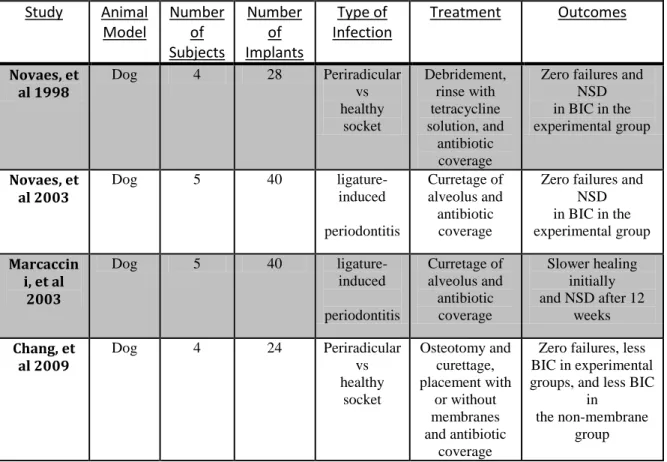
associated with implant placement into sites with infection. Each of the four studies included for review here used a beagle dog model, and either induced periodontal lesions prior to placing implant fixtures, or induced a periradicular type lesion, prior to placing implant fixtures. In all four studies, a split-mouth design was utilized, with approximately half of all implants in the study being designated to either the control or experimental group. Table 2.1 lists each study, as well as the important variables and experimental design.
4
reasonable to suggest that if the presence of an infection precludes an implant to failure, then such a failure would occur within the early phases of the healing and integration processes.
Table 2.1 Animal Studies BIC-Bone-Implant Contact NSD-No Significant Difference
2.4. Human Studies
One of the first reports of placement of implants into sites with the presence of infection in humans was a case report published in 1995 by Novaes, et al14. A total of 3 patients were treated, 2 patients with teeth exhibiting radiographic signs of infection, with clinical signs of root fracture, and 1 patient with a combined periodontal-endodontic lesion. Each of the patients were treated according to the same protocol: Extraction of the involved tooth, careful debridement of the remaining infected osseous tissue, irrigation with sterile saline solution, and administration of pre- and post-operative antibiotics (Penicillin V every 8 hours, for 10 days, beginning 24 hours prior to the procedure, as well as doxycycline once
Study Animal Model Number of Subjects Number of Implants Type of Infection
Treatment Outcomes
Novaes, et al 1998
Dog 4 28 Periradicular
vs healthy socket Debridement, rinse with tetracycline solution, and antibiotic coverage
Zero failures and NSD in BIC in the experimental group
Novaes, et al 2003
Dog 5 40
ligature-induced periodontitis Curretage of alveolus and antibiotic coverage
Zero failures and NSD in BIC in the experimental group Marcaccin
i, et al 2003
Dog 5 40
ligature-induced periodontitis Curretage of alveolus and antibiotic coverage Slower healing initially and NSD after 12
weeks Chang, et
al 2009
Dog 4 24 Periradicular
vs healthy socket Osteotomy and curettage, placement with or without membranes and antibiotic coverage
Zero failures, less BIC in experimental groups, and less BIC
in
5
per day, for 21 days). All implant placements were conducted using a two-stage approach. The follow-up time reported for each case was: 7 months for case report 1, 2 years for case report 2, and 11 months for case report 3. At each follow-up period, an exam and periapical radiograph was taken, to confirm integration of the implant.
The first clinical trial, published in 2005, can be credited to Villa and Rangart16. The objective of their study was to observe implant survival rates for dental implants that were placed into sites with infection, in the interforaminal region of the mandible. A total of 20 patients were enrolled in their study, and received from 4-6 implants. A provisional prosthesis was inserted 3 days later, conforming to an early loading protocol. The final restorations were delivered between 3 and 12 months. The total follow-up time was 44 months. There were no implant failures, accounting for a 100% survival rate.
6 Study Number
of patients Number of implants Type of infection Follow-up Time (months)
Treatment Outcome
Novaes, et al-1995
3 3 Periapical 7-14 Debridement, saline rinse, 31 days of antibiotics
100% survival
Villa and
Rangart-2005
20 97 Periapical and periodontic
15-44 Socket debridement,
curretage, antibiotic (local),
cortisone injection, and post surgical
antibiotics
100% survival
Lindeboom, et al-2006
50 50 Periapical 12 Antibiotics, 1 hour prior to surgery, socket degranulation 92% survival-test group 100% in control group Siegenthaler, et al-2007
29 29 Periapical 12 Antibiotics 1 hour prior to surgery, CHX rinse, socket debridement, GBR, and anti-
biotics 5 days post-surgery
100% survival
Villa and
Rangart-2007
33 100 endodontic, periodontic, or
root fracture
12 Socket debridement,
currettage, irrigation with
antibiotic, cortisone injection into soft
tissue, post-surgery
antibiotics
97.4% survival
Casap, et al-2007
20 30 Periodontal and periapical
12-72 Systemic antibiotics pre-and post-operative intrasocket ostectomy, and GBR 97.7% survival
Naves, et al-2009
1 3 Periapical 36 Antibiotics 1 hr prior to surgery and 7 days
post-surgery, Apical access flap, with debridement 100% survival Del Fabbro, et al-2009
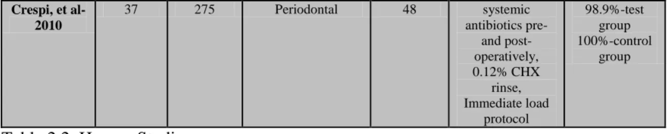
7
Table 2.2 Human Studies CHX-Chlorhexidine, GBR-Guided Bone Regeneration
2.3 Antibiotics and Implant Therapy
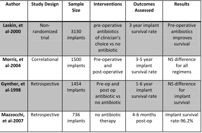
It has traditionally been the standard of practice to provide a pre-loading dose of antibiotic prior to implant placement, and in some instances, a post-operative course of antibiotic therapy subsequent to implant placement. The rational for administration of antibiotics has been the belief that such administration will reduce bacterial loads intra-orally, and thus create an environment that will allow an implant fixture a better opportunity to integrate within the host tissue, at least during the early phase of healing. Becker and Becker have described this in their paper, in addition to a number of other authors24-27. A number of clinical controlled studies have been completed, which have evaluated the efficacy of such practice (Table 2.3). The results have been equivocal.
In a large scale, multi-center prospective analysis, Laskin, et al, compared the efficacy of a pre-operative dose of antibiotic versus no antibiotic24. A total of more than 2900 implants were evaluated in the study, and a minimum follow-up period of 3 years was completed. There were 387 patients (1,743 implants) in the group that received preoperative antibiotics and 315 patients (1,287 implants) in the group that did not receive preoperative antibiotics. Postoperative antibiotics were used in 96% of the total cases. At four different time points, or stages, the implants were assessed for survival status. These time points were as follows: 1) period between the time of implant placement and uncovering (Stage 1); 2) at uncovering (Stage 2); 3) before loading of the prosthesis (Stage 3); and 4) from loading of
Crespi, et al-2010
37 275 Periodontal 48 systemic antibiotics pre-
and post- operatively, 0.12% CHX
rinse, Immediate load
protocol
98.9%-test group 100%-control
8
the prosthesis to 36 months post-placement (Stage 4). Failure was defined as the need to remove the implant at any time for any reason, including clinical mobility, the presence of infection, persistent pain, or the radiographicpresence of pathology. In making their comparison, the authors looked at three different regimens of pre-operative antibiotic regimens: 1) preoperative antibiotic regimen of any type versus no preoperative antibiotic coverage; 2) a sufficient level of preoperative antibiotics as defined by Peterson et al., which is twice the therapeutic level or greater, versus a smaller dose or no preoperative antibiotics; and 3) a sufficient level of preoperative antibiotics as defined by the American Heart
Association (AHA)1 versus an insufficient AHA dose or no preoperative antibiotics. Survival of implants in patients with preoperative antibiotic coverage was 95.4% compared to 90% for those implants placed without coverage. A higher implant survival rate also occurred at each stage of treatment from the time of placement to 36 months. In conducting their statistical analysis, authors found a statistically significant difference between the survival rates among the two groups, stating that the P-value was less than .05. They
concluded that a single pre-operative dose of systemic antibiotic administration has a positive effect on the survival rate implants that are placed.
9
administration of a pre- or post-operative antibiotic regimen has no positive effect on the outcome of implant survival.
Gynther, et al, found similar results in their study26. A total of 1454 implants were placed at two different time periods. A total of 790 implants, which were used to support both fixed and removable prostheses, were placed with a pre- and post-operative course of antibiotics. These implants were followed for a range of 1-6 years, with a mean follow-up of 3 years. A total of 664 implants were placed, almost a decade later, without any type of antibiotic regimen. These implants were followed for a range of 1-5 years, with a mean follow-up period of 3 years. Survival rates for the antibiotic group was 88% in the maxilla, 99% in the mandible, and for the non-antibiotic coverage group, 95% in the maxilla, and 95% in the mandible. The differences were not statistically significant.
Esposito, et al, conducted a meta-analysis of four large clinical trials31. Each of the studies, individually, failed to find a statistically significant difference in implant survival outcomes, when comparing antibiotic administration versus placebo. Their meta-analysis of the four studies together did show that a pre-load dose of 2g of Amoxicillin may be
beneficial in preventing failure of an implant to integrate during the early phases of healing. According to the author of that review, one out of every 33 patients that receive a
pre-operative dose of 2g of Amoxicillin, would prevent early failure of an implant. Although the administration of a preoperative dose of antibiotics, prior to the
10
question: Should we give our healthy patients a pre or post-operative course of antibiotics to improve the outcome of their implant surgery? If we are able, through well-controlled studies, definitively say no, then we can potentially avoid the negative outcomes associated with overuse of antibiotics, such as the creation of strains of bacteria resistant to antibiotic therapy, as well as the potential for development of allergic reactions.
Table 2-3 Studies Comparing Antibiotic Efficacy
2.4 Discussion
The immediate placement of implants into sites with the presence of infection has become an increasingly common procedure. Data from studies that have been published in the past two decades seem to suggest implant survival rates that are equivalent to implants that are placed in native, healthy osseous tissue. While it is not known exactly why the rates are equivalent, some explanations can be proposed. First, when a tooth exhibiting signs of
Author Study Design Sample
Size
Interventions Outcomes Assessed Results Laskin, et al-2000 Non-randomized trial 3130 implants pre-operative antibiotics of clinician's choice vs no antibiotic
3 year implant survival rate Pre-operative antibiotics improves survival Morris, et al-2004
Correlational 1500 implants Pre-operative and post-operative 3-5 year implant survival rate NS difference for all regimens Gynther, et al-1998
Retrospective 1454 Implants Pre-op and post op antibiotic vs no antibiotic 1-6 year implant survival rate NS difference for implant survival Mazzocchi, et al-2007
11
infection is removed, most of the source of that infection is also removed. In some cases, granulation tissue associated with the lesion is also removed with the root of the offending tooth. Any remaining or residual infection is subsequently removed with curettage and irrigation of the socket. Additionally, upon completion of the osteotomy for placement of the implant, more of the infected tissue is removed.
While it has been shown that survival rates for implants placed into sites with infection are high, it is not known whether administration of a pre- and post-course of antibiotic therapy is able to exert any beneficial effect on those rates. In all of the studies included in this review, a course of antibiotic therapy, both a pre-loading dose and post-operative dose were prescribed. Considering that most, if not all, of the infection is removed when a tooth is removed, then it may stand to reason that antibiotic therapy may not be needed when performing immediate placement of implants into such sites. Future studies with larger sample sizes should be completed in order to evaluate the effect of prophylactic antibiotic coverage when immediately placing implants under these circumstances.
Additionally, consideration should be given to conducting trials evaluating the effects of the administration of localized antibiotics. It is possible that the use of local versus
systemic antibiotics could provide a beneficial effect on the outcome, while potentially minimizing the risks that are associated with the use of systemic antibiotics.
2.6 Conclusions
12
III. PROSPECTIVE CLINICAL TRIAL
3.1 INTRODUCTION
The practice of placing dental implants, and immediately loading them after
placement has been studied extensively, and has become a common procedure under certain clinical situations1-5. Advantages to placing and immediately loading dental implants include immediate restoration of function and appearance, decreased morbidity as a result of reduced surgical visits, as well as a reduction in the amount of resorption of soft and hard tissues adjacent to the implant2. Several clinical studies have demonstrated survival rates
comparable to those of implants placed in a conventional manner; that is after osseous and gingival tissues have undergone an appropriate period of healing4.
There is a concern by some practitioners that implants should not be placed immediately within sites that demonstrate periradicular pathology7-9. While no evidence exists to support this claim, there is limited data to suggest that the immediate placement of implants into such sites is possible, and very limited data to suggest that immediate loading of implants placed into such sites is possible as well10-23.
14
success rate. Of importance to note is that 3 of the 29 implants (two experimental and one control) showed signs of infection during the first 13 weeks of healing, which required therapeutic intervention.
Only one clinical trial exists which tested the possibility of immediate loading of immediately placed implants into sites with infection19. A total of 100 implants were placed, 76 being placed into sites with infection, and 24 into normal healthy tissue. Of the implants placed in this study, 2 failed due to periodontal involvement, which represented an overall success rate of 97.4%. Some of the limitations of this study include the lack of identification of health status of patients (i.e., whether patient had controlled or uncontrolled systemic disease, smoker vs. non-smoker, etc), a lack of definition of lesion size/location, the varying types of prosthesis use to restore the implants, such as single crowns, fixed partial dentures, and full arch restorations, and a limited number of implants within the control group.
To our knowledge, there have not been any studies completed which have attempted to determine the need for prophylactic antibiotic coverage under such conditions. Gynther26, et al looked at the effect of administration of preoperative systemic antibiotics on the success rates of implants placed within healthy sites. According to results from their study, implants that were placed in subjects who did not receive preoperative antibiotics exhibited similar rates of success as implants that were placed in subjects receiving preoperative antibiotics.
15
oresistant microorganisms, as well as additional costs associated with use of the medications, are issues that could be avoided if it is determined that such coverage is not necessary28-30.
It would be helpful for clinicians to know, from an evidence-based perspective, whether or not the presence of periradicular infection would preclude the successful outcome of dental implants placed and loaded immediately after an extraction. It would also be helpful for clinicians to know whether or not prophylactic administration of antibiotics during such procedures are necessary for a successful outcome. Thus, one aim of this prospective controlled clinical trial will be to evaluate the rate of success of endosseous dental implants placed into sites with infection, and immediately loaded. A secondary aim will be to evaluate the influence of systemic prophylactic antibiotics on the success rate of implants placed under such circumstances.
We hypothesized that implants placed and immediately loaded within sites that are infected will perform as well as implants that are placed and immediately loaded within healthy sites. We also hypothesized that the use of systemic antibiotics when placing implants according to this protocol would not provide any additional benefit.
3.2 MATERIALS AND METHODS:
16 Table 3.1 Inclusion/Exclusion Criteria
Placement of implants
Upon acceptance into the study, subjects were randomly allocated to either the
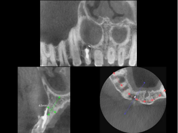
experimental or control groups via block randomization. Full-arch alginate impressions were acquired, and used to record baseline soft tissue levels, as well as provide a matrix for the provisional restoration. For those subjects whose tooth was severely broken down, a direct mock up of the crown was completed using flowable resin. Baseline small volume cone-beam CT (CBCT) scans (Kodak dental systems, Rochester, NY) of each site were acquired prior to extraction and implant placement, and used to evaluate the extent of infection, and presence of remaining osseous tissue. One hour prior to the surgical procedure, each subject received either antibiotic or placebo. Antibiotic coverage consisted of Amoxicillin 2g, PO 1hr before the procedure, and then 500 mg tid, for 7 days following placement. For those patients who were allergic to Amoxicillin, Clindamycin 600mg 1 hour prior to, and then 300mg three times a day, for 7 days was administered. Placebo consisted of sucrose
Inclusion Criteria Exclusion Criteria ASA Class 1 or 2 individuals, to include those
with controlled HTN, diabetes, etc
ASA Class 3 or 4 individuals, or those who are pregnant
Non-smokers and smokers with a reported use of less than 1 pack/day
Age less than 19, over 70
Female/Male, ages 19-70 Patients who are on continuous antibiotic therapy for any medical condition
Presence of at least one pre-molar, canine, or incisor tooth with
site of infection, either of periodontal or endodontic origin
Patients who exhibit gross infection/facial space infection with purulent discharge
Premolar, canine, or incisor tooth deemed non-restorable secondary to vertical root fracture
Patients who use smokeless tobacco, who are unwilling/unable to cease for enrollment into study Patients with sufficient bone quantity for implant
placement, irrespective of infective lesion, and as determined by initial exam and small-volume
CBCT scan
Patients unable to tolerate implant placement with local anesthesia
Presence of stable posterior contacts, bilaterally and distal to the infected site
17
enclosed within a capsule that mimicked the antibiotic. Antibiotic or placebo was
administered by the first author, who was blinded to the randomization schedule. In addition to the pre-operative antibiotic/placebo, all subjects were instructed to rinse for two minutes with 0.12% Chlorhexidine. Anesthesia was administered, and the infected tooth was
extracted, with curettage and irrigation with sterile saline solution and a very copious amount of 0.12% Chlorhexidine. All implants were placed utilizing a flapless procedure. Guided bone regeneration (Bio-Oss, ) with or without barrier membrane (Biomend, Osteohealth, Shirly, NY) was used when it appeared that there was a horizontal deficiency between the implant and alveolus of greater than 2mm.
Loading of implants
After placement, each implant received a pre-fabricated abutment and screw-retained provisional crown (Integrity, Dentsply International, York, PA). The occlusal surface of each crown was adjusted, such that there was no contact during maximum intercuspation or excursive movements of the mandible (non-occlusal loading). Subjects were given a
18 Fig. 3.1 Pre-extraction CBCT of Tooth #7
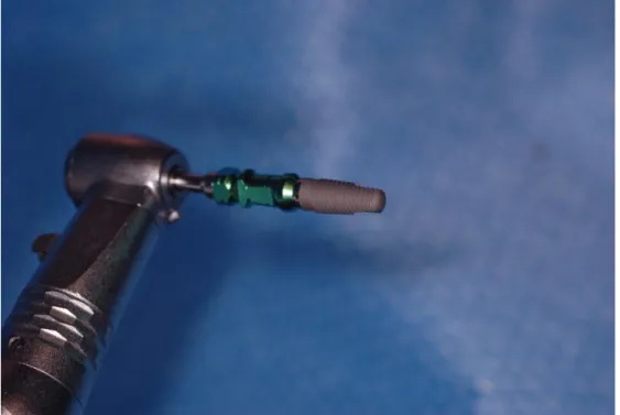
19 Fig 3.4 4.1mm x 11.5mm Zimmer TSV Implant
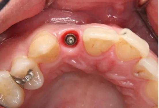
20 Fig 3.6 Occlusal View of Implant Placement
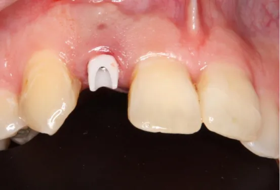
21 Fig 3.8 Definitive All-Zirconia Abutment
22 Fig 3.10- 6 Month Follow-Up Photograph
23
Fig 3.12 Baseline Radiograph Fig 3.13 12-Month Follow-Up Radiograph
Follow-up and Success Criteria
24 Table 3.2 Implant Success Criteria
3.3 RESULTS
A total of 13 implants were placed, in a total of 13 patients (1 implant/patient). Of the 13 implants that were placed, 2 failed to integrate, and were deemed early failures. This represents a survival rate of 84.7%. Table 3.3 lists the distribution of implants based on gender and implant failures. A descriptive analysis was completed, and results are displayed as follows: Table 3.4 lists the distribution of subjects based upon who received antibiotic versus who received placebo, and the distribution of failures among each group. Table 3.5 shows distribution of each subject, tooth number, whether or not the subject received antibiotic or placebo, and whether or not the implant was stable at the 6 month follow-up.
Gender Implants Failures
Male 7 2
Female 6 0
Table 3.3 Implant Distribution and Survival by Gender
Criteria used to determine Implant Success
(Modified from Smith and Zarb) No mobility detected on implant at each follow-up interval
Decrease in size of lesion, from baseline to 12 month follow-up, as
determined by conventional PA radiograph
Vertical bone loss not to exceed 1.5 mm
No persistant pain, discomfort, or infection is attributable to the implant
25
Implants Failures
Antibiotic 5 1
Placebo 8 1
Table 3.4 Implant Survival by Antibiotic or Placebo
Site Total Failed
Anterior 9 2
Posterior 2 0
Table 3.5 Implant Survival by Anterior-Posterior Position
Site Total Failed
Maxillary 8 2
Mandibular 3 0
Table 3.6 Implant Survival by Jaw Location
Subject Tooth # Antibiotic
/Placebo Integrated? Diameter Implant
(mm)
Implant Length
(mm)
1 7 A Yes 4.1 11.5
2 28 A Yes 4.7 11.5
3 7 P Yes 4.7 16
4 10 P Yes 4.7 13
5 7 P Failed 4.1 8
26 Table 3.7 Data Set by Subject
Discussion:
Results from this study are similar to results from other studies investigating placement of implants into sites exhibiting signs of infection, and seem to suggest that the immediate placement of implants into sites exhibiting signs of infection is a viable treatment modality.
Of the 15 subjects enrolled, 2 were unable to receive implants at the time of surgery, due to lack of the buccal plate of bone. It was determined that the possibility to obtain primary stability would be low. For these patients, thorough debridement and irrigation was completed, followed by socket augmentation with Puross Putty and Collagplug. These patients were then given an essix retainer, and informed that they could return after a sufficient period of healing for placement of an implant fixture. Eleven of the thirteen implants, at their respective 6 month follow-up period, have satisfied the criteria for success established within this paper, demonstrating a rate of 84.7%.
7 7 P Yes 4.7 13
8 7 A Yes 4.7 16
9 7 P Yes 4.7 16
10 4 A Yes 4.7 16
11 26 P Yes 3.7 13
12 7 A Failed 4.1 16
13 10 P Yes 4.1 16
14 29 P n/a n/a n/a
27
While the survival rates that have been demonstrated in this study are slightly lower than those from other studies, there are some variables that may account for the discrepancy. One reason might be attributable to the study design, in that the implants in the present study were immediately placed and provisionalized. This can be considered a substantial
difference between this and the majority of the studies that have been published. While careful adherence to the concept of non-occlusal loading (removal of all contacts on the tooth in maximum intercuspation, as well as excursive movements) was followed, it is possible that the implant failures were due to lack of adherence to the strict dietary instructions given to the subjects post-operatively.
In one of the failures, the implant chosen for placement was shorter than average (4.1mm x 8mm). This was chosen, because of the convergence of the adjacent teeth. While we did feel that we were able to obtain primary stability, an objective measure of that stability was not obtained, and thus it is possible that the amount of stability may not have been adequate. Failure of the second implant was determined at the 1 and 4 week follow-up period. This second failure we feel may be attributed to the lack of completion of the control phase of treatment, as well as lack of some posterior support of teeth. Completion of the control phase (i.e., caries, some periodontal pocketing of 4-5mm’s) was to be completed shortly after placement of the implant, however, due to extenuating circumstances
28
seven received placebo. This is particularly interesting, as this seems to suggest that prophylactic antibiotic administration for implant placement may not provide any positive effect on the survival rate of implants placed under these conditions.
It is important to note that the number of subjects enrolled in the present study is low, and that while an exact analysis was performed, results should be interpreted with caution. To satisfy the odds-ratio analysis conducted prior to commencing with study, it was
determined that over 700 subjects would have needed to enroll, to have an accurate assessment of the effects of prophylactic antibiotics on the outcome of survival rates of implants placed within sites previously occupied by infection. Notwithstanding this limitation, we do feel that results from this exploratory study are encouraging, and
recommend that future studies be completed with an identical design protocol, to provide an accurate analysis.
29
cause, such as life-threatening allergic reactions, development of bacteria that are resistant to the antibiotic, etc.
CONCLUSIONS
30
REFERENCES
1. Tarnow DP, et al. Immediate loading of threaded implants at stage 1 surgery in edentulous arches: Ten consecutive case reports with 1-5 year data. Int J Oral Maxillofac Implants. 1997; 12:319-24.
2. Lazzara, et al. Immediate implant placement into extraction sites: surgical and restorative advantages. Int J Periodontics Restorative Dent. 1989;9(5):332-43.
3. Randow K, et al. Immediate functional loading of Branemark dental implants: an 18-month study. Clin Oral Implants Res. 1999; 10:8-15.
4.
Block MS, Mercante DE, Lirette D, Mohamed W, Ryser M, Castellon P. Prospective evaluation of immediate and delayed provisional single tooth restorations. J OralMaxillofac Surg. 2009 Nov;67(11 Suppl):89-107
5. Jivraj S, Reshad M, Chee WW. Immediate loading of implants in the esthetic zone. J Esthet Restor Dent. 2005;17(5):320-5.
6. De Bruyn H, Collaert B. Early loading of machined-surface Branemark implants in completely edentulous mandibles: healed bone versus fresh extraction sites. Clin Implant Dent Relat Res 2002;4:136-42.
7. Quirynen M, Gijbels F, Jacobs R. An infected jawbone site compromising successful osseointegration. Periodontol 2000. 2003;33:129-144.
8. Lekholm U. Immediate/early loading of oral implants in compromised patients. Periodontol 2003;33:94-203.
9. Barzilay, I. (1993) Immediate implants: their current status. International Journal of Prosthodontics 6: 169–175.
10.Novaes AB Jr., Vidigal Junior GM, Novaes AB, Grisi MF, Polloni S, Rosa A. Immediate implants placed into infected sites: A histomorphometric study in dogs. Int J Oral Maxillofac Implants 1998;13:422-427.
11.Novaes AB Jr., Marcaccini AM, Souza SL, Taba M Jr., Grisi MF. Immediate placement of implants into periodontally infected sites in dogs: A histomorphometric study of bone-implant contact. Int J Oral Maxillofac Implants 2003;18:391-398.
31
13.Chang SW, Shin SY, Hong JR, et al. Immediate implant placement into infected and noninfected extraction sockets: A pilot study. Oral Surg Oral Med Oral Pathol Oral Radiol Endod 2009;107:197-203.
14.Novaes AB Jr, Novaes AB. Immediate implants placed into infected sites: a clinical report. Int J Oral Maxillofac Implants. 1995 Sep-Oct;10(5):609-13.
15.Chang SW, Shin SY, Hong JR, Yang SM, Yoo HM, Park DS, Oh TS, Kye SB. Immediate implant placement into infected and noninfected extraction sockets: a pilot study. Oral Surg Oral Med Oral Pathol Oral Radiol Endod. 2009 Feb;107(2):197-203. 16.Villa R, Rangert B. Early loading of interforaminal implants immediately installed after
extraction of teeth presenting endodontic and periodontal lesions. Clin Implant Dent Relat Res 2005;7(Suppl. 1):S28-S35.
17.Lindeboom JA, Tjiook Y, Kroon FH. Immediate placement of implants in periapical infected sites: A prospective randomized study in 50 patients. Oral Surg Oral Med Oral Pathol Oral Radiol Endod 2006;101: 705-710.
18.Siegenthaler DW, Jung RE, Holderegger C, Roos M, Hämmerle CH. Replacement of teeth exhibiting periapical pathology by immediate implants: a prospective, controlled clinical trial. Clin Oral Implants Res. 2007 Dec;18(6):727-37.
19.Villa R, Rangert B. Immediate and early function of implants placed in extraction sockets of maxillary infected teeth: a pilot study. J Prosthet Dent. 2007 Jun;97(6 Suppl):S96-S108.
20.Casap N, Zeltser C, Wexler A, Tarazi E, Zeltser R. Immediate placement of dental implants into debrided infected dentoalveolar sockets. J Oral Maxillofac Surg 2007;65:384-392.
21.Naves Mde M, Horbylon BZ, Gomes Cde F, Menezes HH, Bataglion C, Magalhães D.Immediate implants placed into infected sockets: a case report with 3-year follow-up.Braz Dent J. 2009;20(3):254-8.
22.Del Fabbro M, Boggian C, Taschieri S. Immediate implant placement into fresh extraction sites with chronic periapical pathologic features combined with plasma rich in growth factors: Preliminary results of single-cohort study. J Oral Maxillofac Surg 2009;67: 2476-2484.
23.Crespi, et al. Immediate Loading of Dental Implants Placed in Periodontally Infected and Non-Infected Sites: A 4-Year Follow-Up Clinical Study. J Periodontol 2010;81:1140-1146.
32
25.Morris, HF, et al. AICRG, Part III: The influence of antibiotic use on the survival of a new implant design. Journal of Oral Implantology, 2004, Vol. 30 Issue 3, p144-151
26.Gynther GW, Köndell PA, Moberg LE, Heimdahl A. Dental implant installation without antibiotic prophylaxis. Oral Surg Oral Med Oral Pathol Oral Radiol Endod. 1998 May;85(5):509-11.
27.Mazzocchi A, Passi L, Moretti R. Retrospective analysis of 736 implants inserted without antibiotic therapy. J Oral Maxillofac Surg 2007;65:2321-2323.
28.Andersson DI, Hughes Diarmaid. Antibiotic resistance and its cost: is it possible to reverse resistance? Nature Rev Microbiol. 2010;8:260-271.
29.World Health Organisation. WHO global strategy for containment of antimicrobial resistance. World Health Organisation, Manila 2005.
30.Foster KR, Grundmann H. Do we need to put society first? The potential for tragedy in antimicrobial resistance. PLoS Med 2006; 3: 177–180.
31.Esposito M, Grusovin MG, Talati M, Coulthard P, Oliver R, Worthington HV. Interventions for replacing missing teeth: Antibiotics at dental implant placement to prevent complications. Cochrane Database Syst Rev 2008;CD004152.