Genomic biomarkers of prenatal intrauterine
inflammation in umbilical cord tissue predict
later life neurological outcomes
Sloane K. Tilley1, Robert M. Joseph2, Karl C. K. Kuban3, Olaf U. Dammann4, T. Michael O’Shea5, Rebecca C. Fry1,6*
1 Department of Environmental Sciences and Engineering, Gillings School of Global Public Health, University
of North Carolina, Chapel Hill, North Carolina, United States of America, 2 Department of Anatomy and Neurobiology, Boston University School of Medicine, Boston, Massachusetts, United States of America,
3 Department of Pediatrics, Boston University School of Medicine, Boston, Massachusetts, United States of
America, 4 Department of Public Health and Community Medicine, Tufts University School of Medicine, Boston, Massachusetts, United States of America, 5 Department of Pediatrics, University of North Carolina, Chapel Hill, North Carolina, United States of America, 6 Curriculum in Toxicology, School of Medicine, University of North Carolina, Chapel Hill, North Carolina, United States of America
Abstract
Background
Preterm birth is a major risk factor for neurodevelopmental delays and disorders. This study aimed to identify genomic biomarkers of intrauterine inflammation in umbilical cord tissue in preterm neonates that predict cognitive impairment at 10 years of age.
Study design
Genome-wide messenger RNA (mRNA) levels from umbilical cord tissue were obtained from 43 neonates born before 28 weeks of gestation. Genes that were differentially expressed across four indicators of intrauterine inflammation were identified and their func-tions examined. Exact logistic regression was used to test whether expression levels in umbilical cord tissue predicted neurocognitive function at 10 years of age.
Results
Placental indicators of inflammation were associated with changes in the mRNA expression of 445 genes in umbilical cord tissue. Transcripts with decreased expression showed signifi-cant enrichment for biological signaling processes related to neuronal development and growth. The altered expression of six genes was found to predict neurocognitive impairment when children were 10 years old These genes include two that encode for proteins involved in neuronal development.
Conclusion
Prenatal intrauterine inflammation is associated with altered gene expression in umbilical cord tissue. A set of six of the differentially expressed genes predict cognitive impairment
a1111111111 a1111111111 a1111111111 a1111111111 a1111111111
OPEN ACCESS
Citation: Tilley SK, Joseph RM, Kuban KCK,
Dammann OU, O’Shea TM, Fry RC (2017) Genomic biomarkers of prenatal intrauterine inflammation in umbilical cord tissue predict later life neurological outcomes. PLoS ONE 12(5): e0176953.https://doi.org/10.1371/journal. pone.0176953
Editor: Olivier Baud, Hopital Robert Debre, FRANCE
Received: January 3, 2017
Accepted: April 19, 2017
Published: May 11, 2017
Copyright:©2017 Tilley et al. This is an open access article distributed under the terms of the
Creative Commons Attribution License, which permits unrestricted use, distribution, and reproduction in any medium, provided the original author and source are credited.
Data Availability Statement: Raw data are from
later in life, suggesting that the fetal environment is associated with significant adverse effects on neurodevelopment that persist into later childhood.
Introduction
Preterm birth, defined as delivery at<37 completed weeks gestation, is currently the leading cause of neonatal morbidity and mortality in the United States [1]. Individuals born prema-turely are at increased risk for other adverse health outcomes, and those born at less than 28 weeks gestation are at particularly high risk [2]. Perhaps most important are adverse neurode-velopmental outcomes, which affect an estimated 1 million preterm infants born each year [3].
Preterm birth is thought to be caused by the pathological induction of certain components of the normal parturition process resulting from a combination of environmental, genetic, and behavioral factors [4,5]. Many identified risk factors have the potential to promote inflamma-tory processes [6]. Indicators of intrauterine inflammation are present in as many as 40–70% of preterm births, versus only 1–13% of full term births [7]. These data support the hypothesis that risk of preterm birth is increased by pathological, environmental, and/or genetic factors that contribute to delivery-inducing inflammation [4,8]. Among preterm infants, biomarkers of prenatal inflammation, including inflammatory cytokines in amniotic fluid [9], placental histologic findings [10–12], and inflammation-related proteins in neonatal blood [13–17], are associated with a range of neurodevelopmental impairments [18].
A fetal inflammatory response (FIR) is associated with increased expression of a broad array of genes related to neurodevelopment [19]. In the present study, we aimed to identify whether genomic signaling changes in umbilical cord tissue were associated with a suite of four histologic markers of prenatal inflammation in a subset of infants from the Extremely Low Gestational Age Newborns (ELGAN) cohort. We hypothesized that some of these geno-mic changes would be predictive of neurocognitive function at 10 years of age and could pro-vide novel predictive biomarkers of neurocognitive impairment in preterm infants.
Materials and methods
The ELGAN cohort
The ELGAN cohort was established to identify risk factors for neurodevelopmental impair-ments in extremely low gestational age newborns. Between 2002–2004, 1506 infants were enrolled in the study, and in 1410 cases (94% of the cohort) placentas were collected for patho-logical examination. Placentas were collected at delivery and flash frozen to -70 C. Placentas were examined both grossly and histologically for many parameters, including a subset of intrauterine inflammation markers. The larger ELGAN cohort is described in detail elsewhere [20,21].
RNA isolation and gene expression assessment
The data in this study were generated by Cohenet al. from isolated total RNA from the umbili-cal cord tissue homogenates collected from infants born between 23 and 28 weeks gestation at Brigham and Women’s Hospital, Beth Israel Deaconess Medical Center, or Wake-Forest Med-ical Center between April 1, 2004 and August 31, 2004. RNA was extracted using the Qiagen RNeasy Mini Kit, as described elsewhere [22]. Thirteen samples had insufficient total RNA (<7μg) for hybridization and five infants died before 36 weeks postmenstrual age. These 18
Funding: This research was supported by grants
from the National Institutes of Health (http://www. nih.gov<http://www.nih.gov/>): R01 ES019315 (RF), P42 ES005948 (RF), 5U01NS040069-05 (KK), and 5U01NS040069-09 (KK),
1UG3OD023348-01 (TS) and from the National Institute for Occupational Safety and Health: T42/ OH-008673. The funders had no role in study design, data collection and analysis, decision to publish, or preparation of the manuscript.
Competing interests: The authors have declared
infants were excluded from further analysis. Total RNA from the 54 remaining ELGAN sub-jects were hybridized to the Affymetrix Human Genome U133 Plus 2.0 Array, which assesses gene expression levels across 54,675 probes. All expression data are available at the National Center for Biotechnology Information’s Gene Expression Omnibus repository (GSE8586) [22]. The data were further processed previously, applying quality control assessments to all 54 samples [19]. Six samples that failed these measures were excluded from further analysis [19]. Data were normalized into Affymetrix probesets using fRMA [19]. An additional five subjects were excluded from analysis due to missing clinical information about intrauterine
inflamma-tion markers from the umbilical cord tissue. Probes without annotainflamma-tions toEntrezgene
identi-fiers were removed and only the probeset with the largest inter quartile range perEntrezgene was kept. The final data set consisted of measures of gene expression across 20,155 genes for n = 43 subjects [19] (S1 Table).
Cognitive assessment at 10 years of age
When study participants were 10 years of age, general cognitive ability (or IQ) was assessed with the School-Age Differential Ability Scales–II (DAS-II) Verbal and Nonverbal Reasoning scales [23]. Attention and executive function were assessed with the DAS-II and the NEPSY-II [24]. DAS-II Recall of Digits Backward and Recall of Sequential Order measured verbal work-ing memory. NEPSY-II Auditory Attention and Auditory Response Set evaluated auditory attention, set switching and inhibition. NEPSY-II Inhibition and Inhibition Switching assessed simple inhibition and inhibition in the context of set shifting, respectively. NEPSY-II Animal Sorting measured concept generation and mental flexibility. As a comprehensive measure of cognitive and executive function, we used latent profile analysis (LPA) to identify study partic-ipants with similar distinctive profiles on measures of cognitive and executive functioning. With this approach, four subgroups were identified, corresponding to functioning that was normal (LPA score = 1; 34% of ELGAN cohort), low-normal (LPA score = 2; 41%), moderately impaired (LPA score = 3; 17%), and severely impaired (LPA score = 4; 8%) [25,26].
Relating gene expression to prenatal inflammation measures
Four markers of intrauterine inflammation were selected to test for associations with gene expression: inflammation of the chorionic plate, moderate or severe chorioamnionitis, neutro-philic infiltration of the fetal vessels in the chorionic plate, umbilical cord inflammation [27]. Using ANCOVA analysis, these binary measures were assessed separately. Potential confound-ers were included in the ANCOVA analysis for each placental histologic marker only if they displayed different means (2-sided student t-test p-value<0.20) between subjects with that placental inflammation marker and without that placental marker. Variables tested but that displayed no mean difference in any of the four histologic markers of placenta inflammation included maternal age, maternal race, maternal BMI, maternal education level and infant sex. Exposure to smoke (active or passive) during pregnancy was included in the analysis for chor-ioamnionitis and neutrophilic infiltration of the fetal vessels, and gestational age was also included in the analysis for neutrophilic infiltration of the fetal vessels. In order to control for multiple tests, false discovery rate (FDR) q-values were calculated. Significance was defined as FDR q-value<0.05 and an absolute fold change|2.0|.
Network analysis of genes associated with prenatal inflammation
measures
relationships were assessed among these genes using Ingenuity Network Analysis (IPA)
(Inge-nuity Systems1, Redwood City, CA, USA) and STRING v10.0 [28,29]. Network analyses were
stratified by directionality of gene expression associations. Canonical pathways from IPA and PFAM protein domains enriched among these gene sets were analyzed and reported.
Logistic regression of genomic markers of inflammation to later life
neurological score
We tested whether the expression levels of genes associated with one or more intrauterine inflammation marker predicted later life neurocognitive function using exact logistic regres-sion analysis. Exact logistic regresregres-sion was used due to the small subset of subjects for whom LPA score measured (n = 22). The dependent variable was the child’s LPA score at age 10, with expression levels predicting the binary outcome of (i) no or low impairment (LPA score = 1 or 2, n = 17) or (ii) moderate or severe impairment (LPA score = 3 or 4, n = 5). As potential con-founders had been controlled for in the first step of this analysis, the model was run with gene expression as the sole predictor variable. Significance was defined as an exact p-value<0.05, and exact beta estimates, exact parameter-likelihood odds ratios and 95% confidence intervals for odds ratios are reported.
Results
Characteristics of the study cohort
Maternal and infant demographic and birth clinical data are presented inTable 1for the subset of the ELGAN subjects with gene expression data used in this study (n = 43). In these infants, the majority of mothers were white and reported no smoking during pregnancy. The mean week of delivery was 26.1, and approximately two-thirds of the infants were male (Table 1). The demographic characteristics in this subset of ELGAN infants were similar to those reported for all ELGAN subjects [21], with the exception of a higher proportion of males in this cohort. We calculated the prevalence of four histological markers of prenatal intrauterine inflammation: inflammation of the chorionic plate, neutrophilic infiltration of the fetal vessels in the chorionic plate, and umbilical cord inflammation were all present in approximately 25% of the study subjects, while moderate or severe chorioamnionitis was present in approximately 50% of the study subjects (Table 1). The occurrence of these four histologic markers of inflam-mation were consistent with those reported in larger study (n = 947) of the ELGAN cohort [20].
Markers of prenatal inflammation are associated with umbilical cord
gene expression
Table 1. Maternal and child characteristics. Data are number of study participants (percent) except where indicated.
Characteristic N = 1410 ELGAN Subjects N = 43 ELGAN Subjects N = 22 ELGAN Subjects
Maternal Age at Delivery in years (median; range in parenthesis) 28.6 (13.2–47.3)
32.1 (15.8–43.2)
34 (19.4–43.2) Maternal Race
White 819 (58.1%) 26 (60.5%) 18 (81.8%)
African-American 397 (28.2%) 9 (20.9%) 4 (18.2%)
Other 178 (12.6%) 7 (16.3%) 0 (0%)
Unknown 16 (1.1%) 1 (2.3%) 0 (0%)
Pre-pregnancy BMI (kg/m2) (median; range in parenthesis) 23.9
(13.2–72.1)
23.1 (18.1–46.5)
22.5 (19.1–46.5) Public Health Insurance
No 785 (55.7%) 31 (72.1%) 19 (86.3%)
Yes 558 (39.6%) 12 (27.9%) 3 (13.6%)
Unknown 67 (4.8%)
Education
<= 12 years 224 (15.9) 10 (23.3%) 3 (13.6%)
12–15 years 681 (48.3%) 12 (27.9%) 7 (31.8%)
16+ years 404 (28.7%) 20 (46.5%) 11 (50.0%)
Unknown 101 (7.2%) 1 (2.3%) 1 (4.5%)
Infertility Treatment
No 1063 (75.4%) 31 (72.1%) 14 (63.6%)
Yes 264 (18.7%) 12 (27.9%) 8 (36.4%)
Unknown 83 (5.9%)
Smoking during Pregnancy
No 1133 (80.4%) 40 (93.0%) 20 (90.9%)
Yes 199 (14.1%) 3 (7.0%) 2 (9.1%)
Unknown 78 (5.5%)
Infant Sex
Male 752 (46.7%) 27 (62.8%) 14 (63.6%)
Female 658 (46.7%) 16 (37.2%) 6 (27.3%)
Birth and Later Life Outcomes Gestational Age (weeks)
Median (range) 28.6
(13.2–47.3)
27 (23–27)
27 (23–27)
23–24 weeks 387 (27.5%) 6 (14.0%) 2 (9.1%)
25–26 weeks 618 (43.8%) 14 (32.6%) 8 (36.4%)
27 weeks 405 (28.7%) 23 (53.5%) 12 (54.5%)
Birth weight (g) (median; range in parenthesis) 790 (280–1528)
929.7, 952 (550–1360)
889.5 (550–1360) Inflammation of the chorionic plate (Stage: 3 and Severity: 3)
No 1118 (79.3%) 32 (74.4%) 16 (72.7%)
Yes 265 (18.8) 11 (25.6%) 6 (27.3%)
Unknown 27 (1.9%)
Moderate/Severe Chorioamnionitis
No 879 (62.3%) 23 (53.5%) 13 (59.1%)
Yes 505 (35.8%) 20 (46.5%) 9 (40.9%)
Unknown 26 (1.8%)
Neutrophilic infiltration of fetal vessels in the chorionic plate
No 1034 (73.3%) 29 (67.4%) 15 (68.2%)
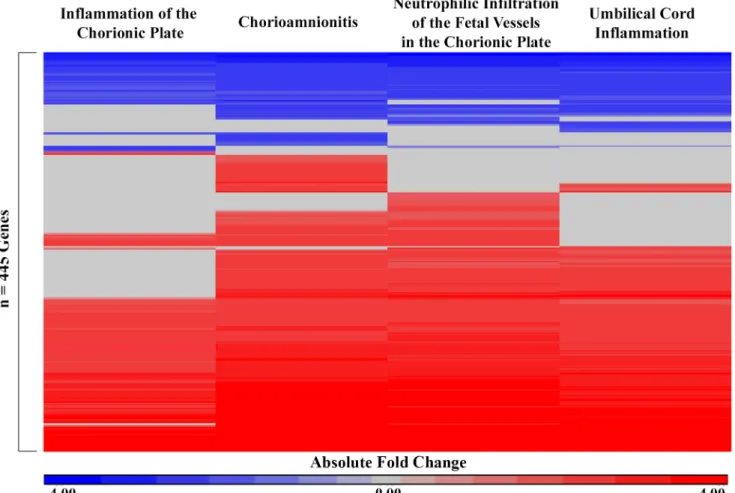
genes were associated with the same directionality across all four intrauterine inflammation markers. Specifically, 168 of these 221 genes (76.0%) were increased in expression in associa-tion with the four intrauterine inflammaassocia-tion markers, 53 of these 221 (24.0%) genes were decreased in expression (S1 Table).
Umbilical cord tissue gene expression profiles are enriched for
inflammation and neuronal development processes
Systems level analysis was used to characterize the canonical pathways and protein domains enriched within the 334 genes with increased expression in association with one or more histo-logical marker of intrauterine inflammation and the 111 genes with decreased expression in association with one or more histological marker of intrauterine inflammation. Notably, dif-ferences in functional signaling between the genes with increased and decreased expression were observed (Table 2). Specifically, the most significant canonical pathway enriched among the genes with increased expression was granulocyte adhesion and diapedesis, and the other top pathways were also associated with inflammation signaling (Table 2). In contrast, the most significant canonical pathways among the genes with decreased expression included gap junc-tion signaling and dopamine-DARPP32 feedback in cAMP signaling, both of which are known to play important roles in early neuronal development (Table 2) [30–32]. Similarly, the protein domains that were significantly enriched among the genes with increased expression in association with one or more histological marker of intrauterine inflammation were small cytokines, metallothionein, and S-100/ ICaBP type calcium binding domain. Proteins contain-ing all of these domains have been demonstrated to play a role in placental inflammation [33– 35]. Interestingly, elevated levels of interleukin-8 (IL-8) in cord blood have been previously associated with a higher incidence with brain injury in preterm infants with placental inflam-mation [35]. Among genes with decreased expression, immunoglobulin I-set protein domains, which are known to function in nervous system development, were found to be significantly enriched [36]. Specifically, among the genes associated with a marker of intrauterine Table 1. (Continued)
Characteristic N = 1410 ELGAN Subjects N = 43 ELGAN Subjects N = 22 ELGAN Subjects
Yes 340 (24.1%) 14 (32.6%) 7 (31.8%)
Unknown 36 (2.6%)
Umbilical cord inflammation (grade 3–5)
No 1136 (80.1%) 31 (72.1%) 16 (72.7%)
Yes 216 (15.3%) 12 (27.9%) 6 (27.3%)
Unknown 58 (4.1%)
LPA Score
1 282 (20.0%) 13 (30.2%) 13 (59.1%)
2 337 (23.9%) 4 (9.3%) 4 (18.2%)
3 134 (9.5%) 4 (9.3%) 4 (18.2%)
4 66 (4.7) 1 (2.3%) 1 (4.5%)
Unknown 591 (41.9%) 21 (48.8%) 0 (0%)
Maternal demographic data, pregnancy characteristics, and data on birth and later in life outcomes are presented for the entire ELGAN sample for which placentas were collected (N = 1410), as well as the N = 43 and N = 22 ELGAN subjects used in this analysis. Data are presented as the number (%) of subjects in the cohort unless otherwise noted. For each of the four histological markers of intrauterine inflammation, there was no significant difference in maternal age, maternal race, maternal BMI, maternal education level and infant sex between subjects with and without each marker (Student’s 2-sided t-test p-value>0.20).
inflammation that encode for a protein with an immunoglobulin I-set protein domain and
that have been previously indicated in neurodevelopment were contactin 1 (CNTN1), neuronal
growth regulator 1 (NEGR1), neurotrophic receptor tyrosine kinase 3 (NTRK3), and receptor tyrosine kinase like orphan receptor 1 (ROR1) [37–40].
Genomic changes in umbilical cord tissue are related to neurocognitive
function
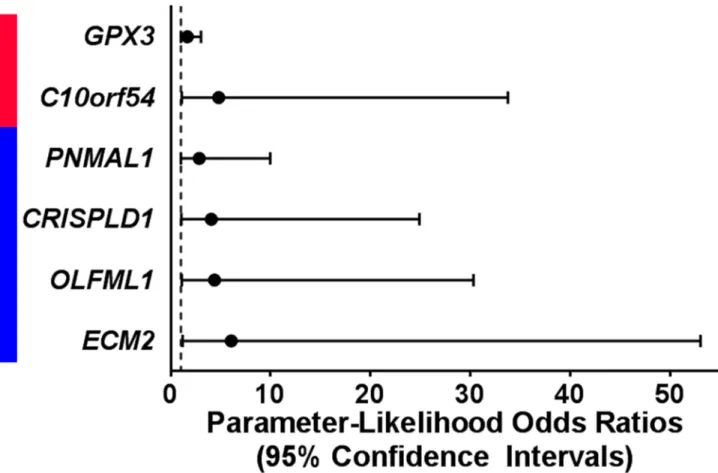
In exact logistic regression models testing all 445 genes, expression levels of 6 genes altered in association with one or more histological marker of prenatal intrauterine inflammation also predicted neurocognitive impairment later in life (Fig 2,S2 Table). Increased expression levels
of chromosome 10 open reading frame 54 (C10orf54, p = 3.64e-2) and glutathione peroxidase
3 (GPX3, p = 4.53e-2) predicted greater neurocognitive impairment later in life and were also increased in association with at least one marker of prenatal intrauterine inflammation (Fig 2). Decreased expression levels of cysteine rich secretory protein LCCL domain containing 1 (CRISPLD1, p = 4.06e-2), extracellular matrix protein 2 (ECM2, p = 2.86e-2), olfactomedin like 1 (OLFML1, p-value = 3.36e-2), and paraneoplastic Ma antigen family like 1 (PNMAL1, p-Fig 1. Heatmap of 445 genes that displayed significant differential expression values across one or more markers of intrauterine inflammation in n = 43 subjects. The absolute fold change (cases/controls) is displayed for each gene significantly associated with each
marker of intrauterine inflammation. Red indicates increased expression in association with a marker of intrauterine inflammation and blue indicates decreased expression in association with a marker of intrauterine inflammation. Rows (genes) were organized by unsupervised hierarchical clustering Euclidean dissimilarity with average linkage. Significance was defined as FDR q-value<0.05 and fold change|2.0|.
value = 4.89e-2) predicted more severe neurocognitive impairment and were also decreased in
association with at least one marker of prenatal intrauterine inflammation (Fig 2).CRISPLD1
andECM2have been suggested to play roles in neuronal development [41,42].
Discussion
In this study, we have demonstrated evidence of genomic signaling changes in the umbilical cord tissue of extremely preterm infants that are associated with multiple markers of intrauter-ine inflammation. Two interesting patterns of gene expression were observed; inflammation-associated genes displayed increased expression in the cord, while among the genes that dis-played decreased expression, several were related to neurodevelopment. Expression levels of six genes altered in umbilical cord tissue in association with one or more intrauterine inflamma-tion marker significantly predict the risk of neurocognitive impairment later in life. In support of our data, several of these genes whose decreased expression predicted more severe cognitive impairment have been previously implicated in neuronal development. Our results indicate that genomic changes observable at parturition in the umbilical cord tissue of extremely low gestational age newborns are associated with neurocognitive function later in life.
Preterm newborns are at increased risk for numerous adverse health effects, many of which are related to prenatal intrauterine inflammation, including neurodevelopmental impairment Table 2. Top canonical pathways and protein domains enriched among the 445 genes associated with intrauterine inflammation markers.
IPA Canonical Pathways
p-value
Associated Genes PFAM Protein Domains
p-value Associated Genes Genes with Increased Expression Levels Granulocyte Adhesion and Diapedesis 1.58e-23
IL1RL1, ITGAM, MMP9, SELL, CSF3R, CCL20, IL1R2, ITGB2, SELE, FPR1, VCAM1, CXCL6, CXCR4, IL1RN, IL1R1,
C5AR1, CXCL1, CXCL5, CCL5, IL1B, TNFRSF1B, CXCL2, PPBP, CXCL8, TNFRSF11B, CCL2, SELPLG, MMP10,
CXCL3, CSF3, ICAM1
Small cytokines (intecrine/ chemokine), interleukin-8 like 1.13e-4 CCL2, CCL20, CCL5, CXCL1, CXCL2, CXCL5, IL8
Hepatic Fibrosis / Hepatic Stellate Cell
Activation
1.58e-16
IL1RL1, MMP9, IL1R2, IL6, VCAM1, IL10RA, IFNGR1, IL1R1, FLT1, IGFBP3,
CCL5, IL1B, VEGFA, PDGFRA, CD14, TNFRSF1B, CXCL8, NFKB2, TNFRSF11B, COL11A1, CCL2, CXCL3,
TIMP1, SERPINE1, ICAM1
Metallothionein 4.27e-2 MT1H, MT1X, MT2A Atherosclerosis Signaling 7.94e-16
CCR2, S100A8, IL1B, MMP9, ITGB2, SELE, IL6, VCAM1, CXCL8, ALOX5, NFKB2, LYZ, CXCR4, IL1RN, CCL2, SELPLG, SERPINA1, F3, PLBD1,
ICAM1, PLA2G2A
S-100/ ICaBP type calcium binding domain
4.27e-2 S100A4, S100A8, S100A9, S100P Genes with Decreased Expression Levels Gap Junction Signaling 2.04e-5
CAV1, GUCY1A2, GUCY1A3, GAB1, PLCE1, PRKAG2, PLCL1
Immunoglobulin I-set domain 1.83e-3 CNTN1, NEGR1, NTRK3, OPCML, ROR1
Cellular Effects of Sildenafil (Viagra)
4.57e-5
MYH3, GUCY1A2, PLCE1, GUCY1A3, PRKAG2, PLCL1
Dopamine-DARPP32 Feedback in cAMP
Signaling
1.55e-4
GUCY1A2, GUCY1A3, PPM1L, PLCE1, PRKAG2, PLCL1
The top three most significant pathways (right-tailed Fisher’s Exact test p-value<0.0001) and significant protein domains (FDR p-value<0.05) are listed. Network analyses were stratified by gene expression directionality. Genes that displayed increased expression levels in association with one or histological markers of intrauterine inflammation were enriched for canonical pathways and protein domains involved in inflammatory and immune processes. Genes that displayed decreased expression levels in association with one or histological markers of intrauterine inflammation were enriched for canonical pathways and protein domains involved early neuronal development.
[43,44]. In our study, we identified that genes that play a role in fetal neurodevelopment had decreased expression levels in the cord of infants exposed to the prenatal inflammatory envi-ronment. Broadly, these genes are associated with gap junction signaling and dopamine-DARPP32 feedback in cAMP signaling, both of which are associated with neurodevelopment. Gap junctions signal for crucial processes in the neonatal cerebral cortex, including neuronal proliferation, migration, and differentiation, while dopamine signaling is known to influence neuronal migration and dendritic growth [30,31]. Through the secondary analysis of genomic prediction of cognitive function, a total of six genes were identified. Two of these genes that
displayed decreased expression in relation to inflammation, namelyECM2andCRISPLD1,
have been previously indicated in neurodevelopment processes.ECM2encodes an
extracellu-lar matrix protein of the small leucine rich glycoprotein family, which are involved in the
regu-lation of many phases of embryonic neurodevelopment [42].CRISPLD1has also been
reported to play a role in extracellular matrix regulation, which is known to play crucial roles in axon growth and guidance [45]. Changes in the expression levels of genes known to play a role in neuronal development in association with markers of prenatal inflammation contribute to the growing body of literature that supports an association between intrauterine inflamma-tion and cognitive impairment in later childhood [18,35].
A previous study of this ELGAN study subset extensively described the robust genomic response that is measurable in umbilical cord tissue in relation to FIR [19]. In that study, FIR Fig 2. Expression levels of six genes predict neurocognitive outcome at 10 years of age. Increased levels (red) of two genes that were
associated with a maker of intrauterine inflammation predicted more severe neurocognitive at 10 years of age. Decreased levels (blue) of 4 genes that were associated with a maker of intrauterine inflammation predicted more severe neurocognitive at 10 years of age. Significance was defined as an exact p-value<0.05 in a logistic regression model.
was defined as the presence of neutrophils in both the umbilical cord and chorionic plate, and the authors reported associations between FIR and gene expression levels of many genes, including 434 of the 445 (97.5%) genes reported here [19]. In support of our findings, this pre-vious analysis similarly found that genes with decreased expression in association with FIR were enriched for roles in neurodevelopment [19]. Providing new information on pathways that drive later life disease, our data indicate that decreased expression levels of genes related to an inflammatory intrauterine environment are associated with adverse neurocognitive development in children who are born prematurely.
A possible limitation of our study is our small sample size due to missing cognitive data for 21 subjects with available gene expression data. The percentage of subjects in our subset of ELGAN infants that were lost to follow-up (21/43 = 48.8%) was much higher than that of the larger ELGAN cohort (8%) [25]. However, previous studies suggest that loss-to-follow-up per-centages below 60% typically are not associated with substantial bias [46]. In order to compen-sate for our sample size, we employed exact logistic regression models, as recommended in cases when data are sparse [47]. Despite the modest sample size, we were able to detect associa-tions between umbilical cord genomic markers and neurocognitive function ten years after birth.
In summary, we identified genes whose expression levels are associated with both intrauter-ine inflammation and later-life neurocognitive impairment. Identification of genes that are associated with adverse neurodevelopment could allow for improved surveillance for neuro-cognitive deficits and earlier intervention for children who are at risk, which has been shown to be an effective treatment method for children with intellectual disabilities [48]. Future research studies could aim to identify the underlying mechanisms for the altered gene expres-sion patterns in order to mitigate the risk for cognitive impairment in extremely low gesta-tional age newborns exposed to intrauterine inflammation.
Supporting information
S1 Table. Processed microarray data and demographical characteristics for the n = 43 ELGAN subjects used in the present analysis.
(TXT)
S2 Table. Genes associated with markers of intrauterine inflammation. 445 genes displayed altered expression levels in association with at least one of the following markers of intrauter-ine inflammation: inflammation of the chorionic plate, moderate or severe chorioamnionitis, neutrophilic infiltration of the fetal vessels in the chorionic plate, umbilical cord inflammation.
Significance was defined as an ANCOVA FDR q-value<0.05 and an absolute fold change|
2.0|. (XLSX)
S3 Table. Results from exact logistic regression analysis. Six genes predicted a significantly greater risk of neurocognitive impairment (LPA score = 3 or 4 versus LPA score = 1 or 2) in exact logistic regression models. Statistics forGPX3andC10orf54are presented for logistic regression models where increased levels of gene expression predicted more severe
neurocog-nitive impairment at 10 years of age. Statistics forPNMAL1,CRISPLD1,OLFML1andECM2
are presented for logistic regression models where decreased levels of gene expression pre-dicted more severe neurocognitive impairment at 10 years of age.
Acknowledgments
This research was supported by grants from the National Institutes of Health (http://www. nih.gov): R01 ES019315, P42 ES005948, 5U01NS040069-05, 2R01NS040069-09, and 1UG3OD023348-01, and from the National Institute for Occupational Safety and Health: T42/OH-008673. The funders of this study had no role in the study design, data collection and analysis, decision to publish, or preparation of the manuscript.
Author Contributions
Conceptualization: ST TS RF.
Data curation: ST TS.
Formal analysis: ST.
Funding acquisition: TS RF.
Investigation: ST RJ KK OD TS.
Methodology: ST RF.
Project administration: TS RF.
Resources: RJ KK OD TS RF.
Software: RF.
Supervision: RJ KK OD TS RF.
Validation: ST.
Visualization: ST RF.
Writing – original draft: ST TS RF.
Writing – review & editing: ST RJ KK OD TS FR.
References
1. Mathews TJ, Macdorman MF, Ph D, Statistics V. Infant Mortality Statistics from the 2009 Period Linked Birth / Infant Death Data Set. Natl Vital Stat Reports From Centers Dis Control Prev Natl Cent Heal Stat Natl Vital Stat Syst. 2013; 54: 0–1.
2. Blencowe H, Cousens S, Chou D, Oestergaard M, Say L, Moller A-B, et al. Born too soon: the global epidemiology of 15 million preterm births. Reprod Health. 2013; 10 Suppl 1: S2.
3. Blencowe H, Lee ACC, Cousens S, Bahalim A, Narwal R, Zhong N, et al. Preterm birth-associated neu-rodevelopmental impairment estimates at regional and global levels for 2010. Pediatr Res. 2013; 74 Suppl 1: 17–34.
4. Romero R, Espinoza J, Kusanovic J, Gotsch F, Hassan S, Erez O, et al. The preterm parturition syn-drome. BJOG An Int J Obstet Gynaecol. 2006; 113: 17–42.
5. Goldenberg RL, Culhane JF, Iams JD, Romero R. Epidemiology and causes of preterm birth. Lancet. 2008; 371: 75–84.https://doi.org/10.1016/S0140-6736(08)60074-4PMID:18177778
6. Goldenberg RL, Culhane JF. Prepregnancy health status and the risk of preterm delivery. Arch Pediatr Adolesc Med. 2005; 159: 89–90.https://doi.org/10.1001/archpedi.159.1.89PMID:15630064
7. Tita A, Andrews W. Diagnosis and Management of Clinical Chorioamnionitis. Clin Perinatol. 2010; 37: 339–354.https://doi.org/10.1016/j.clp.2010.02.003PMID:20569811
9. Yoon BH, Romero R, Park JS, Kim CJ, Kim SH, Choi J-H, et al. Fetal exposure to an intra-amniotic inflammation and the development of cerebral palsy at the age of three years. Am J Obstet Gynecol. 2000; 182: 675–681. PMID:10739529
10. Leviton A, Kuban K, Allred EN, Hecht JL, Onderdonk A, O’Shea TM, et al. Antenatal antecedents of a small head circumference at age 24-months post-term equivalent in a sample of infants born before the 28th post-menstrual week. Early Hum Dev. Elsevier Ireland Ltd; 2010; 86: 515–521.
11. Leviton A, Allred EN, Kuban KCK, Hecht JL, Onderdonk AB, O’Shea TM, et al. Microbiologic and Histo-logic Characteristics of the Extremely Preterm Infant’s placenta predict white matter damage and later cerebral palsy. the ELGAN study. Pediatr Res. 2010; 67: 95–101.https://doi.org/10.1203/PDR. 0b013e3181bf5fabPMID:19745780
12. Helderman JB, O’Shea TM, Kuban KCK, Allred EN, Hecht JL, Dammann O, et al. Antenatal anteced-ents of cognitive impairment at 24 months in extremely low gestational age newborns. Pediatrics. 2012; 129: 494–502.https://doi.org/10.1542/peds.2011-1796PMID:22331342
13. Leviton A, Kuban KCK, Allred EN, Fichorova RN, O’Shea TM, Paneth N. Early postnatal blood concen-trations of inflammation-related proteins and microcephaly two years later in infants born before the 28th post-menstrual week. Early Hum Dev. Elsevier Ireland Ltd; 2011; 87: 325–330.
14. Kuban KCK, O’Shea TM, Allred EN, Paneth N, Hirtz D, Fichorova RN, et al. Systemic Inflammation and Cerebral Palsy Risk in Extremely Preterm Infants. J Child Neurol. 2014; 29: 1692–1698.https://doi.org/ 10.1177/0883073813513335PMID:24646503
15. O’Shea TM, Allred EN, Kuban KCK, Dammann O, Paneth N, Fichorova R, et al. Elevated concentra-tions of inflammation-related proteins in postnatal blood predict severe developmental delay at 2 years of age in extremely preterm infants. J Pediatr. Mosby, Inc.; 2012; 160: 395–401.e4.
16. O’Shea TM, Joseph RM, Kuban KCK, Allred EN, Ware J, Coster T, et al. Elevated blood levels of inflam-mation-related proteins are associated with an attention problem at age 24 mo in extremely preterm infants. Pediatr Res. 2014; 75: 781–787.https://doi.org/10.1038/pr.2014.41PMID:24614800
17. Kuban KCK, O’Shea TM, Allred EN, Fichorova RN, Heeren T, Paneth N, et al. The breadth and type of systemic inflammation and the risk of adverse neurological outcomes in extremely low gestation new-borns. Pediatr Neurol. Elsevier Inc; 2015; 52: 42–48.
18. Dammann O, O’Shea TM. Cytokines and perinatal brain damage. Clin Perinatol. 2008; 35: 643–63, v. https://doi.org/10.1016/j.clp.2008.07.011PMID:19026332
19. Costa D, Castelo R. Umbilical cord gene expression reveals the molecular architecture of the fetal inflammatory response in extremely preterm newborns. Pediatr Res. IOP Publishing; 2015; 1–9.
20. Hecht JL, Allred EN, Kliman HJ, Zambrano E, Doss BJ, Husain A, et al. Histological characteristics of singleton placentas delivered before the 28th week of gestation. Pathology. 2008; 40: 372–6.https:// doi.org/10.1080/00313020802035865PMID:18446627
21. O’Shea TM, Allred EN, Dammann O, Hirtz D, Kuban KCK, Paneth N, et al. The ELGAN study of the brain and related disorders in extremely low gestational age newborns. Early Hum Dev. Elsevier Ireland Ltd; 2009; 85: 719–725.
22. Cohen J, Van Marter LJ, Sun Y, Allred E, Leviton A, Kohane IS. Perturbation of gene expression of the chromatin remodeling pathway in premature newborns at risk for bronchopulmonary dysplasia. Genome Biol. 2007; 8: R210.https://doi.org/10.1186/gb-2007-8-10-r210PMID:17916252
23. Beran TN. Elliott C. D. (2007). Differential Ability Scales (2nd ed.). San Antonio, TX: Harcourt Assess-ment. Can J Sch Psychol. 2007; 22: 128–132.
24. Korkman M, Kirk U, Kemp S. NEPSY-II: Clinical and interpretive manual. San Antonio, TX: The Pyschological Corporation; 2007.
25. Joseph RM, O’Shea TM, Allred EN, Heeren T, Hirtz D, Jara H, et al. Neurocognitive and Academic Out-comes at Age 10 Years of Extremely Preterm Newborns. Pediatrics. 2016; 137: peds.2015-4343-.
26. Kuban KCK, Joseph RM, O’Shea TM, Allred EN, Heeren T, Douglass L, et al. Girls and boys born before 28 weeks gestation: Risks of cognitive, behavioral, and neurologic outcomes at age 10 years. J Pediatr. 2016; 173: 69–75.e1.https://doi.org/10.1016/j.jpeds.2016.02.048PMID:27004675
27. McElrath TF, Hecht JL, Dammann O, Boggess K, Onderdonk A, Markenson G, et al. Pregnancy disor-ders that lead to delivery before the 28th week of gestation: an epidemiologic approach to classification. Am J Epidemiol. 2008; 168: 980–9.https://doi.org/10.1093/aje/kwn202PMID:18756014
28. Szklarczyk D, Franceschini A, Wyder S, Forslund K, Heller D, Huerta-Cepas J, et al. STRING v10: Pro-tein-protein interaction networks, integrated over the tree of life. Nucleic Acids Res. 2015; 43: D447– D452.https://doi.org/10.1093/nar/gku1003PMID:25352553
30. Money KM, Stanwood GD. Developmental origins of brain disorders: roles for dopamine. Front Cell Neurosci. 2013; 7: 260.https://doi.org/10.3389/fncel.2013.00260PMID:24391541
31. Elias LAB, Kriegstein AR. Gap junctions: multifaceted regulators of embryonic cortical development. Trends Neurosci. 2008; 31: 243–250.https://doi.org/10.1016/j.tins.2008.02.007PMID:18403031
32. Swayne LA, Bennett SAL. Connexins and pannexins in neuronal development and adult neurogenesis. BMC Cell Biol. BMC Cell Biology; 2016; 17: 10.https://doi.org/10.1186/s12860-016-0089-5PMID: 27230672
33. Jakovac H, GrebićD, Mrakovcic-sˇutićI, Rukavina D, Radosˇević-stasˇićB. Expression of metallothio-neins in placental and fetal tissues in undisturbed and PGM-Zn treated syngeneic pregnancy. 2015; 3: 1–7.
34. Nair RR, Khanna A, Singh K. Role of inflammatory proteins S100A8 and S100A9 in pathophysiology of recurrent early pregnancy loss. Placenta. Elsevier Ltd; 2013; 34: 824–827.
35. Zhang Q, Lu HY, Wang JX, Mao XQ, Ma JL, Lu JY, et al. Relationship between placental inflammation and fetal inflammatory response syndrome and brain injury in preterm infants. Chinese J Contemp Pediatr. 2015; 17: 217–221.
36. Teichmann S a, Chothia C. Immunoglobulin superfamily proteins in Caenorhabditis elegans. J Mol Biol. 2000; 296: 1367–1383.https://doi.org/10.1006/jmbi.1999.3497PMID:10698639
37. Bizzoca A, Corsi P, Polizzi A, Pinto MF, Xenaki D, Furley AJW, et al. F3/Contactin acts as a modulator of neurogenesis during cerebral cortex development. Dev Biol. Elsevier Inc.; 2012; 365: 133–151.
38. Lee AWS, Hengstler H, Schwald K, Berriel-Diaz M, Loreth D, Kirsch M, et al. Functional inactivation of the genome-wide association study obesity gene neuronal growth regulator 1 in mice causes a body mass phenotype. PLoS One. 2012; 7.
39. Tauszig-Delamasure S, Yu L-Y, Cabrera JR, Bouzas-Rodriguez J, Mermet-Bouvier C, Guix C, et al. The TrkC receptor induces apoptosis when the dependence receptor notion meets the neurotrophin paradigm. Proc Natl Acad Sci U S A. 2007; 104: 13361–13366.https://doi.org/10.1073/pnas. 0701243104PMID:17686986
40. Paganoni S, Ferreira A. Neurite extension in central neurons: a novel role for the receptor tyrosine kinases Ror1 and Ror2. J Cell Sci. 2005; 118: 433–46.https://doi.org/10.1242/jcs.01622PMID: 15654020
41. Chiquet BT, Henry R, Burt A, Mulliken JB, Stal S, Blanton SH, et al. Nonsyndromic cleft lip and palate: CRISPLD genes and the folate gene pathway connection. Birth Defects Res Part A—Clin Mol Teratol. 2011; 91: 44–49.https://doi.org/10.1002/bdra.20737PMID:21254358
42. Dellett M, Hu W, Papadaki V, Ohnuma S ichi. Small leucine rich proteoglycan family regulates multiple signalling pathways in neural development and maintenance. Dev Growth Differ. 2012; 54: 327–340. https://doi.org/10.1111/j.1440-169X.2012.01339.xPMID:22524604
43. Mehren E. Born too soon. J Perinatol. 2012; 13: 393–396.
44. Adams-Chapman I, Stoll BJ. Neonatal infection and long-term neurodevelopmental outcome in the pre-term infant. CurrOpinInfectDis. 2006; 19: 290–297.
45. Kilinc D, Blasiak A, Lee GU. Microtechnologies for studying the role of mechanics in axon growth and guidance. Front Cell Neurosci. 2015; 9: 282.https://doi.org/10.3389/fncel.2015.00282PMID:26283918
46. Kristman V, Manno M, Coˆte´ P. Loss to follow-up in cohort studies: How much is too much? Eur J Epide-miol. 2004; 19: 751–760. PMID:15469032
47. Derr RE. Performing Exact Logistic Regression with the SAS—System. Asymptot Anal. 2000; 254: 1– 19. Available:http://www2.sas.com/proceedings/sugi25/25/st/25p254.pdf