DETERMINANTS OF AAV TRANSPORT ACROSS THE BLOOD-BRAIN BARRIER
Blake Harris Albright
A dissertation submitted to the faculty at the University of North Carolina at Chapel Hill in partial fulfillment of the requirements for the degree of Doctor of Philosophy in the Curriculum of
Genetics and Molecular Biology in the School of Medicine.
Chapel Hill 2018
©2018
ABSTRACT
Blake Harris Albright: Determinants of AAV Transport Across the Blood-Brain Barrier (Under the direction of Aravind Asokan)
Adeno-associated virus (AAV) is currently the most widely used gene therapy vector for treating neurological diseases, showing promising results in preclinical and clinical studies. Nonetheless, many challenges limit effective gene transfer to the central nervous system (CNS) via the vasculature, which requires that AAV vectors cross the blood-brain barrier (BBB). As a consequence of sub-optimal
transduction efficiency, current vectors must be administered at high dosages to achieve therapeutic CNS gene transfer and are limited by off-target tissue transduction/sequestration. Thus, developing vectors with improved efficiency and specificity is critical towards achieving widespread and therapeutic gene transfer to the CNS. This requires a better understanding of the structural determinants for AAV tropism and neurovascular transport – which is the primary aim for this dissertation. To achieve this, we
generated a chimeric capsid library between two highly homologous serotypes – AAVrh.10, which crosses the BBB, and AAV1, which is limited to the vasculature. Through screening individual variants in vivo, computational analyses, and rational design, we mapped a footprint from the AAVrh.10 capsid which confers BBB transport and widespread CNS transduction when grafted onto AAV1. In this way, we engineered the novel and neurotropic AAV1RX capsid, which mediates robust gene transfer throughout the brain, with reduced glial and endothelial transduction, and is detargeted from peripheral tissues (i.e. liver).
To my fiancée, Allison Napier &
ACKNOWLEDGEMENTS
First and foremost, I want to thank my advisor, Aravind Asokan. I am grateful for his mentorship, for investing in me, and for his patience, wisdom, and guidance. More so than an advisor or boss, I have always thought of him as my coach – consistently pushing me to ensure I grow and to expand my
perspective. Above all, he’s taught me to set my sights high and to never let an opportunity pass, focusing not on my perceived limitations, or on the blood, sweat, and tears which come from the path forward, but to see only the fire ahead, focused only on how bright it burns. I want to thank him for training me to be an independent and well-rounded scientist and person. I consider myself very fortunate to have him as a mentor, and it’s something that I will always cherish.
I would like to thank each member of my thesis committee, Drs. Ron Swanstrom, Juan Song, Saskia Neher, and Tal Kafri, for their time and valuable input. Additionally, I want to thank the Genetics & Molecular Biology curriculum, especially John Cornett, for providing support and resources, and for making everything run smoothly throughout graduate school.
Meganck, Ruth Castellanos, Lavanya Rao, and Claire Storey, and to every other person who has been a part of the Asokan lab, thank you for your support, your friendship, for all the fun times, and for playing an important part in my life.
Throughout graduate school, I have had the honor of meeting many wonderful people and developing lasting friendships with a large number of companions through UNC, especially those from my BBSP cohort. You’ve all made grad school an incredibly fun time and I’m thankful for your companionship along the journey. While there are many more, there are a few that I would like to especially thank for their friendship and support through the years - Andy Chan, Temperence Rowell, Amanda Raimer, Rachel McMullan, Anthony Arceci, Kevin Santa Maria, Anne Beall, Heather Vincent, Zach Nash, Chris Holmquist, Raulie Raulerson, and Ray Morales. And a special thanks to those listed in my weekly DnD group for providing some healthy escapism, ridiculousness, and a weekly night full of laughs and adventure. I want to acknowledge and thank Eric Lange and Houston Fullerton, my two best friends of nearly a decade, for their friendship, support, and inspiration. I’m grateful to be able to maintain our friendship despite the distance between us.
I am especially grateful to Ray Morales and Jen Hernandez, as well as their furry companions – Garfield, Masha, and Felix – for their support, friendship, and for generously opening up their home to Allison and me during my last month of grad school, as we’re in between apartments and preparing to move to New York. I can’t thank you enough for both providing us with a temporary home and for providing me with a workspace for writing this dissertation.
Lastly, I want to thank my loving fiancé, Allison. I have acknowledged a lot of people for their support in this dissertation, but you are by far my greatest source of support, strength, and inspiration. Thank you so much for being my best friend, for your perpetual love and support – whether a simple hug after a long and stressful day in lab or coming with me back to lab at night or on a weekend and only minimally complaining, as what I promised would only be a quick 10 minutes in lab somehow turns into two hours. Thanks for the support from afar during the first few years of grad school, as we were long distance, and thank you for facilitating all the long hours of work by taking an unfair amount of responsibilities on your shoulders, and for picking up all my slack during the last couple months of graduate school and while writing this dissertation. I am constantly amazed at what a caring and thoughtful person you are and you inspire me to be a better person every single day. Finally, I am
TABLE OF CONTENTS
LIST OF FIGURES ... xi
LIST OF ABBREVIATIONS ... xiii
CHAPTER 1: INTRODUCTION – AAV BIOLOGY AND USE AS CNS-TARGETED GENE THERAPY VECTOR ... 1
1.1. AAV Biology Overview ... 1
1.2. AAV Genome & Replication ... 1
1.3. AAV Lifecycle ... 3
1.4. AAV Capsid Structure ... 4
1.5. Background on Natural AAV Serotypes ... 7
1.6. Glycan Interactions and Receptor Usage ... 8
1.7. Gene Therapy and AAV Vectors ... 11
1.8. Gene Therapy for the Central Nervous System Using AAV Vectors ... 17
1.9. AAV in the Brain – Phenotypes, Routes of Delivery, & Transport ... 18
1.10. CNS Gene Transfer Across the Blood-Brain Barrier Using AAV Vectors ... 20
1.11. Engineering AAV Capsids for Gene Transfer to the Brain ... 22
CHAPTER 2: MAPPING THE STRUCTURAL DETERMINANTS REQUIRED FOR AAVRH.10 TRANSPORT ACROSS THE BLOOD-BRAIN BARRIER ... 26
2.1. Overview ... 26
2.2. Introduction ... 27
2.3. Methods ... 28
2.4. Results ... 32
2.5. Discussion ... 41
3.1. Overview ... 56
3.2. Introduction ... 57
3.3. Methods ... 59
3.4. Results ... 63
3.5. Discussion ... 72
CHAPTER 4: CONCLUSIONS AND FUTURE DIRECTIONS ... 83
4.1. Overview ... 83
4.2. Mapping Structural Determinants for Transport of AAV across the Blood-Brain Barrier. ... 84
4.3. Influence of Capsid-Glycan Interactions on Blood-Brain Barrier Transport, Neurotropism, and Transduction ... 85
4.4. Future Directions ... 87
LIST OF FIGURES
Figure 1.1. Structure of the AAV Viral Protein monomer ... 24 Figure 1.2. AAV capsid structure and axes of symmetry ... 25 Figure 2.1. Phylogenetic and structural analyses of the AAV1/rh.10 domain swap capsid
library ... 45 Figure 2.2. In vivo screen yields AAV1/rh.10 chimeras capable of crossing the
blood-brain barrier following intravenous administration... 46 Figure 2.3. In vivo screen of AAV1/rh.10 chimeras for their ability to cross the
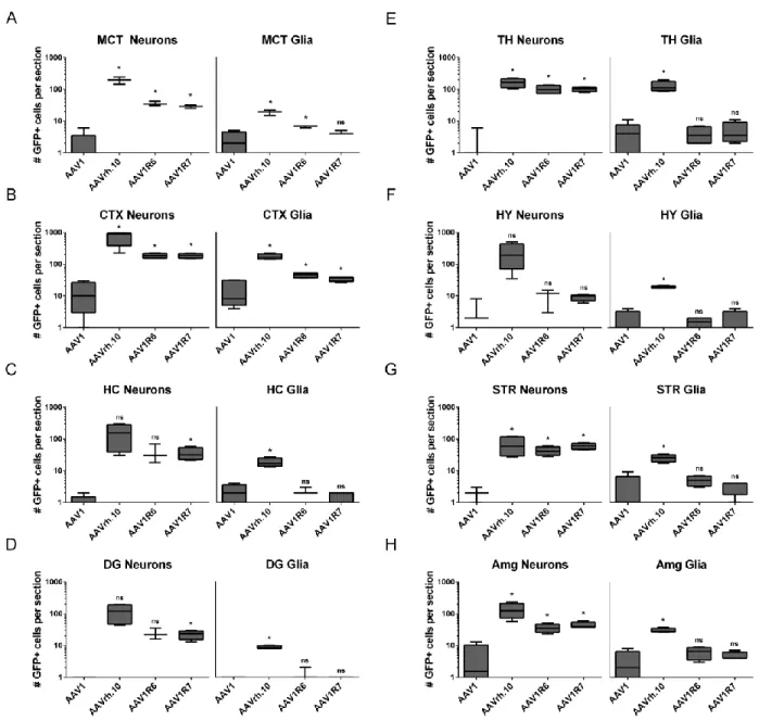
blood-brain barrier and transduce various blood-brain regions following intravascular administration ... 47 Figure 2.4. CNS transduction profile of AAV1R6 and AAV1R7 compared to parental
AAV1 and AAVrh.10 in the brain ... 48 Figure 2.5. Quantitative comparison of neuronal and glial transduction levels for parental
and chimeric capsid variants ... 49 Figure 2.6. Relative cardiac and liver transduction by AAV1R6 and AAV1R7 compared
to parental capsids ... 50 Figure 2.7. Structural analysis of the AAV1R6 chimeric capsid variant ... 51 Figure 2.8. Rational design and functional mapping of a minimal AAVrh.10 footprint for
crossing the BBB ... 52 Figure 2.9. Quantification of neuronal and glial transduction levels mediated by
AAV1RX compared to parental and other chimeric capsid variants ... 53 Figure 2.10. Peripheral tissue transduction and biodistribution of AAV1R6, AAV1R7 and
AAV1RX following intravenous administration ... 54 Figure 2.11. Sequence alignment of 1RX footprint, and Variable Region I (VR-I) across
common AAV serotypes ... 55 Figure 3.1. Structural models of AAV capsids used to interrogate the role of sialic acid
interactions upon blood-brain barrier transport & CNS tropism ... 75 Figure 3.2. Selected capsid variants display intermediate transduction profiles in a cell
type-specific manner in vitro ... 76 Figure 3.3. Capsid variants display differential sensitivities to removal of cell-surface
sialic acid for both transduction and binding ... 77 Figure 3.4. Evaluating the impact of Sialic Acid binding on blood-brain barrier traversal
and CNS transduction in vivo ... 78 Figure 3.5. Quantitation of neuronal and glial transduction profiles for capsid variants
Figure 3.6. Relative liver transduction mediated by capsid variants with differential sialic
acid sensitivities ... 80 Figure 3.7. Structural analysis of the 1RX footprint within the context of the AAV1 Sialic
acid binding pocket ... 81 Figure 3.8. Goldilocks model for the influence of capsid-sialic acid interactions on
blood-brain barrier transport and CNS transduction ... 82 Figure 4.1. Transduction of the choroid plexus and paraventricular nucleus of the
LIST OF ABBREVIATIONS
AADC Amino acid decarboxylase AAP Assembly activating protein AAT Alpha-1-antitrypsin
AAV Adeno-Associated Virus
AAVR Adeno-associated Virus Receptor (aka KIAA0319L) AAVS1 AAV integration site 1
Ad Adenovirus
AD Alzheimer’s Disease
AMG Amygdala
ANOVA Analysis of variation BBB Blood-Brain Barrier
bp base pairs
CA1/2/3 Cornu Ammonis, pyramidal neuron layers 1/2/3 of the hippocampus
cap capsid gene
CBA Chicken beta actin CBh Chicken beta actin hybrid
cDNA Complementary DNA
CMAH cytidine monophosphate-N-acetylneuraminic acid hydroxylase
CMV cytomegalovirus
CNS Central Nervous System
CSF Cerebrospinal fluid
CTX Cortex
DAPI 4’6-diamidino-2-phenylindole
DG Dentate Gyrus
DMD Duchenne muscular dystrophy DMEM Dulbecco’s Modified Eagle Media DMSO Dimethyl sulfoxide
DNA Deoxynucleic acid dsDNA double-stranded DNA
E1a/b Adenovirus early protein 1a/b (transactivator) E2 Adenovirus early protein 2 (DNA-binding protein) E4 Adenovirus early protein 4
ECM extracellular matrix
EM Electron Microscopy
FA Friedrich’s ataxia FBS Fetal bovine serum
FDA Food and drug administration
g gram
GABA Gamma-aminobutyric acid
GAL Galactose
GAPDH Glyceraldehyde-3-phosphate dehydrogenase GFP Green Fluorescent Protein
GMP Good manufacturing practice
HEK293 Human embryonic kidney cell line HIV Human immunodeficiency virus
hr Hour
HS Heparan Sulfate
HSPG Heparan sulfate proteoglycan HSV Herpes Simplex Virus
hSyn human synapsin
HY Hypothalamus
i.v. Intravenous
IACUC Institutional Animal Care and Use Committee IC Intracranial/Intracisternal
ICV Intracerebroventricular
IM Intramuscular
IT Intrathecal
ITR Inverted Terminal Repeats
IV Intravenous
kb kilobases
kg kilogram
LacNac N-acetyl-lactosamine LSD Lysosomal Storage Disease
Luc Luciferase
MB114 Mouse Brain Microvascular Endothelial Cell Line
MCT Motor Cortex
Neuro2a Mouse neuroblastoma cell line NIH National Institutes of Health
nm nanometer
ns Not significant
NVU Neurovascular unit PBS Phosphate Buffered Saline PCR Polymerase chain Reaction PD Parkinson’s Disease PEG Polyethylene glycol
PEI Polyethylenimine
PFA Paraformaldehyde
PSA Polysialic acid qPCR quantitative PCR
rAAV recombinant Adeno-Associated Virus Rep Replication gene
RMS Rostral migratory stream RNA Ribonucleic acid
scGFP Self-complementary Green Fluorescent Protein SCT Somatosensory Cortex
SD Standard Deviation
SEM Standard error of the mean Sf9 Insect cell line
SIA Sialic Acid
SMA Spinal Muscular Atrophy SMN Survival of motor neuron ssDNA single-stranded DNA
ssGFP single-stranded Green Fluorescent Protein ssLuc single-stranded luciferase
STR Striatum
TH Thalamus
TR Terminal Repeat
U87 Human astrocytoma cell line UNC University of North Carolina VA RNA Viral associated RNA
Vg viral genomes
VP Viral protein
VR (I-IX) Variable regions (I-IX)
WT wildtype
αA α-helix A
CHAPTER 1: INTRODUCTION – AAV BIOLOGY AND USE AS CNS-TARGETED GENE THERAPY VECTOR
1.1. AAV Biology Overview
In the famous words of Bruce Lee, “Simplicity is brilliance1.” When it comes to viruses, Adeno-associated virus (AAV) is as simple as they come. Originally discovered as a contaminant in Adenoviral preparations in the 1960s, by Atchison and Rowe, AAV is a small (~20 nm) virus characterized by a non-enveloped icosahedral capsid which contains a 4.7kb single-stranded DNA (ssDNA) genome2–4,. As a helper-dependent member of the Parvoviridae family, AAV requires helper gene functions provided by co-infection with a helper virus such as Adenovirus or various herpesviruses5–8. In addition to its
replication-defective nature, AAV is nonpathogenic, yet is able to persist within the host cell, and several serotypes are prevalent in the human population8–12. These attributes, in part, make it an ideal candidate for gene therapy applications, but also suggest that AAV may have formed an ideal relationship with the human host. Furthermore, because AAV can replicate up to 1 x 10⁶ vg copies per cell, which generally causes cell lysis, only during co-infection with a helper virus, it has been theorized that AAV may possibly have a beneficial effect on the host by providing protection from other viral infections, i.e. Adenovirus or herpesviruses8.
1.2. AAV Genome & Replication
(i.e. transcriptional machinery) in addition to helper gene functions, for replication and transcription of its genome8,15.
Despite its simplicity, the AAV genome possesses additional complexity in its transcripts, accomplished through the use of different promoters, alternative start sites, and alternative splicing8,16,17. There are 3 promoters within the AAV genome, two of which control transcription of the rep gene, with additional variation due to alternative splicing of a single intron to generate a total of 4 transcripts -
Rep78, Rep58, Rep52, and Rep408,16,18. Rep78/58 are both transcribed from the p5 promoter and are the longest of the transcripts, with the sole difference between the two variants being inclusion and exclusion, respectively, of an intron at the 3’-end. The two smaller transcripts, Rep52/40, are transcribed by the p19 promoter and are similarly differentiated by alternative splicing.
The first step for replication of the AAV genome is second-strand synthesis, which converts the ssDNA genome into a transcriptionally competent double-stranded DNA (dsDNA) template8,29,30. The next step is transcription of the rep genes. Transcriptional repression by Rep78/58 is lifted when Ad helper genes allow for Rep-mediated activation of AAV promoters8,31,32. The ITRs serve as the origin of replication for the AAV genome, containing a rep binding site for Rep-mediated activation, and
additionally providing a base-paired free 3’-hydroxyl group which allows DNA synthesis to occur8,32,33. Subsequent replication of the ITR occurs, mediated by binding of Rep and nicking at the terminal
resolution site within the ITR sequence, to facilitate fidelity of ITR replication and to generate a new free 3’-hydroxyl group for subsequent rounds of replication8,29,30. Replication of the AAV genome is known to produce two products due to resolution of the ITR – a dsDNA molecule and a displaced ssDNA molecule.
Productive AAV infection requires helper gene functions which aid in AAV replication, from helper viruses such as Ad and HSV6,8. These essential helper genes are E1a/b, E2a, E4, and the viral associated RNA (VAI RNA)8,15,34. The E1a gene product is a transactivator which relieves repression for the AAV p5 promoter8,15. Briefly, the proteins encoded by these helper genes function to activate,
promote, and ensure processivity of replication and transcription of the AAV genome, while the VA RNA is thought to promote translation of AAV transcripts35. Importantly, these helper functions can be
provided in trans for virus production15. 1.3. AAV Lifecycle
pathway before escaping from early endosomes into the cytosol, a process dependent on the pH-dependent phospholipase A2 domain on VP122–24,47,48. The virion is then thought to traffic through the trans-golgi network before undergoing nuclear import and subsequent trafficking to the nucleolus, where capsid uncoating and viral genome release occurs8,22,37,46,49. Second-strand synthesis, a rate-limiting step in the pathway, then occurs in order to generate a transcriptionally competent template for subsequent transduction and replication steps22. Following replication, both + and – strands are packaged into newly formed AAV capsids at equal efficiencies8. It is also important to note that AAV depends upon the host cell machinery for both transcription and translation.
AAV is capable of persisting in the host cell as an episome, enabling long-term gene expression, and is also able to enter a latent phase which is characterized by targeted genomic integration50,51. The AAV genome integrates into a specific locus on the short arm of chromosome 19, designated the AAVS1 site52. Importantly, integration is dependent on the Rep protein and occurs only for WT virus in the presence of Rep and helper genes, and thus does not occur for recombinant viral vectors53.
There are also several rate-limiting steps along the AAV transduction pathway that have been identified. Briefly, these include trafficking of the capsid to the nucleus22, the rate of capsid uncoating and subsequent release of the viral genome, and second-strand synthesis to generate a dsDNA molecule that can be transcribed8,22,29. The requirement for second-strand synthesis can be overcome through production of self-complementary AAV vectors. These are generated through deletion of the terminal resolution site, a sequence in the ITR, which is required for resolving replication. This deletion enables formation of a partially dsDNA genome packaged into the capsid, bypassing the second-strand synthesis step required for the usual single-stranded genome29,30. Lastly, stability of the vector genome is also known to affect transduction8.
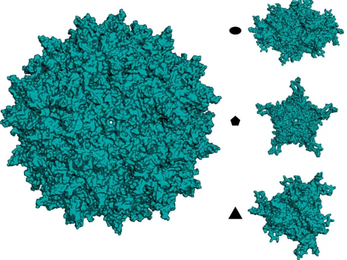
1.4. AAV Capsid Structure
The AAV capsid is a small (~20nm) icosahedron composed of 60 VP monomers - VP1, VP2 & VP3, assembled in a 1:1:10 stoichiometric ratio19. The capsid has a T=1 symmetry (where T is the
predominant VP3 monomer is composed of a highly conserved core β-barrel jellyroll structure which consists of 8 antiparallel βsheets (βB-H) and a similarly conserved α-helix (αA) (Fig. 1.1.)19,20,54. These strands are connected by interdigitating loop regions that are highly variable in sequence identity and are identified based upon the strands they connect. This basic structure is conserved across both
dependoviruses as well as the autonomous parvoviruses19,55,56. The loops are surface-exposed on the full 60mer particle and their interactions dictate the surface topology on the capsid. Consequently, these surface loops are critical determinants of tropism, antigenicity, and receptor usage across capsid serotypes19,57–59.
Furthermore, nine highly variable regions (VRs) are detected in sequence alignments of the primary amino acid sequences across various capsid proteins from various AAV serotypes19,57. They are easily demarcated from the more conserved regions in these alignments. These VR regions, labeled VR-I through VR-IX, vary in length from just a few to a couple hundred amino acids in length. The residues in these VRs overlap with the loop regions between β-strands, and diversity in these VRs dictates nearly all phenotypic differences between various AAV capsid serotypes.
The full length amino acid sequence of VP1 is ~736 amino acids, varying based on relative deletions and/or insertions present across different serotypes. VP1, VP2 and VP3 possess a shared C-terminus; however, VP2 and VP3 are differentially truncated and thus possess different N termini. The VP1 unique N-terminal region is ~140 residues, which is absent in the truncated VP2 sequence (full VP2 length is ~600 amino acids)19,21,23,47,60,61. The further truncated VP3 sequence begins around residue 217 of the VP1 sequence, and consists of the latter ~520 residues of the VP1 sequence, which again varies across serotypes.
crystallographic information for the asymmetrically incorporated VP1 and VP2 monomers, which appear disordered on the inside of the capsid19,68. Thus, structures have been solved for the VP3 capsid
interactions, neglecting the VP1 and VP2 molecules. It is also of note, that the VP1/2 unique regions are not known to directly impact features such as tropism, antigenicity, and receptor usage. Rather, these determinants seem to reside on the VP3 sequence. Nonetheless, the phospholipase A2 domain of the VP1u region is critical for infectivity21,23,47,54,72.
The interactions between the loops of each monomer manifest as various landmark structures that are recognizable on the various axes of symmetry at the capsid surface (Fig. 1.2.). The 5-fold axis of symmetry is a pentamer of VP3 monomers characterized by a cylindrical pore, formed by interactions between the DE loops (corresponding to VR-II) of monomers, and is encircled by a local depression formed largely by interactions involving the HI loop21,73–75 This region is known to be important for capsid assembly and for packaging of the viral genome into the capsid, with the viral genome thought to be inserted through the 5-fold pore54,76.
for the AAV5-SIA complex, and for the AAV9-GAL complex12,36,38,64,68,81–85. Each of these capsids have been found to bind their respective glycan receptors either on or near the three-fold protrusions. The binding pocket for each pair accordingly involves many of the same amino acid residues, most of which are present in VR-IV, VR-V, VR-VI, and VR-VIII, as well as several residues in VR-I. Nonetheless, there is some variation in the precise residues involved, directly and indirectly, in glycan binding.
The 2-fold axis of symmetry is characterized by a topological depression at the interface between two trimers19,20,55,72. This 2-fold channel, also known as the 2-fold dimple, is formed by interactions between VR-I and VR-IX on adjacent monomers, with the α-A helix contributing to formation of the wall lining the 2-fold depression19,57,72,86. The residues composing VR-I, from the B-E loop on the monomer, are also clustered at the shoulder near the base of the three-fold protrusions.
1.5. Background on Natural AAV Serotypes
A large array of AAV variants exist, both natural and synthetic12,20,87–89. The more common of the natural serotypes discovered include AAV1-AAV9. Out of these, AAV2 is by far the most highly
characterized and has historically served as the representative model for most of the studies that have dissected the molecular biology of AAV, elucidating replication and transduction pathways amongst other steps in the AAV lifecycle90. The numerous natural serotypes discovered thus far display broad tropisms at the species, tissue, and cellular levels in addition to broad antigenic profiles12.
In recent years, hundreds of AAV sequences have been isolated from various species. For the most part, these variants were isolated from mostly human and non-human primates (i.e. rhesus
macaques), but new AAV sequences have also been isolated from many other species, including bovine, avian, and reptilian species88,89,91–93. AAV2/3/5/6/9 were all isolated from human hosts, while
varying numbers of human-derived and non-human primate-derived variants. AAV4 and AAV5,
however, diverge significantly from most of the other capsids and fall outside of these clades. Due to their low sequence homology to other AAV serotypes, pre-existing immunity to the AAV4 and AAV5
serotypes is rare in the human population74,96. Additionally, AAV4 and AAV5 have more rounded
protrusions at their 3-fold axes of symmetry, compared to the spikier versions of most other capsids57,71,96. Here, we will briefly discuss the various tropisms and transduction profiles for some of these common serotypes. When delivered systemically, AAV1 transduces mostly the vasculature in various tissues and is generally unable to cross the vascular wall97–99. Nonetheless, AAV1 efficiently transduces skeletal muscle and can also transduce neurons and glia in the CNS, following direct intramuscular or intracranial injections, respectively100–103. AAV2 is similar in that it transduces cells in various tissues, particularly photoreceptors in the eye, with moderate efficiency104–106, but generally relies on direct injections into tissue, performing poorly when administered systemically. AAV2 performs exceptionally well in cell culture, however. AAV3 and AAV8 both transduce the liver at high levels, and are being pursued for liver-targeted gene therapy applications107–110. In humanized murine models, however, AAV3 preferentially transduces human hepatocytes, while AAV8 prefers murine hepatocytes111. AAV8
additionally transduces skeletal muscle as well as pancreatic and cardiac tissues109. Similar to AAV1, AAV6/7 both transduce skeletal muscle at high levels following intramuscular injections; however, AAV7 is also capable of transducing the CNS following systemic administration98,112. AAV4 exhibits a pronounced cardiopulmonary tropism following systemic administration85,112,113. AAV9 is interesting, as it transduces nearly all tissues very efficiently following systemic administration, outperforming nearly all of the other common serotypes in any given tissue112,114,115. Thus, AAV9 is a great example of the tradeoff between efficiency and specificity. Likewise, another serotype, AAVrh.10 (isolated from a rhesus
macaque) behaves very similarly to AAV9 in this regard98. 1.6. Glycan Interactions and Receptor Usage
receptors. Thus, receptor usage is a key determinant for tropism as well as transduction, and AAV capsids are known to utilize several different glycans for attachment19,56. For instance, AAV2 and AAV3 both recognize heparan sulfate proteoglycan (HSPG), and the AAV2-HSPG interaction is historically the most characterized capsid-glycan interaction63,116.
Sialic acid (SIA), in various linkages, is a common receptor for multiple AAV serotypes, including AAV1, AAV4, AAV5, and AAV6. AAV1 recognizes α2,3- and α2,6-N-linked SIA, whereas AAV5 recognizes only α2,3-N-linked SIA117,118. The AAV6 capsid recognizes both HSPG as well as α2,3- and α2,6-N-linked SIA117,118. While these capsid variants all recognize SIA in varying N-linkages, AAV4 is the only variant so far that has been shown to utilize α-2,3-O-linked SIA (also known as mucin)85. Previous studies from our lab have identified galactose (GAL) as the primary receptor for AAV981. A sulfated N-acetyllactosamine (LacNac) was recently identified as the receptor for AAVrh.10, a variant which shares many attributes with AAV9119. Lastly, contrary to the glycoprotein receptors discussed so far, a glycolipid (ganglioside) was found to be the receptor for bovine AAV (BAAV)120,121. Nonetheless, the primary receptors for many other AAV variants remain unknown, i.e. AAV7/8 for instance.
The most important receptor footprint in regards to this dissertation is the SIA binding pocket shared by AAV1 and AAV6, which is located at the base of the three-fold protrusions117. This footprint is comprised of 11 critical amino acid residues, including S268, D270, N271, Y445, G470, S472, V473, N500, T502, and W503. Importantly, only 6/11 of these residues are involved in directly contacting the SIA (N447S G470, S472, V473, N500 T502, and W503). Interestingly, this footprint is analogous to the GAL binding pocket on AAV9. The GAL footprint on AAV9 is localized to the same pocket at the base of the 3-fold protrusions and includes the equivalent residues82. However, the precise amino acid residues of the AAV9 sequence differ at several of these positions (S269, D271, N272, Y446, S448, T471, A473, N474, N501, A503, and W504). Both of these binding pockets involve residues derived from I, VR-IV, and VR-V regions of the VP3 monomer, and the pocket is formed by interactions between loops of two different monomers.
For each of the capsid-glycan interactions described here, the residues and capsid regions involved in binding glycan receptors had also previously been implicated as determinants for
transduction, tropism, and/or antigenicity38,123. This further stresses the role glycan receptor usage has on determining these attributes and reinforces these regions as key structure-function correlates.
In addition to primary glycan receptors, subsequent binding to secondary co-receptors has also been identified as another important step for transduction8,21,38. The nature of such factors have been identified for several AAV variants. These interactions are thought to further facilitate cellular uptake, and generally involve various growth factor receptors or integrins. For example, secondary co-receptors include fibroblast growth factor receptor (FGFR) for AAV2, hepatocyte growth factor receptor (HGFR) for both AAV2 and AAV3, platelet-derived growth factor receptor (PDGFR) for AAV5, and epidermal growth factor receptor (EGFR) for AAV640–42,107,124. Additionally, α5β5 integrins have been identified as important co-receptors for AAV2 and AAV9, while the 37/67-kDa laminin receptor has been found to be a co-receptor for AAV2/3/8/943,46,125. However, no co-receptor has been identified for some serotypes, like AAV4 and AAV7.
In addition to these primary glycan receptors and secondary co-receptors, a previously
uncharacterized transmembrane protein, (KIAA0319L), was recently identified as a universal receptor for AAV. This protein has thus been dubbed the AAV receptor (AAVR)126. While the precise mechanism of how AAVR impacts transduction remains to be clarified, it is thought to facilitate endocytosis and subsequent cellular trafficking. Furthermore, the relative role played by AAVR alongside primary glycan and secondary co-receptors remains contentious.
Lastly, elucidation of these structure-function correlates for glycan receptor usage has afforded our lab and others the ability to rationally engineer these footprints in order to generate novel AAV capsids for both gene therapy applications as well as for basic studies aimed at further expanding our understanding of AAV biology and capsid-glycan interactions38. Historically, such modification of receptor footprints focused on the AAV2 HSPG binding site, but our lab has generated novel AAV4 and AAV9-based vectors with altered mucin and GAL interactions that result in unique phenotypes, which will be discussed in a later section.
1.7. Gene Therapy and AAV Vectors
characterized by an underlying genetic component. For example, in patients afflicted with a disease caused by loss-of-function mutations in a specific gene, the disease may be corrected by delivery of a functional copy of the respective gene. While there are many ways to achieve in vivo gene delivery (i.e. directly administering cDNA, using lipid nanoparticles, etc.), viral vectors are often the most effective means by which to deliver therapeutic transgenes11,12,127–131. Many other viruses have also been evaluated as gene therapy vectors, such as Adenovirus, paramyxoviruses, and the more prominent lentivirus132–137. For the most part, these other viral vectors have been met with concern regarding their safety profile and efficacy; however, lentiviral vectors remain a promising tool for gene therapy applications in which chromosomal integration is required – particularly for ex vivo applications through genetically modifying patient-derived cells for combination gene and cell therapies, such as for hematopoietic stem cells. Nonetheless, AAV is the leading gene therapy vector.
Several of its favorable attributes make AAV an ideal viral vector for gene therapy applications. Chief amongst these is its inherent lack of pathogenicity and replication-defective nature12,138,139.
Additionally, AAV infects both dividing and non-dividing cells, and is able to sustain long-term gene expression in vivo by persisting as an episome within host cells. AAV has an exceptional safety profile, with a lack of integration for the recombinant virus and a favorable immune response8,140. The large array of serotypes which display broad tropisms further provide a large toolbox of capsid reagents for targeting different tissues12. Furthermore, ability to generate high titers of recombinant virus carrying therapeutic transgene cassettes and the relative scalability of production methods further make AAV an ideal viral vector, providing the potential to treat a vast number of monogenetic and familial diseases, or any disease which is treatable by delivery of a therapeutic transgene, afflicting nearly any tissue11,12,141.
European markets with the release of Glybera® (aka Alipogene tiparvovec) a gene therapy for the treatment of familial lipoprotein lipase deficiency, which was developed by uniQure and released in European markets in 2012. Glybera utilizes a single intramuscular injection of an AAV1 capsid to deliver the human lipoprotein lipase gene to muscle cells in order to treat lipoprotein lipase deficiency142–144. Due to current challenges with vector production, it is also important to note that Glybera is currently the most expensive drug in the world, fetching a price around $1,000,000. Furthermore, 2017 became a landmark year for the gene therapy industry, as Luxturna became the first FDA-approved gene therapy using AAV. Luxturna (aka voretigene neparvovec-ryzl) was developed by Spark Therapeutics to treat Leber’s
congenital amaurosis, utilizing subretinal injections of an AAV2-based vector to deliver a functional copy of the RPE65 gene to retinal cells with biallelic RPE65 mutations142,145. Luxturna offers incredible
restoration of vision for patients with this inherited disease; however, like Glybera, is incredibly costly, amounting to over $4,000 per eye146.
Gene therapy using AAV vectors is being pursued for numerous indications which are currently being evaluated in preclinical and clinical studies142. AAV-mediated gene therapy has shown remarkable potential towards the treatment of numerous diseases with a genetic basis. Examples range from
neuromuscular disorders, such as Duchenne muscular dystrophy (DMD) and spinal muscular atrophy (SMA), to neurocardiovascular diseases such as Friedrich’s ataxia, and cardiovascular diseases11,147–151. For example, phase III clinical trials are underway for an AAV9 vector developed by Avexis to treat SMA by delivering a functional copy of the SMN gene to motor neurons following both intravenous and intrathecal administration152–154. AAV vectors are being developed and evaluated by multiple companies to treat Friedrich’s Ataxia (FA) and FA-associated cardiomyopathy by using an AAV vector to deliver a therapeutic frataxin transgene via systemic administration. AAV vectors have also been evaluated in phase I/II clinical trials for the treatment of Alpha-1 antitrypsin (AAT) deficiency, using an AAV1 vector delivered via intramuscular injections to deliver a functioning copy of the Alpha-1 antitrypsin gene155.
Additionally, AAV gene therapies are currently being developed for treating numerous
the most predominant indications being pursued in the CNS space are for Parkinson’s disease and Alzheimer’s disease142. As an example, Voyager has recently developed a drug using an AAV vector to deliver amino acid decarboxylase to improve L-Dopa levels for the treatment of Parkinson’s, which has shown promising safety profiles improvement in patient motor functions following phase 1 clinical studies156. Similarly, gene therapies using AAV vectors could be developed to target Parkinson’s by delivering various other therapeutic genes to replace those identified as disease-associated mutations, such as those involved in lysosomal trafficking defects. Less prominent neurological diseases with a genetic component, such as Gauche’s disease, Pompe disease, and Canavan disease are also being targeted with AAV-based pipelines157–160.
Production and Purification of Recombinant AAV Vectors. Recombinant AAV (rAAV) vectors were first generated in the 1980s using AAV2161. Production of rAAV vectors can be done by replacing the rep & cap genes with a transgene driven by a promoter of choice along with whatever regulatory elements desired, as long as the total size of the transgene cassette is under the 4.5 kb packaging capacity. This transgene cassette is inserted between the flanking ITRs, replacing the rep and cap genes of the WT genome. These ITRs serve as the only cis elements required for packaging. The triple plasmid
transfection method is generally used to produce rAAV vectors. A producer cell line is used to produce the virus. Generally, HEK293 cells are used for this; however, alternative methods such as insect cell-based methods (i.e. using baculovirus/sf9 cells) are also used.
being produced, virus may be harvested from the supernatant and/or the cell pellet, depending on the serotype being made.
Once produced and harvested, a variety of methods are employed to purify rAAV preps141,163–167. In the lab, small-scale preps are purified using PEG precipitation followed by loading onto a density gradient (either cesium chloride or iodixanol) and purified by ultracentrifugation. Depending on the serotype, sonication may be used prior to ultracentrifugation in order to isolate virus from the cells. Following ultracentrifugation, further purification steps such as dialysis or buffer exchange are used; additionally, further purification using a sucrose gradient is employed if more pure virus is needed. Lastly, purified virus preps may be concentrated via centrifugation using size-exclusion columns.
Finally, purified virus preps are titered using qPCR as a gold standard (generally with primers specific for either the transgene or universal primers targeting the ITR) or dot blots. Subsequent biochemical methods can be employed as quality control, including Western blots and electron microscope (to assess purity and ratio of empty:full capsids, respectively). While production of small-scale virus preps at high titer are relatively easy to perform in an academic laboratory setting, scaling up to large-scale good manufacturing practice (GMP) virus production is both challenging and costly. Various methods have been employed to improve these methods, including replacement of adherent HEK293 producer cells with suspension cultures, and further using larger bioreactors and cell stacks to process larger volumes166,168–176. Furthermore, a substantial amount of virus is lost in purification steps, and large-scale manufacturers are employing alternative techniques, such as tangential flow filtration and various forms of chromatography177–179.
suppression are two strategies which have shown promise in circumventing pre-existing immunity. Both the use of less prevalent AAV serotyped capsids and capsid engineering efforts are continually being pursued in order to generate AAV vectors capable of escaping pre-existing immunity while still mediating therapeutic expression levels 57,96,158,182–185.
Second is the limited packaging capacity (4.5 kb) of rAAV vectors, which prevents larger transgenes from being eligible for AAV-mediated gene delivery. Importantly, efforts have been made utilizing dual AAV vectors to deliver these larger transgenes as split versions, packaged within different capsids, which rely on co-infection of the same cell with both vectors and subsequent recombination events, or similarly, dual vectors in which the transcriptional elements are delivered separately from the transgene186–188. These efforts have been met with limited results, but there has been some success in engineering smaller, minimal versions of large transgenes, such as dystrophin. For instance, a smaller micro dystrophin has been engineered which is small enough to be packaged and delivered using rAAV vectors187.
This need for improved efficiency of viral vectors goes hand in hand with a need to improve the (tissue) specificity of vectors. Thus, there is an inherent struggle between optimizing and balancing both efficiency and specificity (i.e. transduction & tropism). For example, vectors that transduce cells in specific target tissues (i.e. the CNS) at a high level (i.e. AAV9 and AAVrh.10) also transduce peripheral tissues, such as the liver, at high levels112,157,193,194. When delivered systemically, these vectors suffer from liver sequestration. Even when these vectors are delivered directly into the CNS through
intracerebroventricular injections, they suffer from systemic leakage, and still transduce the liver195. Efforts of have been made to place transgenes under the control of a cell-type specific promoter in order to optimize specificity, such as the human synapsin (hSyn) promoter, which expresses only in neurons; however, this results in a dramatic drop in transduction efficiency, compared to expression using a more ubiquitous promoter, such as the chicken beta actin (CBA) or CMV promoter196. Thus, this issue is sometimes thought of in terms of an equation in which efficiency plus specificity equals 1, where the two are at odds in a zero-sum game. Nonetheless, capsid engineering efforts in our lab and others, are
continually focused on improving both efficiency and specificity of rAAV vectors, with the goal of achieving therapeutic expression levels at a lower dose.
1.8. Gene Therapy for the Central Nervous System Using AAV Vectors
CNS Gene Therapy and AAV. There has been a lot of success so far in using rAAV vectors to achieve therapeutic CNS gene transfer for targeting multiple neurological diseases, in both preclinical and clinical studies157. AAV possesses many attributes which make it an ideal vector for gene transfer to the CNS and there are many natural and synthetic serotypes with varied phenotypes in regards to CNS transduction and tropism105. Also detailed in previous sections, these attributes are largely the result of structural variation and differential glycan receptor usage. Gene therapy has emerged as an attractive means to treat numerous neurological disorders of the CNS that are characterized by an underlying genetic basis. Indications include examples such as Parkinson’s disease and Alzheimer’s disease. For example, neurodegenerative disorders such as Huntington’s disease and Parkinson’s disease,
through gene replacement using rAAV vectors to restore cellular functions and prevent neurodegeneration157,159.
Examples of CNS Gene Therapy. Additionally, CNS gene therapy is being pursued to treat numerous lysosomal storage disorders (LSDs) such as Krabbe disease, Gaucher disease, Pompe disease, and various mucopolysaccharidoses disorders (MPS)157. These diseases are often caused by loss-of-function mutations which lead to insufficient enzymatic activities, altered metabolism and the
accumulation of toxic products within the cell. This buildup leads to cell death and/or dysfunction, and the resulting neurodegeneration ultimately causes the cognitive and/or motor defects associated with the respective diseases197–199. Similar genetic defects resulting in altered lysosomal storage dysfunctions have also been implicated in causing Parkinson’s disease as well200. Similarly, gene therapy products are currently being developed to treat Parkinson’s disease through delivery of an amino acid decarboxylase (AADC) transgene to increase dopamine levels and are currently undergoing clinical trials156.
1.9. AAV in the Brain – Phenotypes, Routes of Delivery, & Transport
AAV Transport within the CNS. Within the CNS, AAV undergoes several modes of transport, which can also be affected by capsid structure/receptor usage, route of delivery, and the animal models used157. Once in the CNS, AAV vectors can undergo paravascular transport through the CSF and
transport, while AAV2 is only transported in the anterograde direction204,205. AAV8/9, however, can be transported bidirectionally206.
Routes of administration for CNS Gene Delivery. CNS transduction is largely influenced by the tropism of the AAV capsid being used; however, the route of injection also plays a large role. Several routes of administration exist for AAV vectors targeting the CNS, both directly and indirectly, and have been performed in different animal models157,207,208. Direct CNS administration can be achieved through intraparenchymal injections directly into the brain tissue and through administering directly to the
cerebrospinal fluid (CSF). Intra-CSF routes include intracerebroventricular (ICV), intracisternal (IC), and intrathecal (IT) injections into the spinal cord. Intracranial routes generally use stereotaxic injections.
Intraparenchymal injections. Direct injections into the brain parenchyma are the most invasive and generally result in punctate transduction profiles. While many serotypes can effectively transduce a given brain region upon direct intraparenchymal injections at that site, their transduction is limited to the site of injection209. Generally these injections have been performed targeting specific brain regions, such as the thalamus, striatum, hippocampus, etc. Serotypes like AAV2, AAV4, and to a slightly lesser degree, AAV1 and AAV5, show limited spread in the CNS, punctate expression localized to the region injected, and generally show a preferentially neuronal tropism104,210. Thus intraparenchymal injections are desirable for applications in which highly localized and limited gene transfer is desired, but less suited for
applications requiring more global gene transfer.
Intra-CSF administration. Intra-CSF administration, while being similarly invasive, provides the ability to transduce larger areas of the brain. Nonetheless, spread of the virus away from the direct site of injection (i.e. the specific ventricle or cisterna magna) is variable, depending on the capsid. Intra-CSF administration of AAV vectors has been used to successfully achieve CNS gene transfer104,157,211.
Intrathecal (IT) administration via lumbar puncture has proven successful as well157,184,212. AAV1/5/6/8/9 efficiently transduce the spinal cord and brain stem following IT administration, with AAV9
have all been shown to mediate the highest transduction levels and spread more effectively in the CNS than other natural serotypes, following intra-CSF administration through the various routes discussed above105,215,216. Furthermore, AAV1/2/6 capsids mediate preferentially neuronal transduction following intra-CSF or intraparenchymal injections157,210. AAV5 transduces both neurons and glia at low levels while AAV8/9/rh.8/rh.10 transduce both cell types much more efficiently105,216. AAV4, following ICV administration, preferentially transduces ependymal cells, and transduces astrocytes upon
intraparenchymal injection into the subventricular zone and rostral migratory stream (RMS)217,218. However, our lab recently engineered an AAV4 variant, AAV4.18, which was found to have switched receptor usage from O-SIA to α-2,8-polysialic acid (PSA)219. This switch resulted in improved spread in the CNS in addition to selective transduction of migrating neural progenitor cells in the RMS.
Furthermore recent studies from the Song lab at UNC have demonstrated that, following direct microinjections in the dentate gyrus, AAV4 selectively transduces quiescent neural stem cells in this region of the hippocampus220.
1.10. CNS Gene Transfer Across the Blood-Brain Barrier Using AAV Vectors
Systemic Administration. Lastly, AAV vectors can be administered systemically via intravenous injections (IV). This is by far the least invasive route of administration and has the added benefit of promoting a more global distribution of gene transfer across the brain, rather than the more localized expression observed for other routes. Nevertheless, it is also one of the most difficult routes for achieving therapeutic transduction levels, requiring both optimal capsid selection as well as high vector
dosages157,160. This is due to either poor specificity and/or efficiency of vectors targeting the CNS, resulting from tropism for peripheral tissues and sequestration within these tissues, such as the liver221. Effective gene delivery to the CNS is also largely prohibited by the blood-brain barrier157.
cells which line the endothelium, astrocytic end-feet, which provide biochemical support from the brain tissue, and the extracellular matrix (ECM) in which these interact223. The BBB precludes entry of >98% of drugs from entering the brain and the vast majority of all viruses, including AAV. Several pathogenic viruses do possess the ability to cross the BBB and infect cells in the CNS through a few clever
mechanisms224. For instance, HIV can hitchhike along extravasating immune cells225. Others, like Rabies, infect peripheral nerve cells and cross into the brain though axonal transport226. Contrarily, some
pathogenic viruses utilize mechanisms which disrupt the integrity of the BBB in order to gain entry into the CNS, such as the induction of hemorrhages by mouse adenovirus227.
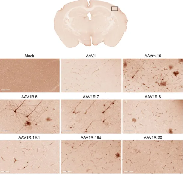
AAV & the BBB. As a very simple virus with a limited coding capacity, both wildtype AAV as well as rAAV vectors are unable to use the more complicated mechanisms employed by pathogenic viruses, as discussed above. Thus, most AAV serotypes are unable to cross the BBB and enter the brain parenchyma following systemic administration. This includes most of the common serotypes (i.e. AAV1-6 and AAV8)98. As an example, AAV1, one of the strains most relevant to this dissertation, effectively transduces and is sequestered within the brain microvasculature following intravenous injection. In the past, strategies have been employed to circumvent the BBB, such as administering vectors in preclinical animal models with a seizure-compromised BBB or through the use of drugs (i.e. mannitol) to disrupt the BBB228,229.
underwent a direct head-to-head comparison evaluating CNS transduction following systemic
administration in adult marmosets230. Here, all three exhibited high transduction levels compared to other serotypes, but AAVrh.8 outperformed both AAV9 and AAVrh.10.
1.11. Engineering AAV Capsids for Gene Transfer to the Brain
Significant efforts in recent years have focused on engineering the AAV capsid in order to achieve improved transduction and tropism for tissues of interest, including the CNS. Strategies to engineer the AAV capsid include peptide insertion, chemical conjugation and mutagenesis231–233.
Combinatorial library-based approaches have seen increased use in recent years to generate new synthetic variants. Often, such libraries are created using either DNA shuffling to recombine cap genes of different serotypes or through targeted mutagenesis strategies228,234. Subsequently, such libraries generally undergo either simple screening or directed evolution in order to identify optimal capsids for the phenotype of interest235–237. An example of a simple screen, from our lab, is the generation of liver-detargeted AAV9 capsid mutants238. Additionally, engraftment of structural features from one capsid to another can generate variants with novel phenotypes. For example, engraftment of the galactose receptor footprint from AAV9 onto the AAV2 capsid generated the AAV2g9 variant, which demonstrated improved neuronal transduction, increased spread, and reduced systemic leakage following ICV injections239. Our lab has recently developed a structure-guided evolution platform, utilizing random mutagenesis of specific capsid regions, informed by structure-function studies, and targeted evolution to generate AAV variants for immune invasion while retaining tropism and transduction properties182. This platform can be extended to carry out structure-guided evolution of glycan receptor footprints as well, which is the focus of future studies.
example, directed evolution of capsid libraries has generated a couple capsid variants which are able to cross the seizure-compromised BBB; however, these are unlikely to cross an intact BBB228. A similar approach was used to generate the synthetic AAV-B1 capsid, which showed improved transduction of the motor cortex and thalamus, as well as the spinal cord, relative to AAV9 following IV injection240. Similar region-focused improvements in gene transfer were observed for the synthetic AAV-AS variant,
generated through peptide-display241. The AAV2-retro vector was engineered through a combination of peptide display and directed evolution and generated a vector with a strong propensity for retrograde axonal transport242. Recently, a Cre recombinase-based AAV targeted evolution (CREATE) method was developed to generate capsids with improved transduction of specific cellular populations in the CNS after IV injections in mice243. This method employed evolution of a peptide-insertion motif inserted into the C-terminus of the AAV9 VP3, and resulted in generation of the AAV-PHP.B vector. AAV-PHP-b was found to improve gene transfer to the astrocytes and neurons in the brain by 40-fold relative to AAV9. Subsequent iterations of this method generated further improved capsids, AAV-PHP.eB and AAV-PHP.S, which exhibit vast improvements relative to AAV9 in terms of CNS and peripheral nervous systems, respectively, following IV injection244. While the AAV-PHP capsids have garnered a lot of attention due to their extremely robust CNS transduction profile, recent studies have shown that these CNS transduction properties are limited to the C57BL/6J mouse background upon which they were selected, and that they fail to transduce BALB/cJ or nonhuman primates245.
This dissertation highlights the efforts I have made towards furthering our understanding of AAV biology in regards to neurovascular transport and CNS tropism/transduction following systemic
CHAPTER 2: MAPPING THE STRUCTURAL DETERMINANTS REQUIRED FOR AAVRH.10 TRANSPORT ACROSS THE BLOOD-BRAIN BARRIER1
2.1. Overview
Effective gene delivery to the central nervous system (CNS) by intravenously administered adeno-associated viral (AAV) vectors requires crossing the blood-brain barrier (BBB). In order to achieve therapeutic CNS transgene expression, high systemic vector doses are often required which pose
challenges such as scale up costs and dose-dependent hepatotoxicity. In order to improve the specificity and efficiency of CNS gene transfer, a better understanding of the structural features that enable AAV transit across the BBB is needed. We generated a combinatorial domain swap library using AAV1, a serotype that does not traverse the vasculature, and AAVrh.10, which crosses the BBB in mice. We then screened individual variants by phylogenetic and structural analyses and subsequently conducted systemic characterization in mice. Using this approach, we identified key clusters of residues on the AAVrh.10 capsid that enabled transport across the brain vasculature and widespread neuronal transduction in mice. Through rational design, we mapped a minimal footprint from AAVrh.10, which, when grafted onto AAV1, confers the aforementioned CNS phenotype, while diminishing vascular and hepatic transduction through an unknown mechanism. Functional mapping of this capsid surface footprint provides a roadmap for engineering synthetic AAV capsids for efficient CNS gene transfer with an improved safety profile.
2.2. Introduction
Adeno-associated viruses (AAV) are non-pathogenic parvoviruses composed of a small, 25 nm icosahedral capsid packaging an approximately 4.7 kb single-stranded DNA genome55. A wide array of AAV capsid sequences have been isolated from human and primate tissues, which have been categorized into several distinct clades based upon sequence and structural diversity246,247. Across these clades, different serotypes display broad tropism at the species, tissue and cellular levels19,246,247. These diverse phenotypes are determined by capsid structure19. The AAV capsid is assembled from 60 viral protein (VP) subunits. The core VP monomer (VP3) has a jellyroll, beta barrel structure comprised of 7 anti-parallel beta strands connected by interdigitating loop regions. Portions of these highly variable loops are surface exposed and define the topology of the AAV capsid, which in turn determines tissue tropism, antigenicity and receptor usage across the various AAV serotypes19. The surface loop residues on the AAV capsid are highly plastic and amenable to modification, affording control over antigenicity, transduction profile and tissue tropism248.
The first step in the AAV lifecycle is recognition of and attachment to cell surface glycan receptors19. These include heparan sulfate (HS) for AAV2, AAV3 and AAV6, α2,3- and α2,6- N-linked Sialic acid (SIA) for AAV1, AAV5 and AAV6, O-linked sialic acid for AAV4, and galactose (Gal) for AAV956. Secondary to glycan binding, cellular uptake of AAV implicates secondary co-receptors,
recently shown that glial-associated lymphatic (glymphatic) transport of CSF influences AAV spread within the mouse brain parenchyma and clearance from the CNS203.
To achieve CNS gene transfer, intravenously administered AAV vectors must first cross the blood-brain barrier (BBB) in order to gain entry into the brain. Comprised of endothelial cell tight junctions along with associated astrocytic end-feet and pericytes, the BBB blocks the diffusion and paracellular flux of macromolecules/particles and regulates the transport of other molecules251. Most viruses which infect the brain do so through disrupting or weakening the BBB; however, some viruses have devised strategies to gain entry into the CNS by methods such as hitchhiking within host immune cells (HIV), by infecting brain endothelial cells or by infecting peripheral nerves and exploiting axonal transport (e.g., Rabies)252,253. In the case of AAV, the BBB prevents entry of most serotypes into the brain with a few notable exceptions. For instance, intravascular administration of AAV serotypes 1-6 and 8 results in poor CNS transduction, while isolates AAV9, AAVrh.8 and AAVrh.10 have been shown, amongst others, to efficiently traverse the BBB in different animal models254–257. In the current study, we generated a chimeric capsid library by shuffling the Cap genes of two isolates, AAV1 and AAVrh.10, which, despite a high degree of structural similarity, display distinct CNS phenotypes. While AAV1 does not appear to cross the BBB efficiently and predominantly transduces the brain endothelium, AAVrh.10 transduces the brain parenchyma by crossing the murine and non-human primate BBB254,258. Using a combination of structural analyses and bioinformatics followed by in vivo screening, we identified a structural footprint on the AAVrh.10 capsid which, when grafted onto AAV1, imparts the ability to traverse the BBB and preferentially transduce neurons within the brain parenchyma.
2.3. Methods
the Cap gene were then used to amplify the library of reassembled full-length Cap sequences and simultaneously insert flanking restriction sites to facilitate subsequent cloning into a pTR plasmid backbone used for virus production.
Phylogenetic and sequence analyses. The amino acid sequences of different AAV capsid isolates were aligned using ClustalW and phylogenetic trees were generated using the MEGAv7.0.21 software package259. The phylogeny was produced using the neighbor-joining algorithm and amino acid distances were calculated using a Poisson correction260. Statistical testing was done by bootstrapping with 1,000 replicates to test the confidence of the phylogenetic analysis and to generate the bootstrap consensus tree261. Branches corresponding to partitions reproduced in less than 50% of bootstrap replicates are collapsed. The percentage of replicate trees in which associated taxa clustered together in the bootstrap test is displayed next to the branches. All sequence alignments were performed using Invitrogen’s Vector NTI Advance 11.5.2 software.
Virus production and titers. An updated triple plasmid transfection protocol was used to produce recombinant AAV vectors195. Specifically, the transfected plasmids include (i) a capsid-specific pXR helper plasmid (i.e. pXR1, pXRrh.10, or various plasmids encoding the various chimeric Cap genes used in this study), (ii) the adenoviral helper plasmid pXX680, and (iii) either pTR-CBh-scGFP or pTR-CBA-Luc plasmids (encoding either a self-complementary green fluorescent protein (GFP) reporter transgene driven by the chicken beta actin hybrid (CBh) promoter or a luciferase reporter transgene (Luc) driven by the chicken beta actin promoter (CBA), respectively, flanked by inverted terminal repeats (TRs) derived from the AAV2 genome. Viral vectors were purified using iodixanol density gradient ultracentrifugation. Vectors packaging a CBh-scGFP transgene were subsequently subjected to buffer exchange and
primers were designed to specifically recognize the AAV2 inverted terminal repeats (forward, 5’- AACATGCTACGCAGAGAGGGAGTGG –3’; reverse, 5’-
CATGAGACAAGGAACCCCTAGTGATGGAG -3’) (IDT Technologies, Ames IA).
Animal studies. All animal experiments were performed using 6-to-8-week-old female C57/BL6 mice purchased from Jackson Laboratories (BAR Harbor, ME). These mice were maintained and treated in compliance with NIH guidelines and as approved by the UNC Institutional Animal Care and Use Committee (IACUC). To investigate the ability of AAV vectors to cross the BBB and transduce CNS cell populations, AAV vectors packaging a CBh-scGFP transgene or 1x PBS (as mock treatment) were administered intravenously (i.v.) via tail vein injection at a dose of 5 x 10¹¹ vg. To assay for GFP reporter transgene expression, animals were sacrificed 21 days post injection with tribromoethanol (Avertin) (0.2 ml of 1.25% solution) followed by transcardial perfusion with 30 ml of 1x PBS followed by 30 ml of 4% paraformaldehyde in PBS. Tissues including the brain, heart and liver were removed and post fixed for 24 h, and 50-μm-thick sections were obtained for each tissue using a Leica VT 1200S vibrating blade
microtome (Leica Biosystems, IL). The mouse brain sections were then immunostained as described below. For in vivo luciferase transduction and viral genome biodistribution experiments, mice were injected with either 1x PBS or viral vectors packaging a CBA-Luciferase transgene at a dose of 1 x 10¹¹ vg. Mice were sacrificed, as described above, at 14 days post injection and various tissues were removed. For these experiments, no fixation with 4% paraformaldehyde in 1x PBS was performed and instead tissues were dissected and frozen at -80⁰C prior to use.
MA). The relative light units obtained for each sample were then normalized to the input tissue weight for each sample, measured in grams. Data was graphed and statistical analyses carried out using an unpaired two-tailed T-test with Welch’s correction as well as ANOVA followed by Tukey’s multiple comparisons test where indicated. These statistical analyses were performed in GraphPad Prism 6® software.
Vector genome biodistribution. Animal studies were performed as described above. At 21 days post injection, mice were sacrificed and tissues were frozen at -80⁰C. Tissues were later thawed and viral genomes were extracted from the tissue lysates using the DNeasy kit (Qiagen, Valencia, CA). Viral genome copy numbers were then determined for each tissue using quantitative PCR with primers specific to the luciferase transgene (forward, 5′- AAAAGCACTCTGATTGACAAATAC -3′; and reverse, 5′- CCTTCGCTTCAAAAAATGGAAC -3′). These viral genome copy numbers were then normalized to the mouse lamin B2 housekeeping gene using the primers (forward, 5′-
GGACCCAAGGACTACCTCAAGGG -3′; and reverse, 5′- AGGGCACCTCCATCTCGGAAAC -3′). The biodistribution of viral genomes are represented as the ratio of vector genomes per cell recovered for each tissue. Data was graphed and statistical analyses carried out as described earlier.
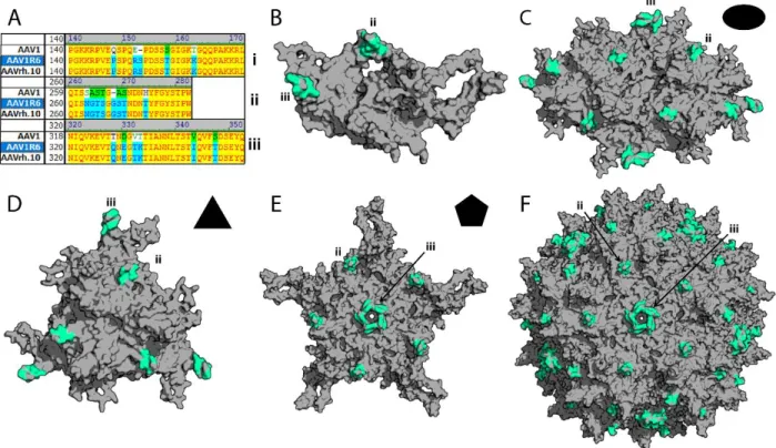
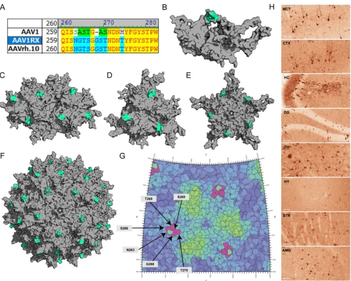
Molecular modeling. Previously published coordinates (PDB ID, 3NG9) were used to generate three-dimensional structures of the AAV1 VP3 trimer/three-fold axis of symmetry.263 Homology models of AAVrh.10 and various AAV1/rh.10 chimeric capsid structures were obtained using the SWISS-Model server (http://swissmodel.expasy.org/)264, with the crystal structure of AAV8 VP3 (PDB ID, 2QA0) used as a template and a structure-based alignment was generated using the secondary structure matching (SSM) application in the WinCoot software59,205,265, with the AAV1 VP3 (PDB ID 3NG9) monomer being used as a template. The VP3 trimers/3-fold symmetry axes, VP3 trimer dimers/2-fold symmetry axes, VP3 pentamers/5-fold symmetry axes, and full capsids were generated using the VIPERdb oligomer generator utility (http://viperdb.scripps.edu/oligomer_multi.php)266. Surface rendered depictions of these models were visualized using PyMOL (the PyMOL Molecular Graphics System, Schrödinger LLC, http://www.pymol.org/). Stereographic roadmap projections of the AAV1RX capsid surface highlighting surface-exposed amino acid residues within the AAVrh.10-derived neurotropic footprint were generated using RIVEM (Radial Interpretation of Viral Electron Density Maps) software267.
2.4. Results
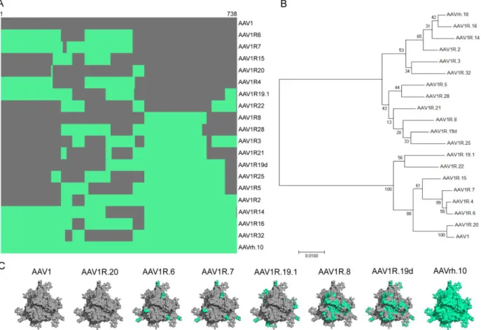
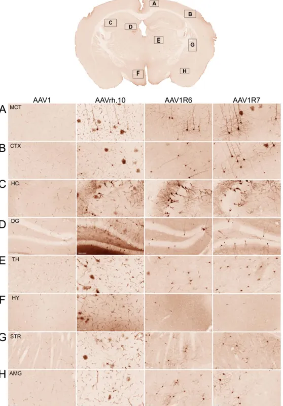
structure-function correlates for traversing the BBB. Correspondingly, we generated an AAV1/rh.10 domain swap library through DNA shuffling. We selected AAV1 and AAVrh.10 as parental capsid sequences for DNA shuffling since they differ markedly in their abilities to cross the BBB and because of the sequence homology (85%) shared by their capsid (Cap)genes. Thirty-six chimeric capsid sequences were then clonally isolated and sequenced. The variants generated from this library displayed substantial diversity at the DNA and amino acid level. Sequence alignment revealed a spectrum of domain swaps, which we then organized in order of increasing homology from AAV1 to AAVrh.10 (Fig. 2.1c), top to bottom). This panel of clones was further characterized phylogenetically by constructing a neighbor-joining tree, which broadly categorized these variants as either more AAV1-like (Clade A) or more AAVrh.10-like (Clade E) (Fig. 2.1b). Small-scale vector production was then used to establish relative titers to exclude capsids defective in assembly or packaging from the study. Homology-based structural models of the parental and representative chimeric capsid trimers highlighting key surface
domains/residues at the three-fold axis of symmetry (Fig. 2.1c) were generated to further narrow the list of chimeric capsid variants for initial screening in vivo. Ten chimeric capsid variants were selected based on structural analyses, out of which six yielded recombinant vectors (packaging scGFP or ssLuc
transgene cassettes) at titers similar to parental AAV1 and AAVrh.10 vectors. These variants were then screened further.
In vivo screening identifies two AAV1/rh.10 chimeric capsids capable of crossing the BBB after
intravenous administration in adult mice. We hypothesized that, based on their structural diversity, the selected panel of capsid variants would differ in their ability to cross the BBB and transduce the CNS after I.V. administration. It is important to note that our approach does not involve directed evolution, as this strategy is generally applicable for selecting optimal capsids and less suited for studying structure-function relationships. We injected 6-8-week-old mice with a dose of 5 x 10¹¹ viral genomes (vg) per mouse of AAV1, AAVrh.10, or one of six different chimeric AAV vectors packaging a