Received 1 Dec 2015
|
Accepted 29 Feb 2016
|
Published 31 Mar 2016
Identification of chemicals that mimic
transcriptional changes associated with autism,
brain aging and neurodegeneration
Brandon L. Pearson
1,2,
*, Jeremy M. Simon
1,2,
*, Eric S. McCoy
1
, Gabriela Salazar
1
, Giulia Fragola
1
& Mark J. Zylka
1,2
Environmental factors, including pesticides, have been linked to autism and
neurodegenera-tion risk using retrospective epidemiological studies. Here we sought to prospectively identify
chemicals that share transcriptomic signatures with neurological disorders, by exposing
mouse cortical neuron-enriched cultures to hundreds of chemicals commonly found in the
environment and on food. We find that rotenone, a pesticide associated with Parkinson’s
disease risk, and certain fungicides, including pyraclostrobin, trifloxystrobin, famoxadone and
fenamidone, produce transcriptional changes
in vitro
that are similar to those seen in brain
samples from humans with autism, advanced age and neurodegeneration (Alzheimer’s
disease and Huntington’s disease). These chemicals stimulate free radical production
and disrupt microtubules in neurons, effects that can be reduced by pretreating with a
microtubule stabilizer, an antioxidant, or with sulforaphane. Our study provides an approach
to prospectively identify environmental chemicals that transcriptionally mimic autism and
other brain disorders.
DOI: 10.1038/ncomms11173
OPEN
1Department of Cell Biology and Physiology, UNC Neuroscience Center, University of North Carolina at Chapel Hill, 111 Mason Farm Road, Chapel Hill,
North Carolina 27599-7545, USA.2Carolina Institute for Developmental Disabilities, University of North Carolina Chapel Hill, Chapel Hill, North Carolina 27599-7255, USA. * These authors contributed equally to this work. Correspondence and requests for materials should be addressed to M.J.Z.
P
owerful new sequencing technologies have been used to
systematically identify hundreds of candidate gene
muta-tions associated with autism spectrum disorder (ASD)
risk
1,2. Heritability studies suggest that environmental factors
also contribute to autism risk
3. Indeed, gestational exposure to
pesticides, including maternal proximity to pesticide applications
and runoff, is reproducibly associated with increased ASD risk in
epidemiological studies
4–6. However, epidemiological studies are
retrospective and cannot ascertain prospectively, precisely or
systematically which chemicals, of the
4
80,000 chemicals
registered for use in the environment, have the greatest
potential to harm the developing or adult brain
7. Existing
in vivo
neurodevelopmental and neurotoxicological assays with
animal models are labour intensive and costly, thus hindering
throughput, whereas higher-throughput toxicological assays
frequently use non-neuronal cells or focus on neuron death as
an end point
8,9. As a result, these tests fail to interrogate
molecular and physiological processes that are unique to neurons
or that differentiate normal from diseased human brains.
There is growing recognition that brain transcriptional changes
are associated with ASD
10,11. This ASD transcriptional signature
is defined by reduced expression of genes involved in synaptic
transmission and elevated expression of genes involved in
immune and microglial function
10,11. Here we hypothesized
that this transcriptional signature might guide the prospective
identification of candidate chemical risks for ASD. To test this
hypothesis, we exposed mouse cortical neuron-enriched cultures
to hundreds of environmental-use chemicals and then monitored
global transcriptional changes. We identify six chemical groups,
one of which mimics the transcriptional changes seen in ASD, but
surprisingly also shares transcriptional similarity to the aged
brain and certain neurodegenerative conditions. Our findings
suggest these neurological conditions share a molecular
pathology, as hypothesized by others
12, despite different ages of
onset and distinct behavioural symptoms. Moreover, our study
shows that a transcriptional approach can be used to
systematically scan a diverse chemical space and identify
potential environmental threats to the human brain.
Results
Mouse cortical cultures transcriptionally model human brain.
To determine whether mouse cortical cultures model cellular and
molecular aspects of the human brain, we performed
immuno-cytochemistry with cellular markers and compared the gene
expression profile of our cultures with brain cell-type-specific
expression data sets and human brain gene expression data sets,
including the Allen BrainSpan atlas (www.brainspan.org) and
GTEx
13,14. Our cultures contained many of the principle cell
types of the brain based on immunocytochemistry with markers
for neurons, astrocytes and microglia (
B
25% of cultures were
non-neuronal cells; Fig. 1a,b). In addition, the expression of
markers for each brain cell type
13in our cultures was highly
correlated with that of whole embryonic (E14.5) brain (Pearson
r
¼
0.8), further suggesting that our culture system represented all
major cell classes in biologically realistic proportions (Fig. 1c;
Supplementary Fig. 1a,b). Globally, gene expression in our
cortical cultures correlated more strongly with each human
brain region (except spinal cord) than to any other tissues of the
human body (Supplementary Fig. 2a). The strongest correlations
were to frontal cortex and anterior cingulate cortex, regions
implicated in ASD
3. Moreover, cultures were most strongly
correlated with frontal cortex from mid-late gestation human
brain (Supplementary Fig. 2b), the developmental time window
implicated in ASD pathogenesis
15. In contrast, terminally
differentiated neurons derived from human embryonic stem
cells
16did not strongly correlate with any brain region, and
instead were most similar to tissues associated with the female
reproductive system (Supplementary Fig. 2c). Taken together, our
cortical cultures show strong transcriptional similarities to the
human brain.
Transcriptional screen with environmental chemicals. We
next measured cytotoxicity of the 294 chemicals in the US
Environmental Protection Agency (EPA) ToxCast Phase I library,
which
includes
common
food-use
pesticides
and
other
environmental chemicals, such as plasticizers
8, to identify a
non-toxic concentration for RNA sequencing (RNA-seq). We
treated cultures in quadruplicate for 24 h at 10
m
M, as this is a
common concentration used in screening studies
17, then used
fluorescent markers to quantify the proportion of live and dead
cells (Supplementary Fig. 3). Most (87%) of the chemicals were
not cytotoxic at 10
m
M, whereas concentrations of the remaining
chemicals had to be reduced to between 10 and 100 nM
(Supplementary Data 1). Fresh cultures were then treated (24 h)
with each chemical at the non-toxic concentration or with vehicle
(equivalent dimethylsulphoxide (DMSO) concentration
r
0.5%),
as the negative control. We also tested three topoisomerase 1
(TOP1) inhibitors, as they reproducibly downregulate long
(
4
100 kb) genes
18.
To identify chemicals that caused concordant gene expression
changes, we performed hierarchical clustering of 5,121 genes
variably expressed across all chemicals, and six chemical groups
emerged (hereafter referred to as clusters 1–6; Fig. 2; see Methods
and Supplementary Fig. 4 for batch correction details). Individual
chemicals were assigned to a cluster using a pairwise correlation
threshold and were validated to be statistically and biologically
Cell marker gene expression
a
c
27% Other
72±2% NeuN+
0.45±0.04% Gfap+ 0.33±0.02% Iba1+
b
S1 pyr.
CA1 pyr.
Interneuron
Astrocyte
Oligo.
Microglia
Mural
Endothelial Ependymal 0
10 20 30 40 50 60
Hoechst Hoechst
Hoechst NeuN Gfap Iba1
Figure 1 | Mouse cortical cultures contain all principle brain cell types.
significant (Supplementary Fig. 5; see Methods for further details
on inclusion/exclusion criteria). To evaluate whether existing
in vitro
ToxCast assay data could identify similar chemical
relationships, we performed clustering analysis across 199
cell-free and cellular (non-neuronal) assays. The resulting
chemical groupings did not resemble those we detected by gene
expression profiling (Supplementary Fig. 6). Thus, transcriptional
profiling with neuronal cells identifies relationships between
chemicals that existing toxicological assays failed to detect.
To determine what these functional relationships were, we
examined specific genes with altered expression in each cluster
(Fig. 2). Cluster 1 chemicals upregulated immediate early genes
(IEGs) while reducing expression of several potassium channel
genes. These expression changes were suggestive of neuronal
hyperexcitability. Supporting this possibility, cluster 1 included
two pyrethroids (cyfluthrin and fenpropathrin) that stimulate
sustained calcium influx in cortical neurons
19. Cluster 2
chemicals upregulated numerous immune and cytoskeletal
transcripts but reduced expression of ion channel and synaptic
genes. Cluster 3 chemicals upregulated IEGs but reduced the
expression of immune and cytoskeletal genes. Cluster 4 chemicals
resembled cluster 3, but without inducing IEGs. Cluster 5
contained
all
three
TOP1
inhibitors,
which
markedly
downregulated long genes and downregulated IEGs, consistent
with one of these inhibitors (topotecan) reducing spontaneous
synaptic
activity
in
cortical
neuron
cultures
20. Deletion
of a related topoisomerase (TOP2B) reduced IEG expression
21.
Cluster 6 chemicals reduced expression of several
neuro-transmitter receptor subunits, including
N
-methyl-
D-aspartate
receptor subunits (
Grin2a
and
Grin2b
), cholinergic receptors
(
Chrm2
and
Chrna7
) and ion channels.
Cluster 2 transcriptionally mimics specific brain disorders. We
next identified the molecular pathways that were differentially
associated with the six chemical clusters using gene set analysis
(GSA). Included in this analysis were 64 gene sets for human
brain disorders and nervous system pathways that we generated
from publications (Supplementary Data 2) alongside 4,722
curated pathways. Over 600 gene sets distinguished the six
clusters (Supplementary Fig. 7; Supplementary Data 3), including
many of the human brain disorder gene sets (Fig. 3). However,
only cluster 2 chemicals mimicked the transcriptional changes of
two post-mortem ASD brain expression data sets in a
bidirec-tional manner. This included downregulation of M12 and Mod1
(largely composed of synaptic genes that are downregulated in
ASD brains), and upregulation of M16 and Mod5 (largely
com-posed of microglial/immune genes that are upregulated in ASD
brains)
10,11. In addition, cluster 2 showed concordance with aging
human brain and two neurodegenerative disorders (Alzheimer’s
disease and Huntington’s disease), suggesting shared aspects of
molecular pathology despite different symptoms (see Discussion).
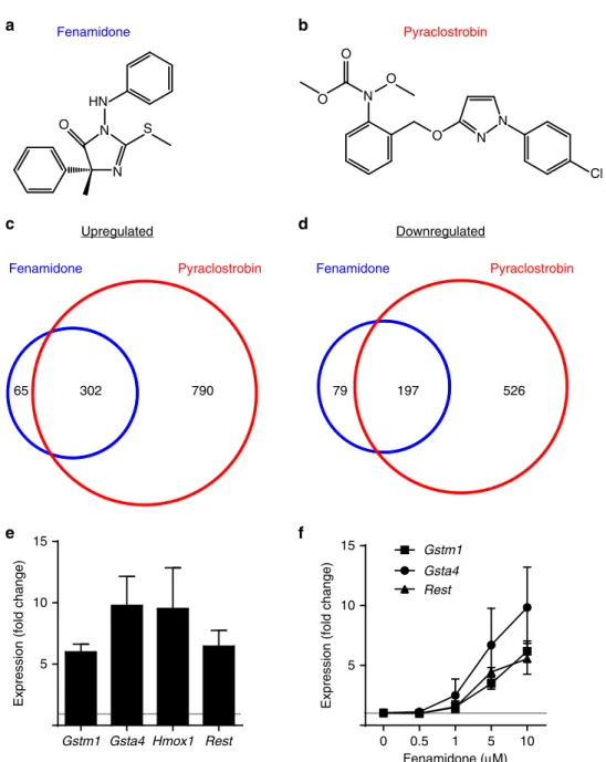
Fenamidone, pyraclostrobin and two other chemicals in cluster
2 (famoxadone and trifloxystrobin) are members of a recently
developed class of fungicides that inhibit mitochondrial complex
III by targeting the quinone outside (Qo) site of cytochrome
bc
1(ref. 22). Since fenamidone and pyraclostrobin are structurally
distinct (Fig. 4a,b), we performed RNA-seq with multiple
replicates of both the chemicals to further validate that expression
of a common set of genes was altered. Each fungicide altered
expression of a largely overlapping set of genes (Fig. 4c,d;
Supplementary Data 4). Upregulated transcripts included
Nrf2
target antioxidant response genes and
Rest
, which we validated
using quantitative real-time PCR (qRT–PCR) (Fig. 4e,f).
5,121 genes
Median-centered expression value 0.5
–0.5
>100 kb
mtDNA
Cytosk.
Acta2 Tagln Cnn1 Cnn2 Lima1
Immune
Tspo Cx3Cr1
Abca1 Mki67 Tlr13
Nrf2 targets
Gstm1 Hmox1
Ion channels
Kcnn1 Kcnn2
IEG/Hormone
Bdnf Crh Vgf Kcnb2 Kcnh3
Fos Fosb Npas4
Gsta4 Nqo1
Synaptic
Nlgn1 Nrxn3 Ctnna2
NMDA
Grin2a Grin2b Kcna3
v
1 2 3 4 5 6
t
Chemicals
Rest
elevation is associated with human brain aging and
neurodegeneration
23. Other compounds in cluster 2 include
fenpyroximate, pyridaben and rotenone, chemicals that target
mitochondrial complex I (ref. 24). Exposure to rotenone is
known to increase risk for Parkinson’s disease
24,25. All cluster 2
chemicals reduced RNA-seq reads arising from the mitochondrial
genome
(Fig.
2),
suggesting
compromised
mitochondrial
function as a common mechanism
26. Note that none of the
mitochondrially encoded transcripts were among the 5,121
variably expressed genes assayed by hierarchical clustering, so
these transcripts did not influence cluster assignment. However,
the influence of chemical treatment on mitochondrial density or
viability was not assessed and could have indirectly modulated
transcriptional profiles.
Cluster 2 induces superoxide and microtubule instability.
Mitochondrial complexes I and III are the main sites of
super-oxide (O
2) production within the electron transport chain, so we
Ontology enrichment score
(FDR < 0.1)
1.0
–1.0
Pathology Set name
Downregulated (Lu)
Upregulated (Lu)
Downregulated (Durrenberger)
Downregulated (Lagier-Tourenne TDP-43 model)
Downregulated (Lederer)
Upregulated (Lederer)
Downregulated (Blalock)
Upregulated (Blalock)
Downregulated (Sekar)
Upregulated (Sekar)
Downregulated (Wu)
Upregulated (Wu)
Downregulated (Gupta Mod1)
Upregulated (Gupta Mod5)
Downregulated (Voineagu M12)
Upregulated (Voineagu M16)
Downregulated (Iwamoto)
Upregulated (Iwamoto)
Downregulated (Durrenberger)
Upregulated (Durrenberger)
Downregulated (Valor, cerebellum)
Upregulated (Valor, cerebellum)
Downregulated (Valor, hippocampus)
Upregulated (Valor, hippocampus)
Multiple sclerosis Downregulated (Durrenberger)
Downregulated (Durrenberger)
Upregulated (Durrenberger)
Downregulated (Simunovic)
Upregulated (Simunovic)
Downregulated (Chahrour Mecp2 null)
Upregulated (Chahrour Mecp2 null)
Downregulated (Samaco Mecp2 null)
Upregulated (Samaco Mecp2 null)
Downregulated (Barnes, BA22 region)
Upregulated (Barnes, BA22 region)
Downregulated (Fillman)
Upregulated (Fillman)
Downregulated (Hwang)
Upregulated (Hwang)
Downregulated (Maycox)
Upregulated (Maycox) Aging brain
Amyotrophic lateral sclerosis
Alzheimer's
Autism spectrum disorders
Bipolar disorder
Huntington's disease
Parkinson's disease
Rett syndrome
Schizophrenia
1 2 34 5 6
Figure 3 | Cluster 2 chemicals show significant gene set enrichment with autism and other brain diseases.The enrichment scores
next tested whether cluster 2 chemicals induced O
2. Fenamidone
produced a concentration-dependent increase in O
2, as
measured with a fluorescent mitochondrial superoxide indicator
dye, and swelling of neuronal soma (Fig. 5a–d). Pretreatment
with the free-radical scavenger vitamin E (
a
-tocopherol) blocked
fenamidone-induced production of O
2(Fig. 5e; using the
concentration of fenamidone that was used for sequencing)
and blocked soma swelling (Fig. 5f). Sulforaphane is a
potent inducer of
Nrf2
target antioxidant gene expression,
it reduces inflammation and it substantially reduces autism
symptoms in humans
27,28. Pretreatment with sulforaphane
attenuated the transcriptional changes, O
2production and
soma swelling caused by fenamidone (Fig. 6). We also monitored
O
2production for each chemical in cluster 2 and for
several chemicals outside of cluster 2 across a seven-point
concentration–response range (Supplementary Fig. 8). All
chemicals that induced O
2at the sequencing concentration
were assigned to cluster 2 based on gene expression.
Fluoxastrobin and azoxystrobin are Qo fungicides that were not
assigned to cluster 2, likely because they did not generate O
2at
the sequencing concentration (Supplementary Fig. 8). Two
additional mitochondrial inhibitors that induce O
2production
in cells (myxothiazol, complex III inhibitor; kresoxim-methyl,
complex III inhibitor and fungicide) altered transcription in a
manner consistent with chemicals in cluster 2 (Supplementary
Fig. 9). Collectively, our data support a relationship between O
2production and the cluster 2 transcriptional signature.
Numerous
genes
associated
with
cytoskeletal
function
have been implicated in autism
29. Moreover, disruption of
microtubules is associated with problems in brain development
0 0.5 1 5 10
5 10 15
Gsta4 Gstm1
Rest
Fenamidone (µM) 5
10 15
Gstm1 Gsta4 Hmox1 Rest
Expression (fold change) Expression (fold change)
Fenamidone Pyraclostrobin
N N HN
S O
N O
O
O
O
N N
Cl
Fenamidone Pyraclostrobin
65 302 790
Fenamidone Pyraclostrobin
79 197 526
Downregulated Upregulated
a
c
b
d
e
f
and with neurodegeneration
30,31. Given that cytoskeletal genes
were altered by cluster 2 chemicals and that rotenone (also in
cluster 2) destabilizes microtubules
31,32, we hypothesized that
microtubule depolymerization triggered the aberrant swelling
morphology of neurons. Consistent with this possibility,
stabilization
of
microtubules
with
paclitaxel
attenuated
fenamidone-induced O
2production and fenamidone-induced
soma swelling (Fig. 5g,h). However, fenamidone did not impair
tubulin polymerization in a cell-free biochemical assay, ruling out
a direct effect of fenamidone on tubulin oligomerization
(Supplementary Fig. 10a). Last, superoxide production and
soma swelling could be phenocopied by depolymerizing
microtubules with vincristine (Supplementary Fig. 10b–e).
Assessment of potential confounds. Cluster 2 compounds
upregulated numerous microglial- (for example,
Cx3cr1
and
Trem2
) and astrocyte- (for example,
Gfap
and
Aqp4
) enriched
genes
33. This increase in markers of proliferating brain cell types
was unlikely to be due to cell division, as our cultures were treated
with an antimitotic on days
in vitro
(DIV) 3. Moreover,
fenamidone (under the same conditions used for sequencing)
caused no significant change in cellular composition, based on
immunocytochemical quantification of neurons, astrocytes and
microglia with markers (NeuN, Gfap and Iba1, respectively;
Supplementary Fig. 11a). However, any contribution by
additional non-neuronal cell types such as oligodendrocytes has
not been accounted for. Neither culture batch nor sequencing
batch effects contributed to cluster identity (Supplementary
Fig. 4). We also assessed RNA quality using the RNA integrity
number (RIN) as a proxy. There were subtle differences in quality
among the six clusters, and RINs were significantly lower for
clusters 2 and 6 (Supplementary Fig. 11b). However, the average
RINs were
Z
9.25 for each of the six clusters, indicating that these
samples were of sufficiently high quality. Collectively, we found
no evidence that technical artefacts or poor RNA quality
contributed to cluster identity. However, we cannot exclude the
possibility that some of the chemicals alter cellular composition at
higher concentrations or after longer exposure times.
Increasing agricultural use of several cluster 2 chemicals.
Epidemiological and human exposure data for most chemicals in
cluster 2 are lacking. We thus sought to evaluate exposure
potential by analysing chemical usage and food commodity
residue data collected by the United States Geological Survey,
the United States Department of Agriculture (USDA) and the
Food and Drug Administration (FDA). All of the mitochondrial
complex III inhibitors in cluster 2 showed positive environmental
usage trends since their EPA registration in 2000 or later
(Fig. 7). Usage of complex I inhibitors (rotenone and pyridaben)
is low and unchanging, with the notable exception of
fenpy-roximate, the most potent superoxide producer we identified
(concentration for half-maximum response (EC50)
¼
0.007
m
M;
Supplementary Fig. 8b). Many cluster 2 residues were found on
conventionally raised food commodities, particularly leafy green
vegetables, and were detected at relatively high levels, up to
20 p.p.m. in the case of pyraclostrobin. These data suggest
significant human exposure potential to many of the chemicals in
cluster 2.
Discussion
By comparing gene expression profiles of cortical cell cultures
with expression data from human brain disorders, we identified a
group of eight chemicals (cluster 2) that transcriptionally
mimicked ASD, brain aging and neurodegeneration. These
chemicals, most of which inhibit mitochondrial complex I
or III, stimulated free radical production and disrupted
micro-tubules. We found that pretreating with a microtubule stabilizer,
an antioxidant, or with sulforaphane could reduce these effects.
–10 –8 –6 –4 –2 –10 –8 –6 –4 –2
2 4 6 8 10
log[Fenamidone], M
****
+
– – +
– – + +
*
1,000 3,000
Vitamin E
Fenamidone
Fluorescence (a.u.)
20 40 60 80 100
log[Fenamidone], M
Aberrant cells (%)
Fluorescence (a.u.)
20 40 60 80 100
Aberrant cells (%)
+
– – +
– – + +
****
****
+
– – +
– – + +
Paclitaxel Fenamidone
1,000 4,000
Fluorescence (a.u.)
****
****
+
– – +
– – + +
20 40 60 80 100
Aberrant cells (%)
****
***
****
Vehicle Fenamidone
O2–
a
b
c
d
e
f
g
h
Vitamin E
Fenamidone
Paclitaxel Fenamidone
Figure 5 | Fenamidone causes mitochondrial superoxide production and microtubule destabilization.(a,b) Superoxide (O2, MitoSOX fluorescent
indicator) and aberrant cell morphology elicited by 2-h treatment with 10mM fenamidone. Scale bars, 10mm. (c) O2 generation and (d) aberrant
morphology is dose dependent. RNA-seq dose is denoted by red circles. (e) Pretreatment with vitamin E (10mM, 2 h) blocked O2 formation and
(f) aberrant morphology elicited by fenamidone (10mM, 2 h). (g) Microtubule stabilization with paclitaxel pretreatment (10mM, 2 h) attenuated O2
Whether this transcriptional and cellular response is related to
the marked clinical efficacy of sulforaphane at treating ASD
symptoms
27remains to be determined.
Numerous studies investigated a link between the inhibition of
mitochondrial complex I, neurotoxicity and neurodegeneration in
animal models
24,34,35. Rotenone (in cluster 2) has been shown
to increase Parkinson’s disease risk in humans
25. Cluster 2
also included a relatively new class of fungicides (quinone
outside, Qo) that inhibit mitochondrial complex III. No evidence
of neurotoxicity was noted for two of these fungicides,
pyraclostrobin and fenamidone, in a set of assays used by
regulatory agencies
36,37. However, a single oral dose of
trifloxystrobin (also in cluster 2) reduced motor activity for
several hours in female rats and for 3 days in males
38, suggesting
a strong interaction with sex. Picoxystrobin (not in ToxCast
Phase I library), marketed as the most rapidly absorbed and most
systemic (in plants) of all Qo fungicides, caused acute
neurotoxicity (reduced motor activity) at the lowest dose tested
in rats
39. Further, mitochondrial complex III-deficient mice
showed severe superoxide-dependent damage to cortical brain
regions and profound motor deficits that were apparent at night,
during their active phase
40. Note that standard acute and chronic
neurotoxicity assays are performed during the day, when rodents
are less active, possibly reducing the power to detect motor
deficits. Mitochondrial complex III inhibitors can additionally
block neuronal differentiation by maintaining embryonic
stem cell pluripotency
41, suggesting a potential for
neuro-developmental effects.
Usage data indicate that Qo fungicides are increasingly
prevalent on food that is consumed by humans of all ages.
At least one cluster 2 chemical (pyraclostrobin) is present in
the environment at levels that affect non-mammalian
organ-isms
42,43and was detected at high levels on foraging honeybees,
further corroborating high levels in the environment
42. To
address whether these levels are a risk to human health, recent
in vitro
reverse dosimetry extrapolations from the EPA found that
food levels of pyraclostrobin exceed the human oral equivalent
dose necessary to affect mitochondrial processes
44. However, Qo
fungicide residues have not been detected on organically
produced foods (EPA and USDA data), suggesting a way to
minimize exposure.
Our finding that cluster 2 chemicals mimic the transcriptional
changes of autism, as well as the aging brain and
neurodegenera-tion was surprising, particularly given the different ages of onset
and disease symptoms. Oxidative stress and cytoskeletal integrity
are implicated in all of these conditions
45–49, suggesting that
overlapping pathological processes might drive the transcriptional
similarities we observed. In support of shared biology, we found
0 20 40 60 80 100 Cytosk.
Acta2 Tagln Cnn1 Cnn2 Lima1 Immune
Tspo Cx3Cr1
Abca1 Mki67 Tlr13
Nrf2 targets Gstm1 Hmox1
Ion channels Kcnn1 Kcnn2
IEG/Hormone Bdnf
Crh Vgf Kcnb2 Kcnh3
Fos Fosb Npas4
Gsta4 Nqo1
Nlgn1 Nrxn3 Ctnna2
NMDA Grin2a Grin2b Kcna3
Sulforaphane Fenamidone +
+ + –
0 2,000 4,000 6,000 8,000
Fenamidone
0 0 1 1 10 10
– + – + – +
Fluorescence (a.u.)
**
****
SULF (µM)
log
2
fold-change expression relative to control 4.0
–4.0 Fenamidone
0 0 1 1 10 10
– + – + – +
SULF (µM)
Aberrant cells (%)
a
b
c
****
***
Synaptic
Figure 6 | Sulforaphane attenuated fenamidone-induced transcriptional and cellular responses in cortical cultures.(a) Genome-wide (RNA-seq) transcriptional changes caused by fenamidone (10mM, 24 h;n¼3 replicates) and fenamidone (10mM, 24 h) after pretreating with sulforaphane (10mM, 18 h;n¼3 replicates). Gene order is identical to Fig. 2. (b) Fenamidone-induced (10mM, 2 h) O2 production and (c) aberrant cell morphology were
0 2 4 6 8 Cilantro (2010)
Spinach (2009)
Kale (2008)
Frozen spinach (2011)
Sweet potato (2008)
0 1 2 3 4 Lettuce (2010)
Frozen spinach (2011)
Spinach (2009) Head lettuce (2011)
Blackberries (2012)
0 0.05 0.1 0.15 Apples (2009)
Pears (2010) Cherry tomatoes (2010)
Currants (2012) Cherry tomatoes (2011)
0 0.01 0.02 0.03 0.04 Cherry tomatoes (2012)
Tomatoes (2012)
Cilantro (2009)
0 0.1 0.2 0.3 0.4 Eggplant, dried or paste (2012)
Grapes (2010) Grapes (2009)
Bok choy dried or paste (2011)
Sweet pepper (2010)
0 0.2 0.4 0.6 Blackberries (2011)
Jicama (2011)
Herbals and botanicals (2012)
0 0.1 0.2 0.3 0.4 0.5 Unspecified vegetable (2012)
Grapes (2009)
Grapes (2010)
Peaches (2008)
Cherry tomatoes (2012) Azoxystrobin
Parts per million
Famoxadone
Fenpyroximate
Fluoxastrobin
Parts per million
Parts per million
Parts per million
Pyridaben
Parts per million
Rotenone
Parts per million
Trifloxystrobin
Parts per million
a
c
b
d
2000 2004 2008 2012 0.2
0.6 1.0 1.4
Year
Millions of kilograms
2000 2004 2008 2012 0.00
0.02 0.04 0.06
Year
Millions of kilograms
2000 2004 2008 2012 0.00
0.01 0.02
Year
Millions of kilograms
2000 2004 2008 2012 0.0
0.1 0.2
Year
Millions of kilograms
2000 2004 2008 2012 0.01
0.02 0.03 0.04
Year
Millions of kilograms
2000 2004 2008 2012 0.000
0.002 0.004 0.006
Year
Millions of kilograms
2000 2004 2008 2012 0.0
0.2 0.4 0.6
Year
Millions of kilograms
1994 1970
0 2 4 6 8 10 Spinach (2009)
Spinach (2009)
Spinach (2010)
Lettuce (2011) Lettuce (2010) Fenamidone
Parts per million
Millions of kilograms
0 5 10 15 20 25 Spinach (2008)
Frozen spinach (2010) Dandelion greens (2009)
Kale (2008)
Frozen spinach (2011) Pyraclostrobin
Parts per million
Millions of kilograms
2000 2004 2008 2012 0.00
0.01 0.02 0.03
2000 2004 2008 2012 0.0
1.0 2.0 3.0
Year Year
e
f
g
h
i
that ASD, the aging brain, Alzheimer’s disease and Huntington’s
disease exhibit altered expression of a common set of genes more
so than any of the other neurological gene sets we tested
(Supplementary Fig. 12a). Many of the genes that were
differentially regulated by pyraclostrobin were also found in the
M16 and M12 ASD gene modules (Supplementary Fig. 12b–e).
Moreover, when focusing specifically on the genes in these ASD
modules, the direction and magnitude by which these genes were
dysregulated in ASD patient samples was strongly correlated with
that of pyraclostrobin treatment in our cortical cultures
(Spearman
r
¼
0.66; Supplementary Fig. 12f), further suggesting
a common mechanism. The fact that these neurological
conditions shared a core set of dysregulated genes may
contribute to the enrichment observed for cluster 2 across these
diseases. However, we cannot exclude the possibility that
molecular pathologies are shared by some but not all of the
conditions. Disentangling these relationships is beyond the scope
of our current study, but suggests a fruitful area for future
research.
Several of the chemicals in cluster 2 unquestionably kill
neurons at higher concentrations (Supplementary Data 1),
consistent with other studies
9,50. This raises the question of
whether cluster 2 reflects the transcriptional signature of ‘sick’
neurons. We identified several chemicals that killed cells at
multiple concentrations (Supplementary Data 1), yet only those
that were
associated
with
O
2production,
microtubule
destabilization and elevated expression of neuroinflammatory
genes were assigned to cluster 2. It thus seems unlikely that
cluster 2 is reflective of chemicals that nonspecifically kill neurons
at high doses and sicken neurons at lower doses. In fact,
chemicals can kill (and presumably sicken) cells via distinct
mechanisms
32, with one of these mechanisms being ‘microtubule
destabilization.’ Rotenone and vincristine fit within this class
32,
likely
providing
additional
insights
into
why
paclitaxel
(a microtubule stabilizer) attenuated the soma swelling and O
2production phenotypes induced by a cluster 2 chemical (Fig. 5).
Our study also shows how systematic transcriptional studies
with neurons can uncover new brain- and disease-relevant
relationships between chemicals that cannot be identified using
existing toxicology assays (Supplementary Fig. 6), including those
that rely on cell death as a readout.
We identified additional brain-relevant relationships between
chemicals within other clusters. Cluster 1 appears to define a
transcriptional signature of neuron hyperexcitability, as evidenced
by upregulation of IEGs, a class of genes that mark recently
depolarized neurons, and downregulation of potassium channels
(which increases neuron excitability when downregulated
51).
Cluster 1 contained two pyrethroids that hyperexcite mammalian
neurons
19. Intriguingly, two recent epidemiological studies found
that pyrethroid exposure doubles the risk for attention deficit
hyperactivity disorder in boys
52,53. Our transcriptional approach
might provide a way to prospectively identify candidate chemical
risks for attention deficit hyperactivity disorder. Cluster 5
contained all three topoisomerase inhibitors, a class of drugs
that downregulated long genes in neurons
18, reduced synaptic
activity
20and reduced IEG expression (Fig. 2). Moreover, cluster
5 is strongly correlated with neurological disease models that
feature dysregulated long synaptic gene expression, particularly
amyotrophic lateral sclerosis
54and Rett syndrome
55,56.
Though estimates vary,
B
50% of cells in the adult human
central nervous system are neurons
57. This is in contrast to the
embryonic culture system employed here, which is comprised of
over 70% neurons. This disparity has the potential to bias
physiological signatures and impair the ability to detect some
disease-relevant processes. Although our cultures show a very
similar representation of brain cell markers relative to E14.5
whole mouse brain (Supplementary Fig. 1), suggesting our
cortical cultures—dissected at E14.5—contain the major brain
cell classes in biologically realistic proportions. Moreover, the
model system employed here is amenable to high-throughput
screens and functional assays
17. Ultimately, candidate chemicals
identified with a cortical culture system will require validation in
animal models.
In summary, our study shows that chemicals that
transcrip-tionally mimic brain disorders can be identified by profiling gene
expression in cortical cultures. This approach may also prove
useful in identifying candidate ASD therapeutics, such as
sulforaphane, or in identifying drugs that normalize long gene
expression, such as topoisomerase inhibitors in Rett syndrome
model neurons
55. While usage and residue levels of cluster 2
chemicals on conventionally grown foods are increasing, in the
absence of causality, it is premature to draw correlations with
the increased prevalence of ASD and other brain disorders.
Nonetheless, greater scrutiny over whether these new Qo
fungicides affect the developing or adult mammalian nervous
system (enteric, peripheral and central) or behaviours seems
warranted, particularly given their striking mechanistic similarity
to rotenone. Ultimately, monitoring steady-state levels in the
environment, assessing exposure levels and pharmacokinetics,
and epidemiological studies will be needed to evaluate whether
any of these chemicals pose real neurological threats to humans
or increase risk for brain disorders, including ASD.
Methods
Cortical neuron culture
.
Primary mouse cortical neuron cultures were prepared as previously described from E14.5 pregnant C57BL/6J (Cat. #000664, Jackson) dams crossed to CAST/EiJ (Cat. #000928, Jackson) males18. Hybrid cultures are thought to better model genetic variation associated with human populations58. Dissociated cells were placed in multiwell plates coated with poly-D-lysine (0.1 mg ml1) in Neurobasal medium (Life Technologies) containing 5% fetal bovine serum (Gibco), B27 (17504-044, Invitrogen), Antibiotic-Antimycotic (15240-062, Invitrogen) and GlutaMAX (35050-061, Invitrogen). At DIV 3, a half medium change was performed with feeding medium identical to the plating medium except that we omitted fetal bovine serum and included 4.84mg ml1uridine 50-triphosphate (U6625, Sigma-Aldrich) and 2.46mg ml15-fluoro-20-deoxyuridine (F0503, Sigma-Aldrich) to inhibit mitosis in dividing cells.Chemicals
.
The chemical library was donated by the EPA ToxCast Program. For verification and replication experiments, the following chemicals were purchased from Sigma-Aldrich:DL-atocopherol acetate (T3376),DL-sulforophane (S4441), vincristine sulfate salt (V8879), oxyfluorfen (35031), rotenone (45656), fenamidone (33965), pyraclostrobin (33696), trifloxystrobin (46447), myxothiazol (T5580), pyridaben (46047), azoxystrobin (31697), fluoxastrobin (33797), fenpyroximate (31684) and kresoxim-methyl (37899). Famoxadone was purchased from Chem Service, Inc (N-11943). Topotecan hydrochloride was purchased from Tocris (4562). Paclitaxel was purchased from Fisher Scientific (AC32842). All chemical stocks were prepared in DMSO unless otherwise noted. Vehicle samples were prepared with an equivalent DMSO concentration ofr0.5% in feeding medium.image to obtain the total number of nuclei and the total number of nuclei from dead cells, respectively. The percentage of dead cells was calculated and averaged across four replicates. All chemicals from the library were initially tested at 10mM. A concentration causing 10% or greater cell death relative to vehicle was considered toxic. Lower doses were tested by reducing an order of magnitude until a non-toxic dose was identified.
RNA-seq
.
Cultures were treated with the non-cytotoxic dose of each chemical, by exchanging half of the original medium with chemicals at 2concentration in prewarmed feeding medium, on DIV 7 for 24 h in 12-well plates at a density of 5105cells per well. RNA was isolated using RNeasy plus mini kit (Cat. #74134, Qiagen). RNA yield and quality were determined using a Nanodrop 1000 Spectrophotometer (Thermo Scientific). Samples were further assessed for quality using either an Agilent Bioanalyzer 2100 or TapeStation 2200 to obtain a RIN. RIN values exceeding 7 were used for sequencing. RNA samples were used to generate and barcode complementary DNA libraries using the TruSeq RNA Library Preparation Kit at the UNC High Throughput Sequencing Facility. Pools of 24 multiplexed samples were sequenced per lane on an Illumina HiSeq 2500 using 50-bp paired-end reads.RNA-seq data processing
.
RNA-seq reads were filtered using TagDust and aligned to the reference mouse genome (mm9) with TopHat using default parameters59,60. Reads aligning to ribosomal RNA genes were removed. Transcript abundance was estimated by computing Reads per kilobase per million mapped reads (RPKM) using RefSeq gene models aggregated by gene symbol61. For differential expression analyses, raw counts over RefSeq exons were used, and then were compared across samples using DESeq62.Hierarchical clustering and cluster membership
.
RPKMs for all RefSeq genes and all samples were filtered such that a given gene had490% of samples with RPKM40 and gene length exceeded 500 bp. RPKMs were then log10-transformed, quantile-normalized, median-centred, and the effects due to culture or sequencing batch were removed using a mixed model analysis of variance. This batch correction step was performed on individual experiments rather than chemical– vehicle ratios to reduce the effects due to variations in culture composition as well as batch-to-batch variations at the level of sequencing. Genes were then median-centred again, and filtered such that their s.d. exceeded 0.1. Chemical replicates were then combined using the median. Classes of chemicals were then discovered in two ways. First, we performed hierarchical clustering using average linkage and Pearson correlation distance measures. The boundaries of the discovered classes from clustering were set by computing pairwise Spearman correlations for all samples. A given cluster had to have at least three members with the minimum pairwise Spearman correlation coefficient exceeding 0.2, though cluster members often exceeded a correlation of 0.6 (Supplementary Fig. 5c,d). Seven chemical clusters were discovered, however one was removed due to failing to have a positive silhouette width, a metric of cluster robustness63. These definitions enabled the three positive control topoisomerase inhibitors to form a cohesive group of chemicals with known structural and functional similarity (cluster 5). None of the culture or sequencing batches contributed to the formation of any one of the final six clusters (Supplementary Fig. 4).Hierarchical clustering of EPA toxicological assays
.
Concentration at 50% maximum activity (AC50) values for all assays and all ToxCast Phase I chemicals were retrieved from the EPA Dashboard. We retained only those assays with data available for 99% of the chemicals and required that a given assay must have at least five active chemicals (AC50r10mM); 199 assays were retained. We then transformed the data values as described in Sipeset al.64and performed hierarchical clustering to find the chemical groups with concordant activity on certain assays.Pathway analysis
.
To detect differentially regulated pathways, chemicals within each of the six clusters were separately compared with all other chemicals tested using GSA65, supplying a modified gene pathway file that contained all MSigDB C2 annotations as well as gene sets from published gene expression studies in human diseases and mouse models. Nearly all added gene sets were derived directly from the associated publication. Raw data derived from Durrenbergeret al.66were analysed by applying quantile normalization, and differential expression was detected using SAM using 500 permutations67. Some brain disease gene expression studies had no significantly differentially expressed genes (qo0.05), and hence could not be included in our analysis. For ‘BLALOCK_ALZHEIMERS_UP’, a gene set included within MSigDB C2, we removed gene symbols that were not included in the RefSeq annotation. The Mod1 pathway (Guptaet al.10) was filtered to include genes with a KM1 score of 0.4 or greater, to reduce the number of genes in this pathway below 1,500 so it could be analysed by GSA. A given pathway (size between 10 and 1,500 genes) was considered if it achieved statistical significance (false discovery rateo0.1) for at least one cluster compared with all other chemicals (500 permutations). Expression of a single pathway was then summarized by taking the median expression value across all genes in that pathway for a given chemical.Once the entire data matrix was assembled, pathways were median-centred and hierarchically clustered, while keeping chemical ordering consistent with Fig. 2. Disease-relevant ontologies were then extracted and plotted separately; colour was applied based on the pathway enrichment score (blue indicates pathway downregulation and red indicates pathway upregulation), but only for the pathways achieving statistical significance (false discovery rateo0.1).
Quantitative real-time PCR
.
Total RNA was extracted and purified using the RNeasy plus mini kit (Qiagen) following the manufacturer’s instructions. RNA was quantified using Nanodrop 1000 (Thermo Scientific) and reverse transcribed using iScript Reverse Transcription Supermix (Bio-Rad). Quantitative real-time PCR analysis was performed in technical duplicates using SYBR Green PCR master mix (Applied Biosystems) in a 7500 Real-Time PCR instrument (Applied Biosystems). An amount of 10 ng of complementary DNA was used in each reaction. Fold change of expression of the target RNA in each treatment condition relative to the untreated sample were calculated by raising 2 to the power of DDCt.DDCt was calculated by subtracting theDCt of the untreated sample to theDCt of each treatment condition. TheDCt of each sample was calculated by subtracting the average Ct ofGapdhto the average Ct of the target RNA. The following primers were used at a final concentration of 0.5mM:Rest(F: 50-GTGCGAACTCACACAGGAGA-30, R: 50-AAGAGGTTTAGGCC CGTTGT-30);Gsta4(F: 50-CGGCTGGAGTGGAGTTTGAG-30, R: 50-CCAAGG GTACTTGGCCGAAA-30);Gstm1(F: 50-CCGTGCAGACATTGTGGAGA-30, R: 50-CTGCTTCTCAAAGTCAGGGTTG-30);Hmox1(F: 50-AGGCTTTAAGCT GGTGATGGC-30, R: 50-GGGGCATAGACTGGGTTCTG-30); andGapdh (F: 50-TATGACTCCACTCACGGCAAAT-30, R: 50-GGGTCTCGCTCCTGGAA GAT-30).
Fluorescence Immunohistochemistry
.
Mouse cortical neuron cultures were prepared identically as for live/dead and RNA-Seq experiments but at an equivalent density of 2.5105cells per well in a 24-well plate containing precoated glass coverslips. At DIV 7, cells were treated with fenamidone at a concentration of 10mM for 24 h. Coverslip-adherent cells were rinsed, paraformaldehyde fixed, blocked with 10% donkey serum and probed with the following cell-type-specific primary antibodies: guinea pig anti-NeuN (1:400, ABN90P, Millipore), goat anti-Gfap (1:1,200, ab53554, Abcam) and rabbit anti-Iba1 (1:400, 019-19741, Wako Pure Chemical Industries). Species-specific AlexaFluor-conjugated donkey secondary antibodies (1:200, Life Technologies) were then applied along with Nucblue (Life Technologies) to counterstain all nuclei. Coverslips were inverted into mounting substance (Fluorogel, 17985-10, Electron Microscopy Sciences) and imaged on a Zeiss LSM 710 laser scanning confocal microscope using a 10 objective tiling scan of entire coverslips including threezplanes to account for uneven coverslips. A researcher blind to the experimental conditions of images utilized maximal projected, spliced images to obtain particle counts for the total Nucblue-positive nuclei and the NeuN-positive neurons using particle analysis in ImageJ. Gfap- and Iba1-positive features containing clear Nucblue-positive nuclei were manually counted using the count function in Adobe Photoshop.Mitochondrial superoxide detection and live-cell imaging
.
Cultures were pre-pared on commercial poly-D-lysine-coated glass coverslips (GG-12-pdl, neuVitro) in 24-well plates at a density of 1.5105cells per well. At DIV 7, cells were treated with chemical or an equivalent DMSO concentration in feeding medium for 2 h. Cells were then treated with MitoSOX Red (M36008, Life Technologies) mito-chondrial superoxide indicator for 10 min at 37°C. Medium containing the dye was aspirated, and the cells rinsed twice with 37°C feeding medium and replaced with 37°C artificial cerebral spinal fluid composed of 150 mM NaCl, 5 mM KCl, 1 mM MgCl2, 2 mM CaCl2, 10 mM HEPES and 10 mM dextrose (pH 7.3). Individual coverslips were transferred to a stage-top perfusion system mounted on an inverted Nikon Ti Eclipse microscope with constant flow of warmed artificial cerebral spinal fluid. Images (20) were collected using an Andora Clara charge-coupled device camera for brightfield and the Texas Red channel to ascertain the mitochondrial superoxide levels with the same exposure across all experiments. ImageJ software (NIH) was used to trace the soma of the cells in bright-field images as regions of interest (ROIs) using the polygon selection tool. ROIs were then superimposed on the fluorescent image and the total cell fluor-escence was calculated per ROI to account for the size of the ROI and normalize to background fluorescent levels, as described68. Fluorescence intensity was then normalized to average corrected vehicle intensity. At least two non-overlapping fields from each coverslip were collected for each well. ROI tracing was performed by a researcher blind to the experimental condition. For select conditions, the proportion of cells showing altered soma morphology was manually calculated per image by the same experimenter performing ROI tracing.minute for 60 min. Equimolar doses of paclitaxel (included in kit) and vincristine (V8879, Sigma-Aldrich) were included as positive and negative modulators of tubulin polymerization, respectively.
Chemical usage and approval data
.
Usage data were acquired from United States Geological Survey. Data are reported as total kilograms applied for each chemical across all US counties sampled per year (2000–2012) and a linear trend line was fit beginning in year 2000 or with the registration year (if after 2000). Registration dates for specific pesticide products were obtained from the National Pesticide Information Retrieval System (NPIRS; http://ppis.ceris.purdue.edu/). The NPIRS is a collection of pesticide-related databases, applications and websites under the administration of the Center for Environmental and Regulatory Information Systems at Purdue University, West Lafayette, Indiana. NPIRS obtains product information on a weekly basis from the EPA Pesticide Product Information System website. This Pesticide Product Information System information, including registered use sites and pests listed on the EPA stamped approved label, is disseminated through the NPIRS member and public websites, and is provided for informational purposes only. To ascertain which foodstuffs had the greatest amount of these chemicals, we ranked the five food commodities in descending order of maximum residue level detected using 2008–2012 data from the USDA Pesticide Data Program and the FDA Pesticide Program Residue Monitoring Program.References
1. De Rubeis, S.et al.Synaptic, transcriptional and chromatin genes disrupted in autism.Nature515,209–215 (2014).
2. Iossifov, I.et al.The contribution ofde novocoding mutations to autism spectrum disorder.Nature515,216–221 (2014).
3. Chen, J. A., Penagarikano, O., Belgard, T. G., Swarup, V. & Geschwind, D. H. The emerging picture of autism spectrum disorder: genetics and pathology.
Annu. Rev. Pathol.10,111–144 (2015).
4. Shelton, J. F.et al.Neurodevelopmental disorders and prenatal residential proximity to agricultural pesticides: the CHARGE study.Environ. Health Perspect.122,1103–1109 (2014).
5. Roberts, E. M.et al.Maternal residence near agricultural pesticide applications and autism spectrum disorders among children in the California Central Valley.Environ. Health Perspect.115,1482–1489 (2007).
6. Rossignol, D. A., Genuis, S. J. & Frye, R. E. Environmental toxicants and autism spectrum disorders: a systematic review.Transl. Psychiatry4,e360 (2014). 7. Grandjean, P. & Landrigan, P. J. Neurobehavioural effects of developmental
toxicity.Lancet Neurol.13,330–338 (2014).
8. Dix, D. J.et al.The ToxCast program for prioritizing toxicity testing of environmental chemicals.Toxicol. Sci.95,5–12 (2007).
9. Regueiro, J., Olguin, N., Simal-Gandara, J. & Sunol, C. Toxicity evaluation of new agricultural fungicides in primary cultured cortical neurons.Environ. Res. 140,37–44 (2015).
10. Gupta, S.et al.Transcriptome analysis reveals dysregulation of innate immune response genes and neuronal activity-dependent genes in autism.Nat. Commun.5,5748 (2014).
11. Voineagu, I.et al.Transcriptomic analysis of autistic brain reveals convergent molecular pathology.Nature474,380–384 (2011).
12. Diaz-Beltran, L., Esteban, F. J. & Wall, D. P. A common molecular signature in ASD gene expression: following Root 66 to autism.Transl. Psychiatry6,e705 (2016).
13. Zeisel, A.et al.Brain structure. Cell types in the mouse cortex and hippocampus revealed by single-cell RNA-seq.Science347,1138–1142 (2015). 14. GTEx Consortium. Human genomics. The Genotype-Tissue Expression
(GTEx) pilot analysis: multitissue gene regulation in humans.Science348,
648–660 (2015).
15. Willsey, A. J.et al.Coexpression networks implicate human midfetal deep cortical projection neurons in the pathogenesis of autism.Cell155,997–1007 (2013).
16. van de Leemput, J.et al.CORTECON: a temporal transcriptome analysis of
in vitrohuman cerebral cortex development from human embryonic stem cells.
Neuron83,51–68 (2014).
17. Huang, H. S.et al.Topoisomerase inhibitors unsilence the dormant allele of Ube3a in neurons.Nature481,185–189 (2012).
18. King, I. F.et al.Topoisomerases facilitate transcription of long genes linked to autism.Nature501,58–62 (2013).
19. Cao, Z., Shafer, T. J. & Murray, T. F. Mechanisms of pyrethroid insecticide-induced stimulation of calcium influx in neocortical neurons.J. Pharmacol. Exp. Ther.336,197–205 (2011).
20. Mabb, A. M.et al.Topoisomerase 1 inhibition reversibly impairs synaptic function.Proc. Natl Acad. Sci. USA111,17290–17295 (2014).
21. Madabhushi, R.et al.Activity-induced DNA breaks govern the expression of neuronal early-response genes.Cell161,1592–1605 (2015).
22. Bartlett, D. W.et al.The strobilurin fungicides.Pest Manag. Sci.58,649–662 (2002).
23. Lu, T.et al.REST and stress resistance in ageing and Alzheimer’s disease.
Nature507,448–454 (2014).
24. Sherer, T. B.et al.Mechanism of toxicity of pesticides acting at complex I: relevance to environmental etiologies of Parkinson’s disease.J. Neurochem.100,
1469–1479 (2007).
25. Tanner, C. M.et al.Rotenone, paraquat, and Parkinson’s disease.Environ. Health Perspect.119,866–872 (2011).
26. Mehrabian, Z., Liu, L. I., Fiskum, G., Rapoport, S. I. & Chandrasekaran, K. Regulation of mitochondrial gene expression by energy demand in neural cells.
J. Neurochem.93,850–860 (2005).
27. Singh, K.et al.Sulforaphane treatment of autism spectrum disorder (ASD).
Proc. Natl Acad. Sci. USA111,15550–15555 (2014).
28. Sandberg, M., Patil, J., D’Angelo, B., Weber, S. G. & Mallard, C. NRF2-regulation in brain health and disease: implication of cerebral inflammation.Neuropharmacology79,298–306 (2014).
29. Chang, J., Gilman, S. R., Chiang, A. H., Sanders, S. J. & Vitkup, D. Genotype to phenotype relationships in autism spectrum disorders.Nat. Neurosci.18,
191–198 (2015).
30. Poirier, K.et al.Mutations in TUBG1, DYNC1H1, KIF5C and KIF2A cause malformations of cortical development and microcephaly.Nat. Genet.45,
639–647 (2013).
31. Choi, W. S., Palmiter, R. D. & Xia, Z. Loss of mitochondrial complex I activity potentiates dopamine neuron death induced by microtubule dysfunction in a Parkinson’s disease model.J. Cell Biol.192,873–882 (2011).
32. Wolpaw, A. J.et al.Modulatory profiling identifies mechanisms of small molecule-induced cell death.Proc. Natl Acad. Sci. USA108,E771–E780 (2011).
33. Zhang, Y.et al.An RNA-sequencing transcriptome and splicing database of glia, neurons, and vascular cells of the cerebral cortex.J. Neurosci.34,
11929–11947 (2014).
34. Greenamyre, J. T., Cannon, J. R., Drolet, R. & Mastroberardino, P. G. Lessons from the rotenone model of Parkinson’s disease.Trends Pharmacol. Sci.31,
141–142 (2010).
35. Mullett, S. J. & Hinkle, D. A. DJ-1 deficiency in astrocytes selectively enhances mitochondrial Complex I inhibitor-induced neurotoxicity.J. Neurochem.117,
375–387 (2011).
36. O’Mullane, M. & Tasheva, M. Fenamidone.JMPR219–270 (2013). 37. Bartholomaeus, A. Pyraclostrobin.JMPR275–319 (2003). 38. Dannan, G. & Tasheva, M. Trifloxystrobin.JMPR387–450 (2004). 39. Dewhurst, I. & Solecki, R. Picoxystrobin.JMPR725–767 (2012).
40. Diaz, F., Garcia, S., Padgett, K. R. & Moraes, C. T. A defect in the mitochondrial complex III, but not complex IV, triggers early ROS-dependent damage in defined brain regions.Hum. Mol. Genet.21,5066–5077 (2012).
41. Pereira, S. L.et al.Inhibition of mitochondrial complex III blocks neuronal differentiation and maintains embryonic stem cell pluripotency.PLoS ONE8,
e82095 (2013).
42. Pettis, J. S.et al.Crop pollination exposes honey bees to pesticides which alters their susceptibility to the gut pathogen Nosema ceranae.PLoS ONE8,e70182 (2013).
43. Bruhl, C. A., Schmidt, T., Pieper, S. & Alscher, A. Terrestrial pesticide exposure of amphibians: an underestimated cause of global decline?Sci. Rep.3,1135 (2013).
44. Wetmore, B. A.et al.Integration of dosimetry, exposure, and high-throughput screening data in chemical toxicity assessment.Toxicol. Sci.125,157–174 (2012).
45. Mota, S. I.et al.Oxidative stress involving changes in Nrf2 and ER stress in early stages of Alzheimer’s disease.Biochim. Biophys. Acta1852,1428–1441 (2015).
46. Valencia, A.et al.Elevated NADPH oxidase activity contributes to oxidative stress and cell death in Huntington’s disease.Hum. Mol. Genet.22,1112–1131 (2013).
47. Salminen, L. E. & Paul, R. H. Oxidative stress and genetic markers of suboptimal antioxidant defense in the aging brain: a theoretical review.Rev. Neurosci.25,805–819 (2014).
48. Bamburg, J. R. & Bloom, G. S. Cytoskeletal pathologies of Alzheimer disease.
Cell Motil. Cytoskeleton.66,635–649 (2009).
49. DiProspero, N. A.et al.Early changes in Huntington’s disease patient brains involve alterations in cytoskeletal and synaptic elements.J. Neurocytol.33,
517–533 (2004).
50. Yap, Y. W.et al.Gene expression profiling of rotenone-mediated cortical neuronal death: evidence for inhibition of ubiquitin-proteasome system and autophagy-lysosomal pathway, and dysfunction of mitochondrial and calcium signaling.Neurochem. Int.62,653–663 (2013).
51. Tsantoulas, C. & McMahon, S. B. Opening paths to novel analgesics: the role of potassium channels in chronic pain.Trends Neurosci.37,146–158 (2014).
53. Wagner-Schuman, M.et al.Association of pyrethroid pesticide exposure with attention-deficit/hyperactivity disorder in a nationally representative sample of U.S. children.Environ. Health14,44 (2015).
54. Lagier-Tourenne, C.et al.Divergent roles of ALS-linked proteins FUS/TLS and TDP-43 intersect in processing long pre-mRNAs.Nat. Neurosci.15,1488–1497 (2012).
55. Gabel, H. W.et al.Disruption of DNA-methylation-dependent long gene repression in Rett syndrome.Nature522,89–93 (2015).
56. Sugino, K.et al.Cell-type-specific repression by methyl-CpG-binding protein 2 is biased toward long genes.J. Neurosci.34,12877–12883 (2014).
57. Azevedo, F. A.et al.Equal numbers of neuronal and nonneuronal cells make the human brain an isometrically scaled-up primate brain.J. Comp. Neurol. 513,532–541 (2009).
58. French, J. E.et al.Diversity outbred mice identify population-based exposure thresholds and genetic factors that influence benzene-induced genotoxicity.
Environ. Health Perspect.123,237–245 (2015).
59. Lassmann, T., Hayashizaki, Y. & Daub, C. O. TagDust--a program to eliminate artifacts from next generation sequencing data.Bioinformatics25,2839–2840 (2009).
60. Trapnell, C., Pachter, L. & Salzberg, S. L. TopHat: discovering splice junctions with RNA-Seq.Bioinformatics25,1105–1111 (2009).
61. Mortazavi, A., Williams, B. A., McCue, K., Schaeffer, L. & Wold, B. Mapping and quantifying mammalian transcriptomes by RNA-Seq.Nat. Methods5,
621–628 (2008).
62. Anders, S. & Huber, W. Differential expression analysis for sequence count data.Genome Biol.11,R106 (2010).
63. Rousseeuw, P. J. Silhouettes: a graphical aid to the interpretation and validation of cluster analysis.J. Comput. Appl. Math.20,53–65 (1987).
64. Sipes, N. S.et al.Profiling 976 ToxCast chemicals across 331
enzymatic and receptor signaling assays.Chem. Res. Toxicol.26,878–895 (2013).
65. Efron, B. & Tibshirani, R. On testing the significance of sets of genes.Tech. Rep.
1–32 (2006).
66. Durrenberger, P. F.et al.Common mechanisms in neurodegeneration and neuroinflammation: a BrainNet Europe gene expression microarray study.
J. Neural. Transm.122,1055–1068 (2014).
67. Tusher, V. G., Tibshirani, R. & Chu, G. Significance analysis of microarrays applied to the ionizing radiation response.Proc. Natl Acad. Sci. USA98,
5116–5121 (2001).
68. Burgess, A.et al.Loss of human Greatwall results in G2 arrest and multiple mitotic defects due to deregulation of the cyclin B-Cdc2/PP2A balance.Proc. Natl Acad. Sci. USA107,12564–12569 (2010).
Acknowledgements
Keith Houck and Richard Judson at the EPA provided the ToxCast library. Margaret Twomey, Alex Carlson and Alice Metz provided technical assistance. Jason Yi provided advice on microtubule assays. This work was supported by NIEHS (DP1ES024088; M.J.Z.), NICHD (5T32HD040127; B.L.P.), NINDS and NICHD (P30NS045892; P30HD03110, J.M.S.).
Author contributions
M.J.Z. and B.L.P. designed the study. B.L.P. performed dosing and RNA preparation experiments. J.M.S. analysed all RNA-seq data. G.S. and E.S.M. generated cortical cultures. B.L.P. and E.S.M. performed live-cell imaging. G.F. performed qRT–PCR experiments. B.L.P., J.M.S. and M.J.Z. wrote the manuscript.
Additional information
Accession codes:All RNA-seq data have been deposited in GEO under accession number GSE70249.
Supplementary Informationaccompanies this paper at http://www.nature.com/ naturecommunications
Competing financial interests:The authors declare no competing financial interests.
Reprints and permissioninformation is available online at http://npg.nature.com/ reprintsandpermissions/
How to cite this article:Pearson, B. L.et al.Identification of chemicals that mimic transcriptional changes associated with autism, brain aging and neurodegeneration. Nat. Commun.7:11173 doi: 10.1038/ncomms11173 (2016).