STRUCTURE AND FUNCTION OF LENTIVIRAL GENOMIC AND MESSENGER RNA
Elizabeth Grace Pollom
A dissertation submitted to the faculty of the University of North Carolina at Chapel Hill in partial fulfillment of the requirements for the degree of Doctor of Philosophy in the Department of Biochemistry and Biophysics.
Chapel Hill 2012
Approved By:
Ronald Swanstrom, Ph.D. Kevin M. Weeks, Ph.D.
© 2012
ABSTRACT
ELIZABETH GRACE POLLOM: Structure and Function of Lentiviral Genomic and Messenger RNA
(Under the direction of Ronald Swanstrom, Ph.D.)
The positive sense lentiviral RNA genome is packaged within the virus as a dimer of two single strands. The RNA of primate lentiviruses human immunodeficiency virus (HIV-1) and simian immunodeficiency virus (SIVmac239) are distantly related and the secondary structures of these viral RNAs share many known biological functions. Using selective 2’-hydroxyl acylation analyzed by primer extension (SHAPE), I present an analysis of the secondary structure of ex virio genomic SIVmac239 RNA in relation to that of HIV-1 as well as an investigation into the secondary structure of the various in vitro mRNA species of HIV-1 resolved using the SHAPE technique.
The lentiviral genomic RNA structures that I have studied do share a few common base pairs, including a small stem-loop at the site of the first splice acceptor (SA1). In the second part, I describe the effect of mutating this structure on viral replication and on the splicing profile of the viral mRNA. To further investigate viral splicing regulation, I determined the SHAPE-derived structures of the most abundant mRNA variants for all of the protein products of HIV-1. Results reveal local interactions that form at regulatory regions in the viral transcripts.
To my family: to Mom and Dad, Katie, Scotty, Anna, and Emily. From your Carolina visits to my trips home for holidays, from the computer conversations to the late-night phone calls, from the sharing of music to the sharing of food, from the sacrifices you have made to the prayers you have said, from the tears to the laughter, from Indianapolis to West Lafayette to Bloomington to Boston to Chicago
to St. Louis to London to Mumbai…
You have always been in North Carolina in my heart helping me along the way.
TABLE OF CONTENTS
LIST OF TABLES ... ix
LIST OF FIGURES ...x
LIST OF ABBREVIATIONS ... xi
Chapter I. LENTIVIRUSES AND RNA STRUCTURE ...1
A. Basic biology of lentiviruses ...1
1. Genome organization and viral replication ...1
2. Viral evolution ...4
B. RNA structure ...6
1. Importance of RNA structure in biological systems ...6
2. Structure of viral RNAs ...9
3. RNA structure determination: SHAPE ...10
4. Functions of known RNA secondary structures in lentiviruses ...11
C. Pre-mRNA splicing ...13
1. General principles of splicing ...13
2. Regulation of splicing ...15
3. Pre-mRNA splicing of HIV-1 ...21
4. HIV-1 splicing regulation ...24
II. COMPARISON OF SIV AND HIV-1 GENOMIC RNA STRUCTURES REVEALS THE IMPACT OF VIRAL SEQUENCE EVOLUTION IN REARRANGING CONSERVED AND NONCONSERVED
STRUCTURAL MOTIFS ...32
A. Introduction ...32
B. Results and Discussion ...34
1. Features of the SIVmac239 RNA structure ...34
2. Overview of Base-pairing within the HIV-1NL4-3 and SIVmac239 RNA Genomes ...44
3. Conserved Structure in the 5’-UTR ...47
4. Conserved Structure in the Gag-Pro-Pol Frameshift Region ...50
5. Conserved Structure in the Rev Response Element (RRE) ...53
6. Conserved Structure at the First Splice Acceptor Site SA1 ...56
7. Conserved Structure in the cPPT and PPT ...58
8. The Role of Base Composition in Defining Structure ...60
C. Summary ...61
D. Materials and Methods ...64
1. Virus production ...64
2. Genomic RNA ...65
3. SHAPE analysis of RNA ...65
4. Primers ...65
5. Primer extension ...66
6. Data processing ...66
7. SHAPE-directed RNA structure modeling ...66
9. Sequence alignment ...68
10. Statistical analyses ...68
11. RNA structure display ...69
12. Grammar predictions of structure ...69
III. THE EFFECT OF RNA SECONDARY STRUCTURE ON SPLICING REGULATION AT THE 3' SPLICE SITE SA1 AND ANALYSIS OF REGULATORY STRUCTURES IN HIV-1 IN VITRO mRNA TRANSCRIPTS ...70
A. Introduction ...70
B. Results ...75
1. The effect of the HIV-1 3'ss SA1 RNA stem loop structure on viral splicing efficiency ...75
2. Similar features of RNA secondary structure in spliced mRNA variants ...80
3. Analysis of cis regulatory structures in spliced mRNA ...88
C. Discussion ...94
1. Altering the structure at SLSA1 has a moderate effect on viral replication and influences the splicing profile of HIV-1 ...94
2. Analysis of regulatory structures that are maintained in spliced mRNA ...95
D. Materials and Methods ...99
1. Cell lines ...99
2. Site-directed mutagenesis ...99
3. Virus production ...100
4. Isolation of viral mRNA from cells ...100
5. Viral mRNA profile ...100
7. HTA ...102
8. Transcription template plasmid construction ...102
9. RNA in vitro transcription ...103
10. RNA purification ...103
11. SHAPE analysis of RNA ...104
12. Primers ...104
13. Primer extension ...104
14. Data processing ...105
15. RNA secondary structure modeling ...105
16. RNA structure display ...105
IV. CONCLUSION...106
LIST OF TABLES
Table
2.1 Sequences of primers used for SHAPE analysis of SIVmac239 ...36 2.2 Start and end points corresponding to regions in the 75-nt moving
window of median SIVmac239 SHAPE reactivities with values lower than 0.3 ...41 2.3 Comparison between SIVmac239 and HIV-1NL4-3 RNA genome
secondary structure models ...46 3.1 Sequences of primers used for SHAPE analysis of HIV-1 mRNA ...85 3.2 Comparison of SRE sequences in genomic and messenger RNA
LIST OF FIGURES
Figures
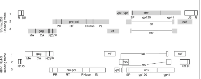
1.1 Organization of SIVmac239 and HIV-1 genomes ...3
1.2 Splicing factors and the impact of RNA structure on splicing regulation ...17
1.3 Schematic showing splice sites and regulatory regions in HIV-1 ...22
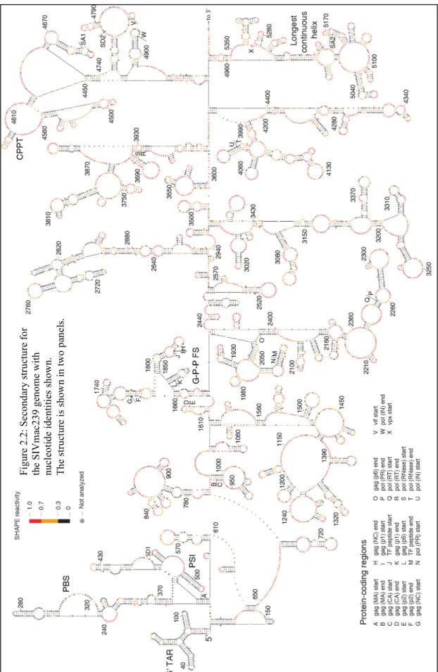
2.1 Model for the structure of the SIVmac239 RNA genome as determined by SHAPE probing and directed RNA structure refinement ...37
2.2 Secondary structure for the SIMvac239 genome with nucleotide identities shown. ...39
2.3 Genomic organization and SHAPE reactivity of SIVmac239 and comparison with HIV-1NL4-3 ...43
2.4 Structural similarity in the 5’ regions of the SIVmac239 and HIV-1NL4-3 genomes. ...49
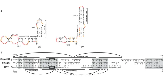
2.5 Codon alignment and predicted pairing partners in the Gag-Pro-Pol frameshift region of SIVmac239 and HIV-1 ...52
2.6 Codon alignment and predicted pairing partners in the RRE region of SIVmac239 and HIV-1 ...55
2.7 Codon alignment and predicted pairing partners in the stem-loop surrounding SA1 ...57
2.8 SHAPE analysis of the polypurine tracts of SIVmac239 and HIV-1 and base composition of both genomes in structured and unstructured regions. ...59
3.1 HIV-1 splicing regulatory sequences ...74
3.2 Mutations to the SLSA1 stem and coculture analysis of viral fitness ...77
3.3 Profiles of HIV-1NL4-3 and HIV-1SLSA1m transcripts ...79
ABBREVIATIONS
∆G change in Gibbs Free Energy
1M7 1-methyl-7-nitroisatoic anhydride 3'ss 3' splice site
33
P-αdCTP 33P-labeled deoxycytidine triphosphate 5'ss 5' splice site
A adenine Å angstrom
AIDS acquired immunodeficiency syndrome
APOBEC apolipoprotein B mRNA-editing, enyme-catalytic, polypeptide- like
b intercept bp base pair C cytosine CA Capsid
cDNA complementary DNA cPPT central polypurine tract DIS dimerization initiation site
DMEM Dulbecco's modified Eagle medium DMSO dimethyl sulfoxide
DNA deoxyribonucleic acid Env Envelope
ESS exonic splicing silencer G guanine
G-P-P FS Gag-Pro-Pol Frameshift Gag Gag polyprotein precursor
Gag-Pro-Pol Gag-Protease-Polymerase polyprotein precursor gp120 glycoprotein 120 (Env surface subunit)
group M main group group N new group group O outlier group
HIV human immunodeficiency virus
hnRNP heterogenous nuclear ribonucleoprotein hr hours
IDT Integrated DNA Technologies IN Integrase
ISE intronic splicing enhancer ISS intronic splicing silencer kb kilobase
m slope M molar MA Matrix
Matlab Matrix laboratory min minutes
mM millimolar NC Nucleocapsid nt nucleotide
PBS primer binding site
PCR polymerase chain reaction
pg41 glycoprotein 41 (Env transmembrane subunit) pH negative log of the hydrogen concentration Pol II cellular polymerase II
PPT polypurine tract PR Protease
Pro-Pol Protease-Polymerase polyprotein precursor py-tract polypyrimidine tract
R repeat
RNA ribonucleic acid RNase ribonuclease
RPMI Roswell Park Memorial Institute medium RRE Rev-response element
rRNA ribosomal RNA RT Reverse Transcriptase SA splice acceptor
SD splice donor sec seconds
siRNA small interfereing RNA
SIV simian immunodeficiency virus SLS stem-loop structure
SLSA1 stem-loop splice acceptor 1 snRNA small nuclear RNA
snRNP small nuclear RNP SP signal peptide
SR serine arginine repeat protein SRE splicing regulatory element SRP signal recognition particle ss single stranded
TAR trans-acting response TBE tris borate EDTA TF transition frame tRNA transfer RNA U uracil
U2AF U2 activating factor U3 unique 3' region U5 unique 5' region UTR untranslated region v/v volume per volume µCi microcuries
CHAPTER I
LENTIVIRUSES AND RNA STRUCTURE
A. Basic biology of lentiviruses
1. Genome organization and viral replication
Human immunodeficiency virus (HIV) and simian immunodeficiency virus (SIV)
belong to the family retroviridae in the genus lentivirus. Retroviruses are defined by their
common replication strategy which involves a reverse-transcribed DNA intermediate that
is integrated into the genomic DNA of the host cell and by virions composed of structural
proteins surrounding and encapsidating viral enzymes that are packaged within the virion
along with the two strands of single-stranded, positive sense RNA. The lentiviruses have
additional accessory proteins that function as adaptor proteins by interacting with host
factors in the cell (1).
The RNA genomes of these viruses largely represent coding domains for genes of
a number of viral proteins (Figure 1.1). The intrinsic function of the genomic RNA is to
be immediately reverse-transcribed into DNA, which is integrated into the cellular
genome and eventually transcribed into full length genomic RNA by the cellular
transcription machinery. This RNA has the baseline functions of acting as a template for
protein translation, both in its full length form and as spliced forms, and packaging into
new virions as genomic RNA (1). These basic functions, however, cannot fully account
translation, but RNA structure has implications in transcription activation, dimerization,
packaging, frameshifting, nuclear export, and splicing regulation. Such functions are
incompletely understood and require a more complete description to explain viral
The lentiviral replication cycle, including the function of many viral proteins,
revolves around RNA. When the envelope protein (Env) interacts with the cellular
receptor CD4 along with either the coreceptor CCR5 or CXCR4, the virus enters the host
cell with a pair of identical, positive sense, single-stranded RNA genomes. Inside the cell,
viral reverse transcriptase (RT) transcribes this approximately 9kb RNA into one copy of
double-stranded DNA, then integrase (IN) incorporates the DNA into the host genome.
The DNA is transcribed to produce genomic RNA, some of which is spliced to form the
various mRNAs, which code for all of the structural and enzymatic viral proteins, as well
as the accessory proteins Vpr and Vpu (in HIV), Vpx (in SIV), Vif, Nef, Tat, and Rev.
The host-initiated transcription of the viral DNA produces full-length 5'-capped and
3’-polyadenylated RNA. The full-length RNA is packaged by and with the structural Gag
and Gag-Pro-Pol polyproteins. The surrounding envelope is composed of cellular lipids
and viral gp120 and gp41 glycoproteins, which are cleaved from the full-length Env
protein by a cellular protease. Viral protease (PR) cleaves Gag and Gag-Pro-Pol into
individual structural nucleocapsid (NC), matrix (MA), and capsid (CA), and the enzymes
PR, RT and IN proteins, creating a virion that is mature and infectious (1).
2. Viral evolution
Two distinct types of HIV are currently circulating in the human population, and
both are causative agents of AIDS. HIV type 1 (HIV-1) is the main type in most of the
world, while HIV type 2 (HIV-2) is localized mainly to West Africa. Although both
viruses use the same cellular receptors and co-receptors for entry, HIV-2 progresses to
mutagenic rate of HIV-1 has propelled its genetic diversity via pressure from host
immune system and restriction factors acting in conjunction with an error-prone reverse
transcriptase and a short life cycle (43). Through this rapid evolution, the virus has been
able to adapt to its host and be successful in high level replication as it has moved from
its original hosts of non-human primates to humans (reviewed in (67)).
The lentiviruses found in non-human primates, SIV, infect African apes or Old
World monkeys with little capacity to cause disease in these animals. One strain of SIV (SIVmac) originating from a species of West African monkeys, called sooty
mangabey, is able to cause illness in a group of Asian monkeys, called macaques,
which were initially infected in captivity. Phylogenetic analysis has grouped HIV-2 in
the same category as SIVmac239 since they both share a common lineage from SIVsm,
which infects but does not lead to disease in the West African primate sooty mangabey
(reviewed in (67)). One of the reference sequences for these HIV-2 and SIV strains is
SIVmac239 (Genbank accession number M33262). HIV-1 originated from SIVcpz,
which is the primate lentivirus found in chimpanzees (55); HIV-1 is further subclassified
into “groups” of genetically similar isolates. These groups are determined based on
geological clustering along with phylogenetic similarity and are labeled group M (main),
group O (outlier), and group N (new). Group M, which is the most prevalent HIV-1
group worldwide, is further divided into subtypes A through K (reviewed in (67)).
HIV-1NL4-3 (Genbank accession number AF324493) belongs to HIV-1 group M subtype B, and
the 3' half of the genome shares a nearly identical sequence to the reference strain of that
Throughout the evolution of the viral sequence, HIV-1, HIV-2, and SIV isolates
have maintained quite similar protein structures. Consider, for example, the lentiviral PR
enzymes, whose sequences and structures are different from cellular proteases in overall
length and conformation of many of their functional regions. Even between HIV-1NL4-3
and SIVmac239, the amino acid sequences of PR differ by approximately 50%. However,
the different viral isolates maintain common structural elements, most of which can be
superimposed almost identically (174). Functional regions in the viral RNA, in contrast,
not only differ in sequence and length, but also in structure. An obvious example is the
trans-acting response element (TAR), which forms a three-helical stem in HIV-2 and
SIVmac239 but a single stem-loop in HIV-1 (12). In these and other instances throughout
the viral genomes, the structure of the proteins seems to be more evolutionarily conserved
than that of the RNA.
B. RNA Structure
1. Importance of RNA structure in biological systems
Ribonucleic acid (RNA), like its counterpart deoxyribonucleic acid (DNA), is a
polymer chain composed of individual nucleotide monomers. Ribonucleotide monomers
are linked together through a ribose-phosphate backbone with each ribose also linked to a
base of either adenine (A), uracil (U), guanine (G), or cytosine (C). RNA is synthesized
as a single strand of these nucleotides, as opposed to DNA which stays base-paired with
its template to form a double-stranded double helix which, for animal cells, stays in the
cellular nucleus. Due to the lack of a complementary strand to stabilize the RNA in
their free energy and stabilize the molecule (66). These interactions occur through
Watson-Crick base pairs where U pairs with A and G pairs with C. The thermodynamic
stability of each pair is determined by the number of hydrogen bonds that form between
the two nucleotides, with the stronger G-C pair forming three bonds and the weaker A-U
pair forming two. In a non-Watson-Crick pair, G can also pair with U by forming two
hydrogen bonds. These interactions can occur when the single-stranded RNA folds back
on itself to create short, irregular stretches of A-form helix. Each turn of the A-form RNA
helix has 11 base pairs with each base pair rising 2.73 Å, compared to the 10 base pairs in
B-form helix made by DNA (148). Variations in the pairing interactions within the RNA
molecule lead to many different motifs that define the RNA structure. A single nucleotide
bulge occurs when one individual nucleotide does not have a pairing partner while the
surrounding nucleotides are forming a helix. A multiple nucleotide bulge has more than
one unpaired nucleotide on the same side of a helix. A hairpin loop or stem-loop is a
structure that forms when the RNA strand folds back in a small loop and allows nearby
nucleotides to form base pairs. RNA helices form mismatch pairs when two nucleotides
directly across from each other do not form canonical G-C, A-U, or G-U pairs. Internal
loops are formed when these mismatch pairs include more than two nucleotides, with
symmetrical loops having the same number of nucleotides on each side of the helix, and
asymmetrical loops having differing numbers of nucleotides on each side of the helix.
Junctions form in RNA structure when two, three, or four stems intersect. RNA also
forms long-range interactions such as pseudoknots which are formed when loop regions
pair with nucleotides to extend the helix of the stem; kissing hairpins are pairing
contacts are pairing interactions between a loop from one stem and a bulge from another
(reviewed in (68)).
These various pairing interactions occur, as stated above, and minimize the free
energy of the system. Pairing happens spontaneously at physiological temperature and pH
and is accomplished by unfavorably lowering the entropy during hydrogen bond
formation but favorably and significantly lowering the enthalpy by ordering the water
around the RNA molecule. This entropy loss increases as the number of consecutive base
pairs increases (reviewed in (68)). The RNA folds into the most energetically favorable
structure, folding into helices by forming base pairs whenever such interactions lower the
free energy. The strength of these helices, however, is not determined solely by the
number of G-C bonds compared to the less energetic A-U or G-U bonds. Instead, the
length of the helix and the surrounding base pairs (or “nearest neighbors”) play a role in
determining free energy of the structure (17). Along with hydrogen bond formation, π
-stacking interactions play an important role in determining the structure of nucleic acids.
The stacking of the aromatic rings between neighboring nucleotide bases helps conserve
the α-helical structure and increases the bases’ ability to hydrogen bond with one another
(116, 119). The strength of a given helix is dictated by not only base pairing interactions,
but also the amount and positioning of loops, bulges, mismatches, and consecutive G-C
pairs (reviewed in (68)).
RNA structure plays an important role in many biological systems. Transfer RNA
(tRNA) is a classic example of the role of RNA structure in biology. Its cloverleaf
structure, which further folds into two pairs of stacked helices, allows biochemical
are required for precise protein synthesis to take place (reviewed in (44)). As part of
larger RNA molecules, riboswitches bind to ligands or aptamers, which force structural
changes and can regulate gene activity and metabolic processes (reviewed in (109)).
Group 1 introns use ribozyme-catalyzed cleavage to self-splice out their own exons as is
the case with certain ribosomal RNAs (rRNAs) in Tetrahymena (90) and can be
engineered to splice a heterologous mRNA such as for human p53, correcting splicing
defects and repairing damaged mRNA (170). The structure of ribosomal RNA allows the
RNA to participate in peptide bond formation and contributes to a number of tertiary
interactions which enable it to efficiently partake in protein synthesis (7, 126) reviewed in
(159) and (127). RNAs that are involved in critical cell processes (and/or have catalytic
function) evolve slowly and in ways that conserve the base pairs involved in the
important features of secondary structure, allowing these structures to be compared over
evolutionary time.
2. Structure of viral RNAs
RNA structure has been implicated in the control of various functions including
transcription, splicing, aminoacylation, translation, and encapsidation in many viruses.
Tobacco bushy stunt virus contains RNA stem-loop structures that modulate mRNA
transcription initiation dependent on base-pairing interactions (168). A single nucleotide
change in some influenza virus H5N1 strains affects the conformation of an RNA
structure, shifting the balance between hairpin formation and pseudoknot formation in
this region, which leads to possible alterations in splicing and impacted virulence of this
mosaic virus contain tRNA-like structures at their 3’ regions that can be aminoacylated
(50, 87). Others, including members of the Dicistroviridae family, use these tRNA-like
structures at their 5' regions to guide translation initiation as part of internal ribosome
entry sites (IRESes) (31). The IRES structure of the RNA in the 5'UTR of this and
members of the Picornaviridae family initiates translation via an internal ribosome entry
site (IRES) in a (m7Gppp) cap-independent way by competing for cellular translation
factors then recruiting and binding them to an internal position in the viral RNA through
their conserved RNA structural motifs (reviewed in (51, 138)). RNA structures have been
shown to influence viral packaging by folding into “panhandle” hairpin structures in
Hantaviruses (120) and by mediating dimerization and protein recognition in Moloney
murine leukemia virus (33).
3. RNA structure determination: SHAPE
Determining the structure of large RNAs is challenging and has been approached
by folding the RNA to obtain the lowest free energy state, comparing related RNA
sequences to identify compensatory changes that can preserve base pairs, and by probing
the structure with chemicals or nucleases to infer the presence of paired versus unpaired
regions. Structural studies are limited by the size of the RNA. Bioinformatics-based
folding programs help determine the lowest free energy of the entire structure, but do not
incorporate chemical data (179). Selective 2' hydroxyl acylation analyzed by primer
extension (SHAPE) is a hybrid approach able to map structure in large RNAs by using
chemical analysis to constrain a folding program (118, 122, 164, 173). The 2' hydroxyl
positions where the base is not paired compared to positions where the base is paired.
After treatment with 1M7, the extent of derivatization at each ribose is assessed as
terminations of DNA synthesis using reverse transcriptase and fluorescent primers. This
approach allows determination of reactivity values for each individual nucleotide and
inputs these reactivity values as pseudo-energy constraints in RNAstructure (146), an
RNA structure prediction program. SHAPEhas been used to map diverse RNA
structures, including the structures near the start codons of all 13 mammalian
mitochondrial RNA open reading frames (72), ribosomal RNA (40), group 1 intron RNA
(45), and even other retroviruses such as Moloney murine leukemia virus (56).
4. Functions of known RNA secondary structures of lentiviruses
The RNA structure in certain regions of the lentiviral genome has been studied in
great detail, and functions have been assigned to various structures throughout the
genome that serve different purposes throughout the viral replication cycle. The 5'
regulatory region of the genome does not code for any proteins, and is therefore termed
the untranslated region (UTR). The UTR is vital to the genome because it contains many
known functional structures. These include the trans-acting response (TAR) hairpin
which interacts with the viral Tat protein to increase the level of transcription of full
length RNA (8, 12, 18), the primer binding site (PBS) which anneals to the tRNALys3,
which serves as primer to initiate negative-strand synthesis during reverse transcription
(85), the dimerization initiation site (DIS) which is contained in the loop region of a
copackaged within the virion (132, 155), and the psi stem which has been implicated in
packaging of the full-length genomic RNA into the virion and in dimerization (26).
Although the coding region of the genome has selective pressure to maintain the
necessary encoded amino acid sequence, the RNA downstream of the UTR still retains a
sequence that is able to form critical structures. Two essential structures in viral
replication are the Gag-Pro-Pol frameshift stem and the Rev-response element (RRE).
The Gag-Pro-Pol frameshift stem is found in only the full-length RNA. The gag gene
encodes a polyprotein, which is subsequently cleaved to yield the MA, CA, and NC
structural proteins. Near the 3' end of the gag gene, a poly(U) sequence directly before
the stable Gag-Pro-Pol frameshift hairpin causes the ribosome to stall and occasionally
slip to the –1 reading frame. This slip shifts the reading frame of the ribosome prior to the
Gag stop codon to form the Gag-Pro-Pol polyprotein, allowing the ribosome not only to
translate the Gag protein (in the 0 reading frame), but also the RT, PR, and IN enzymes,
which are part of the Pro-Pol region (in the -1 reading frame) (69). The downstream RRE
structure is present in all incompletely spliced mRNA, including the vif, vpr, and vpu/env
message, and full-length genomic RNA containing gag and gag-pro-pol genes. The RRE
secondary structure allows for transport of these transcripts out of the nucleus via
recognition by the viral Rev protein (47, 49, 105, 106) in a Crm1-dependent pathway in
which the host nuclear export factor Crm1 helps to transport the mRNA through the
nuclear pore complex into the cytoplasm (32, 52, 139).
The functional RNA structures contained within the TAR, RRE, and frameshift
stem regions fold in ways that facilitate RNA-protein interactions. TAR binds to the viral
undergoes a rearrangement when the arginine side-chains of Tat are bound, creating a
binding pocket for the protein and allowing contact to the other basic residues of Tat
(reviewed in (79)). The viral Rev proteins cooperatively bind to the helical structures that
compose the RRE. The initial binding interaction occurs at stem-loop IIB of the RRE
through binding of a single Rev monomer (27, 34, 162). Subsequently, more Rev proteins
cooperatively assemble via hydrophobic interactions between Rev molecules and
electrostatic interactions between the proteins and the RNA (35, 38, 84, 110). Unlike the
TAR and RRE interactions with viral accessory proteins, the RNA structure at the
Gag-Pro-Pol frameshift stem interacts with cellular factors that compose the ribosome. Instead
of binding sites that encourage interaction, the function of this stable stem is to hinder
translation by the ribosome, discouraging macromolecular interactions between the RNA
structure and the translation machinery (69). The functions of RNA structure described
above are performed by only a fraction of the total lentiviral RNA. Analyzing other
conserved RNA structures may divulge important roles and interactions that are yet
unknown throughout lentiviral replication.
C. Pre-mRNA splicing
1. General principles of splicing
The initial RNA transcript can be modified in several ways including alteration
through removal of long segments of the RNA by splicing. Splicing occurs in the nucleus
and is executed by the spliceosome, a complex machine composed of both protein and
RNA (reviewed in (166)). This mechanism removes segments of RNA, called introns,
the RNA, to be used as a template for protein translation. In total, about 95% of human
cellular RNAs are spliced, and the genes have an average of seven exons (135). Each
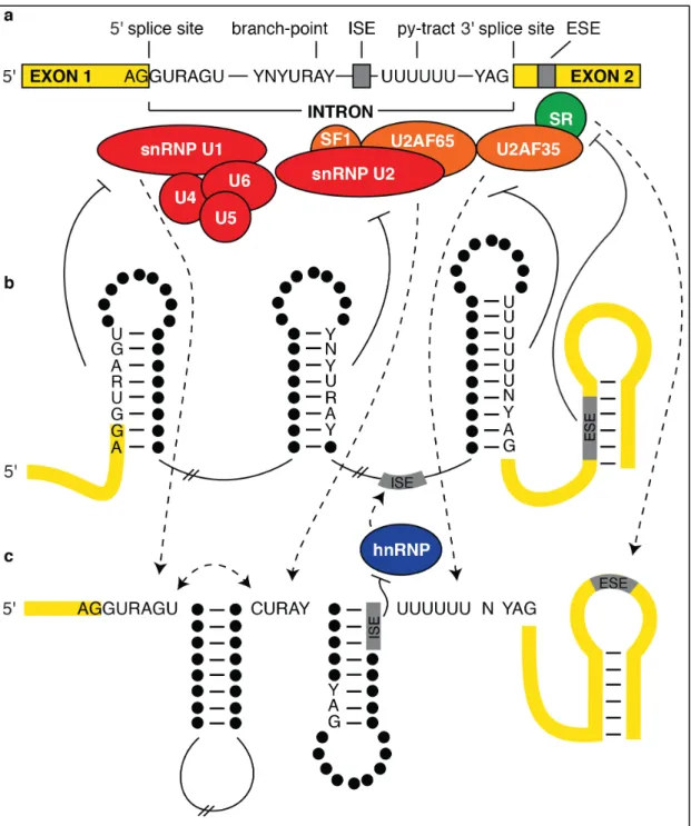
intron starts with a 5' splice site (5'ss) beginning with the sequence GURAGU to serve as
the donor, and a 3' splice site (3'ss) ending with the sequence YAG, which is preceded by
a polypyrimidine tract (py-tract), to accept the ligation event from the donor (Figure
1.2a). The RNA is cut at the donor site and the 5' end of the intron is transferred to the
2'hydroxyl of a conserved adenosine at the branch-point sequence YNYURAY (Figure
1.2a) by a transesterification reaction. The 3'ss is then broken after the AG and linked to
the upstream exon through another transesterification reaction (102, 143). The
spliceosome uses these common sequences to aide in recognition during the splicing
events. Different small nuclear RNAs (snRNAs) combine with proteins to form small
nuclear ribonucleoprotein (snRNP) complexes. These complexes recognize and bind to
the splicing donor, acceptor, or branch-point sequences sequentially. The U1 snRNP
recognizes the 5'ss while the U2 activating factor (U2AF) binds the 3'ss. These elements
then act cooperatively to recruit U2 snRNP (Figure 1.2a). Although these factors work to
excise the introns, these assembling complexes do not recognize the introns in their
entirety. Instead, they bind to and define both sides of the individual exons before joining
them together (11, 57, 147).
When a single transcript is spliced in two or more alternative ways, it can give
rise to more than one protein or protein isoforms with different activities. This alternative
splicing is accomplished in different ways: i) The same transcript is spliced at completely
different exons leading to completely different proteins, ii) the spliceosome intermittently
middle of a protein, or iii) multiple exons are available and the machinery can chose any
of them to include in the message (115). Exons range in size and can potentially be rather
small. When the gene includes even a few extra residues due to alternative exon
inclusion, the protein structure and function can be drastically affected. In this way, the
approximately 25,000 genes in the human genome can give rise to around 500,000
proteins, or a short viral genome can generate the many necessary viral proteins for
efficient replication, allowing greater diversity from a small amount of genetic
information.
2. Regulation of splicing
The positioning of introns relative to the genetic code appears to be random.
However, the ability of the spliceosome to recognize these introns and splice them
properly is vital to proper cellular function. The spliceosomal machinery, especially the
snRNPs, must be able to distinguish genuine splice sites from cryptic or fortuitous sites.
The RNA binding factors must also be able to recognize and use the correct alternative
sites given the needs of the cell or virus. Therefore, many factors are involved to assure
this process is precise. The spliceosome accurately splices the transcribed RNA based on
many factors that are encoded in the mRNA itself. These are displayed as sequences
termed splicing regulatory elements (SREs) and include exonic splicing enhancer (ESE)
sequences which enhance splicing of the exon they are part of, exonic splicing silencer
(ESS) sequences which are located in the exon but inhibit splicing at a certain acceptor or
donor site, intronic splicing enhancer (ISE) sequences which amplify a splicing event that
silencer (ISS) sequences which are also located in the potentially discarded intron but act
Functional SREs seem to inhabit single-stranded regions more often than
base-paired regions, allowing binding proteins to identify and interact with these particular
sequences. It has been found that enhancer-dependent regions (those with weak splicing
sequences) are particularly marked with single-stranded ESE motifs, and regions that
depend on silencing have a stronger occurrence of single-stranded ESS motifs (65).
These SRE sequences work by binding specific proteins, which in turn inhibit or enhance
splicing factors from recognizing the 5'ss or 3'ss sequences in close proximity. The
acceptor sites are enhanced by the ability of surrounding sequences to bind SR proteins
(Figure 1.2a), which are defined by their RNA recognition motifs and their
carboxy-terminal domain that contains several serine and arginine repeats (reviewed in (58, 156).
These proteins interact with different parts of the spliceosome: regions of the U1 snRNP,
U2AF, and U4/U6.U5 tri-snRNP (reviewed in (58)). Interactions between parts of the
spliceosome and the corresponding splice sites are inhibited by ISS or ESS sequence
recognition by heterogeneous nuclear ribonucleoprotein (hnRNP) complexes (Figure
1.2b), blocking recognition and disallowing splicing at a given site (41). In the cell,
however, the splicing factors, including SR proteins and hnRNP complexes, are present
at varying concentrations, which leads to the concept that cells are able to regulate the
utilization of different splicing pathways by controlling the amounts of these complexes.
This allows for more complexity in splicing regulation through cellular control factors
that dictate the ratios of splicing enhancement and silencing factors (156).
RNA structure itself can have a regulatory effect on splicing. In a broad sense,
structures that form within large introns to bring distant splice sites closer together allow
range (24) (Figure 1.2c). At a closer scale, structure formation occurs
co-transcriptionally, allowing the RNA to make short-range interactions as each nucleotide
is added to the polymer (150). These short interactions, particularly stem-loop structures,
have been implicated in controlling recognition of splicing factors (Figure 1.2b and c).
The minimal structure needed is a small stem-loop as short as 7-bp, which is adequate to
enclose and seclude an enhancer sequence, impeding its activity (98). An analysis of
splicing in mammalian cells showed conserved structures around splice sites that conceal
certain sites where regulation is necessary for control of gene expression and repression
of various disease phenotypes (152). Furthermore, GC content, which is implicated in
stronger pairing interactions, has been shown to be enriched around alternative splice
sites, particularly those for the first possible exon, suppressing usage of such sites by the
splicing machinery (178). These analyses imply that stable secondary structures around
splice sites serve to seclude these sequences from their binding proteins and allow other
alternative sites to be used at higher frequency. A stem that includes the py-tract and the
AG 3'ss precludes binding of that sequence to U2AF, while a stem that makes pairing
interactions with the sequences involved in the 5'ss precludes U1 snRNP binding (Figure
1.2b). Even though the U2AF and U1 snRNP help recruit U2 snRNP, if the branch point
sequence is paired in a stem structure, the binding of U2 snRNP to that site will be
disrupted. In contrast, when these sequences are in single-stranded or loop regions, they
increase the binding interactions to their respective proteins (Figure 1.2c). Additionally,
cryptic acceptor sites, when paired in structure, are hidden from U2AF recognition and
Splicing regulation depends not only on the accessibility of actual splice sites but
also the RNA structures around cis-acting SRE sequences. These motifs bind certain
proteins that allow or disallow splicing to occur. ESE and ISE motifs bind to SR proteins
which, when bound, enhance the splicing efficiency of the nearby 3'ss. If enhancer
elements are secluded in base-pairing interactions, the SR proteins are not able to bind
and thus do not impact splicing at the 3'ss (reviewed in (169)) (Figure 1.2c). In a
converse event, an hnRNP can bind to either an ISS or ESS (Figure 1.2b). Once bound,
this causes the structure around the exon to form a loop and disallows U1 snRNP binding
at the 3'ss near the silencer motif, thus avoiding the given exon altogether (125),
(reviewed in (19)). Although they do not incorporate the given regulatory element,
structures that occur upstream of SRE regions have been shown by computational
analysis to enhance the function of the given SRE. These stable structures near the region
of regulation could potentially function to interfere with any RNA conformation that
might have otherwise been part of the SRE (98). Taken together, these concepts
strengthen the idea that structure of the pre-mRNA around the splice site sequences,
which includes enhancer and silencer elements, helps determine whether the given site
will be recognized and spliced by the spliceosome (reviewed in (169)). The general
model for splicing regulation by RNA structure is the following: The single-stranded
SREs are recognized by and bind their regulatory proteins, and the base-pairing of these
3. Pre-mRNA splicing of HIV-1
Cellular genomes are vast in size compared to viral genomes, especially those of
RNA viruses. These RNAs need to strike a balance between being small enough to allow
efficient replication in a cell yet large enough to contain all of the genetic material to
produce the necessary viral proteins. One way lentiviruses accomplish this is by their
ability to be spliced in a multifaceted manner. The proviral DNA is transcribed by
cellular polymerase Pol II to produce a long unspliced RNA that can either be used as the
genomic RNA in the packaged virus, as a transcript for Gag and Gag-Pro-Pol translation,
or spliced to produce the over 40 different mRNA species used to generate the remaining
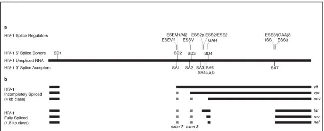
viral proteins (142). For HIV-1, the cellular splicing machinery utilizes four 5'ss donors
The first 5'ss SD1 is the major splice donor and is used in all spliced products.
When SD1 is used in conjunction with the splice acceptors SA1, SA2, or SA5 with no
downstream splicing events, the vif, vpr, or vpu/env mRNA transcripts are produced,
respectively. These are categorized as the 4 kb class of transcripts (or singly spliced
mRNAs) because they incorporate the RRE-containing sequence between SD4 and SA7
(Figure 1.3b). The splice variants that excise this intron are grouped as the 1.8 kb class of
transcripts (or small, multiply spliced mRNAs), which is comprised of mRNAs spliced
once from SD1 to SA3, SA4(a,b,c), and SA5 and then again from SD4 to SA7 creating
tat, rev, and nef mRNAs, respectively (142) (Figure 1.3b). Although the transcripts that
are spliced directly from the SD1 donor to the closest upstream acceptors are the most
abundant variants, smaller exons can still be incorporated into the mRNA, including exon
2 from SA1 to SD2 and exon 3 from SA2 to SD3 (Figure 1.3b). The start codons for Vif
and Vpr are after SD2 and SD3, respectively. Therefore, any splicing event that uses
these donors effectively excises those start codons, producing the corresponding
transcript of the next acceptor that is used (142).
An important feature of unspliced and singly spliced (4 kb class) mRNA is the
RNA structure in the SD4-SA7 intron, the RRE. Typically, unspliced or incompletely
spliced mRNA remains in the nucleus until it is fully spliced (32). If incompletely spliced
pre-mRNA interacts with U1snRNA alone with no other splicing factors, it is retained in
the nucleus and disallowed to either be fully spliced or exit into the cytoplasm (21, 95).
HIV-1 overcomes this nuclear retention by encoding the nuclear-export protein Rev.
Once Rev is translated in the cytoplasm (using the multiply spliced and efficiently
binds the RRE of these incompletely spliced mRNA molecules, and shuttles them out of
the nucleus (32, 139).
4. HIV-1 splicing regulation
The diverse mRNA splicing that occurs in HIV-1 transcripts is a result of
complicated alternative splicing (142). The splicing events must be highly regulated to
allow the necessary amounts of each transcript to be produced. For example, if the
nascent transcript were completely spliced to the smallest mRNA form, or if any of the
splice sites were preferentially used, one protein product would dominate. Instead, the
pre-mRNA contains many weak splice acceptor and donor sites along with weak
branch-point sequences, leveling the potential for all to be recognized by the splicing machinery.
The acceptor sites contain purines, causing them to be weaker than the ideal
polypyrimidine tracts that enhance splicing (4, 46, 129, 153, 157). Maintaining the
balance for splicing at each acceptor remains complicated, however, because the RNA
also contains cis-acting regulatory elements in the form of sequences and secondary
structures that enhance or repress splicing at particular sites (reviewed in (160).
Regulation of HIV-1 RNA splicing begins with the major splice donor (SD1).
This donor has the capacity to splice to any of the downstream acceptors, and when
splicing occurs within the primary transcript, the splicing machinery will always use SD1
(142). The SD1 sequence is composed of the loop region of a conserved stem-loop
structure in the 5' region of the genome which, when mutated, alters the efficiency of
splicing using SD1 (2). SD1 becomes a less efficient 5'ss when the structure around SD1
suggesting that accessibility of SD1 in a loop is imperative for SD1 recognition (2). The
mechanism that seems to be at work in this region is one that allows the SD1 site to be
visible to the splicing machinery by placing it in a loop instead of in a paired interaction.
Splicing to the first splice acceptor SA1 with no further downstream splicing
events results in the mRNA transcript for the Vif protein. This protein is necessary for
cellular APOBEC3-G and -F cytadine deaminase downregulation, therefore diminishing
the cellular restriction factors’ mutagenic effects on the newly reverse-transcribed
negative strand DNA (29). Regulation of the SA1 site is crucial for usage of downstream
splice acceptor sites. This site has been determined to include the strongest splice
acceptor site of all the HIV-1 3’ss sequences (76). However, this 3'ss must be used in
moderation to allow for splicing from SD1 to the other possible splice acceptors.
Following the idea that splicing complexes recognize the entire exons instead of
the introns (11), it has been shown that production of the vif message is regulated at the
second splice donor site (SD2) (48, 76), located 50 nucleotides downstream of SA1 and
78 nucleotides upstream of the vif start codon. If used, SD2 splices to a downstream 3’ss,
creating a small exon 2 and excluding as an intron the sequence that would begin Vif
translation (142). In this way, even if SA1 is the acceptor used for SD1, the vif message is
not necessarily the final product. A weak 5'ss sequence at the SD2 site has been shown to
enhance the production of the vif message (104, 108). Another enhancer, ESEVif, occurs
in the region between SA1 and SD2 along with a GGGG splicing silencer that follows
SD2. These are in competition with one another to systematically regulate splicing at this
site and, ultimately, expression of Vif (48). The regulatory sequences ESEM1 and
the use of SD2 (76). These elements have been identified through mutagenesis analysis
that specifically disrupted the sequences, however, this was done with no regard to the
structures that may have been formed in these regions. As of yet, the RNA structure
around SA1 has not been implicated in any silencing or enhancing function for splicing at
this region.
A splicing event to the second splice acceptor site (SA2) produces a message that
encodes Vpr, a protein important for both promoting infection in myeloid cells (6, 28)
and arresting the cell cycle of dividing cells (64, 74). A regulatory sequence termed
ESSV has been identified in this region that helps to repress the use of SA2 (13). This
silencing mechanism functions when the ESSV sequence binds hnRNP A/B proteins and
inhibits binding of U2AF65 to SA2 (42). Specific mutagenesis of ESSV has localized the
interacting element to a 16-nt sequence (103).
The splicing events that use SA3 will produce the tat transcript. The usage of SA3
is usually followed by excision of the intron from SD4 to SA7, and this is a requirement
for the production of functional Tat (142). The Tat protein recognizes the TAR hairpin
structure in the 5' UTR and recruits transcription factors to upregulate RNA Pol II
function during mRNA transcription (15, 93). However, Tat has an apoptotic effect on
the cell (16, 79, 97) and its production is therefore well regulated. An upstream silencer
called ESS2p (70) and ESS2, a silencer downstream of SA3 (153), work against a
relatively stronger py-tract compared to most others on the RNA (71) and splicing
enhancer sequence ESE2 (176) to accomplish this regulation. These sequences are
located on two stem-loop structures SLS2 (which contains ESS2p (70)) and SLS3 (which
in the vicinity of SA3. Various regions on these stem-loop structures interact with
enhancing and silencing proteins to modulate splicing at SA3. The proteins recognize
sequences within these RNA structures that seem to be shared binding sites between
many of them, thus leading to competition of regulation at this site (61).
Splicing from SD1 to any of the splice acceptors SA4c, SA4a, and SA4b followed
by the SD4-SA7 splicing event leads to the rev mRNA transcript. Use of SA5 by SD1
leads to the transcript for either nef or vpu/env depending on if the intron between SD4
and SA7 is excised (nef) or included (vpu/env) (142). An ESE element termed GAR is
located directly downstream of SA5 and has been shown to regulate usage of the splice
acceptor sites SA4c/a/b and SA5 and the downstream donor SD4 (20, 75). This
regulation is accomplished in two ways: binding SR proteins in a bidirectional manner to
allow sufficient usage of the upstream acceptors, and binding U1 snRNP to SD4 which
amplifies expression of unspliced and incompletely spliced mRNA (75). An RNA
structure at this site has yet to be identified.
Perhaps one of the most intuitive splicing regulatory regions is that pertaining to
the splicing event that occurs between SD4 and SA7. When this intron is excised, the 1.8
kb class of mRNA products is produced. Even in the absence of Rev, these products are
able to exit the nucleus freely. If the SD4-SA7 intron is not spliced from the transcript,
the 4 kb class of mRNAs and the unspliced product are generated and require Rev for
efficient transport from the nucleus into the cytoplasm. The sequences that serve as SREs
around this splicing interaction have been studied in detail, and the RNA structures that
include these SREs are known. An ISS at this site consists of a sequence that makes up
base-paired in the structure SLS3, and ESE3/(GAA)3 has been described as a large bulge
region in the structure SLS2 (37, 111). As with the different silencers and enhancers that
act upon SA3, these various SREs must cooperatively and competitively bind different
hnRNP factors and SR proteins to regulate splicing at this site (111).
D. Thesis Overview
Regions of RNA secondary structure play essential roles in the replication cycle
of HIV-1. The SHAPE technique has been applied to determine the RNA secondary
structure of the full-length HIV-1NL4-3 genome, and this analysis has shown many
elements of RNA structure (172), but only a fraction of these have been previously
studied. One tool to assess the importance of these structures is to determine the extent to
which they are conserved over evolutionary time and the extent to which they are
maintained after mRNA splicing.
The second chapter describes the application of SHAPE technology to develop a
secondary structure model for the genomic RNA of a second primate lentivirus, simian
immunodeficiency virus (SIVmac239), which shares 50% sequence identity at the
nucleotide level with HIV-1. In both genomes approximately 60% of the nucleotides are
paired within the coding region (8,738 nucleotides). However, only about half of these
paired nucleotides are paired in both sequences, and only 58 base pairs form with the
same pairing partner in the coding region of both sequences. Thus on average the RNA
secondary structure is evolving at a much faster rate than the sequence. Some structures
are conserved between HIV-1 and SIVmac239, including in the 5' untranslated region (5'
polyadenylation sequence, the polypurine tracts (PPT and cPPT) that begin plus-strand
synthesis, and the stem-loop structure that includes the first splice acceptor site. Structure
at the Gag-Pro-Pol frameshift site is maintained but in a significantly altered form. As
with all lentiviruses, the HIV-1 and SIVmac239 genomes are adenosine-rich and
cytidine-poor. Approximately two-thirds of the cytidines, uridines, and guanosines are
base-paired while only one-third of adenosines are base-paired, leading to the
concentration of adenosines in single-stranded regions (55% of the unpaired nucleotides).
Thus the base composition of the structured regions is very different from either the
unpaired regions or the genome as a whole. Structures with adenosine content equal to or
greater than the number of guanosines had higher SHAPE reactivity and were not
conserved between the two genomes. By contrast, those structures in which guanosines
were more abundant than adenosines had lower SHAPE reactivity and structure was
maintained, although still undergoing significant evolution. This leads to the conclusion
that much of the secondary structure reflects pairing in a state which allows the RNA to
form and reform interactions throughout evolution of the sequence. However, regions of
the structure that perform necessary functions within the viral replication cycle seem to
have a high guanosine content, which stabilizes these structures and allows them to
remain intact even through the course of sequence evolution.
The work in the third chapter examines regulation of splicing due to RNA
secondary structure in the HIV-1NL4-3 transcript mRNA. I evaluate the importance of an
evolutionarily conserved stem-loop structure whose pairing interactions at the base of the
stem were kept constant between HIV-1NL4-3 and SIVmac239 genomic RNA structures.
Mutations to this stem that disrupted the pairing interaction while keeping surrounding
ESE sequences intact as well as the corresponding amino acid sequence were introduced
to the HIV-1NL4-3 genome, creating the mutant virus SLSA1m. In a virus coculture assay,
the wild-type virus outcompeted the SLSA1m by a small margin. Separately, the mutant
and wild-type viruses were passaged in cells and the mRNA profiles from these cells
showed a difference in splicing pattern. Taken together, these data indicate a decreased
viral fitness to SLSA1m and a change in usage of the splice sites based on the disruption
of this stem structure. To examine other splicing regulatory features in the context of
entire transcripts of fully spliced and partially spliced mRNA, I performed SHAPE
analysis on in vitro transcribed RNAs representing the most abundant versions of spliced
mRNA for all of the viral proteins. These structures exhibit maintenance of known motifs
around splice sites SD1, SA2, SA3, and SA7, but with slightly altered conformations,
emphasizing the importance of analyzing these structures and pairing interactions in a
whole-molecule context. I observed maintenance of some previously unreported
structures around the known SRE sequence at SA4c/a/b, SA5, and SD4, implying a role
of RNA structure in regulation of splicing at this region. Many of these known and newly
identified structures are preserved even after splicing events excise large regions of
sequence, however, some structures are altered based on initial splicing events. This leads
to the conclusion that most RNA regulatory structures affecting splicing of HIV-1 are
formed through local interactions and are thus made impervious to large sequence
changes or deletions because of the need to maintain these structures intact in the mRNA
after the initial splicing event, but some structures are altered to modify the occurrence of
In the fourth chapter, I will summarize the results of my thesis work and discuss
CHAPTER 2
COMPARISON OF SIV AND HIV-1 GENOMIC RNA STRUCTURES REVEALS IMPACT OF SEQUENCE EVOLUTION ON CONSERVED AND NON-CONSERVED
STRUCTURAL MOTIF1
A. Introduction
RNA secondary structures play fundamental roles in the replication of all
positive-strand RNA viruses. Because of their small genomes (which are largely devoted
to encoding viral proteins), these viruses use available sequence space highly efficiently.
The genomic RNA of viruses forms structures necessary for various functions. For
example, internal ribosome entry site elements interact with the cellular translation
initiation machinery, diverse structural signals direct packaging of viral RNA into viral
particles, and RNA structure can provide control signals for differential viral gene
expression. The human immunodeficiency virus type 1 (HIV-1) is no exception and
well-characterized RNA structures within the coding domains of the genome play critical roles
in regulation of replication. These include a structure in the env gene, the Rev response
element (RRE), that binds the viral protein Rev leading to the transport of unspliced and
singly-spliced viral mRNA out of the nucleus (80, 130), and a hairpin structure preceded
by a poly(U) slippery sequence that mediates a frameshift during synthesis of the
Gag-Pro-Pol polyprotein (136). The untranslated regions (UTRs) of HIV-1 and simian
immunodeficiency virus (SIV) contain the TAR hairpin, which recruits the Tat protein to
modulate transcription (63, 124) (reviewed in (80)) and other stem-loop structures that
are important for dimer initiation (DIS) (155), splicing (123, 142), and viral RNA
packaging (10, 62) (reviewed in (101)). Several lines of evidence emphasize that the
HIV-1 genome contains extensive RNA secondary structures whose functional roles are
not yet fully understood (99, 167, 172).
The structures of large RNAs, like viral RNA genomes, are too complex to be
predicted with confidence from first principles or thermodynamic-based algorithms
alone. Useful working models can often be obtained when additional information is used
to restrain the number of possible secondary structure elements. Two such approaches are
to compare evolutionarily related sequences to identify RNA motifs that co-vary to
preserve base pairs, and to experimentally probe the RNA structure with chemicals or
nucleases to infer the presence of paired versus unpaired regions. In the selective
2'-hydroxyl acylation analyzed by primer extension (SHAPE) chemical probing approach,
nucleotide reactivities show a strong inverse correlation with the probability that a
nucleotide is base-paired. SHAPE-directed prediction of RNA folding has been used to
develop secondary structure models for diverse RNAs (40, 45, 56, 72, 173) including the
full-length genomic RNA structure of HIV-1NL4-3 (172). This HIV-1 model shows a very
strong correlation between regions that can be targeted by siRNAs to inhibit viral
replication (99) and regions that are predicted to be single-stranded, suggesting that
global structural features are likely correct.
One approach to evaluating the broader significance of these structures is to examine the
conservation of these structures in a related virus. To this end, we analyzed the secondary
representative of the SIVsm/HIV-2 lineage of primate lentiviruses. HIV-2 evolved from a
different primate reservoir than did HIV-1. HIV-2 arose in the sooty mangabey
(Cercocebus atys), and SIVsm has also infected rhesus macaques in primate centers and
to cause an AIDS-like illness. SIVmac239 (144) now serves as a prototype reference
sequence for comparative analysis of the HIV-2/SIVsm lineage (22). SIVmac239 has a
large evolutionary distance from HIV-1, and conservation of structures between HIV-1
and SIVmac239 represents an especially stringent test for functional relevance. In this
analysis, we describe areas where RNA structure is maintained between HIV-1NL4-3 and
SIVmac239, where it is divergent, and outline possible mechanisms for understanding the
interplay between rapid sequence evolution in the context of selection for maintenance of
function of RNA structural motifs.
B. Results and Discussion
1. Features of the SIVmac239 RNA structure
To develop an experimentally-based secondary structure model for the genomic
RNA structure of SIVmac239 (GenBank accession M33262), we used a strategy similar
to that used to develop a model for the secondary structure of genomic HIV-1 RNA
(172). Viral RNA was purified from SIVmac239 particles and derivatized with the
SHAPE reagent 1-methyl-7-nitroisatoic anhydride (1M7) under physiologically relevant
conditions to discriminate between single-stranded (generally reactive) positions versus
(unreactive) nucleotides constrained by base-pairing or other interactions (118, 122, 164,
173). The derivatized positions were identified as terminations of DNA synthesis by
9,605 nucleotides, 99.6% of the genome. These data were used as pseudo-free energy
change constraints to direct RNA secondary structure prediction. In the secondary
structure model for the SIVmac239 RNA genome (Figures 2.1 and 2.2), 4,970
nucleotides were predicted to be base-paired (51.5%), whereas 4,676 nucleotides were
Name Primer Sequence
SIV309 SIV443
TCCTTCAAGTCCCTGTTCAGGC AACCGGAGGCCTCTTCCTCTCC SIV593 CTTTCCGTTGGGTCGTAGCCT SIV897 GATGGTGCTGTTGGTCTACTTG SIV1193 GTCCTTGTTGTGGAGCTGGTTG SIV1475 GTTTGAGTCATCCAATTCTTTAC SIV1761 TCCAGCATCCCTGTCTTCTTG SIV2095 CTGTATCCAGTAATACTTCTAC SIV2138 GTGGACCTAACTCTATTCCTG SIV2386 GCCACTGCTTCAATTTTGGTCC SIV2674 CTAGAGGTATGGAGAAATATGC SIV2952 CCCTATGCTATTCAAGAGTTCC SIV3225 CTCATATTCTGCTTCTGCCATC SIV3505 CCCATACATCCTTCTCAACTGG SIV3780 CCCTGAGTCTGTCAATGCCATG SIV4027 CATGTTCTTCTTGTGCTGGCTC SIV4289 AATAGTGCTGTCTGTCTTCCTG SIV4576 GAGTCATATCCCCTATTCCTCC SIV4831 AACTGCTATCCACCTCTTTTCC SIV5112 TAGTTTGGTGTTACATCTGTCC SIV5382 CCGCCTCTCTGTTTATCTCCTC SIV5647 TGTGGTCCTTCATTTTCTGGAG SIV5904 TAGAGGGCGGTATAGCTGAGAG SIV6184 ATTGTCGCATTCCTCCAAGCTG SIV6350 CTCAAAGAGTTGCCATACATCC SIV6635 TGCAGATGACCAAGTTTCATTG SIV6894 AGCCAAACCAAGTAGAAGTCTG SIV7170 CAGTATACCTGGGATGTTTGAC SIV7462 AGACTGGTCACTGTGGAGTTAC SIV7745 CCCAGCCAATAAAGTTCGGGAC SIV8001 AGTCAACCTTTCGCTCCCACTC SIV8261 GAAATAAGAGGGTGGGGAAGAG SIV8536 TGTAGGTAGGTCAGTTCAGTCC SIV8830 CCAAGTCATCATCTTACTCATC SIV9107 TCATCCTCCTGTGCCTCATCTG SIV9282 TAGCCTTCTTCTAACCTCTTCC SIV9485 GAACCTCCCAGGGCTCAATCTG SIV9621 TTTTTACTTCTAAAATGGCAGC
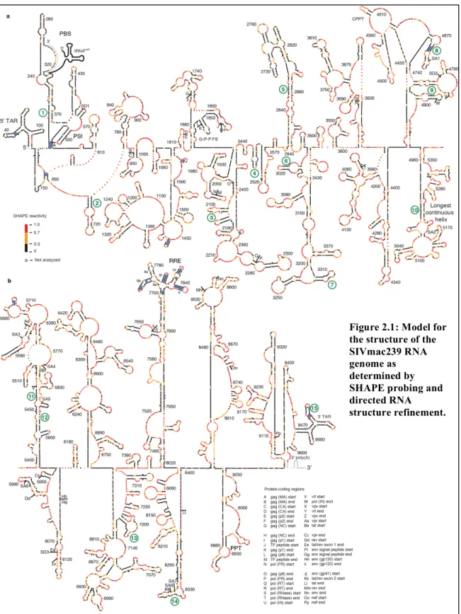
Figure 2.1 (Next Page): Model for the structure of the SIVmac239 RNA genome as determined by SHAPE probing and directed RNA structure refinement. The genome is divided into (a) 5' and (b) 3' halves. Colors of nucleotides indicate SHAPE reactivity on the scale shown on the left. Each sphere corresponds to a nucleotide, and side-by-side spheres indicate a base pair. Protein coding region boundaries are indicated by letters with the code shown at the bottom. Splice acceptor and donor sites(165) are labeled SA and SD, respectively. tRNALys3 interaction is shown in gray. Heavy blue bars
indicate base pairs in stems that are conserved between codon-aligned SIVmac239 and HIV-1NL4-3 RNA
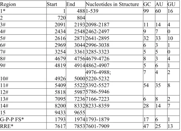
Region Start End Nucleotides in Structure GC AU GU
1* 1 488 1-539 99 60 16
2 720 804
3# 2091 2193 2098-2187 11 14 4
4# 2434 2548 2462-2497 9 7 0
5# 2616 2871 2641-2895 32 33 10
6# 2969 3044 2996-3038 6 3 1
7# 3254 3361 3285-3323 5 5 0
8# 4679 4756 4679-4726 8 3 4
9# 4819 4914 4862-4907 5 6 1
10# 4926 5000
4976-4988; 5220-5232
7 4 2
11# 5409 5522 5392-5527
5786-5946
54 35 8
12# 5818 5987
13# 7095 7236 7166-7223 6 8 2
14# 8200 8332 8233-8359 28 14 7
15 9433 9655
G-P-P FS* 1793 1974 1793-1879 17 6 1
RRE* 7617 7853 7601-7909 47 25 13
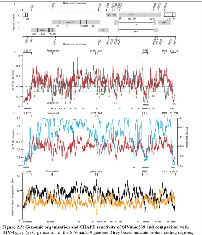
Highly structured regions in an RNA can be inferred in a model-free way by
identifying regions with low overall median SHAPE reactivities. Many areas of the
SIVmac239 RNA genome have low median SHAPE reactivity (defined as less than 0.3
on a scale from 0 to ~1.5) over a 75 nucleotide window, and these correspond to regions
of structure with both known and unknown function (Figure 2.3). The lowest median
SHAPE reactivity values occurred at the 5' and 3' ends of the genome. The highly
structured 5' region extends until nucleotide 539 (Figures 2.1a and 2.3b, motif 1), and the
structured 3' region begins at position 9462 at the start of the 3' TAR structure within the
terminal repeat (R) regions (Figures 2.1b and 2.3b, motif 15). In addition, the
Gag-Pro-Pol frameshift (G-P-P FS) element (Figures 2.1a and 2.3b; positions 1852-1879) and the
RRE are highly structured (Figures 2.1b and 2.3b). By comparison, when we used RNA
Decoder (a program that predicts evolutionarily conserved RNA secondary structure in
the context of the protein-coding sequence of the RNA (137)) with an HIV-2/SIVsm
sequence dataset to infer conserved regions of secondary structure, we found that the 5'
and 3' UTRs and the RRE showed the strongest signal for conservation of structure
(Figure 2.2). Using the RNA Decoder approach we conclude that major features of
secondary structure are not conserved within the coding region at the level of the RRE.
Other regions, however, have low median SHAPE reactivities, yet currently unknown
RNA functions (Figures 2.1a, 2.1b, and 2.3b, motifs 2-14). In the following sections, we
examine these structures and infer biological importance based on their conservation with
Figure 2.3: Genomic organization and SHAPE reactivity of SIVmac239 and comparison with HIV-1NL4-3. (a) Organization of the SIVmac239 genome. Grey boxes indicate protein coding regions, dark lines indicate the boundaries of the mature viral proteins.(b) SIVmac239 median SHAPE reactivity values calculated over a 75 nucleotide sliding window (red). Green dashed line indicates SHAPE reactivity of 0.3. Regions with SHAPE reactivities below 0.3 are numbered (and listed explicity in Table 2.2). SHAPE reactivity values of HIV-1NL4-3 (gray) are shown as medians calculated over a 75
2. Overview of Base-pairing within the HIV-1NL4-3 and SIVmac239 RNA Genomes
SIVmac239 is the second full-length genomic primate lentivirus RNA evaluated
by SHAPE-directed modeling; the first was that of HIV-1NL4-3 (172). Comparison of the
structural models of these two distantly related retroviral RNA genomes should reveal
conserved structural elements. For this analysis we have used updated folding parameters
that result in modest changes in the previous HIV-1 model (see Methods). Visually the
patterns of 1M7 reactivity in the 5' noncoding region, the frameshift site, and the RRE —
all regions with well-established conserved functions — are similar for SIVmac239 and
HIV-1 RNAs (Figure 2.3b). A bootstrapping analysis (see Methods) showed that the
measured SHAPE profiles across both genomes were significantly more similar than
expected by chance (10,000 trials, p < 0.0001). Thus, in a broad view, there appears to be
a strong propensity to conserve the overall level of local RNA structure across the same
regions of these two genomes.
The RNA folding algorithm employed for structure prediction included a
pseudo-free energy change term to account for the SHAPE reactivity (see Methods). Newly
optimized parameters for calculating the pseudo-free energy term were used to predict a
revised secondary structure model for HIV-1NL4-3 based on the original reactivity data
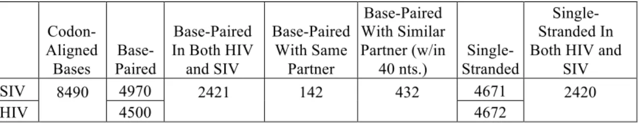
(172). We then compared the codon-aligned sequences of HIV-1NL4-3 and SIVmac239 for
equivalency in terms of base-pairing. The two genomes share 50% identity at the
nucleotide level; however, if these sequences were randomized, they would appear to
have 24% identity, emphasizing the extent of divergence at 50% identity. We found that
roughly half of the nucleotides predicted to be base-paired in the HIV-1NL4-3 sequence
predicted to be single-stranded in the HIV-1NL4-3 sequence were also single-stranded in
the SIVmac239 sequence (Table 2.3). In spite of the limited conservation of paired bases,
we did observe regions in similar locations within the genomes with low SHAPE
reactivity (defined as median reactivity below 0.4 over a 75 nucleotide window). These
areas (Figure 2.3b, grey dashes) largely fold into structures of unknown function. None of
these structures have conserved base pairs, and in only a few examples are there even