Homologous Recombination Repair Factors Rad51 and BRCA1 Are
Necessary for Productive Replication of Human Papillomavirus 31
William H. Chappell,*Dipendra Gautam, Suzan T. Ok, Bryan A. Johnson, Daniel C. Anacker, Cary A. Moody
Lineberger Comprehensive Cancer Center and Department of Microbiology and Immunology, University of North Carolina at Chapel Hill, Chapel Hill, North Carolina, USA
ABSTRACT
High-risk human papillomavirus 31 (HPV31)-positive cells exhibit constitutive activation of the ATM-dependent DNA damage
response (DDR), which is necessary for productive viral replication. In response to DNA double-strand breaks (DSBs), ATM
activation leads to DNA repair through homologous recombination (HR), which requires the principal recombinase protein
Rad51, as well as BRCA1. Previous studies from our lab demonstrated that Rad51 and BRCA1 are expressed at high levels in
HPV31-positive cells and localize to sites of viral replication. These results suggest that HPV may utilize ATM activity to increase
HR activity as a means to facilitate viral replication. In this study, we demonstrate that high-risk HPV E7 expression alone is
suf-ficient for the increase in Rad51 and BRCA1 protein levels. We have found that this increase occurs, at least in part, at the level of
transcription. Studies analyzing protein stability indicate that HPV may also protect Rad51 and BRCA1 from turnover,
contrib-uting to the overall increase in cellular levels. We also demonstrate that Rad51 is bound to HPV31 genomes, with binding
in-creasing per viral genome upon productive replication. We have found that depletion of Rad51 and BRCA1, as well as inhibition
of Rad51’s recombinase activity, abrogates productive viral replication upon differentiation. Overall, these results indicate that
Rad51 and BRCA1 are required for the process of HPV31 genome amplification and suggest that productive replication occurs in
a manner dependent upon recombination.
IMPORTANCE
Productive replication of HPV31 requires activation of an ATM-dependent DNA damage response, though how ATM activity
contributes to replication is unclear. Rad51 and BRCA1 play essential roles in repair of double-strand breaks, as well as the
re-start of stalled replication forks through homologous recombination (HR). Given that ATM activity is required to initiate HR
repair, coupled with the requirement of Rad51 and BRCA1 for productive viral replication, our findings suggest that HPV may
utilize ATM activity to ensure localization of recombination factors to productively replicating viral genomes. The finding that
E7 increases the levels of Rad51 and BRCA1 suggests that E7 contributes to productive replication by providing DNA repair
fac-tors required for viral DNA synthesis. Our studies not only imply a role for recombination in the regulation of productive
HPV replication but provide further insight into how HPV manipulates the DDR to facilitate the productive phase of the
viral life cycle.
H
uman papillomaviruses (HPVs) are small double-stranded
DNA viruses approximately 8 kb in size that exhibit a
prefer-ential tropism for epithelial cells. High-risk mucosal HPV
sub-types are the causative agents of cervical cancer and have been
increasingly associated with anogenital, oropharyngeal, and head
and neck cancers (
1
). The life cycle of HPV is intimately linked to
the differentiation of its host cell, the keratinocyte (
2
). After
expo-sure through a microwound in the stratified epithelium, HPV
infects the actively dividing basal cells. Upon infection, viral
ge-nomes are amplified transiently to 50 to 100 copies per cell, which
are subsequently maintained by replicating once per cell cycle,
along with cellular DNA. As infected daughter cells migrate out of
the basal stratum into the suprabasal cell layers to undergo
differ-entiation, expression of viral E7 and E6 proteins prevents the
nor-mal exit from the cell cycle and promotes reentry of infected cells
into S phase, providing a cellular environment conducive for viral
DNA synthesis. Upon differentiation, the productive phase of the
viral life cycle is induced, resulting in amplification of viral
ge-nomes to thousands of copies per cell, late gene expression, and
virion assembly and release from the outermost surface of the
epithelium (
3
).
Previous studies demonstrated that high-risk HPV31
pro-motes the constitutive activation of an ATM (ataxia
telangiecta-sia-mutated kinase)-dependent DNA damage response and that
ATM activity is necessary for productive viral replication (
4
).
Ac-tivation of ATM is instrumental in the cellular response to certain
types of genomic damage, particularly DNA double-strand breaks
(DSBs), one of the most harmful types of DNA lesions if left
un-repaired (
5
,
6
). Phosphorylation of ATM sets in motion signaling
events that temporarily stop progression of the cell cycle, activate
downstream repair factors, and, if necessary, initiate apoptosis
(
7
). Earlier studies demonstrated that although ATM kinase
activ-ity is critical for productive amplification of HPV31 genomes,
Received29 September 2015Accepted16 December 2015
Accepted manuscript posted online23 December 2015
CitationChappell WH, Gautam D, Ok ST, Johnson BA, Anacker DC, Moody CA. 2016. Homologous recombination repair factors Rad51 and BRCA1 are necessary for productive replication of human papillomavirus 31. J Virol 90:2639 –2652.
doi:10.1128/JVI.02495-15. Editor:S. R. Ross
Address correspondence to Cary A. Moody, [email protected].
*Present address: William H. Chappell, BASF Plant Science LP, Research Triangle Park, North Carolina, USA.
episomal maintenance is not affected with inhibition of ATM in
undifferentiated cells (
4
). These studies suggest that HPV induces
ATM activation specifically for productive replication, although
how HPV utilizes this activity for viral replication is unclear.
Previous studies by our lab and others demonstrated the
re-cruitment of ATM-dependent DNA damage response factors
(
␥
H2AX, Chk2, 53BP1, MRN complex [Mre11, Rad50, Nbs1]) to
sites of HPV DNA synthesis (
8
,
9
). Furthermore, increased levels
of the homologous recombination (HR) proteins Rad51 and
BRCA1, as well as their colocalization to sites of viral DNA
repli-cation, were observed (
8
). Coupled with the detection of the DNA
damage marker
␥
H2AX and phosphorylated RPA32, a marker of
DNA damage and/or perturbed replication (
10–12
), at HPV
rep-lication foci (
8
), these results suggest that HPV could utilize the
ATM DNA damage response pathway to upregulate, activate, and
recruit DNA repair factors to viral genomes to promote
amplifi-cation through DNA repair mechanisms.
Restoration of proper cellular genomic structure from DNA
double-strand breaks is facilitated through two principal
path-ways of repair that function at different stages of the cell cycle.
Nonhomologous end joining (NHEJ) can occur throughout the
cell cycle, while HR repair is found predominantly in the late S/G
2phase due to the requirement of a sister chromatid as a template
for repair (
13
). Unlike NHEJ, which is capable of inadvertently
removing large regions of DNA during the repair process, HR is
generally viewed as an error-free system of DNA damage repair.
Central to the HR repair process is the recombinase Rad51. Initial
recognition and resection of DSBs by the MRN complex and the
nuclease CtIP result in coating of single-stranded DNA (ssDNA)
by the tripartite RPA complex (
7
,
14
). RPA is then replaced by
Rad51, resulting in the formation of a Rad51-ssDNA
nucleofila-ment that facilitates strand annealing and invasion of the
neigh-boring homologous template, leading to the displacement of the
identical DNA strand (D-loop) (
15
).
The tumor suppressor BRCA1 (breast cancer 1, early onset)
functions to maintain genomic stability through the assembly of
multiple protein complexes involved in DNA repair, cell cycle
arrest, and transcriptional regulation (
16
). BRCA1 is significantly
involved at multiple steps along the HR repair pathway (
17
).
In-teractions of BRCA1 with the CtIP endonuclease and the MRN
complex are important for the initial processing of DSB ends (
18
,
19
), which stimulates HR repair while repressing NHEJ (
20
). In
addition, the association of BRCA1 with PALB2 and BRCA2 aids
in the recruitment of Rad51 to sites of DNA damage (
21
).
Mounting evidence indicates that viruses exploit the cellular
DNA damage response in order to facilitate the replication of their
own genetic material (
22
,
23
), and recombination-dependent
rep-lication has been previously described as a viable and efficient
method for production for some virus types (
24
). In fact, DNA
viruses such as simian virus 40 (SV40) (
25
), herpes simplex virus 1
(HSV-1) (
26–28
), Epstein-Barr virus (EBV) (
29
,
30
), and Kaposi’s
sarcoma-associated herpesvirus (KSHV) (
31
) have exhibited the
use of replication-associated recombination in the synthesis of
their genomes during infection. We recently demonstrated the
importance of members of the DNA double-strand break
recog-nition complex, MRN, in the HPV productive life cycle (
32
).
MRN facilitates the activation of ATM in response to DSBs and
also functions downstream of ATM to facilitate HR repair, as
mentioned above. We found that disruption of the MRN complex
through depletion of Nbs1 abrogates productive replication of
HPV31 and blocks the recruitment of Mre11 and Rad50, as well as
Rad51, to viral genomes (
32
). Given the importance of ATM, the
MRN complex, Rad51, and BRCA1 to DSB repair through HR,
these results suggest that recombination may play a role in the
amplification of HPV genomes.
In this work, we investigate the mechanisms by which Rad51
and BRCA1 are regulated in HPV-infected cells, as well as whether
they contribute to viral replication throughout the life cycle. We
have found that E7 expression alone is sufficient to increase the
protein levels of Rad51 and BRCA1. Importantly, we have
dem-onstrated that Rad51 binds to HPV31 genomes, with binding per
genome increasing during productive replication. Knockdown of
Rad51 and BRCA1, as well as inhibition of Rad51 activity using a
small-molecule inhibitor, is sufficient to block productive
replica-tion but not viral genome maintenance, similarly to previously
published studies after loss of ATM and MRN activity (
4
,
32
).
Overall, these studies indicate that HPV may utilize ATM activity
to facilitate recombination at viral genomes through Rad51 and
BRCA1.
MATERIALS AND METHODS
Cell culture.Primary human foreskin keratinocytes (HFKs) were isolated from neonatal foreskins as previously described and cultured in Der-maLife keratinocyte growth medium (KGM; Lifeline Cell Technology) (33). CIN612 9E, HFK-31, HFK31-E6, HFK31-E7, HFK31-E6E7, and HFK31/DR-GFP cells were cultured in E medium supplemented with 5 ng/ml mouse epidermal growth factor (EGF; BD Biosciences) in the pres-ence of mitomycin C-treated J2 3T3 fibroblast feeder cells, as described previously (33). 293T cells were cultured in Dulbecco’s modified Eagle’s medium (DMEM; Life Technologies) supplemented with 10% bovine growth serum (BGS; ThermoFisher Scientific). When necessary, J2 feed-ers were removed from HPV-positive cells by incubation with 1 mM EDTA in phosphate-buffered saline (PBS).
Plasmids and chemicals.The HPV31 E6-, HPV31 E7-, and HPV31 E6E7-pLSXN vectors have been previously described (34). The pBR322-HPV31 plasmid containing the wild-type pBR322-HPV31 genome has been pre-viously described (34,35). The p1203 PML2d HPV16 plasmid containing the wild-type HPV16 genome was obtained from Addgene (plasmid no. 10869). The homologous recombination reporter vector pHPRT-DRGFP was previously described (36) and obtained from Addgene (plasmid no. 26476). The pHFUW-ISceI lentiviral plasmid was a kind gift from David M. Weinstock and has been described previously (37). Luciferase reporter constructs containing the wild-type proximal promoter of Rad51 or BRCA1 directly upstream of the firefly luciferase gene have been previ-ously described and were a kind gift from Peter M. Glazer (38,39). The promoterless pGL3-basic and pRL-SV40 (Renillaluciferase) reporter con-structs were obtained from Promega. B02 was obtained from Calbiochem.
Generation of HPV31- and HPV16-positive HFKs.HFKs stably maintaining HPV31 and HPV16 genomes were created as described pre-viously (33). Briefly, the HPV31 and HPV16 genomes were excised from plasmid backbones using HindIII and BamHI, respectively. Excised ge-nomes were religated using T4 DNA ligase (Life Technologies). Primary HFKs were transfected with 1g of ligated HPV genomes along with 1g of pSV2-Neo using FuGene 6 per the manufacturer’s instructions (Pro-mega). Stable transfectants were generated through 8 days of G418 selec-tion (Sigma), and surviving populaselec-tions were pooled and expanded for analysis.
with supplements for 16 h. Cells were then incubated in keratinocyte basal medium (KBM; Lonza) without supplements but with 1.8 mM CaCl2 (Sigma). Cells were allowed to differentiate for 48, 72, or 96 h after addi-tion of high-calcium medium. DNA and protein were harvested at each time point, and viral genome amplification was measured by Southern blotting for each experiment to ensure activation of the productive phase of the viral life cycle.
Luciferase assays.HFK or CIN612 9E cells (1⫻105) were seeded in 6-well plates. The next day, cells were transfected in triplicate with 500 ng of either pGL3-basic, Rad51WT-luc, or BRCA1WT-luc reporter plasmids along with 100 ng of theRenilla-luc plasmid (Promega) using polyethyl-eneimine (PEI) in Opti-MEM (Gibco). Sixteen hours later, transfection medium was replaced with fresh KGM for HFKs and E medium for CIN612 cells. Seventy-two hours later, cells were harvested using 1⫻ pas-sive lysis buffer per the manufacturer’s instructions (Promega). Luciferase activity was determined using the Dual-Glo substrate kit per the manu-facturer’s instructions (Promega), and samples were read on the LMax luminometer (Molecular Devices) with parameters of a 2-s delay followed by a 10-s read. Promoter activity was determined by normalizing firefly luciferase activity toRenillaluciferase activity and calculating the fold change over pGL3-basic control values.
Lentivirus production.Lentivirus was produced as previously de-scribed (40). Plasmids carrying the short hairpin RNA (shRNA)
se-quences for Rad51 (TRCN0000018876 and TRCN0000018879) or
BRCA1 (TRCN0000009824andTRCN0000039833) or a scramble non-target control shRNA cloned into the pLKO.1-puro background were obtained from the UNC Lentiviral Core Facility (Chapel Hill, NC). Each of these plasmids (5g) was transiently transfected into 293T cells, along with 3.37g Gag-Pol-Tet-Rev plasmid DNA and 1.66g vesicular sto-matitis virus G (VSV-G) plasmid DNA using polyethyleneimine (PEI) (VWR). Supernatants containing lentivirus were harvested 72 h post-transfection, sterile filtered, and stored at⫺80°C until used. I-SceI lenti-virus was packaged according to the same procedure. CIN612 9E and HFK-31 cells were transduced with 5 ml viral supernatant in the presence of 4.8g/ml hexadimethrine bromide (Polybrene) (Sigma-Aldrich).
Southern blot analysis.DNA was isolated and Southern blot analysis was performed as described previously (41). Briefly, cells were harvested in buffer containing 400 mM NaCl, 10 mM Tris (pH 7.5), and 10 mM EDTA. Cells were lysed by the addition of 30l of 20% SDS and subse-quently treated with 15l of 10-mg/ml proteinase K overnight at 37°C. DNA was then extracted with phenol chloroform and precipitated using sodium acetate and ethanol. Five micrograms of resultant DNA was di-gested with BamHI (which does not cut the HPV31 genome) or HindIII (which linearizes the HPV31 genome) (New England BioLabs), separated on an 0.8% agarose gel for 15 h at 40 V, and subsequently transferred to a positively charged nylon membrane (Immobilon-Ny⫹; Millipore).32 P-labeled linearized HPV31 genome was used as a probe.
Western blot analysis.Whole-cell lysates were prepared by lysing cell pellets in radioimmunoprecipitation assay (RIPA) buffer supplemented with Complete Mini protease inhibitor and PhosSTOP phosphatase in-hibitor tablets (Roche). Total cellular protein concentrations were deter-mined by the Bio-Rad protein assay (Bio-Rad). Equal amounts of total cellular protein were separated via SDS-PAGE and subsequently trans-ferred to polyvinylidene difluoride (PVDF) membranes (Immobilon-P; Millipore). Proteins were visualized using primary antibodies as fol-lows: Rad51, involucrin, glyceraldehyde-3-phosphate dehydrogenase (GAPDH) (Santa Cruz); BRCA1 (Calbiochem); cleaved caspase-7 D198, caspase-7 (Cell Signaling Technology); phospho-ATM S1981 (Ab-cam); ATM (Bethyl Laboratories). Secondary antibodies were as follows: horseradish peroxidase (HRP)-conjugated anti-rabbit (Cell Signaling Technology) and HRP-conjugated anti-mouse (GE Life Sciences). Clarity Western enhanced chemiluminescence (ECL) blotting substrate (Bio-Rad) was used to detect antibody binding.
Cell cycle analysis.CIN612 9E cells transduced with shRNAs for Scramble, Rad51, or BRCA1 were harvested as atT0or suspended in
methylcellulose to induce differentiation. At the indicated time points, cells were fixed in 70% ice-cold ethanol and stored at 4°C overnight. The next day, cells were washed once with 1⫻PBS (Gibco), suspended in 1⫻ PBS containing 10g/ml RNase A and 20g/ml propidium iodide, and incubated for 20 min at 37°C. Stained cells were analyzed for total DNA content using the CyAn (Beckman Coulter Inc.) flow cytometer. Cell cycle profiles were visualized, and percent values were calculated using FlowJo software v10.0.7 (FlowJo).
qPCR analysis.Total cellular RNA was isolated from HFKs and HFK-31 and CIN612 9E cells, as well as HFK-E6, HFK-E7, and HFK-E6E7 cells using RNA Stat 60 (Tel-Test) followed by DNase digestion according to the manufacturer’s instructions (Promega). cDNA was generated by reverse transcription using the iScript reverse transcription kit per the manufacturer’s instructions (Bio-Rad). Fifty nanograms of cDNA was analyzed in triplicate reactions using quantitative PCR (qPCR) with 375 nM primers and iTaq Universal SYBR green Supermix (Bio-Rad) in a total reaction volume of 20l. Reactions were carried out in an Applied Bio-systems QuantStudio 6 Flex real-time PCR thermal cycler (Life Technol-ogies). Reaction profiles were set up as follows: initial denaturation at 95°C for 10 min followed by 40 cycles of 95°C for 15 s, 63°C for 1 min, and 72°C for 30 s. Melt curves were subsequently performed to ensure proper primer annealing. Relative transcript amounts were calculated using the threshold cycle method (⌬⌬CT) with GAPDH as the reference gene and normalized to uninfected HFK samples. Gene-specific primers were as
follows: Rad51 forward, 5=-TCTCTGGCAGTGATGTCCTGGA-3=;
Rad51 reverse, 5=-TAAAGGGCGGTGGCACTGTCTA-3=; BRCA1
for-ward, 5=-CTGAAGACTGCTCAGGGCTATC-3=; BRCA1 reverse, 5=-AG
GGTAGCTGTTAGAAGGCTGG-3=; GAPDH forward, 5=-CTGTTGCT
GTAGCCAAATTCGT-3=; GAPDH reverse, 5=-ACCCACTCCTCCACCT
TTGAC-3=.
Measurement of protein half-life.HFK or CIN612 9E cells (2⫻105) (plus J2 feeders) were seeded in 60-mm dishes. Forty-eight hours later, one culture dish was harvested for the 0-h point and the rest of the culture dishes were treated with 50g/ml cycloheximide (Sigma-Aldrich). Cells were harvested for total cellular protein at 2, 4, 6, 8, 12, and 24 h after cycloheximide addition. At harvest, 3T3-J2 feeder cells were removed with Versene prior to harvesting of CIN612 cells. Western blot analysis was performed as described above using 100g of lysate from HFKs and 50g of lysates from CIN612 cells.
ChIP.At the indicated time points, CIN612 cells were cross-linked with 1% formaldehyde at 37°C for 10 min, subsequently quenched with 0.125 M glycine, and centrifuged at 240⫻gfor 4 min at room tempera-ture. Cells were washed with 1⫻PBS and permeabilized in cell lysis buffer (10 mM EDTA, pH 8.0, 0.5 mM EGTA, 10 mM HEPES, pH 6.5, 1% Triton X-100, 1% NP-40) on ice for 10 min. Cells were washed in wash buffer (pH 8.0; 0.5 mM EGTA, 10 mM HEPES, pH 6.5), resuspended in 500l of sonication buffer (10 mM Tris, pH 8, 100 mM NaCl, 1 mM EDTA, 0.5 mM EGTA, 0.1% sodium deoxycholate, 0.5%N-lauroylsarcosine) sup-plemented with 1⫻protease inhibitor cocktail (PIC) (Roche) and 1 mM phenylmethylsulfonyl fluoride (PMSF), and incubated for 20 min on ice. Sonication was performed with a Bioruptor-300 at 22 cycles of 30 s on and 30 s off. Four hundred micrograms of cleared chromatin was subjected to immunoprecipitation overnight with 10g of Rad51 (Santa Cruz) or IgG antibodies. One percent of the sample used for immunoprecipitation was saved as the input. The next day, samples were incubated with 25l of Protein AG UltraLink resin beads (Thermo Scientific). The beads were washed for 2 min with the following buffers: low-salt wash buffer (0.1% SDS, 1% Triton X-100, 2 mM EDTA, 150 mM NaCl, 20 mM Tris [pH 8.1]) with 1⫻PIC freshly added, high-salt wash buffer (0.1% SDS, 1% Triton X-100, 2 mM EDTA, 500 mM NaCl, 20 mM Tris [pH 8.1]), LiCl wash buffer (0.25 M LiCl, 1% NP-40, 1% deoxycholate, 1 mM EDTA, 10 mM Tris [pH 8.1]), and two TE buffer (10 mM Tris [pH 8.1] and 1 mM EDTA) washes. DNA was eluted from the beads with 200l of elution buffer (1% SDS, 0.1 M NaHCO3) followed by reverse cross-linking with 4
Input chromatin was also subjected to similar treatment to reverse cross-link. DNA was harvested after proteinase K digestion using spin columns (Qiagen) and eluted with water. Input and immunoprecipitated DNAs were assayed in triplicate reactions by quantitative PCR (qPCR) in an Applied Biosystems QuantStudio 6 Flex real-time PCR thermal cycler (Life Technologies). Reaction profiles were set up as follows: initial dena-turation at 95°C for 10 min followed by 40 cycles of 95°C for 15 s, 63°C for 1 min, and 72°C for 30 s. Melt curves were subsequently performed to ensure proper primer annealing. The following primer pairs specific to the HPV31 upstream regulatory region (URR) (bp 7708 to 7784) were used
for qPCR: forward primer 5=TCCTACACACCTTAAACTGCTTTTAGG
3=and reverse primer 5=GCAAAAGCCAGCACTGCAATC 3=. Chroma-tin immunoprecipitation (ChIP) signals were represented as a percentage of input chromatin calculated by normalization against signals of nonspe-cific IgG.
Homologous recombination reporter assay.To generate the HFK31/ DR-GFP cell line, the DR-GFP fragment was isolated from the pHPRT-DRGFP plasmid by digestion with SacI and KpnI (New England BioLabs) and subsequent gel purification. One microgram of isolated DR-GFP fragment was transfected into HFK-31 cells using FuGene 6 per the man-ufacturer’s instructions (Promega), followed by stable selection using pu-romycin (Sigma) and 6-thioguanine (Sigma) as previously described (36). Surviving populations were pooled and expanded for analysis. For analy-sis of homologous recombination activity, 2⫻105HFK31/DR-GFP cells were plated onto 100-mm tissue culture dishes seeded with mitomycin C-treated 3T3-J2 feeder cells in E medium supplemented with EGF. The following day, cells were transduced with I-SceI viral supernatant for 16 h. After transduction, viral supernatant was replaced with fresh E medium and mitomycin C-treated 3T3-J2 feeder cells on each culture plate. B02 was added to respective plates with the medium change, and plates were incubated at 37°C for 72 h. At harvest, feeder cells were removed using EDTA-PBS (Versene) and HFK31/DR-GFP cells were removed using trypsin and pelleted. Cells were then fixed in 3% paraformaldehyde (Sigma), analyzed on a CyAn (Beckman Coulter Inc.) flow cytometer, and visualized using Flowing software v2.5.1 (University of Turku, Finland) for percent green fluorescent protein (GFP) expression. Homologous re-pair frequencies were calculated by subtracting the percentage of cells expressing GFP from nonspecific homologous recombination events (i.e., without I-SceI virus treatment) and then determining percent GFP ex-pression relative to I-SceI virus-treated cells without B02.
RESULTS
Homologous recombination factors Rad51 and BRCA1 are
in-creased in HPV-positive cells.
Previous studies by our laboratory
demonstrated that HPV31-positive CIN612 9E (referred to as
CIN612) keratinocytes, which are derived from a cervical
intra-epithelial neoplasia grade 1 (CIN1) lesion, exhibit increased
pro-tein levels of the homologous recombination factors Rad51 and
BRCA1 compared to uninfected keratinocytes derived from
neo-natal human foreskins (HFKs) (
8
). We also found that Rad51 and
BRCA1 colocalize to sites of viral replication in undifferentiated
cells, as well as to sites of productive viral replication upon
differ-entiation, indicating that HPV may increase levels of these factors
to drive viral replication. To determine if HPV31 lines generated
in the laboratory exhibit a similar increase in Rad51 and BRCA1
protein levels, HFKs were transfected with either recircularized
HPV31 (HFK-31) or HPV16 (HFK-16) genomes, along with a
neomycin resistance cassette. Stable lines were generated through
selection in neomycin. Similar to CIN612 cells, HFK-31 as well as
HFK-16 cells exhibited increased Rad51 and BRCA1 protein levels
compared to uninfected HFKs, across multiple HFK backgrounds
(
Fig. 1A
). As shown in
Fig. 1B
, BRCA1 levels were similar between
CIN612 and HFK-31 cells and were significantly increased over
those in HFKs. Although HFK-31 and CIN612 cells also exhibited
a significant increase in Rad51 protein levels (
Fig. 1B
), CIN612
cells consistently displayed higher levels of Rad51 than did
HFK-31 cells. While we did observe some variation in Rad51 and
BRCA1 protein levels between different HFK backgrounds,
HPV31-positive cells (CIN612 and HFK-31) consistently
exhib-ited significantly higher levels of Rad51 and BRCA1 protein,
re-gardless of the background utilized (
Fig. 1B
).
A recent study by Park et al. demonstrated that expression of
HPV16 E7 in oral keratinocytes is sufficient to increase protein
levels of Rad51 (
42
). To determine if HPV31 E7 is also capable of
increasing the levels of Rad51, as well as BRCA1, we examined
HFKs retrovirally transduced and stably expressing HPV31 E6,
E7, or E6/E7 in combination. As shown in
Fig. 1C
, expression of
E7 alone was sufficient to significantly increase protein levels of
Rad51 and BRCA1 compared to HFKs, as well as E6-expressing
cells, and this effect was maintained in E6/E7-expressing cells. In
contrast, Rad51 and BRCA1 levels in E6-expressing cells were on
average more similar to those found in HFKs (
Fig. 1C
). As shown
in
Fig. 1D
, levels of Rad51 and BRCA1 were maintained in E7- and
E6/E7-expressing cells upon differentiation induced by
suspen-sion in methylcellulose, which is commonly used to study the
productive phase of the viral life cycle. Similar results were
ob-served with HFKs expressing HPV16 E7 (data not shown). These
results suggest that E7 is a key factor in maintaining the increased
Rad51 and BRCA1 levels observed in HPV-positive cells.
HPV31-positive cells exhibit increased transcript levels of
Rad51 and BRCA1.
Our studies demonstrate that
HPV31-posi-tive cells maintain elevated levels of Rad51 and BRCA1 protein. To
determine if HPV regulates Rad51 and BRCA1 at the level of
tran-scription, we first examined the promoter activity of Rad51 and
BRCA1 using a luciferase reporter system. Expression vectors
con-taining the proximal promoter sequence of either Rad51 or
BRCA1 directly upstream of the firefly luciferase gene were
tran-siently transfected into undifferentiated HFKs or HPV31-positive
CIN612 cells. Forty-eight hours posttransfection, cells were
har-vested and luciferase activity was measured. As shown in
Fig. 2A
,
the promoter activity of Rad51 and BRCA1 was significantly
higher in CIN612 cells than in uninfected cells, exhibiting an
ap-proximately 7.5-fold increase in Rad51 promoter activity and an
approximately 9-fold increase in BRCA1 promoter activity. These
results suggest that HPV31 may increase Rad51 and BRCA1
pro-tein levels through increased transcription.
To examine whether increased promoter activity correlated
with increased transcription, we examined Rad51 and BRCA1
mRNA levels in CIN612 cells compared to HFKs. As shown in
Fig.
2B
, CIN612 cells exhibited a significant increase in transcript
lev-els of Rad51 and BRCA1 compared to uninfected HFKs, across
multiple HFK backgrounds. Rad51 and BRCA1 transcript levels
were also significantly increased in HFK-31 cells compared to the
matched HFKs, across several HFK backgrounds (
Fig. 2C
). To
determine if these results could be attributed to the expression of
E7, we examined Rad51 and BRCA1 message levels in the HPV31
E6, E7, and E6/E7 HFK lines. As shown in
Fig. 2D
, transcript levels
of Rad51 and BRCA1 significantly increased upon expression of
E7, as well as E6/E7 in combination, compared to HFKs, as well as
E6-expressing cells. Interestingly, despite similar levels of Rad51
and BRCA1 protein being observed in E6-expressing cells
com-pared to HFKs (
Fig. 1B
), the mRNA levels of Rad51 and BRCA1
were significantly increased compared to the HFK control
(
Fig. 2D
). This observation suggests the possibility that the
regu-lation of Rad51 and BRCA1 levels in HPV31-positive cells is not
due simply to increased transcript levels.
HPV31 increases the half-life of Rad51 and BRCA1 proteins.
The expression of E7 is well known to lead to the degradation of
key cellular proteins necessary for progression of the viral life cycle
(i.e., Rb, p107, and p130); however, other proteins have been
re-ported to have increased half-lives due to the presence of E7 (i.e.,
p53) (
43
). E6 is best known for targeting proteins for degradation
(i.e., p53, BAX, and PDZ domain-containing proteins) through
interaction with the E6AP ubiquitin ligase (
44
). To determine if
HPV31 has an effect on the half-life of Rad51 and BRCA1, we
inhibited
de novo
protein synthesis by treating HFKs and CIN612
cells with cycloheximide for the indicated times (
Fig. 3
). As shown
in
Fig. 3
, the half-lives of both Rad51 and BRCA1 proteins were
significantly extended in CIN612 cells compared to HFKs, across
multiple HFK backgrounds. These data support the possibility
that Rad51 and BRCA1 are regulated in a transcriptional as well as
posttranscriptional manner. The finding that E7 expression
sig-nificantly increases protein levels of Rad51 and BRCA1 while E6
does not suggests that E7 may impart an additional level of
regu-lation through enhancing protein stability.
Rad51 and BRCA1 are required for productive viral
replica-tion.
The increase in Rad51 and BRCA1 in HPV31-positive cells
and their localization to viral replication foci suggest that they may
be important for viral replication. To test this, we utilized small
hairpin RNAs (shRNAs) containing a control sequence
(Scram-ble; shScram) or sequences that target Rad51 (shRad51) or
BRCA1 (shBRCA1). The efficiency of Rad51 and BRCA1
knock-down is shown in
Fig. 4A
. We first examined the effect of
tran-siently knocking down Rad51 and BRCA1 expression on the
abil-ity of viral episomes to be maintained in undifferentiated cells.
CIN612 cells were left untreated or were transduced with
lentivi-rus particles containing a scramble control shRNA (shScram),
Rad51 shRNA #1, or BRCA1 shRNA #1 (
Fig. 4B
). Since stable
knockdown of Rad51 and BRCA1 proved to be toxic to
HPV31-positive cells, we analyzed episome copy number 4 days
posttrans-duction. Despite localization to viral genomes, transient
knock-down of Rad51 or BRCA1 had little to no effect on the ability of
viral episomes to be maintained (
Fig. 4B
). To determine if Rad51
and BRCA1 were required for productive replication upon
differ-entiation, CIN612 cells were left untreated or transduced with
lentiviruses containing shScram, shRad51, or shBRCA1.
Forty-eight hours posttransduction, undifferentiated cells were either
harvested (T
0) or differentiated in methylcellulose for 24 and 48 h
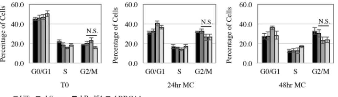
HPV requires infected cells to be maintained in a G
2/M-like
phase upon differentiation in order to properly amplify viral DNA
and produce infectious virions (
45
). In addition to DNA repair
through HR, BRCA1 is important in regulating cell cycle
check-points in response to DNA damage (
16
). To ensure that the block
in productive replication observed with BRCA1 as well as Rad51
knockdown was not due to an effect on the cell cycle, we analyzed
DNA content by flow cytometry. As shown in
Fig. 5
, untreated
CIN612 cells, as well as CIN612 cells expressing shScram,
exhib-ited a shift in the cell cycle to the G
2/M peak upon differentiation
in methylcellulose. However, knockdown of Rad51 and BRCA1
had no significant effect on the G
2/M shift compared to the
Scram-ble control after 24 or 48 h of differentiation in methylcellulose
(P
⬎
0.05). These results indicate that the lack of viral genome
amplification observed in CIN612-shRad51 and
CIN612-sh-BRCA1 cells is not due to an indirect effect on the cell cycle and
further suggest a direct role for these factors in facilitating viral
replication.
Inhibition of Rad51 function prevents productive viral
rep-lication.
The ability of Rad51 to bind single-stranded DNA
(ssDNA) is central to its role in HR (
46
). Single-stranded DNA
is generated by resection of DSBs or by stalled replication forks,
resulting in the binding of the tripartite RPA complex. RPA is
ultimately replaced by Rad51, which forms nucleofilaments that
mediate strand invasion into homologous sequences. We
previ-ously demonstrated that the RPA32 subunit localizes to HPV31
genomes (
8
), suggesting that resection occurs on viral DNA. In
addition, RPA32 is phosphorylated on Ser33 at viral genomes
upon differentiation, indicative of DNA damage and/or
replica-tion perturbareplica-tion (
10–12
). To determine if Rad51 is bound to viral
DNA, rather than just localized to sites of HPV replication, we
performed chromatin immunoprecipitation (ChIP) analysis on
CIN612 cells that were undifferentiated or differentiated in
high-calcium medium for 72 h to induce the productive phase of the
viral life cycle. As shown in
Fig. 6A
, we found that Rad51 is bound
to the upstream regulatory region (URR) of HPV31 genomes in
FIG 2Rad51 and BRCA1 promoter activity and transcript levels are increased in HPV31-positive cells. (A) HFKs and HPV31-positive CIN612 cells were transiently transfected with luciferase reporter constructs containing either Rad51 or BRCA1 proximal promoter sequences, along with aRenilla luciferase-expressing control vector. Luciferase assays were performed 72 h posttransfection. Luciferase activity was normalized toRenillaluciferase activity, and values were calculated as fold change over luciferase activity in uninfected HFKs. Values are averages from five independent experiments⫾standard errors of the means. *, Pⱕ0.03. (B and C) RNA was extracted from uninfected HFKs (B and C), HPV31-positive CIN612 cells (B), and HFK-31 cells (C). Quantitative reverse transcription-PCR analysis was performed using primers specific to Rad51 and BRCA1. Fold change was calculated using the 2⫺⌬⌬CTmethod. Shown is the foldundifferentiated cells. Interestingly, upon differentiation, Rad51
binding to HPV DNA increased per viral genome, suggesting the
formation of Rad51 nucleofilaments. To confirm the importance
of Rad51 activity in productive viral replication, we utilized the
Rad51-specific small-molecule inhibitor B02, which blocks Rad51
binding to DNA and inhibits DNA strand exchange into
homol-ogous sequences (
47
,
48
). As demonstrated in
Fig. 6B
, CIN612
cells differentiated for 72 h in high-calcium medium with
di-methyl sulfoxide (DMSO) resulted in amplification of viral
ge-nomes, consistent with activation of productive replication.
How-ever, treatment of CIN612 cells with increasing concentrations of
B02 resulted in a significant dose-dependent decrease in viral
ge-nome amplification (
Fig. 6B
and
C
). B02 treatment did not affect
the levels of Rad51 until the highest concentration (30
M) (
Fig.
6B
), which corresponded with an increase in cellular toxicity (data
not shown). Similarly to knockdown of Rad51 protein levels, there
was no appreciable effect on the differentiation-specific marker
involucrin upon treatment with B02 (
Fig. 6B
), indicating that
dif-ferentiation was not affected. In addition, we also measured the
effect of B02 treatment on the productive replication of HFKs
stably maintaining HPV31 genomes (HFK-31) upon
differentia-tion in high-calcium medium (
Fig. 7
). Similarly to treatment of
CIN612 cells, a dose-dependent effect on productive replication
was also observed in HFK-31 cells upon exposure to increasing
concentrations of B02 (
Fig. 7A
and
B
). To confirm that HR activity
is targeted by B02 treatment, we utilized HFK-31 cells containing
a chromosomally integrated HR reporter (DR-GFP), which
mea-sures the efficiency of recombination of two GFP alleles after
gen-eration of a double-strand break by the I-SceI restriction enzyme
(
Fig. 7C
). As shown in
Fig. 7D
, treatment of HFK-31 cells with
increasing concentrations of B02 resulted in a dose-dependent
decrease in HR activity. These results further support the idea that
the ability of Rad51 to bind viral DNA and form nucleofilaments is
important for efficient viral genome synthesis. Overall, our
find-ings indicate that HPV increases levels of Rad51 and BRCA1 to
drive HR at viral genomes for efficient DNA synthesis upon
dif-ferentiation.
DISCUSSION
point activation (
16
). However, no significant effects on the cell
cycle were observed upon BRCA1 or Rad51 knockdown. The
lo-calization of BRCA1 to HPV replication foci, coupled with the
binding of Rad51 to viral genomes and the requirement for
BRCA1 and Rad51 in productive replication, supports a role for
BRCA1 and Rad51 in facilitating recombination at HPV genomes
during productive replication.
Recombination can be stimulated by resection of DSBs, as
(
8
,
9
). In support of this, multiple components of the ATM
response, in addition to Rad51 and BRCA1, are required for
productive replication, including the MRN complex (Mre11,
Rad50, Nbs1) and Chk2 (
4
,
32
). ATM can stimulate
recombi-nation through two homology-directed pathways:
Rad51-de-pendent strand invasion (referred to as HR) and single-strand
annealing (SSA), both of which rely on a resection intermediate
(
49
). SSA, which is inhibited by Rad51 assembly on ssDNA, has
recently been implicated in recombination-dependent
replica-tion of HSV-1 (
26
). The recruitment of Rad51 and BRCA1 to
viral genomes suggests that HPV utilizes ATM activity to repair
replication-associated DSBs through HR. As HR involves the
use of the sister chromatid as a template for repair (
50
) and is
generally considered an error-free process, HPV may
preferen-tially use this method of repair for high-fidelity genome
ampli-fication upon differentiation. In addition, productive
replica-FIG 5Cell cycle distribution is not affected by knockdown of Rad51 or BRCA1. CIN612 cells were transduced with control shRNA (shScram) or Rad51 (shRad51) or BRCA1 (shBRCA1) shRNA containing lentiviral particles and either harvested atT0(undifferentiated) or differentiated in methylcellulose for 24 and 48 h. Cells from each time point were ethanol fixed, stained with propidium iodide (20g/ml), and analyzed for total cellular DNA content by flow cytometry. Graphed values are the averages of three independent experiments. Error bars represent means⫾standard errors. Statistics were performed using a two-tailedttest comparing shScram to shRad51 and shBRCA1 for the G2/M phase at each time point, with aPvalue of⬎0.05. N.S., not significant; MC, methylcellulose.tion is thought to occur in a G
2/M-like environment (
45
), at a
time when HR is most active (
45
).
End resection is a key step in the choice of DSB repair pathway
utilized, promoting pathways that use homology while
suppress-ing error-prone NHEJ (
20
). Previous studies from our lab indicate
that DNA-activated protein kinase (DNA-PK), a key factor in
classic NHEJ, does not localize to viral genomes (
8
), suggesting
that HR factors are specifically recruited. ATM and BRCA1 each
play essential roles in directing repair through recombination by
initiating resection of DSBs. ATM accomplishes this through
phosphorylation of the endonuclease CtIP (
18
), while BRCA1
an-tagonizes the resection inhibitor 53BP1 (
51–53
) and recruits CtIP
to the MRN resection complex (
54–56
). We previously
proximity of DSBs during S phase (
53
). BRCA1 may serve the
same function on HPV genomes during productive replication.
Future studies will focus on determining if and how HPV utilizes
ATM activity and BRCA1 to influence the DSB repair pathway of
choice, as has recently been shown for SV40 (
58
).
Our studies support a role for HR during productive
replica-tion, potentially for repair of replication-associated DNA breaks.
However, increasing evidence supports a role for HR proteins,
including Rad51, BRCA1, and BRCA2, as well as resection
en-zymes (CtIP and MRN), in replication fork stabilization
indepen-dently of DSB repair (
59–64
). In response to replication stress,
BRCA1 and BRCA2 stabilize Rad51 at stalled forks, protecting
nascent strands against nucleolytic degradation (
59–61
). Fork
re-start requires Rad51, which catalyzes Holliday junction formation
by invading a homologous molecule (
65
). HPV16 E7 induces
rep-lication stress due to unscheduled entry into the cell cycle,
result-ing in a paucity of nucleotides to support DNA synthesis (
66
).
Upon differentiation, E7 pushes postmitotic cells back into the cell
cycle to provide cellular factors required for productive
replica-tion (
67
). Whether cell cycle reentry upon differentiation also
in-duces replication stress is unclear but could potentially impact
viral genome amplification if cellular substrates are not
immedi-ately available. Alternatively, viral genome amplification could in
itself lead to replication stress due to the high levels of cellular
substrates required to meet the demand for increased viral DNA
synthesis. HPV may utilize ATM activity and HR factors to
pre-serve replication fork integrity, preventing the collapse of
replica-tion forks into DSBs. Understanding how HR factors are utilized
during viral replication will be an important area of future study.
Our studies indicate that the transient loss of Rad51 and
BRCA1 expression does not impact episomal copy number in
un-differentiated cells. Although we cannot rule out a role for these
HR factors in long-term episomal maintenance, these results
mir-ror our previous studies in which inhibition of ATM activity (
4
),
as well as disruption of the MRN complex (
32
), had minimal effect
on maintenance replication but was specifically required for
pro-ductive viral replication. Furthermore, our previous studies
dem-onstrated that knockdown of Nbs1 affected only the localization
of Rad51 to viral genomes upon differentiation (
32
). This may
suggest that in undifferentiated cells, Rad51 is not localized to viral
genomes for repair but may be present at single-stranded DNA
formed through transient uncoupling of the replication fork from
the viral E1 helicase (
59
,
68
). These results also argue that perhaps
there are inherent differences in the way HPV replicates and
uti-lizes DNA repair pathways during different phases of the viral life
cycle. In support of this, Flores and Lambert provided evidence
that HPV16 and HPV31 undergo a shift in the mode of replication
upon differentiation (
69
). In undifferentiated cells, HPV
repli-cates bidirectionally; however, two-dimensional (2D) gel analysis
revealed that productively replicating genomes exhibit branched
structures, including X structures and simple Y arcs, consistent
with strand invasion (Holliday junction formation) and
unidirec-tional (rolling-circle) replication, respectively (
70
,
71
). These
studies offer support for our findings that recombination is
im-portant for successful viral genome amplification and suggest that
productive replication may occur through rolling-circle
replica-tion and concatemer formareplica-tion. If viral concatemers are indeed
formed during productive replication, recombination may
addi-tionally be required to resolve concatemers into individual
ge-nomes.
We have found that Rad51 and BRCA1 are increased in
HPV31-positive cells at the level of transcription and protein
sta-bility. Increased levels of Rad51 have been documented in a wide
range of human tumors, including HPV-positive head and neck
cancer cell lines, as well as HPV-positive cervical cancer lines (
42
,
72
). Moreover, increased expression of Rad51 is correlated with
increased genomic instability through an elevation in
spontane-ous and aberrant HR (
73
,
74
). Although we can detect HR activity
in HPV31-positive cell lines using an HR-specific reporter assay,
whether this activity is increased relative to uninfected cells is
cur-rently unclear. How HPV infection affects DSB repair at cellular
sites of damage is also unknown. HPV may increase expression of
Rad51 and BRCA1 to drive productive replication, but in turn,
this increase could be detrimental to the genomic stability of
HPV-infected cells and contribute to cancer progression.
Alterna-tively, HPV may sequester these factors for viral replication,
pre-venting HR repair of cellular DSBs, resulting in increased genomic
instability.
Our studies indicate that both HPV31 E6 and E7 are able to
facilitate an increase in Rad51 and BRCA1 transcript levels,
though the mechanism remains unclear. Both Rad51 and BRCA1
are regulated in a manner dependent on E2F transcription factors
(
38
,
39
,
75–77
). The ability of E7 to target Rb and the related
pocket proteins p130 and p107 for degradation, in turn resulting
in constitutive activation of E2F transcription factors, may
facili-tate increased expression of Rad51 and BRCA1. E6 has also been
shown to induce expression of E2F-responsive genes (
78
). In
ad-dition, p53 can repress Rad51 expression (
79
), and E6’s ability to
promote p53 degradation may account for the increase in Rad51
and BRCA1 transcripts that we observed. While E7 was also able to
significantly increase Rad51 and BRCA1 protein levels compared
to uninfected HFKs, this was not consistently observed for E6,
despite the increase in Rad51 and BRCA1 transcript levels. These
data suggest an additional level of regulation for the increased
levels of Rad51 and BRCA1 protein observed in HPV-positive
cells. In support of this, we have found that the protein half-life of
Rad51 and BRCA1 is increased in HPV31-positive cells, although
the mechanism is unclear. HPV31 induces an ATM- as well as
ATR-dependent DNA damage response (
4
), which may in turn
stabilize Rad51 and BRCA1 through phosphorylation (
7
,
16
). In
addition, HPV16 E7 has been shown to bind BRCA1 (
80
), which
may be sufficient to protect BRCA1 from degradation. Whether
E7 also binds Rad51 is unknown. While HPV16 E6 also interacts
with BRCA1 (
80
), E6 has been shown to bind to the ubiquitin
ligase HERC2 (
81
), which can target BRCA1 for degradation (
82
).
Understanding the regulation of Rad51 and BRCA1 gene
expres-sion and protein stability in HPV-positive cells is an important
area for future study.
how HPV utilizes the DNA damage response to regulate
produc-tive viral replication.
ACKNOWLEDGMENTS
We thank David Weinstock, Peter Glazer, and Jeremy Stark for their gen-erous gifts of reagents.
This project was supported by NIH grant 1R01CA181581 and Amer-ican Cancer Society grant A14-0113 (to C.A.M.) and NIH grant 5T32 AI007419 (to W.H.C.).
FUNDING INFORMATION
HHS | NIH | National Cancer Institute (NCI) provided funding to Cary A Moody under grant number 1R01CA181581. HHS | NIH | National Can-cer Institute (NCI) provided funding to William H Chappell under grant number 5T32 AI007419. American Cancer Society (ACS) provided fund-ing to Cary A Moody under grant number A14-0113.
REFERENCES
1.zur Hausen H.2009. Papillomaviruses in the causation of human can-cers—a brief historical account. Virology384:260 –265.http://dx.doi.org /10.1016/j.virol.2008.11.046.
2.Longworth MS, Laimins LA.2004. Pathogenesis of human papillomavi-ruses in differentiating epithelia. Microbiol Mol Biol Rev68:362–372.
http://dx.doi.org/10.1128/MMBR.68.2.362-372.2004.
3.Moody CA, Laimins LA.2010. Human papillomavirus oncoproteins: pathways to transformation. Nat Rev Cancer10:550 –560.http://dx.doi .org/10.1038/nrc2886.
4.Moody CA, Laimins LA.2009. Human papillomaviruses activate the ATM DNA damage pathway for viral genome amplification upon differ-entiation. PLoS Pathog 5:e1000605. http://dx.doi.org/10.1371/journal .ppat.1000605.
5.Agarwal S, Tafel AA, Kanaar R.2006. DNA double-strand break repair and chromosome translocations. DNA Repair (Amst)5:1075–1081.http: //dx.doi.org/10.1016/j.dnarep.2006.05.029.
6.Helleday T, Lo J, van Gent DC, Engelward BP. 2007. DNA double-strand break repair: from mechanistic understanding to cancer treatment. DNA Repair (Amst)6:923–935.http://dx.doi.org/10.1016/j.dnarep.2007 .02.006.
7.Ciccia A, Elledge SJ.2010. The DNA damage response: making it safe to play with knives. Mol Cell40:179 –204.http://dx.doi.org/10.1016/j.molcel .2010.09.019.
8.Gillespie KA, Mehta KP, Laimins LA, Moody CA.2012. Human papil-lomaviruses recruit cellular DNA repair and homologous recombination factors to viral replication centers. J Virol86:9520 –9526.http://dx.doi.org /10.1128/JVI.00247-12.
9.Sakakibara N, Chen D, Jang MK, Kang DW, Luecke HF, Wu SY, Chiang CM, McBride AA.2013. Brd4 is displaced from HPV replication factories as they expand and amplify viral DNA. PLoS Pathog9:e1003777.
http://dx.doi.org/10.1371/journal.ppat.1003777.
10. Murphy AK, Fitzgerald M, Ro T, Kim JH, Rabinowitsch AI, Chowd-hury D, Schildkraut CL, Borowiec JA.2014. Phosphorylated RPA re-cruits PALB2 to stalled DNA replication forks to facilitate fork recovery. J Cell Biol206:493–507.http://dx.doi.org/10.1083/jcb.201404111. 11. Shiotani B, Nguyen HD, Hakansson P, Marechal A, Tse A, Tahara H,
Zou L.2013. Two distinct modes of ATR activation orchestrated by Rad17 and Nbs1. Cell Rep3:1651–1662.http://dx.doi.org/10.1016/j.celrep.2013 .04.018.
12. Vassin VM, Wold MS, Borowiec JA.2004. Replication protein A (RPA) phosphorylation prevents RPA association with replication centers. Mol Cell Biol24:1930 –1943.http://dx.doi.org/10.1128/MCB.24.5.1930-1943 .2004.
13. Rothkamm K, Kruger I, Thompson LH, Lobrich M.2003. Pathways of DNA double-strand break repair during the mammalian cell cycle. Mol Cell Biol23:5706 –5715.http://dx.doi.org/10.1128/MCB.23.16.5706-5715 .2003.
14.Moynahan ME, Jasin M. 2010. Mitotic homologous recombination maintains genomic stability and suppresses tumorigenesis. Nat Rev Mol Cell Biol11:196 –207.http://dx.doi.org/10.1038/nrm2851.
15. Baumann P, Benson FE, West SC. 1996. Human Rad51 protein promotes ATP-dependent homologous pairing and strand transfer
re-actions in vitro. Cell 87:757–766. http://dx.doi.org/10.1016/S0092 -8674(00)81394-X.
16. Savage KI, Harkin DP.2015. BRCA1, a ‘complex’ protein involved in the maintenance of genomic stability. FEBS J282:630 – 646.http://dx.doi.org /10.1111/febs.13150.
17. Prakash R, Zhang Y, Feng W, Jasin M.2015. Homologous recombina-tion and human health: the roles of BRCA1, BRCA2, and associated pro-teins. Cold Spring Harb Perspect Biol 7:a016600.http://dx.doi.org/10 .1101/cshperspect.a016600.
18. Wang H, Shi LZ, Wong CC, Han X, Hwang PY, Truong LN, Zhu Q, Shao Z, Chen DJ, Berns MW, Yates JR, III, Chen L, Wu X.2013. The interaction of CtIP and Nbs1 connects CDK and ATM to regulate HR-mediated double-strand break repair. PLoS Genet9:e1003277.http://dx .doi.org/10.1371/journal.pgen.1003277.
19. Chen L, Nievera CJ, Lee AY, Wu X.2008. Cell cycle-dependent complex formation of BRCA1.CtIP.MRN is important for DNA double-strand break repair. J Biol Chem283:7713–7720.http://dx.doi.org/10.1074/jbc .M710245200.
20. Chapman JR, Taylor MR, Boulton SJ.2012. Playing the end game: DNA double-strand break repair pathway choice. Mol Cell47:497–510.http: //dx.doi.org/10.1016/j.molcel.2012.07.029.
21. Sy SM, Huen MS, Chen J.2009. PALB2 is an integral component of the BRCA complex required for homologous recombination repair. Proc Natl Acad Sci U S A106:7155–7160.http://dx.doi.org/10.1073 /pnas.0811159106.
22. Weitzman MD, Lilley CE, Chaurushiya MS.2010. Genomes in conflict: maintaining genome integrity during virus infection. Annu Rev Microbiol
64:61– 81.http://dx.doi.org/10.1146/annurev.micro.112408.134016. 23. Turnell AS, Grand RJ.2012. DNA viruses and the cellular DNA-damage
response. J Gen Virol 93:2076 –2097. http://dx.doi.org/10.1099/vir.0 .044412-0.
24. Lo Piano A, Martinez-Jimenez MI, Zecchi L, Ayora S.2011. Recombi-nation-dependent concatemeric viral DNA replication. Virus Res160:1– 14.http://dx.doi.org/10.1016/j.virusres.2011.06.009.
25. Boichuk S, Hu L, Hein J, Gjoerup OV.2010. Multiple DNA damage signaling and repair pathways deregulated by simian virus 40 large T an-tigen. J Virol84:8007– 8020.http://dx.doi.org/10.1128/JVI.00334-10. 26. Schumacher AJ, Mohni KN, Kan Y, Hendrickson EA, Stark JM, Weller
SK.2012. The HSV-1 exonuclease, UL12, stimulates recombination by a single strand annealing mechanism. PLoS Pathog8:e1002862.http://dx .doi.org/10.1371/journal.ppat.1002862.
27. Wilkinson DE, Weller SK.2004. Recruitment of cellular recombination and repair proteins to sites of herpes simplex virus type 1 DNA replication is dependent on the composition of viral proteins within prereplicative sites and correlates with the induction of the DNA damage response. J Virol78:4783– 4796.http://dx.doi.org/10.1128/JVI.78.9.4783-4796.2004. 28. Wilkinson DE, Weller SK.2003. The role of DNA recombination in herpes simplex virus DNA replication. IUBMB Life55:451– 458.http://dx .doi.org/10.1080/15216540310001612237.
29. Kudoh A, Iwahori S, Sato Y, Nakayama S, Isomura H, Murata T, Tsurumi T.2009. Homologous recombinational repair factors are re-cruited and loaded onto the viral DNA genome in Epstein-Barr virus replication compartments. J Virol 83:6641– 6651. http://dx.doi.org/10 .1128/JVI.00049-09.
30. Dheekollu J, Deng Z, Wiedmer A, Weitzman MD, Lieberman PM.
2007. A role for MRE11, NBS1, and recombination junctions in replica-tion and stable maintenance of EBV episomes. PLoS One2:e1257.http: //dx.doi.org/10.1371/journal.pone.0001257.
31. Dheekollu J, Chen HS, Kaye KM, Lieberman PM. 2013. Timeless-dependent DNA replication-coupled recombination promotes Kapo-si’s sarcoma-associated herpesvirus episome maintenance and termi-nal repeat stability. J Virol87:3699 –3709.http://dx.doi.org/10.1128 /JVI.02211-12.
32. Anacker DC, Gautam D, Gillespie KA, Chappell WH, Moody CA.
2014. Productive replication of human papillomavirus 31 requires DNA repair factor Nbs1. J Virol88:8528 – 8544.http://dx.doi.org/10 .1128/JVI.00517-14.
33. Wilson R, Laimins LA.2005. Differentiation of HPV-containing cells using organotypic “raft” culture or methylcellulose. Methods Mol Med
119:157–169.
for the life cycle of human papillomavirus type 31. J Virol78:3533–3541.
http://dx.doi.org/10.1128/JVI.78.7.3533-3541.2004.
35. Hubert WG, Laimins LA.2002. Human papillomavirus type 31 replica-tion modes during the early phases of the viral life cycle depend on tran-scriptional and posttrantran-scriptional regulation of E1 and E2 expression. J Virol76:2263–2273.http://dx.doi.org/10.1128/jvi.76.5.2263-2273.2002. 36. Pierce AJ, Hu P, Han M, Ellis N, Jasin M.2001. Ku DNA end-binding
protein modulates homologous repair of double-strand breaks in mam-malian cells. Genes Dev 15:3237–3242. http://dx.doi.org/10.1101/gad .946401.
37. Fung H, Weinstock DM.2011. Repair at single targeted DNA double-strand breaks in pluripotent and differentiated human cells. PLoS One
6:e20514.http://dx.doi.org/10.1371/journal.pone.0020514.
38. Bindra RS, Glazer PM.2007. Repression of RAD51 gene expression by E2F4/p130 complexes in hypoxia. Oncogene26:2048 –2057.http://dx.doi .org/10.1038/sj.onc.1210001.
39. Bindra RS, Gibson SL, Meng A, Westermark U, Jasin M, Pierce AJ, Bristow RG, Classon MK, Glazer PM.2005. Hypoxia-induced down-regulation of BRCA1 expression by E2Fs. Cancer Res65:11597–11604.
http://dx.doi.org/10.1158/0008-5472.CAN-05-2119.
40. Mighty KK, Laimins LA.2011. p63 is necessary for the activation of human papillomavirus late viral functions upon epithelial differentiation. J Virol85:8863– 8869.http://dx.doi.org/10.1128/JVI.00750-11. 41. Fehrmann F, Klumpp DJ, Laimins LA.2003. Human papillomavirus
type 31 E5 protein supports cell cycle progression and activates late viral functions upon epithelial differentiation. J Virol77:2819 –2831.http://dx .doi.org/10.1128/JVI.77.5.2819-2831.2003.
42. Park JW, Nickel KP, Torres AD, Lee D, Lambert PF, Kimple RJ.2014. Human papillomavirus type 16 E7 oncoprotein causes a delay in repair of DNA damage. Radiother Oncol113:337–344.http://dx.doi.org/10.1016/j .radonc.2014.08.026.
43. Roman A, Munger K.2013. The papillomavirus E7 proteins. Virology
445:138 –168.http://dx.doi.org/10.1016/j.virol.2013.04.013.
44. Howie HL, Katzenellenbogen RA, Galloway DA.2009. Papillomavirus E6 proteins. Virology 384:324 –334. http://dx.doi.org/10.1016/j.virol .2008.11.017.
45. Banerjee NS, Wang HK, Broker TR, Chow LT.2011. Human papillo-mavirus (HPV) E7 induces prolonged G2following S phase reentry in differentiated human keratinocytes. J Biol Chem286:15473–15482.http: //dx.doi.org/10.1074/jbc.M110.197574.
46. Morrical SW.2015. DNA-pairing and annealing processes in homolo-gous recombination and homology-directed repair. Cold Spring Harb Perspect Biol7:a016444.http://dx.doi.org/10.1101/cshperspect.a016444. 47. Huang F, Mazina OM, Zentner IJ, Cocklin S, Mazin AV.2012. Inhibi-tion of homologous recombinaInhibi-tion in human cells by targeting RAD51 recombinase. J Med Chem 55:3011–3020. http://dx.doi.org/10.1021 /jm201173g.
48. Huang F, Motlekar NA, Burgwin CM, Napper AD, Diamond SL, Mazin AV.2011. Identification of specific inhibitors of human RAD51 recombi-nase using high-throughput screening. ACS Chem Biol6:628 – 635.http: //dx.doi.org/10.1021/cb100428c.
49. Gunn A, Bennardo N, Cheng A, Stark JM.2011. Correct end use during end joining of multiple chromosomal double strand breaks is influenced by repair protein RAD50, DNA-dependent protein kinase DNA-PKcs, and transcription context. J Biol Chem286:42470 – 42482.http://dx.doi .org/10.1074/jbc.M111.309252.
50. Johnson RD, Jasin M.2001. Double-strand-break-induced homologous recombination in mammalian cells. Biochem Soc Trans29:196 –201.http: //dx.doi.org/10.1042/bst0290196.
51. Bouwman P, Aly A, Escandell JM, Pieterse M, Bartkova J, van der Gulden H, Hiddingh S, Thanasoula M, Kulkarni A, Yang Q, Haffty BG, Tommiska J, Blomqvist C, Drapkin R, Adams DJ, Nevanlinna H, Bartek J, Tarsounas M, Ganesan S, Jonkers J.2010. 53BP1 loss rescues BRCA1 deficiency and is associated with triple-negative and BRCA-mutated breast cancers. Nat Struct Mol Biol17:688 – 695.http://dx.doi .org/10.1038/nsmb.1831.
52. Bunting SF, Callen E, Wong N, Chen HT, Polato F, Gunn A, Bothmer A, Feldhahn N, Fernandez-Capetillo O, Cao L, Xu X, Deng CX, Finkel T, Nussenzweig M, Stark JM, Nussenzweig A. 2010. 53BP1 inhibits homologous recombination in Brca1-deficient cells by blocking resection of DNA breaks. Cell141:243–254.http://dx.doi.org/10.1016/j.cell.2010.03 .012.
53.Chapman JR, Sossick AJ, Boulton SJ, Jackson SP. 2012.
BRCA1-associated exclusion of 53BP1 from DNA damage sites underlies temporal control of DNA repair. J Cell Sci125:3529 –3534.http://dx.doi.org/10 .1242/jcs.105353.
54. Yu X, Wu LC, Bowcock AM, Aronheim A, Baer R.1998. The C-terminal (BRCT) domains of BRCA1 interact in vivo with CtIP, a protein impli-cated in the CtBP pathway of transcriptional repression. J Biol Chem
273:25388 –25392.http://dx.doi.org/10.1074/jbc.273.39.25388. 55. Zhong Q, Chen CF, Li S, Chen Y, Wang CC, Xiao J, Chen PL, Sharp
ZD, Lee WH.1999. Association of BRCA1 with the hRad50-hMre11-p95 complex and the DNA damage response. Science285:747–750.http://dx .doi.org/10.1126/science.285.5428.747.
56. Sartori AA, Lukas C, Coates J, Mistrik M, Fu S, Bartek J, Baer R, Lukas J, Jackson SP.2007. Human CtIP promotes DNA end resection. Nature
450:509 –514.http://dx.doi.org/10.1038/nature06337.
57. Langsfeld ES, Bodily JM, Laimins LA.2015. The deacetylase sirtuin 1 regulates human papillomavirus replication by modulating histone acet-ylation and recruitment of DNA damage factors NBS1 and Rad51 to viral genomes. PLoS Pathog11:e1005181. http://dx.doi.org/10.1371/journal .ppat.1005181.
58. Sowd GA, Mody D, Eggold J, Cortez D, Friedman KL, Fanning E.2014. SV40 utilizes ATM kinase activity to prevent non-homologous end join-ing of broken viral DNA replication products. PLoS Pathog10:e1004536.
http://dx.doi.org/10.1371/journal.ppat.1004536.
59. Schlacher K, Christ N, Siaud N, Egashira A, Wu H, Jasin M.2011. Double-strand break repair-independent role for BRCA2 in blocking stalled replication fork degradation by MRE11. Cell145:529 –542.http: //dx.doi.org/10.1016/j.cell.2011.03.041.
60. Schlacher K, Wu H, Jasin M.2012. A distinct replication fork protection pathway connects Fanconi anemia tumor suppressors to RAD51-BRCA1/2. Cancer Cell22:106 –116.http://dx.doi.org/10.1016/j.ccr.2012 .05.015.
61. Hashimoto Y, Ray Chaudhuri A, Lopes M, Costanzo V.2010. Rad51 protects nascent DNA from Mre11-dependent degradation and promotes continuous DNA synthesis. Nat Struct Mol Biol17:1305–1311.http://dx .doi.org/10.1038/nsmb.1927.
62. Yeo JE, Lee EH, Hendrickson EA, Sobeck A.2014. CtIP mediates repli-cation fork recovery in a FANCD2-regulated manner. Hum Mol Genet
23:3695–3705.http://dx.doi.org/10.1093/hmg/ddu078.
63. Lomonosov M, Anand S, Sangrithi M, Davies R, Venkitaraman AR.
2003. Stabilization of stalled DNA replication forks by the BRCA2 breast cancer susceptibility protein. Genes Dev17:3017–3022.http://dx.doi.org /10.1101/gad.279003.
64. Bryant HE, Petermann E, Schultz N, Jemth AS, Loseva O, Issaeva N, Johansson F, Fernandez S, McGlynn P, Helleday T.2009. PARP is activated at stalled forks to mediate Mre11-dependent replication restart and recombination. EMBO J 28:2601–2615.http://dx.doi.org/10.1038 /emboj.2009.206.
65. Petermann E, Orta ML, Issaeva N, Schultz N, Helleday T.2010. Hy-droxyurea-stalled replication forks become progressively inactivated and require two different RAD51-mediated pathways for restart and repair. Mol Cell37:492–502.http://dx.doi.org/10.1016/j.molcel.2010.01.021. 66. Bester AC, Roniger M, Oren YS, Im MM, Sarni D, Chaoat M, Bensimon
A, Zamir G, Shewach DS, Kerem B.2011. Nucleotide deficiency pro-motes genomic instability in early stages of cancer development. Cell145:
435– 446.http://dx.doi.org/10.1016/j.cell.2011.03.044.
67. Cheng S, Schmidt-Grimminger DC, Murant T, Broker TR, Chow LT.
1995. Differentiation-dependent up-regulation of the human papilloma-virus E7 gene reactivates cellular DNA replication in suprabasal differen-tiated keratinocytes. Genes Dev9:2335–2349.http://dx.doi.org/10.1101 /gad.9.19.2335.
68.Hashimoto Y, Puddu F, Costanzo V. 2012. RAD51- and MRE11-dependent reassembly of uncoupled CMG helicase complex at collapsed replication forks. Nat Struct Mol Biol19:17–24.http://dx.doi.org/10.1038 /nsmb.2177.
69. Flores ER, Lambert PF.1997. Evidence for a switch in the mode of human papillomavirus type 16 DNA replication during the viral life cycle. J Virol
71:7167–7179.
70. Preiser PR, Wilson RJ, Moore PW, McCready S, Hajibagheri MA, Blight KJ, Strath M, Williamson DH.1996. Recombination associated with replication of malarial mitochondrial DNA. EMBO J15:684 – 693. 71. Backert S.2002. R-loop-dependent rolling-circle replication and a new
72. Raderschall E, Stout K, Freier S, Suckow V, Schweiger S, Haaf T.2002. Elevated levels of Rad51 recombination protein in tumor cells. Cancer Res
62:219 –225.
73. Richardson C, Stark JM, Ommundsen M, Jasin M.2004. Rad51 over-expression promotes alternative double-strand break repair pathways and genome instability. Oncogene23:546 –553.http://dx.doi.org/10.1038/sj .onc.1207098.
74. Klein HL.2008. The consequences of Rad51 overexpression for normal and tumor cells. DNA Repair (Amst)7:686 – 693.http://dx.doi.org/10 .1016/j.dnarep.2007.12.008.
75. Bindra RS, Glazer PM.2006. Basal repression of BRCA1 by multiple E2Fs and pocket proteins at adjacent E2F sites. Cancer Biol Ther5:1400 –1407.
http://dx.doi.org/10.4161/cbt.5.10.3454.
76. Iwanaga R, Komori H, Ohtani K.2004. Differential regulation of expres-sion of the mammalian DNA repair genes by growth stimulation. Onco-gene23:8581– 8590.http://dx.doi.org/10.1038/sj.onc.1207976.
77. Wang A, Schneider-Broussard R, Kumar AP, MacLeod MC, Johnson DG.2000. Regulation of BRCA1 expression by the Rb-E2F pathway. J Biol Chem275:4532– 4536.http://dx.doi.org/10.1074/jbc.275.6.4532.
78. Shai A, Brake T, Somoza C, Lambert PF.2007. The human papilloma-virus E6 oncogene dysregulates the cell cycle and contributes to cervical carcinogenesis through two independent activities. Cancer Res67:1626 – 1635.http://dx.doi.org/10.1158/0008-5472.CAN-06-3344.
79. Arias-Lopez C, Lazaro-Trueba I, Kerr P, Lord CJ, Dexter T, Iravani M, Ashworth A, Silva A.2006. p53 modulates homologous recombination by transcriptional regulation of the RAD51 gene. EMBO Rep7:219 –224.
http://dx.doi.org/10.1038/sj.embor.7400587.
80. Zhang Y, Fan S, Meng Q, Ma Y, Katiyar P, Schlegel R, Rosen EM.2005. BRCA1 interaction with human papillomavirus oncoproteins. J Biol Chem280:33165–33177.http://dx.doi.org/10.1074/jbc.M505124200. 81. Vos RM, Altreuter J, White EA, Howley PM.2009. The
ubiquitin-specific peptidase USP15 regulates human papillomavirus type 16 E6 protein stability. J Virol83:8885– 8892.http://dx.doi.org/10.1128/JVI .00605-09.