Cellular Mechanisms of Immune Dysfunction Following Severe Burn Injury
Crystal J. Neely
A dissertation submitted to the faculty of the University of North Carolina at Chapel Hill in partial fulfillment of the requirements for the degree of Doctor of Philosophy in the
Department of Microbiology and Immunology.
Chapel Hill 2013
Approved by:
Bruce A. Cairns, MD, FACS
Rob Maile, PhD
Bo Wang, PhD
Thomas H. Kawula, PhD
Mark T. Heise, PhD
ii ABSTRACT
CRYSTAL J. NEELY: Cellular Mechanisms of Immune Dysfunction Following Severe Burn Injury
(Under the direction of Bruce A. Cairns)
The immune system protects the body against infection and disease. Severe burn injury induces profound immune dysfunction rendering patients extremely susceptible to infection. Although progress has been made in reducing the incidence of infection, burn wound infection and pulmonary sepsis are still major causes of mortality. Therefore, in order to improve patient outcome it is important to understand the immune response to infection after burn injury. Furthermore, the immune response following burn injury is dynamic and changes over time. Therefore, elucidating the immune response both early and late after burn is also crucial.
Using a murine model of thermal injury, we characterized the pro-inflammatory CD4+ T cell response at various timepoints after burn. We detected Th17 cells in wound-draining lymph nodes at 3, 7, and 14 days post burn. Also, there was bimodal skewing of the Th1/Th17 T cell balance with an early predominance of Th1 cells Th17 cells late.
The innate immune response to a clinically relevant pathogen was then assessed early after burn injury. Burn mice were susceptible to an early wound inoculation with
iii
with polarization of neutrophils into an anti-inflammatory (N2; IL-10+ IL-12-) phenotype. Administration of flagellin after burn injury skewed the neutrophil response towards a pro-inflammatory neutrophil (N1; IL-10-IL-12+) phenotype resulting in increased bacterial clearance.
Additional studies evaluated susceptibility to pulmonary P. aeruginosa infection late after burn injury. In contrast to early wound infection, burn mice exhibited enhanced
clearance of a delayed pulmonary P. aeruginosa challenge compared to non-burn controls. This appeared to result from to a burn-induced accumulation of neutrophils within the lungs. Collectively, these data suggests that neutrophil responses vary after burn injury where they exhibit an anti-inflammatory phenotype early after burn followed by a late and
pro-inflammatory phenotype.
iv
v
ACKNOWLEDGEMENTS
First and foremost I would like to thank Bruce Cairns and Rob Maile for being outstanding mentors. I am extremely grateful for all of your advice and encouragement, as well as the many opportunities you provided me.
A special thanks to members of the Cairns lab, both present and past. It has been a privilege to get to know and to collaborate with so many amazing people.
My committee members: Bo Wang, Mark Heise, Tom Kawula, and Yisong Wan. Thank you for always being available to talk.
I would also like to thank Ed Collins, Jeff Frelinger, Matt Wolfgang, and Nat Moorman. I have always appreciated your feedback and recommendations.
To my parents, I can’t begin to express how much your love and encouragement has meant to me. I would not be where are I am today without you.
To Matt Powers, thank you for your patience, understanding, and support. I am so grateful you were by my side these last four years.
vi
TABLE OF CONTENTS
LIST OF TABLES……….…..…ix
LIST OF FIGURES……….……...x
LIST OF ABBREVIATIONS………..……...xii
1. Introduction ...1
1.1 Burn injury ...1
1.2 Incidence of burn ...1
1.3 Cost of burn care ...2
1.4 Grading of burn wounds ...2
1.5 Systemic response to burn injury ...3
1.6 Wound infection ...3
1.7 Pulmonary infection ...4
1.8 Pseudomonas aeruginosa ...5
1.9 P. aeruginosa virulence factors ...5
1.10 Immune response to infection ...6
1.11 Toll-like receptors ...6
1.12 TLR and burn injury ...7
1.13 Neutrophils ...8
1.14 Neutrophils and burn injury ...9
1.15 Macrophages ...9
vii
1.17 Dendritic cells ...11
1.18 Dendritic cells and burn injury ...11
1.19 Th17 cells ...12
1.20 Th17 cells and burn injury ...12
1.21 Remaining questions ...13
1.22 References ...14
2. Th17 (IFNγ- IL-17+) CD4+ T cells generated after burn injury may be a novel cellular mechanism for post-burn immunosuppression ...21
2.1 Summary ...21
2.2 Introduction ...22
2.3 Methods and materials ...24
2.4 Results ...27
2.5 Discussion ...31
2.6 References ...44
3. Flagellin treatment prevents increased susceptibility to systemic bacterial infection after injury by inhibiting IL-10+ IL-12- neutrophil polarization ...49
3.1 Summary ...49
3.2 Introduction ...50
3.3 Methods and materials ...52
3.4 Results ...56
3.5 Discussion ...63
3.6 References ...77
4. Burn-induced “innate-priming” enhances clearance of bacterial challenge late after injury ...82
viii
4.2 Introduction ...83
4.3 Methods and materials ...84
4.4 Results ...88
4.5 Discussion ...92
4.6 References ...104
5. Conclusions and Future Directions ...107
5.1 A clinical need ...107
5.2 Characterization of CD4+ T cell response after burn injury...107
5.3 Future directions: defining the role of Th17 cells after burn injury ...108
5.4 Cellular mechanisms of increased susceptibility to early wound infection ...109
5.5 Future directions: characterization of neutrophil response early after burn injury ...110
5.6 Neutrophil accumulation in the lungs late after burn injury ...112
5.7 Future directions: characterization of neutrophil response late after burn injury ...113
5.8 Closing remarks ...114
ix
LIST OF TABLES
x
LIST OF FIGURES
Figure 2.1……….43
Figure 2.2…...44
Figure 2.3……….…45
Figure 2.4…...46
Figure 2.5……….47
Figure 2.6……….48
Figure 2.7……….49
Figure 2.8……….50
Figure 3.1…...76
Figure 3.2……….78
Figure 3.3……….79
Figure 3.4……….80
Figure 3.5……….81
Figure 3.6……….82
Figure 3.7……….83
Figure 3.8……….84
Figure 4.1…...102
Figure 4.2………...103
Figure 4.3………...104
Figure 4.4………...105
Figure 4.5…...106
Figure 4.6………...107
Figure 4.7…...108
xi
xii
LIST OF ABBREVIATIONS ALI: acute lung injury
B6: C57BL/6 mouse strain BAL: brochoalveolar lavage CD: cluster of differentiation CFU: colony forming units
COPD: chronic obstructive pulmonary disease DAMP: damage associated molecular pattern DC: dendritic cell
DHR: dihydrorhodamine Exo: exoenzyme
Foxp3: forkhead box P3 hpi: hours post infection IC: immune complexes IFN-γ: interferon gamma IL: interleukin
IP: intraperitoneal IT: intratracheal
KC: keratinocyte-derived cytokine LPS: lipopolysaccharide
M1: pro-inflammatory macrophage M2: anti-inflammatory macrophage
xiii MFI: mean fluorescence intensity
MHC: major histocompatibility complex N1: pro-inflammatory neutrophil
N2: anti-inflammatory neutrophil NET: neutrophil extracellular trap NO: nitric oxide
NOI: neutrophil oxidative index
PAMP: pattern associated molecular pattern pDC: plasmacytoid dendritic cell
PGE2: prostaglandin E2 PLN: peripheral lymph nodes
PMA: phorbol 12-myristate 13-actetate RNI: reactive nitrogen intermediates ROI: reactive oxygen intermediates T2SS: type II secretion system T3SS: type III secretion system TBSA: total body surface area TCR: T cell receptor
TGF-β: transforming growth factor beta Th: T helper
TLR: toll-like receptor
2 CHAPTER 1
Introduction
1.1 Burn injury
The International Society of Burn Injuries defines burn as damage to the skin or other tissues primarily caused by thermal injury. This typically occurs when hot liquids (scalds), hot solids (contact burns), or flames (flame burns) induce subtotal or total destruction of cells within the localized area of exposure. The etiology of burn is not limited to the
aforementioned modes of trauma; it also includes radiation, electric, friction and chemical injuries.
1.2 Incidence of burn
2 1.3 Cost of burn care
Individuals with severe burn injury require immediate specialized care in order to minimize morbidity and mortality. This specialized care includes aggressive fluid
resuscitation and prevention of fluid loss, regulation of body temperature, management of severe pain, wound debridement, early excision and wound closure, and prevention of infections5. The cost of initial hospitalization and the surgical care of a burn patient is approximately $200,0006,7. However, this estimate does not include expenses incurred by rehabilitation, vocational services, late complications, and disability. Although the overall hospitalization rates from minor burn injuries have significantly declined over the last 50 years, the proportion of patients admitted to burn centers has increased7. This has led to an annual cost of more than $18 billion for specialized burn care in the United States7.
1.4Grading of burn wounds
3
Lastly, a fourth-degree burn involves other organ tissue below the skin and soft tissue, such as muscle and bone5. Burn injuries are also measured by size or area of injury. The percent total body surface area (TBSA) of an adult patient is typically assessed by the rule of nines, while the Berkow formula is used on children5. Taken together these classification schemes guide fluid resuscitation and wound management.
1.5 Systemic response to burn injury
Severe burn injuries covering greater than 30% TBSA are typically followed by a period of hypermetabolism, altered hemodynamics, vascular permeability and edema, decreased renal blood flow, and increased gut mucosal permeability8. The massive release of
inflammatory mediators in the wound, as well as in other tissues, is believed to impact and/or trigger this multi-organ dysfunction. There is also immune suppression demonstrated by prolonged allograft skin survival on burn wounds5. Since burn injury impairs all parts of the immune system, patients are extremely susceptible to infection. With burns of more the 20% TBSA, the magnitude of immune impairment is proportional to the size of burn5.
1.6Wound infection
Since the skin is a barrier against invading pathogens, it is not surprising that the risk of subsequent burn wound infection also correlates with the size of the burn injury9,10.
4
health care worker's hands11-13. Gram-negative bacteria are currently the most common infectious agents seen in burn centers due to their numerous virulence factors and
antimicrobial resistance traits14. Burn wound infection delays epidermal maturation and leads to additional scar tissue formation15,16. Invasion of microbes into the tissue layers below the dermis may also result in bacteremia and sepsis where progression to multiple organ failure is usually fatal17,18.
1.7Pulmonary infection
Pulmonary bacterial infections are a major cause of mortality in burn patients. Hypostatic pneumonia is common in patients with large burns due to hyperventilation and decreased lung expansion19. Hematogenous pneumonia, where the infection is spread by the blood from another part of the body, is much less common in burn patients due to aggressive antibiotic therapies20. Nevertheless, both can be fatal. Inhalation injury, which occurs in approximately one-third of all major burns, damages the airway mucosa, impairs immune cell function, and requires intubation for airway protection21,22. This significantly increases a burn patient’s risk of developing a pulmonary bacterial infection and in particular ventilator-associated pneumonia (VAP)21,23. VAP occurs when a mechanically ventilated patient develops
5 1.8Pseudomonas aeruginosa
Pseudomonas aeruginosa, a gram negative extracellular bacterium, is one of the most commonly encountered infectious agents in burn centers across the United States25. The organism can be isolated from environmental sources, such as freshwater and soil. It can also survive on surfaces contaminating the floors, bed rails, and sinks of hospitals26,27. Innate immune responses are essential for controlling P. aeruginosa. Clinically, the vast majority of cases of P. aeruginosa infection are found in patients with comprised immune systems28.
1.9 P. aeruginosa virulence factors
6
aeruginosa and induces rapid necrotic cell death30,31. Also, ExoS and ExoT can inhibit macrophage and neutrophil migration and phagocytosis32,33.
1.10 Immune response to infection
The immune system is the body’s defense against infection and disease. It detects a wide variety of antigens derived from invading pathogens and distinguishes them from the host’s own tissue. The immune response can be divided into innate and adaptive immunity. The innate immune system is non-specific, meaning it recognizes and responds to pathogens in a generic way. More specifically, it depends upon germline-encoded receptors (ie. TLRs) to recognize features that are common to many microbes. Most pathogens are detected and destroyed within minutes to hours of invasion by innate immune cells, which includes the neutrophils and macrophages. However if a pathogen persists, the adaptive immune response ensues. The adaptive immune system is specific and consists of T and B lymphocytes. It targets a precise pathogen by utilizing pathogen-specific receptors, such as T cell receptors (TCR), which are acquired during the lifetime of the host. Induction of an adaptive immune response leads to immunological memory, which ensures a more rapid and effective response on subsequent encounters with the same pathogen.
1.11 Toll-like receptors
danger-7
associated molecular patterns (DAMPs). In humans there are 10 known TLRs, whereas 12 have been characterized in mice34. Each TLR recognizes and is activated by a small assortment of microbe-derived molecules. For example, TLR2, TLR4, and TLR5 are involved in control of P. aeruginosa infection by recognizing outer membrane lipoproteins, LPS, and flagellin, respectively35-37. TLRs are expressed by immune cells and a wide variety of non-immune cells. TLR signaling induces the expression of hundreds of genes required for the inflammatory response, including inflammatory cytokines, chemokines, antimicrobial molecules, and major histocompatibility complex (MHC) and costimulatory molecules important for adaptive immune activation34. Although TLRs act primarily to initiate an innate immune response, some adaptive immune responses are elicited by TLR signaling.
1.12 TLR and burn injury
8
secretion upon stimulation39. Conversely, there is upregulation in TLR4 expression on memory CD4+ and CD8+ T cells at 14 days after burn41.
1.13 Neutrophils
Neutrophils are considered the first responders of the immune system since they are quickly recruited from the vasculature to the site of infection. Non-activated human
9 1.14 Neutrophils and burn injury
Neutrophil function is significantly impaired after burn injury although the timing and extent is still unclear. One study showed that neutrophils have decreased Fc
receptor-mediated phagocytosis, as well as a 50% reduction in intracellular killing, after burn injury. This group also showed that the ability of circulating neutrophils to undergo oxidative burst gradually declines during the first two weeks after burn injury48. Another study reported that there is an increased number of neutrophils in the peritoneal cavity and an increase in
neutrophil oxidative burst at one day after burn49. Neutrophils have also been reported to be immunosuppressive after burn injury as demonstrated by their secretion of IL-10, a potent anti-inflammatory cytokine, upon TLR2 stimulation50.
1.15 Macrophages
Macrophages reside in every tissue of the body although they are sometimes called by other names such as microglia, Kupffer cells, and osteoclasts. Upon infection, circulating monocytes are rapidly recruited to the tissue, where they differentiate into tissue
10
remarkably plastic and can change their functional phenotype depending on the
environmental cues they receive. For example, a macrophage can be polarized towards a inflammatory phenotype (M1) marked by production of IL-12, as well as other
pro-inflammatory mediators, when activated in the presence of interferon-gamma (IFN-γ)54. However, if a macrophage is later exposed to IL-10, glucocorticoids, or immune complexes in the presence of the TLR ligands, it can exhibit an anti-inflammatory phenotype (M2, IL-10+ IL-12-)55,56.
1.16 Macrophages and burn injury
Macrophages are reported to be hyper-responsive following burn injury. Upon stimulation, macrophages exhibit exaggerated production of IL-1, IL-6, transforming growth factor (TGF-β), prostaglandin E2 (PGE2), and reactive nitrogen intermediates (RNI)57-59. Previous studies have implicated macrophage hyperactivity in the increased susceptibility to bacterial sepsis following burn injury59,60. In addition, recent evidence suggests that severe burn injury results in a shift of macrophage polarization towards an M2 phenotype61. These M2 macrophages have reduced capacity to kill bacterial pathogens. Soluble factors released by these cells inhibit macrophage conversion of resident macrophages to a M1
11 1.17 Dendritic cells
Similar to macrophages, dendritic cells (DCs) are capable of both phagocytosis and antigen presentation. However, DCs are not directly involved in immediate pathogen
clearance; instead their primary function is antigen presentation63. Immature DCs are present in all peripheral tissue and constantly sample the surrounding environment for invading pathogens. When an immature DC phagocytoses a pathogen, it begins to mature by upregulating cell-surface receptors that act as co-receptors in T-cell activation. It also upregulates CCR7, a chemotactic receptor that directs DCs to secondary lymphoid organs63. Once inside a lymphoid organ, DCs act as antigen-presenting cells to initiate antigen-specific immune responses. DCs also secrete a variety of cytokines, such as IL-12, that are protective against a number of infections64,65.
1.18 Dendritic cells and burn injury
12 1.19 Th17 Cells
Until recently, CD3+CD4+ T cells were divided into pro-inflammatory Th1 (IFN-γ-producing) or anti-inflammatory Th2 (IL-4-(IFN-γ-producing) cells69. However, a newly discovered subset of CD4+ T cells, called Th17 cells, have been shown to secrete 17, 21, and IL-22, but do not secrete IFN- γ or IL-470-72. IL-17 activates macrophages and promotes the production of inflammatory cytokines, which includes tumor necrosis factor-α (TNF-α) and IL-1β. Subsequently, these cytokines recruit neutrophils to sites of infection73. For example, IL-17 receptor deficient mice have reduced neutrophil recruitment, increased bacterial loads, and high mortality rates after challenge with Klebsiella pneumoniae74. Furthermore,
overexpression of IL-17 in the lungs results in a localized increase in TNF-α and IL-1β, as well as enhanced neutrophil accumulation and K. pneumoniae clearance75. IL-17 receptor deficient mice are also more susceptible to infection with Candida albicans76. IL-21, another cytokine secreted by Th17 cells, acts on epithelial cells and is involved with antimicrobial peptide production77-79, tissue repair80, and epithelial cell proliferation81, differentiation82, and survival83. Together, these findings suggests that the main function of Th17 cells is to
promote antimicrobial immunity at mucosal interfaces.
1.20 Th17 cells and burn injury
13
and pediatric burn patient serum has shown that IL-17 is elevated within 1 week after injury87,88. Another recent study examined Th17 cell development in patients with full thickness burns and revealed that peripheral blood mononuclear cells isolated from these patients had a decreased production of IL-17 in response to C. albicans antigen stimulation compared to healthy controls89. Since burn patients are highly susceptible to C. albicans infection, enhancing the IL-17 response after burn injury could be beneficial.
1.21 Remaining questions
14 1.22 References
1. Who Health Organization. The global burden of disease: 2004 update. (WHO Press, Geneva, Switzerland; 2008).
2. Peck, M.D. Epidemiology of burns throughout the world. Part I: Distribution and risk factors. Burns : journal of the International Society for Burn Injuries 37, 1087-1100 (2011).
3. Miller, S.F. et al. National burn repository 2007 report: a synopsis of the 2007 call for data. Journal of burn care & research : official publication of the American Burn Association 29, 862-870; discussion 871 (2008).
4. American Burn Association. Burn incidence and treatment in the United States: 2011 Fact Sheet. http://ameriburn.org/resources_factsheet.php; 2011.
5. Cotlar, A.M. Surgery - Pocket companion to Sabiston Textbook of Surgery, 16th ed: Handheld software. Jama-J Am Med Assoc 290, 3142-3142 (2003).
6. Dimick, A., Cope, D.N., Barillo, D., Gillespie, R. & Mozingo, D. Report on a more rational approach to managed care for burns. The Journal of burn care &
rehabilitation 26, 14-18 (2005).
7. Roth, J.J., and W. B. Hughes. The essential burn unit handbook. (Quality Medical Publishing, St. Louis, MO; 2004).
8. Hettiaratchy, S. & Dziewulski, P. ABC of burns: pathophysiology and types of burns. BMJ 328, 1427-1429 (2004).
9. Santaniello, J.M. et al. Ten year experience of burn, trauma, and combined burn/trauma injuries comparing outcomes. The Journal of trauma 57, 696-700; dicussion 700-691 (2004).
10. Sheridan, R.L. Evaluating and managing burn wounds. Dermatology nursing / Dermatology Nurses' Association 12, 17-18, 21-18; quiz 30-11 (2000).
11. Erol, S., Altoparlak, U., Akcay, M.N., Celebi, F. & Parlak, M. Changes of microbial flora and wound colonization in burned patients. Burns : journal of the International Society for Burn Injuries 30, 357-361 (2004).
15
13. Manson, W.L., Pernot, P.C., Fidler, V., Sauer, E.W. & Klasen, H.J. Colonization of burns and the duration of hospital stay of severely burned patients. The Journal of hospital infection 22, 55-63 (1992).
14. Smith, D.J., Jr. & Thomson, P.D. Changing flora in burn and trauma units: historical perspective--experience in the United States. The Journal of burn care &
rehabilitation 13, 276-280 (1992).
15. Singer, A.J. & McClain, S.A. Persistent wound infection delays epidermal maturation and increases scarring in thermal burns. Wound repair and regeneration : official publication of the Wound Healing Society [and] the European Tissue Repair Society 10, 372-377 (2002).
16. Edwards, R. & Harding, K.G. Bacteria and wound healing. Current opinion in infectious diseases 17, 91-96 (2004).
17. Horton, J.W., Maass, D.L., White, J. & Minei, J.P. Reducing susceptibility to
bacteremia after experimental burn injury: a role for selective decontamination of the digestive tract. J Appl Physiol 102, 2207-2216 (2007).
18. Baker, C.C., Miller, C.L. & Trunkey, D.D. Predicting fatal sepsis in burn patients. The Journal of trauma 19, 641-648 (1979).
19. Fabian, T.C. Empiric therapy for pneumonia in the surgical intensive care unit. American journal of surgery 179, 18-23 (2000).
20. Church, D., Elsayed, S., Reid, O., Winston, B. & Lindsay, R. Burn wound infections. Clinical microbiology reviews 19, 403-434 (2006).
21. Moylan, J.A. & Alexander, L.G., Jr. Diagnosis and treatment of inhalation injury. World journal of surgery 2, 185-191 (1978).
22. Demling, R.H. Smoke inhalation lung injury: an update. Eplasty 8, e27 (2008). 23. Baughman, R.P. Diagnosis of ventilator-associated pneumonia. Microbes and
infection / Institut Pasteur 7, 262-267 (2005).
24. Shorr, A.F., Sherner, J.H., Jackson, W.L. & Kollef, M.H. Invasive approaches to the diagnosis of ventilator-associated pneumonia: a meta-analysis. Critical care medicine 33, 46-53 (2005).
25. Mayhall, C.G. The epidemiology of burn wound infections: Then and now. Clin Infect Dis 37, 543-550 (2003).
16
27. Chitkara, Y.K. & Feierabend, T.C. Endogenous and exogenous infection with Pseudomonas aeruginosa in a burns unit. International surgery 66, 237-240 (1981). 28. Lyczak, J.B., Cannon, C.L. & Pier, G.B. Establishment of Pseudomonas aeruginosa infection: lessons from a versatile opportunist. Microbes and Infection 2, 1051-1060 (2000).
29. Palmer, K.L., Mashburn, L.M., Singh, P.K. & Whiteley, M. Cystic fibrosis sputum supports growth and cues key aspects of Pseudomonas aeruginosa physiology. Journal of bacteriology 187, 5267-5277 (2005).
30. Finck-Barbancon, V. et al. ExoU expression by Pseudomonas aeruginosa correlates with acute cytotoxicity and epithelial injury. Molecular microbiology 25, 547-557 (1997).
31. Sutterwala, F.S. et al. Immune recognition of Pseudomonas aeruginosa mediated by the IPAF/NLRC4 inflammasome. The Journal of experimental medicine 204, 3235-3245 (2007).
32. Garrity-Ryan, L. et al. The arginine finger domain of ExoT contributes to actin cytoskeleton disruption and inhibition of internalization of Pseudomonas aeruginosa by epithelial cells and macrophages. Infect Immun 68, 7100-7113 (2000).
33. Rocha, C.L., Coburn, J., Rucks, E.A. & Olson, J.C. Characterization of Pseudomonas aeruginosa exoenzyme S as a bifunctional enzyme in J774A.1 macrophages. Infect Immun 71, 5296-5305 (2003).
34. Moresco, E.M., LaVine, D. & Beutler, B. Toll-like receptors. Current biology : CB 21, R488-493 (2011).
35. Feldman, M. et al. Role of flagella in pathogenesis of Pseudomonas aeruginosa pulmonary infection. Infect Immun 66, 43-51 (1998).
36. Hajjar, A.M., Ernst, R.K., Tsai, J.H., Wilson, C.B. & Miller, S.I. Human Toll-like receptor 4 recognizes host-specific LPS modifications. Nature immunology 3, 354-359 (2002).
37. Balloy, V. et al. The role of flagellin versus motility in acute lung disease caused by Pseudomonas aeruginosa. The Journal of infectious diseases 196, 289-296 (2007). 38. Paterson, H.M. et al. Injury primes the innate immune system for enhanced Toll-like
receptor reactivity. Journal of Immunology 171, 1473-1483 (2003).
17
40. Schwacha, M.G. & Daniel, T. Up-regulation of cell surface Toll-like receptors on circulating gammadelta T-cells following burn injury. Cytokine 44, 328-334 (2008). 41. Cairns, B., Maile, R., Barnes, C.M., Frelinger, J.A. & Meyer, A.A. Increased toll-like
receptor 4 expression on T cells may be a mechanism for enhanced T cell response late after burn injury. Journal of Trauma-Injury Infection and Critical Care 61, 293-298 (2006).
42. Maung, A.A. et al. Enhanced TLR4 reactivity following injury is mediated by increased p38 activation. J Leukocyte Biol 78, 565-573 (2005).
43. Pillay, J. et al. In vivo labeling with 2H2O reveals a human neutrophil lifespan of 5.4 days. Blood 116, 625-627 (2010).
44. Nathan, C. Points of control in inflammation. Nature 420, 846-852 (2002). 45. Nordenfelt, P. & Tapper, H. Phagosome dynamics during phagocytosis by
neutrophils. J Leukocyte Biol 90, 271-284 (2011).
46. Borregaard, N. Development of neutrophil granule diversity. Annals of the New York Academy of Sciences 832, 62-68 (1997).
47. Brinkmann, V. et al. Neutrophil extracellular traps kill bacteria. Science 303, 1532-1535 (2004).
48. Bjerknes, R., Vindenes, H. & Laerum, O.D. Altered neutrophil functions in patients with large burns. Blood cells 16, 127-141; discussion 142-123 (1990).
49. Adediran, S.G. et al. Early infection during burn-induced inflammatory response results in increased mortality and p38-mediated neutrophil dysfunction. American journal of physiology. Regulatory, integrative and comparative physiology 299, R918-925 (2010).
50. Noel, G. et al. Neutrophils, not monocyte/macrophages, are the major splenic source of postburn IL-10. Shock 36, 149-155 (2011).
51. Auffray, C., Sieweke, M.H. & Geissmann, F. Blood monocytes: development,
heterogeneity, and relationship with dendritic cells. Annual review of immunology 27, 669-692 (2009).
52. Fujiwara, N. & Kobayashi, K. Macrophages in inflammation. Current drug targets. Inflammation and allergy 4, 281-286 (2005).
18
54. Jeannin, P., Duluc, D. & Delneste, Y. IL-6 and leukemia-inhibitory factor are involved in the generation of tumor-associated macrophage: regulation by IFN-gamma. Immunotherapy 3, 23-26 (2011).
55. Ambarus, C.A. et al. Soluble immune complexes shift the TLR-induced cytokine production of distinct polarized human macrophage subsets towards IL-10. PloS one 7, e35994 (2012).
56. Martinez, F.O., Sica, A., Mantovani, A. & Locati, M. Macrophage activation and polarization. Frontiers in bioscience : a journal and virtual library 13, 453-461 (2008).
57. Molloy, R.G. et al. Mechanism of increased tumor necrosis factor production after thermal injury. Altered sensitivity to PGE2 and immunomodulation with
indomethacin. Journal of Immunology 151, 2142-2149 (1993).
58. Ogle, C.K., Mao, J.X., Wu, J.Z., Ogle, J.D. & Alexander, J.W. The 1994 Lindberg Award. The production of tumor necrosis factor, interleukin-1, interleukin-6, and prostaglandin E2 by isolated enterocytes and gut macrophages: effect of
lipopolysaccharide and thermal injury. The Journal of burn care & rehabilitation 15, 470-477 (1994).
59. Schwacha, M.G. & Somers, S.D. Thermal injury-induced immunosuppression in mice: the role of macrophage-derived reactive nitrogen intermediates. J Leukocyte Biol 63, 51-58 (1998).
60. O'Riordain, M.G. et al. Modulation of macrophage hyperactivity improves survival in a burn-sepsis model. Arch Surg 127, 152-157; discussion 157-158 (1992).
61. Shigematsu, K., Asai, A., Kobayashi, M., Herndon, D.N. & Suzuki, F. Enterococcus faecalis translocation in mice with severe burn injury: a pathogenic role of CCL2 and alternatively activated macrophages (M2aMphi and M2cMphi). J Leukocyte Biol 86, 999-1005 (2009).
62. Katakura, T., Miyazaki, M., Kobayashi, M., Herndon, D.N. & Suzuki, F. CCL17 and IL-10 as effectors that enable alternatively activated macrophages to inhibit the generation of classically activated macrophages. Journal of Immunology 172, 1407-1413 (2004).
63. Savina, A. & Amigorena, S. Phagocytosis and antigen presentation in dendritic cells. Immunological reviews 219, 143-156 (2007).
19
65. Johnson, L.L. & Sayles, P.C. Interleukin-12, dendritic cells, and the initiation of host-protective mechanisms against Toxoplasma gondii. The Journal of experimental medicine 186, 1799-1802 (1997).
66. D'Arpa, N. et al. Circulating dendritic cells following burn. Burns : journal of the International Society for Burn Injuries 35, 513-518 (2009).
67. Fujimi, S. et al. Murine dendritic cell antigen-presenting cell function is not altered by burn injury. J Leukocyte Biol 85, 862-870 (2009).
68. Shen, H., de Almeida, P.E., Kang, K.H., Yao, P. & Chan, C.W. Burn injury triggered dysfunction in dendritic cell response to TLR9 activation and resulted in skewed T cell functions. PloS one 7, e50238 (2012).
69. Mosmann, T.R., Cherwinski, H., Bond, M.W., Giedlin, M.A. & Coffman, R.L. Two types of murine helper T cell clone. I. Definition according to profiles of lymphokine activities and secreted proteins. Journal of Immunology 136, 2348-2357 (1986). 70. Harrington, L.E. et al. Interleukin 17-producing CD4+ effector T cells develop via a
lineage distinct from the T helper type 1 and 2 lineages. Nature immunology 6, 1123-1132 (2005).
71. Park, H. et al. A distinct lineage of CD4 T cells regulates tissue inflammation by producing interleukin 17. Nature immunology 6, 1133-1141 (2005).
72. Miossec, P., Korn, T. & Kuchroo, V.K. Interleukin-17 and type 17 helper T cells. The New England journal of medicine 361, 888-898 (2009).
73. Dragon, S., Saffar, A.S., Shan, L. & Gounni, A.S. IL-17 attenuates the anti-apoptotic effects of GM-CSF in human neutrophils. Molecular immunology 45, 160-168 (2008).
74. Ye, P. et al. Requirement of interleukin 17 receptor signaling for lung CXC chemokine and granulocyte colony-stimulating factor expression, neutrophil recruitment, and host defense. The Journal of experimental medicine 194, 519-527 (2001).
75. Ye, P. et al. Interleukin-17 and lung host defense against Klebsiella pneumoniae infection. American journal of respiratory cell and molecular biology 25, 335-340 (2001).
20
77. Liang, S.C. et al. Interleukin (IL)-22 and IL-17 are coexpressed by Th17 cells and cooperatively enhance expression of antimicrobial peptides. The Journal of experimental medicine 203, 2271-2279 (2006).
78. Wolk, K. et al. IL-22 increases the innate immunity of tissues. Immunity 21, 241-254 (2004).
79. Zheng, Y. et al. Interleukin-22 mediates early host defense against attaching and effacing bacterial pathogens. Nature medicine 14, 282-289 (2008).
80. Eyerich, S. et al. Th22 cells represent a distinct human T cell subset involved in epidermal immunity and remodeling. The Journal of clinical investigation 119, 3573-3585 (2009).
81. Aujla, S.J. et al. IL-22 mediates mucosal host defense against Gram-negative bacterial pneumonia. Nature medicine 14, 275-281 (2008).
82. Wolk, K. et al. IL-22 regulates the expression of genes responsible for antimicrobial defense, cellular differentiation, and mobility in keratinocytes: a potential role in psoriasis. European journal of immunology 36, 1309-1323 (2006).
83. Radaeva, S., Sun, R., Pan, H.N., Hong, F. & Gao, B. Interleukin 22 (IL-22) plays a protective role in T cell-mediated murine hepatitis: IL-22 is a survival factor for hepatocytes via STAT3 activation. Hepatology 39, 1332-1342 (2004).
84. Sasaki, J.R., Zhang, Q. & Schwacha, M.G. Burn induces a Th-17 inflammatory response at the injury site. Burns : journal of the International Society for Burn Injuries 37, 646-651 (2011).
85. Oppeltz, R.F., Zhang, Q., Rani, M., Sasaki, J.R. & Schwacha, M.G. Increased expression of cardiac IL-17 after burn. J Inflamm (Lond) 7, 38 (2010).
86. Finnerty, C.C., Przkora, R., Herndon, D.N. & Jeschke, M.G. Cytokine expression profile over time in burned mice. Cytokine 45, 20-25 (2009).
87. Finnerty, C.C. et al. Cytokine expression profile over time in severely burned pediatric patients. Shock 26, 13-19 (2006).
88. Finnerty, C.C. et al. Temporal cytokine profiles in severely burned patients: a comparison of adults and children. Mol Med 14, 553-560 (2008).
21 CHAPTER 2
Th17 (IFNγγγγ
IL-17+) CD4+ T cells generated after burn injury may be a novel cellular mechanism for post-burn immunosuppression
2.1 Summary
The mechanism responsible for initiating and controlling the immunosuppressive response following burn injury remains unknown. Interleukin-17 (IL-17) secreting Th17 (interferon (IFN)-γ – IL-17+) cells are a novel subset of CD4+ T cells associated with a weak pro-inflammatory response that antagonizes the pro-inflammatory Th1 (IFN-γ+IL-17-) response. Given that transforming growth factor (TGF)-β and IL-6 mediate Th17 cell development, we hypothesized that burn injury may generate Th17 cells that could mediate post-burn immunosuppression. Following a 20% total body surface area burn in female C57BL/6 mice, wound-draining lymph nodes were harvested 3, 7 or 14 days after injury. CD4+ T cells were enriched by magnetic selection, and flow cytometry was used to identify intracellular IL-17 and IFN-γ in CD3+CD4+ T cells. Additional purified CD3+CD4+ T cells were cultured with Th17- polarizing IL-6 and TGF-β for four days, and flow cytometry was again used to identify intracellular IL-17 and IFN-γ in CD4+ T cells. The number and
22
(IFN-γ+IL-17-) cells, these results suggest a novel mechanism for initiating and controlling post-burn immunosuppression that deserves further investigation.
2.2 Introduction
CD4+ T-helper (Th) cells have been classically divided into pro- (Th1) or anti-inflammatory (Th2) T cells based on the type of cytokines they produce after stimulation1. However, a newly discovered subset of CD4+ T cells, called Th17 cells, has been shown to secrete interleukin (IL) -17 but not interferon (IFN)-γ or IL-42-4. Recent studies have
suggested that the main functions of IL-17 are to activate macrophages and to promote their production of inflammatory cytokines. Subsequently these cytokines, which include tumor necrosis factor-α (TNF-α) and IL-1β, recruit neutrophils to sites of infection5. For example, IL-17-receptor deficient mice have reduced neutrophil recruitment, increased bacterial loads, and high mortality rates following challenge with Klebsiella pneumoniae6. Furthermore, overexpression of IL-17 in the lungs results in a localized increase in TNF-α and IL-1β, as well as enhanced neutrophil accumulation and K. pneumoniae clearance7. IL-17-receptor deficient mice are also more susceptible to infection with Candida albicans8. Since these pathogens primarily cause infection in individuals with suppressed immune systems, this has led to the belief that Th17 cells are weak pro-inflammatory cells.
23
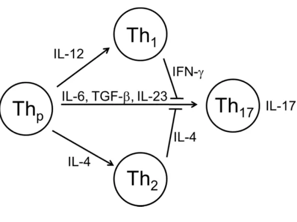
differentiation process requires a unique transcription factor, ROR-γt, to induce transcription of the IL-17 gene12. Additionally, cells genetically deficient in the master transcription factors required for Th1 and Th2 differentiation have either undiminished or enhanced Th17 development2. Together these findings indicate that Th17 cells are a lineage distinct from Th1 and Th2 cells. Furthermore, cytokines secreted by differentiated Th1 and Th2 cells inhibit formation of Th17 cells (Figure 2.1). Conversely, when naïve CD4+ T cells are
exposed to TGF-β in the absence of IL-6, they express forkhead box P3 (Foxp3), which is the master transcription factor that drives induction of Foxp3+ regulatory T cells (Treg)13.
Therefore, the presence of IL-6 switches the development of naïve CD4+ T cells from a Treg pathway to a Th17 pathway.
Severe burn injury causes a dangerous immune dysfunction. Much research effort has been expended into defining the immune consequence of burn in terms of lymphocyte
cytokine identity14-33. One model of burn injury suggests there is a rapid onset of a systemic inflammatory response characterized by the production of pro-inflammatory cytokines23, 34. If this pro-inflammatory state is uncontrolled, patients can experience early multiple organ dysfunction syndrome and death35. Patients surviving this period are then thought to develop a compensatory anti-inflammatory response characterized by immune suppression and decreased resistance to infection36.
We and others have defined CD4+ and CD8+ T cell population changes both early and late after burn in patients and animal models. After burn injury, there is an early (hours to days) pro-inflammatory response followed by a shift towards an anti-inflammatory
24
which are defining characteristics of Th1 cells. In addition, macrophages produce TNF-α, IL-1β, and IL-6 as well as other pro-inflammatory cytokines37. Furthermore, the level of TGF-β increases gradually in the burn wound throughout this phase36, 38. Following this phase after burn, the pro-inflammatory state of the immune system switches to an anti-inflammatory response characterized by over-production of TGF-β36, 38, as well as a switch in CD4+ T cell responses from a Th1 to a Th2 (IL-4) phenotype39. This manifests itself as decreased antigen-specific proliferation, diminished cytokine secretion and cytotoxic T lymphocyte activity18, 22, 40, 41
.
Since most patients develop immune failure days to weeks after injury, we have been interested in both CD8+ and CD4+ T cell function during the first two weeks after injury. We have previously demonstrated profound alterations in the cytokine profile late after burn injury. CD8+ T cells, crucial for anti-pathogen and anti-allograft immune responses, are impaired immediately post-burn but experience increased proliferation and have a unique and dramatic altered cytokine profile later after burn (14 days)18, 22, 40, 41. This functional
enhancement is characterized by a selective peripheral T cell lymphopenia that drives a homeostatic increase in “spontaneous” memory-like CD8+ and CD4+ T cells in the periphery14, 22. Since elevated TGF-β and IL-6 levels are found following bury injury, we hypothesize that CD4+ Th17 cells are generated after burn injury in a murine model of burn.
2.3 Methods and materials
Animals
25
Association of Laboratory Animal Care-accredited University of North Carolina Department of Laboratory Animal Medicine Facilities.
Mouse Burn Injury
Six to eight week old (15-20g weight) female B6 mice were used as subjects in all experiments. All protocols were performed in accordance with the National Institutes of Health guidelines and approved by the University of North Carolina IACUC as previously described15. Briefly, animals were anesthetized with inhalation of isoflurane vapor (Pitman-Moore, Washington Crossing, NJ) and their dorsal and flank hair clipped. A full-contact burn of approximately 20% total body surface area (TBSA) was produced by applying a copper rod, heated in boiling water, to the animal's dorsum and flank for 10 seconds. Four
applications of a 65g rod (1.9 cm in diameter) were used to produce the wound, and previous biopsies of the wounds demonstrate full-thickness cutaneous burn with visible unburned muscle beneath. Mice were resuscitated with an intraperitoneal (IP) injection of Lactated Ringer's solution (0.1 ml/g body weight) and were given a subcutaneous injection of buprenorphine (2 mg/kg body weight) for pain control immediately after burn injury.
26 CD4+ T cell purification
Cell suspensions were prepared from PLN of mice. CD4+ T cells were negatively selected by depletion of CD8+, MHC Class II+, CD11b+ and other cell types using the BD iMag Mouse CD4+ T Lymphocyte Enrichment Set according to the manufacturer’s instructions (Becton Dickinson, San Diego, CA). This method routinely provides us with greater than 90% pure CD4+ T cell populations.
T cell stimulation for cytokine analysis
Splenocytes, bulk PLN cells or purified CD4+ T cells from burn and sham mice (1 x 10⁶ cells/ml) cells were stimulated with PMA (1ug/ml; Simga Aldrich, St. Louis, MO) and ionomycin (1ug/ml; Sigma Aldrich, St. Louis, MO) for a total of 4 hours in 1ml of complete RPMI (10% fetal calf serum) in 24 well flat-bottom plates. Brefeldin A (3.0ug/ml;
eBioscience, San Deigo, CA) was added for the final 2 hours of culture to retain cytokines within the cell.
Th17 in vitro cell polarization
27 Flow cytometric analysis
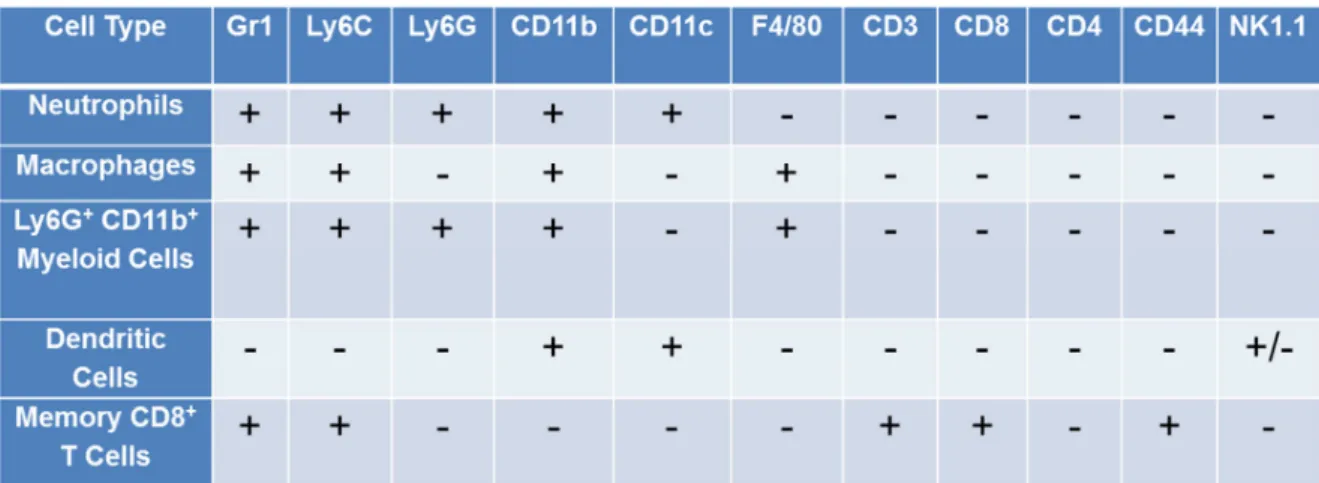
The panel of monoclonal antibodies used for flow cytometric analyses were anti-ROR-γt (AFKJS-9), anti-CD8α (53-6.7), anti-CD3ε (145-2C11), anti-CD4 (L3T4), anti-IFN-γ (XMG-1.2) (BD Pharmingen, San Diego, CA) and anti-IL17 (eBio17B7) (eBioscience, San
Diego, CA). Intracellular staining for cytokines and ROR-γt was performed using standard methods 15. Four and five-color analysis was performed using standard methods. List mode data were collected on a FACS Cyan (Dako, Ft. Collins, CO) and analyzed using Summit software (Dako, Ft. Collins, CO).
Statistical Analysis
Data were analyzed using Student's t-test for intracellular cytokine, absolute number and ratio differences. Statistical significance was defined as p<0.05 unless indicated
otherwise.
2.4 Results
Th17 cells exist in wound-draining lymph nodes but not in spleen following burn injury
Wound-28
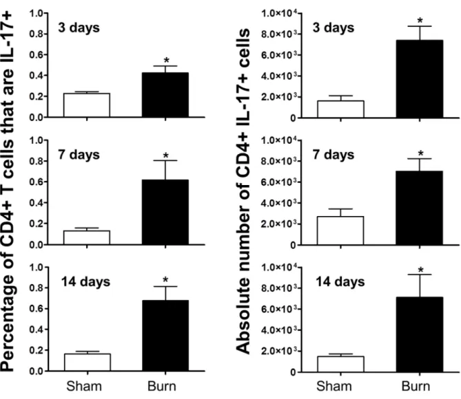
draining PLN have been previously shown to exhibit marked lymphocyte alterations after burn injury25, 42, 43. T cells were stimulated with mitogen, and standard intracellular cytokine staining employed to identify CD3+ CD4+ IL-17+ (Th17), CD3+ CD4+ IFN-γ+ (Th1), CD3+ CD8+ IL-17+ (Tc17) and CD3+ CD8+ IFN-γ+ (Tc1) cells using flow cytometry. At 3 days following burn or sham injury, we found an increased percentage of Th1 cells in the spleen (as reported previously15) and inguinal PLN but no increase in IL-17 producing CD4+ T cells (Figure 2.2). In contrast, there was a significant increase in the amount of Th17 cells found in the axillary PLN of the burn mice when compared to sham mice (Figure 2.3). We did not observe any expression of IL-17 by CD8+ T cells (data not shown). At 14 days post-burn and sham injury, we found a burn-dependent increase in the percentage of Th17 cells present in both the axillary and inguinal PLN but not the spleen (Figure 2.3). The percentage of Th1 cells in all sites studied was significantly lower at day 14 versus day 3, corresponding to the suppressed pro-inflammatory state observed late after burn injury. Again, we did not observe any expression of IL-17 by CD8+ T cells at day 14 (data not shown). At all timepoints and in all organs studied, we did not observe at dual positive CD3+ CD4+ IL-17+ IFN-γ+ T cells.
29
cells that were Th17 cells, as well as in the absolute number of Th17 cells, when compared to sham (Figure 2.4).
We also confirmed ROR-γt expression using an anti-ROR-γt antibody at these timepoints. ROR-γt staining was concurrent with IL-17 expression, but not with IFN- γ expression at day 7 post burn, further confirming these are Th17 cells (Figure 2.5). ROR-γt was also expressed in Th17 cells at day 14, as expected, but there was also significant
expression within Th1 cells. The plasticity of the Th1/Th17 lineage is an emerging concept in the current literature (12), and we are actively pursuing this as a mechanism for Th17
production within burn mice.
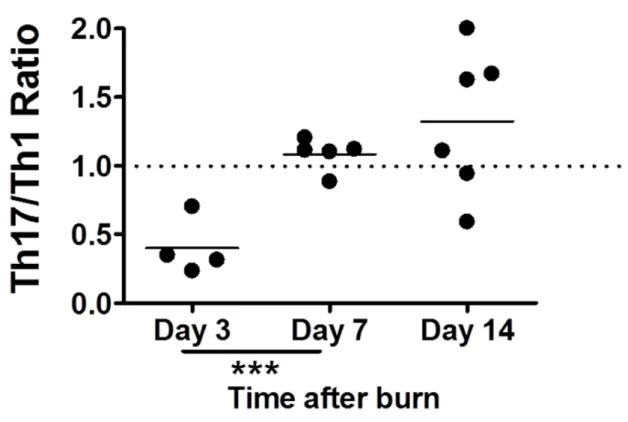
Burn injury induces a dynamic alteration in the Th1/Th17 balance
30
(Figure 2.6). These data suggest that although we observe an increase in Th17 cell number and percentage at all timepoints, there are increases of Th1 cells at day 3 and day 7 after burn, with day 3 having an overt burn-dependent Th1 response. We predict that day 14 represents a period after burn where the pro-inflammatory CD4+ T cell response is beginning to bias towards the weaker pro-inflammatory Th17 phenotype.
CD4+ T cells from burn and sham mice have a similar ability to polarize towards a Th17 phenotype
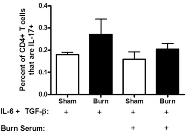
Various cytokines are known to polarize a pro-inflammatory response towards a Th17 phenotype, such as IL-6 and TGF-β. IL-23 is known to “fix” this phenotype in place. As a possible cellular mechanism for increased Th17 development, we assessed the ability of CD4+ T cells isolated from burn and sham mice to polarize in vitro towards a
Th17-phenotype. We also tested whether serum collected from burn mice was able to aid in the ability to polarize. Serum was collected and CD4+ T cells were purified from PLN 3 and 14 days following burn or sham injury. The CD4+ T cells were activated in vitro via T Cell Receptor (TCR) ligation with anti-CD3 antibody and anti-CD28 costimulation in presence of IL-6 and TGF-β for four days, which has been previously shown to promote Th17
differentiation2, 3. Sham and burn T cells were also stimulated in the presence or absence of burn serum. Th17 and Th1 CD4+ T cells were identified after each culture condition utilizing intracellular cytokine staining and flow cytometry. At both 3 and 14 days, CD4+ T cells from burn mice had an equivalent ability to polarize to Th17 cells when compared to sham in the presence of know polarizing conditions (Figure 2.7). We predicted that as the Th17
31
effect on Th17 polarization / stabilization of sham or burn CD4+ T cells (Figure 2.8), nor was there no significant difference between the response to serum alone and to any of the
treatments shown in Figure 2.8. These data suggest that burn injury has no inherent effect on the ability of CD4+ T cells to bias towards a Th17 phenotype under in vitro polarizing cytokine conditions. For example, serum from burn mice may not contain any soluble factors, such as IL-23, which affects Th17 polarization2, 9-11.
2.5 Discussion
Many variables contribute to the development, regulation, and effector functions of CD4+ Th1, Th17, and regulatory T cells. We hypothesized that burn injury would have a profound effect on the balance of Th1/Th17 T cells. The main reasoning behind this was that certain cytokines known to promote Th17 polarization are present after burn injury, namely IL-6 in the serum and TGF-β in the burn wound.
32
corresponding lymph nodes are approximately equidistant to the burn wound. However, it does appear that proximity to the wound corresponds to Th17 expansion (Figure 2.2).
To eliminate the possibility of underestimating the absolute number of Th17, due to likely downregulation of the CD4 co-receptor during T cell stimulation, we purified the CD4+ T cell population prior to culturing. In order to generate enough cells for this selection process, various wound-draining PLN were collected and pooled together for each mouse. With the addition of the CD4+ T cell-purification, we also saw a significant increase in the number of Th17 T cells in wound draining PLN across all timepoints compared to sham (Figure 2.4). While the percentage and cell number differences between burn and sham are statistically significant, the values are modest. This is likely due to the inefficiency and low sensitivity of intracellular cytokine staining at revealing polarized T cell subsets. We identified ROR-γt staining within these cells to confirm their phenotype.
While the proportion and absolute number of Th17 cells are important to study, the role of the Th1/Th17 balance has been postulated as being key for overall pro-inflammatory status in human and animal studies. Indeed, after burn we observed a dynamic shift in this ratio. To account for variability between intracellular cytokine staining between experiments, we defined the sham as having a Th1/Th17 ratio of 1.0 at each case so that data from
33
we observed meaning that Th1-promoting effects of burn over-ride the Th17-polarizing effects of IL-6 and TGF-β produced in response to burn injury.
These results suggest that day 3 represents a period of overt burn-dependent Th1 responses to antigen. Since IFN-γ is known to inhibit naïve CD4+ T cells development towards a Th17 pathway, this would likely impact the ability of the patient to recruit
neutrophils to sites of infections. Likewise, IL-12 is another cytokine driving differentiation into Th1 cells. While it has been shown that inducing a stronger Th1-response by IL-12 administration can protect against cecal ligation and puncture sepsis in mice45, such a response has not been specifically studied with respect to the role of Th17 cells and pathogens that require rapid neutrophil clearance (e.g. Pseudomonas, Klebsiella and Candida) mediated effectively by Th17 cells. Indeed, when burn mice are challenged with Pseudomonas aeruginosa, they experience high levels of mortality within 2 to 3 days following injury correlated with an overt pro-inflammatory response46. Therefore, driving differentiation at these early tim points towards a Th17 differentiation pathway could be a potential benefit for patients.
In contrast to the early pro-inflammatory Th1 phase, it appears that day 14 in our mouse model represents a period after burn where the pro-inflammatory CD4+ T cell
34
The mechanism for this Th17-dominance is currently unclear. The driving force for the Th1/Th2 cytokine imbalance at day 14 after burn injury appears to be intense homeostatic expansion of “spontaneous” memory-like CD8+ and CD4+ T cells as a consequence of lymphopenia early after burn injury. Th17 cells, also often defined as possessing a memory-like phenotype, have not yet been studied as being a consequence of homeostatic
proliferation. Another potential mechanism is that Th17 polarization can be driven in vitro and in vivo by innate stimulation through Toll-like Receptors. (TLR)47. We have shown altered Toll-like Receptor (TLR) expression on innate cells (e.g. macrophages express lower TLR) and adaptive cells (T cells express higher levels of TLR) late after burn injury48, 49. Therefore, we predict that T cells are stimulated directly or indirectly via innate cells to various innate stimuli produced by burn injury, such as endogenous innate signaling molecules50-58 released from the burn wound. While the results suggest that burn serum did not affect Th17 polarization of sham CD4+ T cells during in vitro stimulation under TGF-β and IL-6 conditions, the serum levels of such endogenous innate signaling molecules might not be high enough to detect any differences. Experiments to test the role of innate
stimulation after burn in affecting adaptive T cell responses are underway.
Attempts to improve the T cell response to burn injury by manipulating cytokines has been largely been unsuccessful and are not generally employed in the clinic. Our data
suggests why cytokine manipulation may be unsuccessful. For example, the CD4+ T cell phenotype after burn injury is dynamic and may already be set along a certain
35
36
37
38
39
40
41
Figure 2.6 Th17/Th1 ratio is dynamic following burn injury. Cells were harvested from various peripheral lymph nodes at 3, 7 or 14 days following a 20% total burn surface area, full-thickness burn or sham injury. Negative magnetic selection was performed to enrich the CD4+ T cell population. CD4+ cells were stimulated in vitro before intracellular cytokine staining was performed, and flow cytometry used to quantify IL-17+ CD4+ T cells. For each timepoint, the average sham Th17/Th1 ratio was set at 1.0. For each burn mouse, its
42
Figure 2.7. CD4+ T cells from burn and sham mice have similar abilities to polarize to Th17 cells. Cells were harvested from various peripheral lymph nodes at 3 or 14 days following a 20% total burn surface area, full-thickness burn or sham injury. CD4+ T cells were enriched by negative magnetic selection and cultured with plate bound anti-CD3, soluble anti-CD28, IL-6, and TGF-β for 4 days. IL-17 and IFN-γ production by CD3+ CD4+ cells was analyzed using flow cytometric staining. Representative examples of the
43
44 2.6 References
1. Mosmann, T.R., Cherwinski, H., Bond, M.W., Giedlin, M.A. & Coffman, R.L. Two types of murine helper T cell clone. I. Definition according to profiles of lymphokine activities and secreted proteins. J Immunol 136, 2348-2357 (1986).
2. Harrington, L.E. et al. Interleukin 17-producing CD4+ effector T cells develop via a lineage distinct from the T helper type 1 and 2 lineages. Nat Immunol 6, 1123-1132 (2005). 3. Park, H. et al. A distinct lineage of CD4 T cells regulates tissue inflammation by producing interleukin 17. Nat Immunol 6, 1133-1141 (2005).
4. Miossec, P., Korn, T. & Kuchroo, V.K. Interleukin-17 and type 17 helper T cells. N Engl J Med 361, 888-898 (2009).
5. Dragon, S., Saffar, A.S., Shan, L. & Gounni, A.S. IL-17 attenuates the anti-apoptotic effects of GM-CSF in human neutrophils. Mol Immunol 45, 160-168 (2008).
6. Ye, P. et al. Requirement of interleukin 17 receptor signaling for lung CXC
chemokine and granulocyte colony-stimulating factor expression, neutrophil recruitment, and host defense. J Exp Med 194, 519-527 (2001).
7. Ye, P. et al. Interleukin-17 and lung host defense against Klebsiella pneumoniae infection. Am J Respir Cell Mol Biol 25, 335-340 (2001).
8. Huang, W., Na, L., Fidel, P.L. & Schwarzenberger, P. Requirement of interleukin-17A for systemic anti-Candida albicans host defense in mice. J Infect Dis 190, 624-631 (2004).
9. Bettelli, E. et al. Reciprocal developmental pathways for the generation of pathogenic effector TH17 and regulatory T cells. Nature 441, 235-238 (2006).
10. Mangan, P.R. et al. Transforming growth factor-beta induces development of the T(H)17 lineage. Nature 441, 231-234 (2006).
11. Veldhoen, M., Hocking, R.J., Atkins, C.J., Locksley, R.M. & Stockinger, B. TGFbeta in the context of an inflammatory cytokine milieu supports de novo differentiation of IL-17-producing T cells. Immunity 24, 179-189 (2006).
12. Ivanov, II et al. The orphan nuclear receptor RORgammat directs the differentiation program of proinflammatory IL-17+ T helper cells. Cell 126, 1121-1133 (2006).
45
14. Buchanan, I., Maile, R., Frelinger, J., Meyer, A.A. & Cairns, B.A. The effect of burn injury on CD8+ and CD4+ T cells in an irradiation model of homeostatic proliferation. J Trauma (2006).
15. Cairns, B.A. et al. CD8 T cells express a T-helper 1-like phenotype after burn injury. Surgery 130, 210-216. (2001).
16. Cho, K., Adamson, L.K., Park, J. & Greenhalgh, D.G. Burn injury-mediated
alterations in cell cycle progression in lymphoid organs of mice. Shock 19, 138-143 (2003). 17. Guo, Z. et al. Burn injury promotes antigen-driven Th2-type responses in vivo. J Immunol 171, 3983-3990 (2003).
18. Hultman, C.S., Cairns, B.A., deSerres, S., Frelinger, J.A. & Meyer, A.A. Burn injury impairs second-set rejection and CTL reactivity in mice primed by cultured keratinocyte allografts. Transplantation 60, 584-589 (1995).
19. Kavanagh, E.G. et al. Burn injury primes naive CD4+ T cells for an augmented T-helper 1 response. Surgery 124, 269-276; discussion 276-267 (1998).
20. Kell, M.R., Kavanaugh, E.G., Goebel, A., Soberg, C.C. & Lederer, J.A. Injury primes the immune system for an enhanced and lethal T-cell response against bacterial superantigen. Shock 12, 139-144 (1999).
21. Maekawa, T., Kajihara, H., Okabayashi, K., Otani, M. & Yuge, O. Impairment of splenic B and T lymphocytes in the early period after severe thermal injury:
immunohistochemical and electron microscopic analysis. Burns 28, 329-339 (2002).
22. Maile, R. et al. Lymphopenia-Induced Homeostatic Proliferation of CD8+ T Cells Is a Mechanism for Effective Allogeneic Skin Graft Rejection following Burn Injury. Journal of Immunology 176, 6717-6726 (2006).
23. Mannick, J.A., Rodrick, M.L. & Lederer, J.A. The immunologic response to injury. J Am Coll Surg 193, 237-244 (2001).
24. Murphy, T.J., Choileain, N.N., Zang, Y., Mannick, J.A. & Lederer, J.A. CD4+CD25+ regulatory T cells control innate immune reactivity after injury. J Immunol 174, 2957-2963 (2005).
25. Organ, B.C. et al. Changes in lymphocyte number and phenotype in seven lymphoid compartments after thermal injury. Ann Surg 210, 78-89 (1989).
26. O'Sullivan, S.T. et al. Major injury leads to predominance of the T helper-2
46
27. Patenaude, J., D'Elia, M., Hamelin, C., Garrel, D. & Bernier, J. Burn injury induces a change in T cell homeostasis affecting preferentially CD4+ T cells. J Leukoc Biol (2004). 28. Schwacha, M.G. Macrophages and post-burn immune dysfunction. Burns 29, 1-14 (2003).
29. Teodorczyk-Injeyan, J.A., Cembrzynska-Nowak, M., Lalani, S., Peters, W.J. & Mills, G.B. Immune deficiency following thermal trauma is associated with apoptotic cell death. J Clin Immunol 15, 318-328 (1995).
30. Teodorczyk-Injeyan, J.A., Sparkes, B.G., Mills, G.B. & Peters, W.J.
Immunosuppression follows systemic T lymphocyte activation in the burn patient. Clin Exp Immunol 85, 515-518 (1991).
31. Zang, Y. et al. Burn injury initiates a shift in superantigen-induced T cell responses and host survival. J Immunol 172, 4883-4892 (2004).
32. Zedler, S., Bone, R.C., Baue, A.E., von Donnersmarck, G.H. & Faist, E. T-cell reactivity and its predictive role in immunosuppression after burns. Crit Care Med 27, 66-72 (1999).
33. Zedler, S., Faist, E., Ostermeier, B., von Donnersmarck, G.H. & Schildberg, F.W. Postburn constitutional changes in T-cell reactivity occur in CD8+ rather than in CD4+ cells. J Trauma 42, 872-880; discussion 880-871 (1997).
34. Kelly, J.L. et al. Is circulating endotoxin the trigger for the systemic inflammatory response syndrome seen after injury? Ann Surg 225, 530-541; discussion 541-533 (1997). 35. Deitch, E.A. Multiple organ failure. Pathophysiology and potential future therapy. Ann Surg 216, 117-134 (1992).
36. Al-Qattan, M.M. 'Late' multiorgan failure in major burns: a "three-event" construct rather than a "two-event" construct. Burns 33, 268-270 (2007).
37. Ogle, C.K., Mao, J.X., Wu, J.Z., Ogle, J.D. & Alexander, J.W. The 1994 Lindberg Award. The production of tumor necrosis factor, interleukin-1, interleukin-6, and
prostaglandin E2 by isolated enterocytes and gut macrophages: effect of lipopolysaccharide and thermal injury. J Burn Care Rehabil 15, 470-477 (1994).
38. Wei, D., Ge, S., Chen, Y., Dai, F. & Su, B. Expression of endogenous transforming growth factor-beta and its type I and type II receptors in rat burn wounds. Wound Repair Regen 5, 229-234 (1997).
47
40. Hultman, C.S., Cairns, B.A., deSerres, S., Frelinger, J.A. & Meyer, A.A. Early, complete burn wound excision partially restores cytotoxic T lymphocyte function. Surgery 118, 421-429; discussion 429-430 (1995).
41. Hultman, C.S. et al. The 1995 Moyer Award. The effect of burn injury on allograft rejection, alloantigen processing, and cytotoxic T-lymphocyte sensitization. J Burn Care Rehabil 16, 573-580 (1995).
42. Choileain, N.N. et al. Enhanced Regulatory T Cell Activity Is an Element of the Host Response to Injury. J Immunol 176, 225-236 (2006).
43. Kataranovski, M. et al. Post-traumatic activation of draining lymph node cells. II. Proliferative and phenotypic characteristics. Burns 20, 403-408 (1994).
44. Richie, E.R., McEntire, B., Phillips, J. & Allison, J.P. Altered expression of
lymphocyte differentiation antigens on phorbol ester-activated CD4+8+ T cells. J Immunol 140, 4115-4122 (1988).
45. O'Suilleabhain, C. et al. Interleukin-12 treatment restores normal resistance to bacterial challenge after burn injury. Surgery 120, 290-296 (1996).
46. Murphey, E.D. & Sherwood, E.R. Bacterial clearance and mortality are not improved by a combination of IL-10 neutralization and IFN-gamma administration in a murine model of post-CLP immunosuppression. Shock 26, 417-424 (2006).
47. Jyonouchi, H., Geng, L., Cushing-Ruby, A. & Monteiro, I.M. Aberrant responses to TLR agonists in pediatric IBD patients; the possible association with increased production of Th1/Th17 cytokines in response to candida, a luminal antigen. Pediatr Allergy Immunol (2009).
48. Cairns, B., Maile, R., Barnes, C.M., Frelinger, J.A. & Meyer, A.A. Increased Toll-like receptor 4 expression on T cells may be a mechanism for enhanced T cell response late after burn injury. J Trauma 61, 293-298; discussion 298-299 (2006).
49. Cairns, B.A., Barnes, C.M., Mlot, S., Meyer, A.A. & Maile, R. Toll-like receptor 2 and 4 ligation results in complex altered cytokine profiles early and late after burn injury. J Trauma 64, 1069-1077; discussion 1077-1068 (2008).
50. Anders, H.J., Banas, B. & Schlondorff, D. Signaling danger: toll-like receptors and their potential roles in kidney disease. J Am Soc Nephrol 15, 854-867 (2004).
51. Dinarello, C.A. Infection, fever, and exogenous and endogenous pyrogens: some concepts have changed. J Endotoxin Res 10, 201-222 (2004).
48
53. Iliev, A.I., Stringaris, A.K., Nau, R. & Neumann, H. Neuronal injury mediated via stimulation of microglial toll-like receptor-9 (TLR9). Faseb J 18, 412-414 (2004).
54. Johnson, G.B., Brunn, G.J., Kodaira, Y. & Platt, J.L. Receptor-mediated monitoring of tissue well-being via detection of soluble heparan sulfate by Toll-like receptor 4. J Immunol 168, 5233-5239 (2002).
55. Johnson, G.B., Brunn, G.J. & Platt, J.L. Cutting edge: an endogenous pathway to systemic inflammatory response syndrome (SIRS)-like reactions through Toll-like receptor 4. J Immunol 172, 20-24 (2004).
56. Kirschning, C.J. & Schumann, R.R. TLR2: cellular sensor for microbial and endogenous molecular patterns. Curr Top Microbiol Immunol 270, 121-144 (2002).
57. Marshak-Rothstein, A., Busconi, L., Rifkin, I.R. & Viglianti, G.A. The stimulation of Toll-like receptors by nuclear antigens: a link between apoptosis and autoimmunity. Rheum Dis Clin North Am 30, 559-574, ix (2004).
49 CHAPTER 3
Flagellin treatment prevents increased susceptibility to systemic bacterial infection after injury by inhibiting IL-10+ IL-12- neutrophil polarization
3.1 Summary
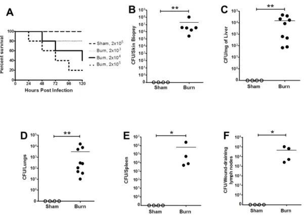
Severe trauma renders patients susceptible to infection. In sepsis, defective bacterial clearance has been linked to specific deviations in the innate immune response. We
hypothesized that the innate immune modulations observed during sepsis also contribute to increased bacterial susceptibility after severe trauma. A well-established murine model of burn injury, which was used to replicate infection following trauma, showed that wound inoculation with Pseudomonas aeruginosa quickly spreads systemically. This correlated with apoptosis of dendritic cells and memory CD8+ T cells, as well as differential Toll-like
receptor expression on a variety of innate immune cells. The systemic IL-10/IL-12 axis was also skewed after burn injury with infection as indicated by a significant elevation in serum IL-10 and polarization of neutrophils into an anti-inflammatory (N2; IL-10+ IL-12-)
phenotype. Infection with an attenuated P. aeruginosa strain (∆cyaB) was cleared more efficiently than the wildtype strain and was associated with an increased pro-inflammatory neutrophil (N1; IL-10-IL-12+) response in burn mice. This suggests that neutrophil
50
findings, for the first time, detail specific alterations in innate cell populations after burn injury that contribute to increased susceptibility to bacterial infection. In addition, it identifies neutrophil polarization as a therapeutic target for the reversal of bacterial susceptibility after injury.
3.2 Introduction
Each year traumatic injury accounts for over 40 million emergency room visits and 2 million hospital admissions across the United States1. Severe trauma predisposes patients to infection resulting in an overall infection rate of 37%2. Infectious complications, such as sepsis and pneumonia, increase the length of hospitalization and cost of treatment 3, 4. Furthermore, infection increases the mortality rate of trauma patients by 5-fold5.