Research Paper
Predicting the influence of liposomal lipid composition on liposome size,
zeta potential and liposome-induced dendritic cell maturation using a
design of experiments approach
Peter C. Soema
a,⇑, Geert-Jan Willems
a, Wim Jiskoot
b, Jean-Pierre Amorij
a, Gideon F. Kersten
a,b aIntravacc (Institute for Translational Vaccinology), Bilthoven, The Netherlands
b
Division of Drug Delivery Technology, Leiden Academic Centre for Drug Research, Leiden University, Leiden, The Netherlands
a r t i c l e
i n f o
Article history:
Received 17 May 2015 Revised 25 June 2015
Accepted in revised form 29 June 2015 Available online 2 July 2015
Keywords:
Liposomes Cationic lipids Design of experiments Prediction models Dendritic cells Maturation markers
a b s t r a c t
In this study, the effect of liposomal lipid composition on the physicochemical characteristics and adju-vanticity of liposomes was investigated. Using a design of experiments (DoE) approach, peptide-containing liposomes containing various lipids (EPC, DOPE, DOTAP and DC-Chol) and peptide concentrations were formulated. Liposome size and zeta potential were determined for each formulation. Moreover, the adjuvanticity of the liposomes was assessed in anin vitrodendritic cell (DC) model, by quantifying the expression of DC maturation markers CD40, CD80, CD83 and CD86. The acquired data of these liposome characteristics were successfully fitted with regression models, and response contour plots were generated for each response factor. These models were applied to predict a lipid composition that resulted in a liposome with a target zeta potential. Subsequently, the expression of the DC matura-tion factors for this lipid composimatura-tion was predicted and tested in vitro; the acquired maturation responses corresponded well with the predicted ones. These results show that a DoE approach can be used to screen various lipids and lipid compositions, and to predict their impact on liposome size, charge and adjuvanticity. Using such an approach may accelerate the formulation development of liposomal vaccine adjuvants.
Ó2015 The Authors. Published by Elsevier B.V. This is an open access article under the CC BY-NC-ND license (http://creativecommons.org/licenses/by-nc-nd/4.0/).
1. Introduction
Many vaccines are based on purified or synthetic antigens derived from their respective pathogens. These include antigens, such as peptides and proteins, which are poorly immunogenic on their own. Adjuvants, based on delivery systems and/or immunopotentiators, are used frequently to improve the immuno-genicity of antigens[1]. Liposomes are important delivery systems for vaccines because of their high versatility, which enables them to be suited for many types of antigens[2].
Numerous lipid compositions and preparation methods for lipo-somes can be chosen, which affect several liposomal characteristics, such as size, zeta potential, bilayer fluidity and encapsulation or association of antigen or adjuvant. In turn, these characteristics can influence the adjuvant effect of liposomes[3]. The adjuvanticity of liposomes is attributed to several mechanisms, such as antigen
depot formation, induction of local inflammation and increased antigen uptake by antigen presenting cells.
Antigen presenting cells, with dendritic cells (DCs) in particular, play a pivotal role in the induction of adaptive immune responses. DCs recognize, internalize and process antigens, and ultimately present them to naïve CD4+or CD8+T cells[4]. The uptake of anti-gens by DCs is affected by several antigen characteristics, of which size and surface charge are the most influential. Generally, the size of most subunit antigens is too small for the DC to be taken up effi-ciently. Incorporation of an antigen into a particulate delivery sys-tem such as a liposome, whose size is comparable to that of a virus particle, can therefore significantly increase antigen uptake by DCs through endocytosis[5].
The surface charge density of a liposome influences its zeta potential, and thereby its electrostatic interaction with the surface of a DC. Since cellular membranes are anionic, cationic liposomes are ideally suited to increase antigen uptake by DCs[6]. It is gener-ally accepted that anionic and neutral liposomes are less suited for the induction of immune responses[7]. The cationic liposome for-mulation CAF01 is currently advancing through clinical trials in
http://dx.doi.org/10.1016/j.ejpb.2015.06.026
0939-6411/Ó2015 The Authors. Published by Elsevier B.V.
This is an open access article under the CC BY-NC-ND license (http://creativecommons.org/licenses/by-nc-nd/4.0/). ⇑ Corresponding author at: Antonie van Leeuwenhoeklaan 9, 3721 MA Bilthoven,
The Netherlands.
E-mail address:[email protected](P.C. Soema).
Contents lists available atScienceDirect
European Journal of Pharmaceutics and Biopharmaceutics
combination with HIV and tuberculosis antigens, indicating the potency of cationic liposomes[8,9].
For the successful priming of naïve B- or T cells by DCs, more is needed than efficient antigen uptake and processing. During antigen presentation by the DCs to naïve lymphocytes, costimulatory signals are required. These are provided by the DCs, which can express cos-timulatory molecules such as CD40 (for B cells), CD80 and CD86 (for T cells) after maturation[10]. The maturation of DCs is considered to be of vital importance for the overall immunogenicity of a vaccine antigen[11].In vitroDC maturation models can therefore be used as preclinical screening tools for vaccine formulations[12].
Immunostimulatory signals, which are often provided by pathogen-associated molecular patterns (PAMPs), are required for the activation of DCs. Inclusion of PAMPs such as Toll-like receptor (TLR) ligands or other molecules in liposomes is therefore a popular strategy to increase liposome adjuvanticity [13,14]. Cationic lipids also seem to affect DC maturation [15]. Besides the positive charge, other physical characteristics, such as lipid bilayer fluidity, may affect DC maturation [16]. Chemical differ-ences between cationic lipids indeed have shown to affect DC mat-uration, underlining the significance of the lipid composition of cationic liposomes.
Design of experiments (DoE) is a statistical method to screen, identify and optimize important factors in various processes, such
as pharmaceutical formulation development[17,18]. It uses a min-imal number of experiments to model the effects of each formula-tion parameter, which significantly accelerates the identificaformula-tion of optimal conditions. A DoE approach was recently employed to optimize the formulation process of itraconazole-loaded liposomes
[19]. The authors were able to predict drug loading with a mathe-matical model obtained with DoE, and identify critical formulation parameters affecting drug loading. However, no attempts have been made yet to predict biological parameters, such as the adju-vanticity of liposomes, with DoE-like approaches.
In this study, the effects of liposomal lipid composition and pep-tide incorporation on the physicochemical characteristics and the adjuvanticity of liposomes were studied. To gain insight into the effects of each component with a minimal number of experiments, a DoE approach was used. The physicochemical characteristics of the liposomes were determined as the liposome size and zeta potential, while the liposome adjuvanticity was determined as liposome-induced in vitro expression of DC maturation factors CD40, CD80, CD83 and CD86. To this end, four lipids, i.e., egg-phosphatidylcholine (EPC), 1,2-dioleoyl-sn-glycero-3-pho sphoethanolamine (DOPE), 1,2-dioleoyl-3-trimethylammonium-propane (DOTAP) and 3ß-[N-(N0,N0 -dimethylaminoethane)-carbamoyl]cholesterol (DC-Chol), and the HLA-A2.1-restricted influenza peptide GILGFVFTL (M158-66), were used to generate
peptide-loaded liposomes with different lipid compositions. Liposome size and zeta potential were determined for each formu-lation, and prediction models for these parameters were generated by using a DoE approach. Simultaneously, the ability of these liposomes to maturate DCs was evaluated by determining the expression of DC maturation markers CD40, CD80, CD83 and CD86. With DoE, the most influential lipids were identified, and prediction models were generated for each maturation marker. Finally, the prediction models were validated by selecting a lipo-some with a previously untested lipid composition. A complete overview of the study is depicted inFig. 1.
2. Materials and methods
2.1. Reagents
The influenza peptide GILGFVFTL (M158-66) was synthesized at
the Dutch Cancer Institute (NKI). All lipids (EPC, DOPE, DOTAP, DC-Chol) were purchased from Avanti Polar Lipids, bovine serum albumin (BSA), ethylenediaminetetraacetic acid (EDTA), 4-(2-hyd roxyethyl)-1-piperazineethanesulfonic acid (HEPES) and sodium chloride from Sigma–Aldrich, Iscove’s Modified Dulbecco’s Medium (IMDM) from Invitrogen, human granulocyte macrophage colony-stimulating factor (GM-CSF) from Peprotech, human IL-4 from Sanquin, anti-human CD40-PE and CD80-FITC from BD Pharmingen, anti-human CD83-APC and CD86-Pacific Blue from Biolegend, phosphate-buffered saline (PBS; 155 mM NaCl, 1.5 mM potassium phosphate monobasic, 2.7 mM sodium phos-phate dibasic, pH 7.2) and live/dead-Aqua from Life Technologies, lipopolysaccharide (LPS)E. coliK12 from Invivogen and Hyclone fetal calf serum (FCS) from Thermo Scientific.
2.2. Experimental design
To investigate the effect of the liposome composition on lipo-some size, zeta potential and lipolipo-some-induced DC maturation, a linear mixture model was selected with MODDE 10 (Umetrics) software. Boundaries for EPC, DOPE, DOTAP and DC-Chol fractions were set at 0 and 1 (with 1 being 100% of total lipid content). GILGFVFTL peptide content was set between 10 and 100
l
g/mL. A D-optimal design was selected, which was composed of 18 runs, including a quadruple center point [20]. After the runs were completed, models for liposome zeta potential and DC maturation factors CD40, CD80, CD83 and CD86 were created with apartial-least square (PLS) regression. Data were log-transformed, if needed, and non-significant factors were removed from the model untilR2(model fit) andQ2(model prediction power) were
optimal.
2.3. Liposome formulation
Lipids were admixed (ratios according to the experimental design) to a total amount of 7.5
l
mol in 10 ml chloroform. The lipid mixture was transferred to a 50 mL round bottom flask, and the chloroform was evaporated under reduced pressure at 40°C with a rotary evaporator (Buchi Rotavapor R-3). The obtained lipid film was subsequently rehydrated for 2 h, room temperature atTable 1
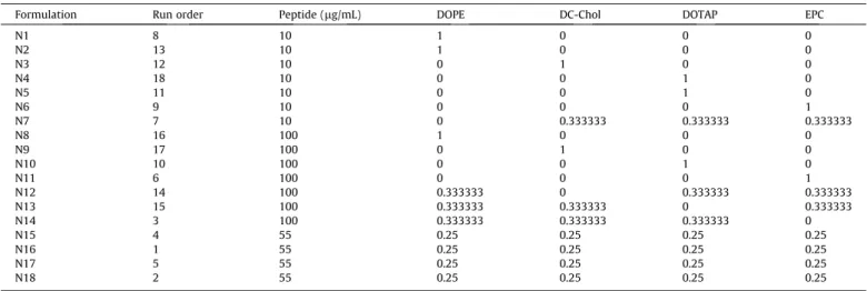
Design of experiments worksheet. Lipids (DOPE, DC-Chol, DOTAP, EPC) are presented as fraction of total lipid content (1 = 100%).
Formulation Run order Peptide (lg/mL) DOPE DC-Chol DOTAP EPC
N1 8 10 1 0 0 0
N2 13 10 1 0 0 0
N3 12 10 0 1 0 0
N4 18 10 0 0 1 0
N5 11 10 0 0 1 0
N6 9 10 0 0 0 1
N7 7 10 0 0.333333 0.333333 0.333333
N8 16 100 1 0 0 0
N9 17 100 0 1 0 0
N10 10 100 0 0 1 0
N11 6 100 0 0 0 1
N12 14 100 0.333333 0 0.333333 0.333333
N13 15 100 0.333333 0.333333 0 0.333333
N14 3 100 0.333333 0.333333 0.333333 0
N15 4 55 0.25 0.25 0.25 0.25
N16 1 55 0.25 0.25 0.25 0.25
N17 5 55 0.25 0.25 0.25 0.25
N18 2 55 0.25 0.25 0.25 0.25
250 rpm with a shaker (Edmund Bühler Swip KS-10) after addition of GILGFVFTL peptide (concentrations according to experimental design) dissolved in 1.5 mL buffer (10 mM HEPES, 100 mM NaCl, pH 7.4). After rehydration, crude liposomes were extruded five times through a 0.2-
l
m Nucleopore Track-Etch membrane (Whatman) with a 10-mL Lipex extruder (Northern Lipids Inc.). Each liposome formulation from the experimental design was made in duplicate.2.4. Characterization of liposomes
Liposome size and polydispersity index (PDI) were determined by dynamic light scattering (DLS) using a Nanosizer ZS (Malvern Instruments). The zeta potential of the liposomes was determined after a 5 fold dilution in MilliQ water by laser Doppler velocimetry using a Nanosizer ZS with a universal dip cell (Malvern Instruments).
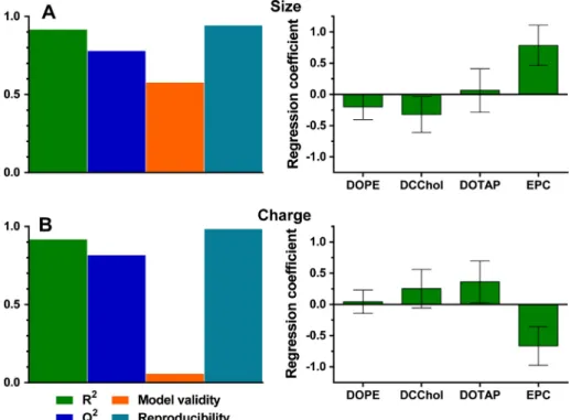
Fig. 3.Regression models for liposome size (A) and zeta potential (B). On the left summaries of fit for the models are displayed. Model fit (R2
, >0.5 indicates a good model fit to the data), prediction power (Q2, >0.5 indicates sufficient prediction power), model validity (>0.25 indicates that the model error is smaller than the experimental error) and reproducibility (>0.5 indicates a small experimental error) are shown. On the right, normalized model regression coefficients are displayed. Coefficients with a 95% confidence interval that does not cross zero are significant terms.
2.5. Maturation of human dendritic cells
Human CD14+monocytes were isolated from fresh donor blood
as described previously[21]. Monocytes were plated at a concen-tration of 0.4⁄106cells/mL in 24-wells plates in IMDM medium
containing 1% FCS, 500 U/mL GM-CSF and 800 U/mL IL-4. Monocytes were differentiated to immature dendritic cells (iDCs) after 6 days. iDCs were subsequently stimulated with either med-ium, LPS or liposomes in duplicate. After 24 h incubation, cells were transferred to a 96-wells plate and washed twice with FACS buffer (PBS, pH 7.2, 0.5% BSA, 0.5 mM EDTA). DCs were stained with anti-human CD40, CD80, CD83, CD86 and live/dead staining for 30 min, and subsequently washed twice with FACS buffer. Samples were measured on a FACS Canto II flow cytometer (BD). Data were analyzed by using FlowJo 10 software for Mac OSX (Tree Star Inc.). Surface markers are reported as % of mean fluores-cent intensity (MFI) relative to that induced by LPS.
3. Results
3.1. Liposome characteristics
A linear mixture model was selected to screen the effects of the lipids EPC, DOPE, DC-Chol and DOTAP on the size distribution and zeta potential of the liposomes and their ability to induce DC mat-uration. A D-optimal design was chosen, which generated a work-sheet with 18 formulations (Table 1). Physical characteristics of the liposomes, i.e., size, PDI and zeta potential, were determined. Formulations N1, N2 and N8, which all contained DOPE as the only lipid, did not yield liposomes and were excluded from further experiments. Liposome sizes ranged from 150 to 194 nm, with an average of 170 nm (Fig. 2A) and a low PDI (<0.2), indicating that the liposomes were relatively monodisperse. As expected, the zeta potential of the liposomes containing a cationic lipid (DOTAP and/or DC-Chol) was positive, whereas formulations lacking a cationic lipid (N6, N11) showed a zeta potential close to zero (Fig. 2B).
Based on the experimental results, PLS regression models were fitted for both liposome size and zeta potential data using MODDE software. These regression models allowed the identification and qualification of input parameters (being peptide, DOPE, DC-Chol, DOTAP and EPC) which significantly contributed to the output parameters (size and zeta potential). The model regression coeffi-cients reflect the influence of the particular input parameter on the response of the output parameter. Valid models were obtained for both output parameters (Fig. 3). Liposome size was influenced the most by EPC (Fig. 3A), which increased liposome size when pre-sent in high amounts. Model validity for the zeta potential model was low (avalue > 0.25 indicates a good model fit), which is likely
a model artifact caused by the high reproducibility [20]. DOTAP and EPC were the most significant model terms, with DOTAP increasing the zeta potential, and EPC decreasing it (Fig. 3B). The incorporation of the peptide antigen had no influence on both lipo-some size and zeta potential, and was thus removed as a model term.
Response contour surface plots were generated for both liposome size and zeta potential after the fitting of the regression models (Fig. 4). These surface plots visualize the predicted value of a response factor according to the corresponding lipid composi-tion at that specific position in the plot (due to the two-dimensional nature of these plots and the multi-dimensional nature of the regression models, one input parameter is kept con-stant). As expected, both cationic lipids (DOTAP and DC-Chol) increased to zeta potential of the liposomes, whereas the zwitteri-onic lipids (EPC and DOPE) decreased it.
3.2. DC maturation by liposomes
The effect of the liposomal lipid composition on DC maturation was evaluated by measuring four DC maturation markers (CD40, CD80, CD83 and CD86) on maturated DCs 24 h after stimulation with the liposome formulations from the experimental design. The formulations were tested in duplicate on immature DCs iso-lated from two different donors (donors 1 and 2). LPS was taken as a positive control and reference sample in both experiments. The expression of CD40, CD80, CD83 and CD86 by DCs (derived from donor 1) after stimulation with the liposomes is presented inFig. 5.
Datasets from both experiments were fitted with PLS regression models per individual maturation marker, and the resulting mod-els and their coefficients are summarized in Fig. 6 (maturation experiment on DCs derived from donor 1) and Supplementary
Fig. S1 (maturation experiment on DCs derived from donor 2).
While the resulting models differed between experiments (most likely due to donor variability), the models showed similar trends. Since this study concerned a proof-of-principle, we opted to inves-tigate the models obtained with DCs derived from donor 1 in more detail, since overall fit of the models from donor 1 were better than those of donor 2.
For both CD40 and CD80 responses models were yielded with a high model fit, validity and reproducibility. Formulation N14 was statistically found to be an outlier, and was subsequently removed from all models. For both CD40 and CD80, the DOTAP lipid was found to be the most significant model term, indicating that the presence of DOTAP in the liposomes induces CD40 and CD80 expression by DCs. The model for CD83 had a relatively low model validity, which again might be a model artifact caused by the high reproducibility. The two cationic lipids, DC-Chol and DOTAP, were
the most significant model terms for CD83. The model for CD86 was valid, but suffered overall from a relatively low model fit, pre-dictability and reproducibility. This was confirmed with the model coefficients, which all have a non-significant contribution to the CD86 response, indicating that no single lipid had a great effect. Similar to the models for liposome size and zeta potential, the pep-tide content was a non-significant model term in all the models for the maturation markers. The response contour surface plots of all four maturation markers are displayed inFig. 7. From these figures it can be clearly seen that in general, a high fraction of DOTAP and
to a lesser extent DC-Chol, has a positive effect on the expression of all maturation markers. Furthermore, the inclusion of DOPE gener-ally had a negative effect on the maturation. The lipid EPC was non-influential for most responses, and is therefore still suited as a helper lipid to produce stable liposomes.
3.3. Prediction power of obtained models
As described previously, valid prediction models were obtained for liposomal size, zeta potential and all four DC maturation
markers. The prediction power of the models was tested by select-ing a liposomal formulation that was not yet included in the D-optimal design. For proof-of-principle purposes, an initial target response factor was set for the liposome formulation. In this case, a
target liposomal zeta potential was set. Since most formulations in the experimental design showed a zeta potential of either above 60 mV or 0 mV, a target zeta potential of 30 mV was chosen. The zeta potential prediction model subsequently gave a lipid compo-sition (Table 2) which should yield liposomes with a zeta potential of 30 mV. The liposome formulation (N19) was made, and size and zeta potential were determined (Table 3), which indeed correlated with the predicted values. Subsequently, the selected lipid compo-sition could now be used as an input for the previously acquired prediction models for liposomal adjuvanticity.
Next, liposome formulation N19 was added to immature DCs, and DC maturation markers were determined. The experimentally
Fig. 7.Response contour plots for DC maturation markers induced by liposomes. Lipid amounts are displayed as a fraction of 100% total lipid. The fraction of the least influential lipid was set at a constant fraction of 0.25. The values in the boxes and associated color regions represent the predicted response (either CD40, CD80, CD83 or CD86, all in % of LPS-induced expression) for that particular lipid composition.
Table 2
Lipid composition of a liposome formulation predicted to have a zeta potential of 30 mV by the zeta potential model.
Formulation Peptide (lg/mL) DOPE DC-Chol DOTAP EPC
acquired data were similar to the predicted means (Table 3), indi-cating that the predictions made by the models were accurate.
4. Discussion
From a historical perspective, most researchers are inclined to vary one factor at a time (OFAT) when systematically screening or optimizing a certain system or formulation. Such OFAT approaches however are ineffective, since the number of experi-ments increases exponentially when a variable is added to the design. Another drawback of OFAT is that important interactions between the parameters can be missed. Utilizing a DoE approach instead solves some of these OFAT-associated constraints, by decreasing the number of experiments needed to screen multiple variables, and to visualize interactions with the aid of statistical models. Furthermore, prediction models can be generated from the existing data, which can predict inter- or extrapolated variables that have not been tested yet.
In the current study, a DoE approach was used to investigate a five-component (one antigen and four lipids) liposomal system with respect to physicochemical properties and biological activity: size, zeta potential and liposome-induced DC maturation. While DoE approaches are increasingly used for the formulation and pro-cess development of pharmaceuticals[4], they have been rarely used in studies involving liposomes. Two previous studies investi-gated the role of different liposome formulation processes on the encapsulation efficiency of either a poorly soluble drug molecule
[19], itraconazole, or a small peptide[22]. These studies proved that the DoE approach is applicable for the development and opti-mization of liposomal formulations. In this current study, it was found that the liposomal lipid composition affected liposomal characteristics such as size, zeta potential and liposome-induced DC maturation. The inclusion of a peptide antigen, however, was not of influence on any of these factors.
It is clear from our results that the liposomal lipid composition influenced the expression of DC maturation markers. However, not much is yet known on the individual effects of these lipids on expression of CD40, CD80, CD83 or CD86. Vangasseri et al. pre-viously demonstrated that liposomes containing DOTAP effec-tively induced CD80 and CD86 expression by DC2.4 cells [16]. When the cationic head group of DOTAP was replaced by anionic or neutral head groups, the liposomes lost their ability to induce DC maturation. Similarly, replacement of the unsaturated fatty acid chain of DOTAP with saturated analogues was detrimental to the maturation response. Addition of counter ions to the cationic liposomes also did not affect their ability to induce DC maturation. Another study showed similar results with DOTAP:DOPC liposomes; a higher molar ratio of DOTAP correlated with increased CD83 and CD86 expression by human monocyte-derived DCs [23]. From these results, it was
hypothesized that not only the zeta potential of the liposomes, but also the chemical composition of the lipids influenced the immunostimulatory properties of liposomes.
Our results confirm that liposomes containing cationic lipids, particularly DOTAP, were able to induce DC maturation. Contrarily to the expression of CD80, CD86 and CD40, the CD86 marker expression was more sensitive to DC-Chol than to DOTAP. It has been previously reported that DC-Chol liposomes also have an immunostimulatory effect on DCs[15]. The difference in expression of the maturation markers with these two cationic lipids might be related to the chemical and structural differences between the lipids, as mentioned earlier. Further studies are needed to elucidate the underlying mechanisms for these differ-ences, in order to support the rational design of optimal cationic liposomes for the induction of DC maturation and subsequent immune responses.
The maturation experiments in the current study were per-formed on immature DCs derived from human blood monocytes isolated from donors. This introduces a donor variety into the DC studies, which can have a large effect on the prediction models. Indeed, the obtained prediction models from experiments using two differed iDC donor sources showed some differences due to biological donor variety. To eliminate this biological variability from the models, future investigations could be performed on immortalized DC cell lines. Human-derived DC cell lines such as MUTZ-3 have been used to screen vaccine immunogenicity, and showed consistent maturation responses opposed to monocyte-derived DCs from fresh blood, which showed a large donor variabil-ity[24]. Using such cell lines would probably yield prediction mod-els that can be used continually on the same cell line, which is a huge advantage for the reproduction of the experiments. When combined, the current DoE approach and established DC cell lines could form an effective platform to rapidly screen liposomal (and other) vaccine formulations without the use of animal studies
[12].
Aside from the liposome-induced DC maturation responses, the effects of lipid composition on liposome size and zeta potential were investigated and modeled. While the size of the liposomes is mostly dictated by the formulation method (e.g., extrusion and sonication), the lipid composition does influence the size to some extent. This may be accredited to differences in lipid tail length, molecular shape and membrane fluidity, but also the incorporation of charged lipids. Nonetheless, the size variations observed in this study were small, and therefore most likely did not influence size-dependent mechanisms, such as uptake by DCs[25]. The zeta potential of the liposomes was influenced by the lipid composition. The cationic lipids DC-Chol and DOTAP both increased the zeta potential of the liposomes, while EPC had a neutralizing effect on the zeta potential. The acquired model for zeta potential could accurately predict a suitable lipid composition of a liposome with a zeta potential of 30 mV. The ability to predict the zeta potential of a liposome according to its lipid composition could be a power-ful tool, since the zeta potential of liposomes affects several factors
[26], such as their colloidal stability (electrostatic repulsion), encapsulation efficiency of a drug or antigen (electrostatic attrac-tion) and depot formation at the injection site.
In conclusion, this study shows the usefulness of a DoE approach to investigate the influence of the lipid composition and antigen content of liposomes on their physicochemical charac-teristics (size and zeta potential) and biological effect (maturation of DCs). The obtained models were able to accurately predict lipo-some size, zeta potential, and relative levels of lipolipo-some-induced DC maturation factors CD40, CD80, CD83 and CD86. This approach could be a valuable method for the development of liposome-based vaccine adjuvants.
Table 3
Assessment of the validity of the prediction models. The liposome size, zeta potential and liposome-induced maturation markers were predicted by the acquired models for formulation N19. Prediction is expressed as mean ± 95% confidence intervals. Measured values are given as mean ± upper/lower values,n= 2.
Predicted mean Lower Upper Measured
Size (nm) 188.5 183.3 193.7 181.1 ± 8.7
PDI n.a.a
n.a. n.a. 0.12 ± 0.01
Zeta potential (mV) 30.0 17.1 39.5 30.3 ± 6.2
CD40 (% MFILPS) 52.3 40.1 64.5 46.2 ± 16.8
CD80 (% MFILPS) 32.9 30.9 34.7 31.1 ± 3.9
CD83 (% MFILPS) 13.1 9.6 17.7 13.0 ± 4.0
CD86 (% MFILPS) 24.8 19.7 29.8 26.1 ± 6.8
a
Acknowledgment
This work was supported by the Center for Translational Molecular Medicine grant AMPVACS.
Appendix A. Supplementary material
Supplementary data associated with this article can be found, in the online version, athttp://dx.doi.org/10.1016/j.ejpb.2015.06.026.
References
[1]J.P. Amorij et al., Towards tailored vaccine delivery: needs, challenges and perspectives, J. Control. Release 161 (2) (2012) 363–376.
[2]R.A. Schwendener, Liposomes as vaccine delivery systems: a review of the recent advances, Ther. Adv. Vaccines 2 (6) (2014) 159–182.
[3]D.S. Watson, A.N. Endsley, L. Huang, Design considerations for liposomal vaccines: influence of formulation parameters on antibody and cell-mediated immune responses to liposome associated antigens, Vaccine 30 (13) (2012) 2256–2272.
[4]P.C. Soema et al., Development of cross-protective influenza A vaccines based on cellular responses, Front. Immunol. 6 (2015) 237.
[5]J.M. Brewer et al., Lipid vesicle size determines the Th1 or Th2 response to entrapped antigen, J. Immunol. 161 (8) (1998) 4000–4007.
[6]D. Christensen et al., Cationic liposomes as vaccine adjuvants, Expert Rev. Vaccines 10 (4) (2011) 513–521.
[7]G. Gregoriadis et al., A role for liposomes in genetic vaccination, Vaccine 20 (Suppl. 5) (2002) B1–B9.
[8]J.T. van Dissel et al., A novel liposomal adjuvant system, CAF01, promotes long-lived Mycobacterium tuberculosis-specific T-cell responses in human, Vaccine 32 (52) (2014) 7098–7107.
[9]V.R. Roman et al., Therapeutic vaccination using cationic liposome-adjuvanted HIV type 1 peptides representing HLA-supertype-restricted subdominant T cell epitopes: safety, immunogenicity, and feasibility in Guinea-Bissau, AIDS Res. Hum. Retroviruses 29 (11) (2013) 1504–1512.
[10]L. Chen, D.B. Flies, Molecular mechanisms of T cell stimulation and co-inhibition, Nat. Rev. Immunol. 13 (4) (2013) 227–242.
[11]L. Cohn, L. Delamarre, Dendritic cell-targeted vaccines, Front. Immunol. 5 (2014) 255.
[12]R. Vandebriel, M.M. Hoefnagel, Dendritic cell-based in vitro assays for vaccine immunogenicity, Hum. Vaccin. Immunother. 8 (9) (2012) 1323–1325. [13]E.M. Varypataki et al., Cationic liposomes loaded with a synthetic long peptide
and poly(I:C): a defined adjuvanted vaccine for induction of antigen-specific T cell cytotoxicity, AAPS J. 17 (1) (2015) 216–226.
[14]D. Christensen et al., Liposome-based cationic adjuvant formulations (CAF): past, present, and future, J. Liposome Res. 19 (1) (2009) 2–11.
[15]C. Barnier Quer et al., Cationic liposomes as adjuvants for influenza hemagglutinin: more than charge alone, Eur. J. Pharm. Biopharm. 81 (2) (2012) 294–302.
[16]D.P. Vangasseri et al., Immunostimulation of dendritic cells by cationic liposomes, Mol. Membr. Biol. 23 (5) (2006) 385–395.
[17]B. Singh, R. Kumar, N. Ahuja, Optimizing drug delivery systems using systematic ‘‘design of experiments.’’ Part I: fundamental aspects, Crit. Rev. Ther. Drug Carrier Syst. 22 (1) (2005) 27–105.
[18]B. Singh et al., Optimizing drug delivery systems using systematic ‘‘design of experiments.’’ Part II: retrospect and prospects, Crit. Rev. Ther. Drug Carrier Syst. 22 (3) (2005) 215–294.
[19]A. Curic et al., Formulation optimization of itraconazole loaded PEGylated liposomes for parenteral administration by using design of experiments, Int. J. Pharm. 448 (1) (2013) 189–197.
[20] L. Eriksson, E.J., N. Kettaneh-Wold, C. Wikström, S. Wold, Design of Experiments: Principles and Applications, Umetrics Academy, 2008. [21]P.C. Soema et al., Influenza T-cell epitope-loaded virosomes adjuvanted with
CpG as a potential influenza vaccine, Pharm. Res. 32 (4) (2015) 1505–1515. [22]E. Ducat et al., The experimental design as practical approach to develop and
optimize a formulation of peptide-loaded liposomes, AAPS PharmSciTech 11 (2) (2010) 966–975.
[23]Y. Ma et al., The role of surface charge density in cationic liposome-promoted dendritic cell maturation and vaccine-induced immune responses, Nanoscale 3 (5) (2011) 2307–2314.
[24]M.H. Hoefnagel et al., Response of MUTZ-3 dendritic cells to the different components of the Haemophilus influenzae type B conjugate vaccine: towards an in vitro assay for vaccine immunogenicity, Vaccine 29 (32) (2011) 5114– 5121.
[25]R.R. Shah et al., The impact of size on particulate vaccine adjuvants, Nanomedicine (Lond) 9 (17) (2014) 2671–2681.