RESEARCH ARTICLE
The Rheumatoid Arthritis Risk Variant
CCR6DNP Regulates
CCR6
via PARP-1
Gang Li1*, Pierre Cunin1, Di Wu1,2,3¤a, Dorothée Diogo1,4,5¤b, Yu Yang1, Yukinori Okada6,7,8, Robert M. Plenge1¤b, Peter A. Nigrovic1,9*
1Division of Rheumatology, Immunology and Allergy, Brigham and Women’s Hospital, Boston, Massachusetts, United States of America,2Department of Statistics, Harvard University, Cambridge, Massachusetts, United States of America,3Centre for Cancer Research, Monash Institute of Medical Research, Monash University, Clayton, Victoria, Australia,4Division of Genetics, Brigham and Women's Hospital, Harvard Medical School, Boston, Massachusetts, United States of America,5Program in Medical and Population Genetics, Broad Institute, Cambridge, Massachusetts, United States of America,
6Department of Human Genetics and Disease Diversity, Graduate School of Medical and Dental Sciences, Tokyo Medical and Dental University, Tokyo, Japan,7Laboratory for Statistical Analysis, RIKEN Center for Integrative Medical Sciences, Yokohama, Japan,8Department of Statistical Genetics, Osaka University Graduate School of Medicine, Osaka, Japan,9Division of Immunology, Boston Children’s Hospital, Boston, Massachusetts, United States of America
¤a Current address: Department of Periodontology, University of North Carolina at Chapel Hill, Chapel Hill, North Carolina, United States of America
¤b Current address: Merck Research Laboratories, Boston, Massachusetts, United States of America
*[email protected](GL);[email protected](PAN)
Abstract
Understanding the implications of genome-wide association studies (GWAS) for disease biology requires both identification of causal variants and definition of how these variants alter gene function. The non-coding triallelic dinucleotide polymorphism CCR6DNP is asso-ciated with risk for rheumatoid arthritis, and is considered likely causal because allelic varia-tion correlates with expression of the chemokine receptor CCR6. Using transcripvaria-tion activator-like effector nuclease (TALEN) gene editing, we confirmed that CCR6DNP regu-latesCCR6. To identify the associated transcription factor, we applied a novel assay, Flank-ing Restriction Enhanced Pulldown (FREP), to identify specific association of poly (ADP-ribose) polymerase 1 (PARP-1) with CCR6DNP consistent with the established allelic risk hierarchy. Correspondingly, manipulation of PARP-1 expression or activity impaired CCR6 expression in several lineages. These findings show that CCR6DNP is a causal variant through which PARP-1 regulatesCCR6, and introduce a highly efficient approach to interro-gate non-coding genetic polymorphisms associated with human disease.
Author Summary
Genome-wide association studies (GWAS) identify loci associated with human disease risk, but bridging the gap between locus and mechanism has proven particularly difficult in cases where associated variants do not alter coding. We aimed to develop a generalizable approach to this problem. Previously, a dual nucleotide polymorphism within the first
a11111
OPEN ACCESS
Citation:Li G, Cunin P, Wu D, Diogo D, Yang Y, Okada Y, et al. (2016) The Rheumatoid Arthritis Risk Variant CCR6DNP RegulatesCCR6via PARP-1. PLoS Genet 12(9): e1006292. doi:10.1371/journal. pgen.1006292
Editor:Barbara E. Stranger, University of Chicago Department of Medicine, UNITED STATES
Received:March 1, 2016
Accepted:August 10, 2016
Published:September 14, 2016
Copyright:© 2016 Li et al. This is an open access article distributed under the terms of theCreative Commons Attribution License, which permits unrestricted use, distribution, and reproduction in any medium, provided the original author and source are credited.
Data Availability Statement:All data are contained within the paper and/or Supporting Information files.
intron ofCCR6(termed the CCR6DNP) had been associated with risk for rheumatoid arthritis, but the pathway by which this variant altered gene expression could not be deter-mined. Here, we employed sequence perturbation to confirm a regulatory role for the CCR6DNP. Next, using a new technique termed Flanking Restriction Enhanced Pulldown
(FREP), we identified PARP-1 as the protein that regulatesCCR6expression through
alle-lic association with the CCR6DNP, a finding confirmed by chromatin immunoprecipita-tion and funcimmunoprecipita-tional assays. These findings reveal an unexpected regulatory pathway for
CCR6implicated in rheumatoid arthritis and other disease by human genetics, and more
generally introduce a novel approach to identifying regulatory protein-DNA interactions.
Introduction
Rheumatoid arthritis (RA) is an autoimmune disease that affects 0.5–1% of the population, resulting in destructive inflammation of the joints and other tissues. The pathogenesis of RA
remains incompletely understood [1]. GWAS have identified over 100 associated loci,
confirm-ing remarkable genetic complexity [2]. For many of these loci, the responsible genetic
polymor-phism remains ambiguous, in particular for loci that are not in linkage dysequlibrium (LD) with any variant that affects protein sequence. This ambiguity complicates the optimal utiliza-tion of human genetics to understand disease pathogenesis and to identify new therapeutic tar-gets [3].
For some loci, however, specific non-coding variants have been implicated in disease risk. One example is the RA risk locus at 6q37. While several genes reside in this locus, integrative
bioinformatics approaches implicateCCR6as the likely risk gene [2],[4]. This suggestion is
supported by biological plausibility. CCR6 is expressed by T cells, including Th17 and Treg subtypes, dendritic cells, and B cells, and plays a role in cell recruitment during inflammation
[5]. CCL20, the only known ligand for CCR6, is produced within the inflamed joint by cells
including fibroblast-like synoviocytes, neutrophils, and Th17 cells [5]. Murine studies confirm
that CCR6 antagonism can attenuate experimental arthritis [6]. Thus, understanding how
genetic variants aroundCCR6influence RA risk could shed new light on disease pathogenesis.
In 2010, Kochi, Okada et al. identified a novel triallelic dinucleotide polymorphism,
CCR6DNP, as the likely non-coding variant regulatingCCR6expression [4]. CCR6DNP alleles
enhanced CCR6 expression in a luciferase reporter assay and correlated with greater expression of CCR6 in Epstein-Barr virus-transformed lymphoblastoid cell lines, in parallel with the order
of RA risk (TG>CG>CA). RA patients carrying higher risk alleles were more likely to have
detectable circulating levels of IL-17. Finally, binding of nuclear protein(s) in an allele-specific manner was observed using an electrophoretic mobility shift assay (EMSA). However, bioin-formatic and candidate approaches were unsuccessful in defining a specific transcriptional reg-ulator. These data provide strong, but still correlative, support for the hypothesis that
CCR6DNP regulatesCCR6.
Identification of the specific protein or protein complex that regulatesCCR6through
CCR6DNP would be important. First, it would confirm that CCR6DNP is indeed the direct
regulatory variant. Second, it would define the cellular pathway that regulatesCCR6expression
and thereby open up the potential for therapeutic targeting, not only for RA but also for other
inflammatory diseases associated with the CCR6DNP, including Crohn’s disease, Graves’
dis-ease, and systemic sclerosis [4,7].
However, identification of specific regulatory proteins is difficult. Traditional DNA pull-down assays are complicated by extensive binding of non-specific DNA binding proteins. This Competing Interests:The authors have declared
technical limitation represents an important roadblock in the effort to“bridge the gap”from GWAS to mechanism for polymorphisms that do not alter protein coding.
Here, we sought to identify the mechanism by which the CCR6DNP regulatesCCR6. We
first confirmed its regulatory function through TALEN gene editing at the CCR6DNP locus. We then applied a novel DNA pulldown method, termed Flanking Restriction Enhanced Pull-down (FREP), that reduces non-specific binding to the DNA probe through sequential enzy-matic restriction and that can further be employed in an allelic competition assay to test sequence-specific binding. Using FREP, we identified a specific allelic association between the
CCR6DNP and PARP-1, a protein previously implicated in murine arthritis [8]. This result
was confirmed by chromatin immunoprecipitation (ChIP) and genetic targeting as well as enzymatic inhibition of PARP-1. These findings begin to define the mechanism through which
CCR6DNP regulatesCCR6and more generally model an efficient approach to proceed from
regulatory polymorphism to molecular mechanism.
Results
Mutations at the CCR6DNP locus alter CCR6 expression
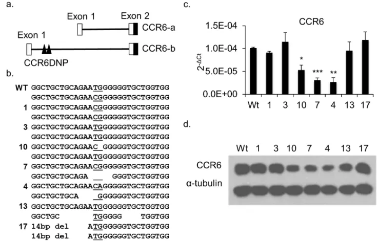
The CCR6DNP resides in intron 1 ofCCR6(Fig 1a). To confirm the role of the CCR6DNP in
gene expression we employed TALEN gene editing [9]. For these experiments we employed
Fig 1. Characterization of mutated HCT116 clones generated by TALENs targeting the CCR6DNP.(a) Partial genomic arrangement of
CCR6showing alternative transcripts CCR6-a and CCR6-b and the location of CCR6DNP. Only CCR-b could be detected in HCT116 and Jurkat T cells by real time PCR (seeS3 Fig). (b) Sequence of the three alleles of CCR6DNP in 7 representative targeted HCT116 clones. The CCR6DNP is underlined. (c) and (d) Expression of CCR6 in the 7 targeted clones by qPCR and Western blot. For qPCR, data are shown as mean±s.d. (n= 3). Sequence, sequence trace and expression data for all 17 mutated HCT116 clones are inS1andS2Figs. Statistical significance in 1c reflects comparison to WT.
doi:10.1371/journal.pgen.1006292.g001
HCT116 cells, a human colon cancer line that expresses CCR6 at a high level and is easily transfectable. Cells were co-transfected with both left and right TALEN constructs together with a puromycin selection marker. After selection, we screened more than 100 puromycin-resistant clones by Sanger sequencing of a 122bp PCR fragment flanking the CCR6DNP. Clones positive for mutations at the CCR6DNP locus were sub-cloned. Identical 122bp PCR fragments from these sub-clones were cloned into TA vectors for sequencing to define the mutations on each allele. In total, we obtained 17 mutated clones, including 18 deletions and 4
mutated alleles (Fig 1bandS1 Fig). We then measured CCR6 expression by both qPCR and
Western blot, confirming that in most clones sequence perturbation at CCR6DNP modulated
gene expression (Fig 1c and 1d;S2a Fig). By contrast, expression of the control geneCD40was
unchanged (S2b Fig). This result confirms that CCR6DNP participates directly in the
regula-tion ofCCR6and is thus a plausible causal variant at the RA-associated 6q27 locus.
Flanking Restriction Enhanced Pulldown (FREP) identifies the
association of PARP-1 with the CCR6DNP
We assumed that CCR6DNP, as a causal allele located in an intron, required a transcription factor or enhancer to carry out its regulatory function. To identify such a protein, we developed
a novel DNA pulldown assay termed FREP (Fig 2). An 82bp biotinylated DNA fragment was
conjugated to streptavidin-coated magnetic Dynabeads (Invitrogen). This fragment was engi-neered to include a 31bp sequence containing the CCD6DNP (TG allele, termed CCR6DNP/ TG), flanked by restriction enzyme cleavage sites for BamH I proximally and EcoR I distally, as
Fig 2. Flanking Restriction Enhanced Pulldown (FREP).(a) An 82 bp biotinylated DNA fragment is conjugated to streptavidin-coated magnetic Dynabeads (Invitrogen). This fragment is engineered to include a 31bp gene specific (“bait”) sequence (black box), flanked by restriction enzyme cleavage sites for BamH I proximally (blue box) and EcoR I distally (red box), as well as 20bp DNA fragments to allow PCR amplification of the whole unit. (b) DNA-beads are mixed with nuclear extract. A free 82 bp non-biotinylated DNA fragment can be included in the control reaction at this stage as a specific competitor. (c) Magnetic separation and wash to remove non-DNA binding proteins. (d) EcoR I digestion to release 3’DNA end-binding proteins, yielding the EcoR I fraction. (e) BamH I digestion to separate the sequence specific DNA binding proteins, BamH I fraction, from proteins that bind 5’DNA and Dynabeads. (f) Mass spectrometry (MS) to identify proteins remaining within the BamH I fraction.
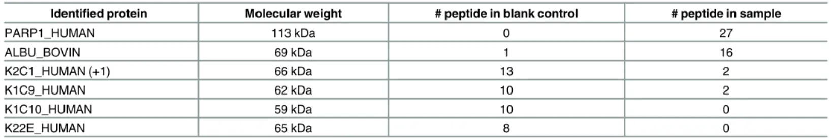
well as 20bp sequences to allow PCR amplification of the whole unit. This configuration allows pulldown of DNA-binding proteins specific to the sequence of interest and also elimination of proteins that bind non-specifically to DNA. CCR6DNP/TG beads were incubated with nuclear extract from THP-1 cells, a human monocyte leukemia line expressing of CCR6. Following magnetic separation and wash, the CCR6DNP/TG beads were cut with EcoR I and the super-natant was collected as the EcoR I fraction. After a second round of magnetic separation and wash, the DNA beads were cut with BamH I to release the CCR6DNP/TG fragment together with any binding proteins. In parallel, we performed a control FREP in the presence of a 40-fold excess of free CCR6DNP/TG as a binding competitor. BamH I fractions from these experiments were resolved by SDS-PAGE and silver stained to identify potential CCR6DNP/
TG binding proteins (Fig 3). A dominant band emerged at approximately 120kD (lane 1,
indi-cated by an arrow) but was much lower in intensity in the presence of competitor (lane 2). This band was cut and analyzed by mass spectrometry, revealing 27 peptide fragments from a single
protein, PARP-1 (Table 1).
Fig 3. FREP to identify the binding of PARP-1 with CCR6DNP.(a) Silver stain and (b) densitometry to show proteins pulled down from CCR6DNP/TG-beads mixed with nuclear extract from THP-1 cells. Lane 1, BamH I fraction; Lane 2, BamH I fraction where pulldown was performed in the presence of a 40x excess of competitor (free CCR6DNP/TG). Arrow indicates the specific band sent for mass spectrometry analysis.
doi:10.1371/journal.pgen.1006292.g003
Table 1. Mass spectrometry analysis identifies PARP-1 as the CCR6DNP binding protein.
Identified protein Molecular weight # peptide in blank control # peptide in sample
PARP1_HUMAN 113 kDa 0 27
ALBU_BOVIN 69 kDa 1 16
K2C1_HUMAN (+1) 66 kDa 13 2
K1C9_HUMAN 62 kDa 10 2
K1C10_HUMAN 59 kDa 10 0
K22E_HUMAN 65 kDa 8 0
doi:10.1371/journal.pgen.1006292.t001
The PARP-1 association with CCR6DNP exhibits sequence and allele
specificity
PARP-1 is a well-known DNA repair protein [10]. It is a DNA end-binding protein and can
also bind single-stranded DNA without sequence specificity [11]. However, FREP minimizes
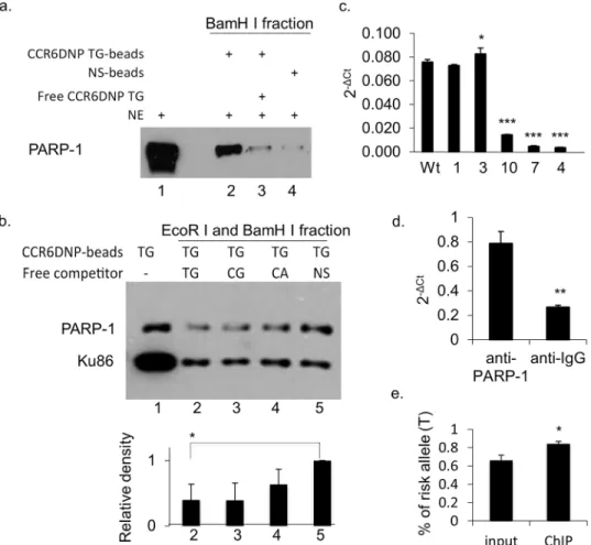
detection of such non-specific binding through restriction enzyme digestion, because the 3’ free end is cleaved off by EcoR I, while single-stranded DNA is not cleaved by BamH I and is therefore discarded along with the bead. To confirm sequence-specific association of PARP-1 with CCR6DNP/TG, we combined FREP with Western blot, adapting a method developed by
Wu [12]. We compared FREP using CCR6DNP/TG with FREP using a non-specific 31bp
DNA sequence, and probed the Western blot of BamH I fractions with an PARP-1
anti-body (Fig 4a). Our results showed that PARP-1 associated much more strongly with
CCR6DNP/TG (lane 2) than with the non-specific DNA sequence (lane 4). This association could be specifically competed away by the free CCR6DNP/TG (lane 3). In this assay, some PARP-1 was also observed with the non-specific DNA sequence, likely reflecting end binding to DNA uncut by EcoR I. Together, these results demonstrate that PARP-1 binds CCR6DNP/ TG with sequence specificity.
We then investigated whether PARP-1 exhibited allele-specific association with CCR6DNP
in accordance with the order of RA risk TG>CG>CA [4]. For this purpose, we repeated FREP
using CCR6DNP/TG beads in the presence of excess free competitors CCR6DNP/TG,
CCR6DNP/CG, CCR6DNP/CA, or a non-specific DNA sequence (Fig 4b; lane 2, 3, 4 and 5,
respectively). In these experiments, CCR6DNP/TG-beads were mixed with nuclear extract and different competitors, after which the beads were washed and separated. The bead-bound CCR6DNP/TG was cut directly with BamH I and the supernatant was resolved on SDS-PAGE. Western blots were probed with an anti-PARP-1 antibody. In triplicate experiments, the asso-ciation of PARP-1 with CCR6DNP/TG was competed away by free CCR6DNP/TG more
effi-ciently than by non-specific DNA (p<0.05, n = 3), with a trend toward TG>GC>CA in line
with the established CCR6DNP risk hierarchy (Fig 4b; lane 2, 3, and 4, respectively).
Non-spe-cific DNA also competed for PARP-1 binding, but less efficiently, consistent with the known
end-binding activity of PARP-1 (Fig 4b; lane 5) [11,13]. To ensure equivalent concentrations
of competitors, we probed simultaneously against Ku86, a double-stranded DNA end-binding protein without known sequence specificity. All four competitors inhibited Ku86 binding equally. Together, these data show that PARP-1 associates with the CCR6DNP in an allelic
manner consistent with the RA risk hierarchy TG>CG>CA.
To further confirm the association of PARP-1 with CCR6DNP, we performed ChIP assays using an anti-PARP-1 antibody (Genetex GTX100573). First, we performed ChIP using wild
type HCT116 cells and the mutated HCT116 clones #1, 3, 4, 7 and 10 as shown inFig 1. In
brief, cells from different clones were cross-linked using 1% formaldehyde, fragmented with sonication, and followed by immunoprecipitation. After the cross-linked DNAs were reversed and purified, the endogenous association of PARP-1 with CCR6DNP was detected by qPCR
using primers recognizing a shared sequence 5’of the CCR6DNP and outside of the mutated
regions. Our data showed that the binding of PARP-1 to CCR6DNP corresponds with the levels
of CCR6 expression in these mutants inFig 1(Fig 4c). Second, we performed ChIP with human
Jurkat T cells, confirming that the association of PARP-1 with CCR6DNP is evident not only in
HCT116 but also in T cells (Fig 4d). Further, we sequenced the CCR6DNP fragments from the
ChIP with WT HCT116 cells inFig 4c, which as shown inFig 1bare heterozygous TG/CG at
binding of PARP-1 to the TG allele (Fig 4e). Together, these results support the conclusion that PARP-1 associates with the CCR6DNP in an allele-specific manner.
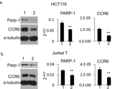
PARP-1 regulates CCR6 expression in human HCT116 cells and Jurkat
T cells
The association of PARP-1 with CCR6DNP suggests that PARP-1 is a regulatory protein that
regulatesCCR6. To test this hypothesis, we generated PARP-1-deficient HCT116 cells by
shRNA knockdown. PARP-1-specific shRNA, but not scrambled shRNA, induced a striking
reduction of CCR6 expression by both Western blot (Fig 5a, left) and qPCR analysis (Fig 5a,
Fig 4. The association of PARP-1 with CCR6DNP is sequence dependent and allele specific.(a) FREP +Western blot for PARP-1 with BamH I fractions. Lane 1: nuclear extract (NE) input, lane 2: NE with CCR6DNP/TG-beads, lane 3: NE with CCR6DNP/TG-beads with cold competitor (Free TG), and lane 4: NE with a non-specific DNA sequence (NS-beads). (b) To detect allele specificity, FREP was modified to cut directly with BamH I only, yielding a combined BamH I+EcoR I fraction, and SDS-PAGE was probed with anti-PARP-1. Anti-Ku86 antibody was used as the internal loading control to ensure comparable amounts of competitor DNA. CCR6DNP/TG-beads were incubated with NE and competed with equal amount of free competitors. Lane 1: no competitor; lane 2: free CCR6DNP/TG; lane 3: CCR6DNP/CG; lane 4: free CCR6DNP/CA; and lane 5: free non-specific DNA. Upper: Western blot; Lower: relative density from the Western blot. (c) ChIP assay with an anti-PARP-1 antibody on WT HCT116 cells and 5 mutants as indicated inFig 1showing impaired binding of PARP-1 to the CCR6DNP. (d) ChIP assay with an anti-PARP-1 antibody on WT Jurkat T cells showing significant enrichment of the CCR6DNP fragments comparing to anti-IgG antibody. (e) Sequencing of the CCR6DNP fragments from the ChIP with WT HCT116 cells showing the significant enrichment of the T allele over the C allele by an anti-PARP-1 antibody. Results from (a), (b), (c), and (d) representative of 3 experiments. Data are shown as mean±s.d. (n= 3).
doi:10.1371/journal.pgen.1006292.g004
middle and right). To ensure that this mechanism is not restricted to HCT116 cells, we per-formed siRNA knockdown of PARP-1 in human Jurkat T cells and confirmed a similar result (Fig 5b). These results demonstrate that PARP-1 is a novel regulator of CCR6 expression across human cell types.
The (ADP-ribose) polymerase activity of PARP-1 participates in the
regulation of
CCR6
in human cells
PARP-1 is a poly(ADP-ribose)polymerase that transfers ADP-ribose groups to target proteins
and can thereby alter gene transcription [10]. To test if poly(ADP-ribose)polymerase activity is
required for the regulation of CCR6 expression, we treated human Jurkat T cells, HCT116 cells and Hela cells with a PARP-1 inhibitor, 3-aminobenzamide (3-AB), that inhibits PARP-1
enzy-matic activity by competing for the NAD+ binding site on PARP-1 [14]. Cells were treated
with 0, 5 and 10 mM 3-AB for 72 h and analyzed for CCR6 expression by qPCR and Western
blot (Fig 6a and 6b). At the 10 mM concentration, our results showed a significant decrease of
CCR6 expression on Jurkat T cells and HCT116, while Hela cells exhibited a trend in the same direction. Under these conditions, treated cells grew at a slower rate (~75% of control), but no apoptosis was observed morphologically or by Western blot, as PARP-1 degradation is a
marker of apoptosis (Fig 6b). This result confirms the identification of PARP-1 in the
regula-tion of CCR6 expression and implicates its poly(ADP-ribose)polymerase activity in this function.
Discussion
GWAS have identified a role for numerous genetic loci in the modulation of complex pheno-types, including common diseases such as RA. Dissecting the associated mechanistic pathways remains a major challenge, especially for non-coding variants, even when a putative causative Fig 5. CCR6 expression in PARP-1 knockdown human cells.HCT116 cells (a) and Jurkat T cells (b) by Western blot (left), and qPCR analysis on PARP-1 (middle) and on CCR6 (right). For Western blot, whole cell extract was isolated and separated on SDS-PAGE gel. Western blot was detected with mouse anti-human PARP-1, CCR6 andα-tubulin antibodies simultaneously. Lane 1: negative control for shRNA. Lane 2: shRNA treatment for HCT116 cells or siRNA treatment for Jurkat T cells. For qPCR, data are shown as mean±s.d. (n= 3).
polymorphism has been established [3]. In this report, we introduce FREP as a technically straightforward tool to identify DNA binding proteins, and use this method to define the
path-way by which a functional polymorphism, the CCR6DNP, regulates expression ofCCR6.
Spe-cifically, we show that PARP-1 associates with the CCR6DNP in sequence- and allele-specific manner; that interference with PARP-1 expression impairs expression of CCR6 at mRNA and protein levels; and that chemical inhibition of PARP-1 exerts a similar effect, indicating that the enzymatic activity of PARP-1 is relevant for its regulatory function, while confirming that the pathway spans cellular lineages.
Compared with conventional DNA pulldown assays [15], FREP reduces non-specific
bind-ing through distinct restriction enzyme cleavage sites on either side of the bait sequence. An identical DNA construct, but without bead linkage, can be employed as a competitor to
high-light proteins that bind bait DNA with sequence specificity (seeFig 3). We further adapted a
method developed by Wu [12] to combine DNA pulldown with Western blot, in the setting of
varying competitor sequences, to assess allelic specificity (seeFig 4b). Together, these
tech-niques provide a new approach to explore regulatory DNA-protein associations identified by genetic association studies.
FREP and other pulldown assays cannot distinguish direct DNA binding from participation in a larger DNA-binding protein complex. Previous reports have implicated PARP-1 in
tran-scriptional regulation of a number of human genes includingHFE,CXCL1,cTnT,iNOS,SNCA
andTcirg1[16–24]. In these reports, PARP-1 interacted with the TGTTG sequence in the
cTnTpromoter, GCTGTGGGAA in theTcirg1promoter, and the palindrome-like sequence
ATGGTcttACCTA in theHFEpromoter. The lack of sequence specificity could suggest that
PARP-1 serves as part of a larger protein complex, or alternately that it recognizes a secondary
structure such as cruciform DNA [25,26]. Indeed, our present data do not establish that
PARP-1 itself directly recognizes the CCR6DNP sequence. However, it is interesting to note that the
CCR6DNP risk allele GCAGAATGGGGGGT bears some similarity to the binding sequences
atcTnTandTcirg1, suggesting that—if in fact PARP-1 binds directly to these sequences—then TG might play an important role in DNA recognition.
Fig 6. CCR6 expression in human cells treated with 3-aminobenzamide.(a) qPCR and (b) Western blot to show expression of CCR6 and PARP-1 in Jurkat T cells, HCT116 and Hela cells. Cells were treated with 0 mM (lane 1), 5 mM (lane 2) and 10 mM (lane 3) 3-AB for 72 hrs. For qPCR, data are shown as mean±s.d. (n= 3).
doi:10.1371/journal.pgen.1006292.g006
PARP-1 is the founding member of a family of enzymes engaged in transfer of poly ADP-ribose (PAR) to a wide range of nuclear and cytoplasmic proteins, including PARP-1 itself
[10,27]. PARP-1 is a multifunctional protein that plays essential roles in the cell, including
DNA repair, translation, transcription, telomere maintenance, genomic stability, and
chroma-tin remodeling [28]. PARP-1-deficient mice are viable but show increased sensitivity to
geno-toxic stress, resistance to DNA damage-induced cell death, and increased susceptibility to
chemically- or genetically-induced tumors [29]. Recently, accumulating data suggest that
PARP-1 is an important regulator of gene transcription [28–30]. This role may be
accom-plished in multiple ways, including binding to DNA, decondensation of chromatin [31,32],
alteration of mRNA splicing through regulation of the splicing silencers or enhancers [33],
recruitment of transcription factors [34], and post-translational modulation of RNA
transla-tion [35,36]. Some of these studies implicate the enzymatic activity of PARP-1 [24,30],
although in other contexts this activity appears dispensable [37]. Our findings add to this
litera-ture by identifyingCCR6as a specific target of PARP-1, defining a disease-associated DNA
polymorphism that modulates this association, and confirming a role for PARP-1 enzymatic
activity in the transcriptional regulation ofCCR6. Our data do not define the relevant
PARyla-tion target, which could include another protein in theCCR6regulatory complex, associated
histones, or PARP-1 itself.
Identification of the role for PARP-1 as a regulator ofCCR6began with experimental
confir-mation by TALEN that CCR6DNP was directly involved in modulation of gene expression, as
had been suggested previously [4]. These experiments targeted the general locus, rather than
the dinuclotide motif itself, and revealed clone-to-clone variability that we presume reflects the effect of each mutation on the larger binding sequence, not defined here. For example,
dele-tions flanking the CCR6DNP in clone 9 translated into impairedCCR6expression despite an
intact CCR6DNP (S1andS2Figs), while gene expression was hardly altered by mutations
introduced in clones 13 and 17 (Fig 1). Despite this variability, these results helped to engender
confidence that CCR6DNP was indeed a suitable“bait”region, a conclusion borne out by the
subsequent identification of the allelic association of PARP-1 with this locus.
One strength of FREP is its utility to test allele specificity through a unique assay in which
FREP is performed in the presence of allelic competitors (Fig 4b). This assay can be technically
challenging if allelic impact on binding affinity is partial. For example, in triplicate repeats quantitated using densitometry, we were able to distinguish statistically between the high-risk TG allele and non-specific DNA, but found only a trend with respect to intermediate risk alleles GC and CA, though it is notable that no such trend was evident in the binding of loading control Ku86.
The effect of PARP-1 onCCR6is consistent with its role as an activator of inflammation
mediated by NFκB, AP-1, and MAP kinases [38–40]. Its impact onCCR6via a RA-associated
functional polymorphism is also consistent with the role of PARP-1 in inflammatory arthritis
[8], although limited knowledge of signal transduction pathways downstream of CCR6
con-strains the understanding of how the interaction between PARP-1 and CCR6 contributes to
RA. Nevertheless, both genetic deficiency [41] and chemical inhibition [42–44] of PARP-1
attenuates experimental arthritis in the mouse. The latter is accompanied by significant reduc-tions in pro-inflammatory cytokines, chemokines, inflammatory mediators, and endothelial expression of key adhesion molecules VCAM-1 and ICAM-1. Our finding that PARP-1
regu-latesCCR6–work originating in human genetics—lends further support to the hypothesis that
More generally, our studies demonstrate a solution to the challenge of bridging the gap from GWAS to biological mechanism for regulatory polymorphisms, considered to represent a
substantial fraction of GWAS hits [2,3]. First, TALEN or other targeted gene editing technique
(such as clustered regularly-interspaced short palindromic repeats, CRISPR) is used to confirm that a specific locus is active in regulating a target gene. Second, FREP is employed to identify associated transcriptional enhancers or repressors. This approach is applicable both to the identification of regulatory proteins, as demonstrated here, and also to the validation of candi-date regulatory proteins implicated through bioinformatic or other approaches.
In conclusion, we confirm that CCR6DNP is a causal allele that controls expression of
CCR6, and identify PARP-1 as a direct regulator ofCCR6through the RA risk polymorphism
CCR6DNP. Our strategy, encompassing sequence perturbation and FREP, represents a novel approach to defining pathways by which non-coding genetic polymorphisms alter disease risk. A comprehensive understanding of such pathways will represent an important step forward in the development of individualized therapeutic strategies for patients with RA and other diseases.
Materials and Methods
Cells and culture
The human colon cancer line HCT116 and the human monocyte line THP-1 were purchased
from ATCC and cultured in McCoy’s 5A and RPMI medium supplemented with 10% FBS,
respectively. Mouse anti-human PARP-1 antibody (Cat#: 14-6666-92) and CCR6 antibody
(Cat#: 14-1969-82) were purchased from eBioscience and used at 1μg/ml.
Construction of TALEN expression plasmids
TALEN expression plasmids were constructed exactly as described [9]. The TALEN cloning
backbone and TALE monomer template were purchased from Addgene (TALEN Kit #1000000019). All primers were synthesized and purchased from IDT. The TALEN target sequence for CCR6 is AGCACCCCCCATTCTGCAGC. Sequences for the left and right TALENs are TCATTCATGTTAGATCCACC and AGCCACAGCCCCCAGGGTGA.
Generation of mutations by TALENs in HCT116 cells
50μg of right and left TALENs were transfected together with 5μg of pGK/puro (Addgene,
plas-mid 11349) using standard calcium phosphate transfection. After 48 h incubation, cells were
re-plated and selected against 0.8μg/ml puromycin. Single puromycin- resistant clones were
harvested and genomic DNA isolated to amplify the CCR6 fragment using forward primer
5’-ACATGTCTCCCAAACTTCTCACCC-3’and reverse primer: 5’-TTTGTGCAGGGAGGTTG
GGATGAA-3’. The PCR fragments were either sequenced directly with the reverse primer or cloned into TA vector and sequenced with T7 primer (Genewiz). Clones positive for mutations were subcloned and their DNA was cloned into TA vector to sequence both alleles. Each clone was sequenced at least 10 times to establish heterozygosity or homozygosity.
RNA isolation, reverse transcription and real time PCR
RNA was isolated using the RNeasy Mini Kit (Qiagen). cDNA was synthesized with Super-Script III First-Strand Synthesis System and cDNA was amplified by real time PCR with Power SYBR Green PCR Master Mix (Life Technologies). For detection of whole CCR6 mRNA (both
a and b variants), 5’primer: TGAGCGGGGAATCAATGAATT and 3’primer: TCCTGCAAG
GAGCACAGTAACAT were used. For detection of the transcript variant a, 5’primer:
TAGGAAGTGGCAATCCAGAAC and 3’primer: ATGACTCCAGCTCACCAATG were
used. For detection of the transcript variant b, 5’primer: CCACGTGTATATGCTGGTGAA
and 3’primer: GGAGCTGTCTGTTCCACAAA were used.
Flanking Restriction Enhanced Pulldown (FREP)
An 82bp DNA fragment biotinylated at the 5’end by IDT was conjugated to
streptavidin-coated Dynabeads as per manufacturer’s instructions (Invitrogen). This fragment was
engi-neered to include a 31bp target sequence (see below) flanked by restriction enzyme cleavage sites with BamH I proximally and EcoR I distally. Outside these two cleavage sites were intro-duced 20bp DNA fragments for PCR amplification of the whole unit (proximal: 5’- AATGATA
CGGCGACCACCGA-3’; distal: 5’- CAAGCAGAAGACGGCATACGA-3’). Bead-linked DNA
was mixed with 50μg THP-1 nuclear extract in 1x binding buffer (10 mM Tris pH 7.5, 50 mM
KCl, 5mM MgCl2, 2.5% glycerol, 0.5% NP-40 and 1μg polydI-dC) at room temperature for 20
min. Nuclear extract was generated by NE-PER nuclear and cytoplasmic extraction reagents (Thermo Scientific). After magnetic selection and wash with PBS+0.05% Tween 20, DNA beads with bound proteins were digested with EcoR I at 37C for 30 min. The supernatant was collected as the EcoR I fraction. After another magnetic selection and wash, the DNA beads were digested with BamH I at 37C for 30 min and the supernatant was collected as the BamH I fraction. Both EcoR I and BamH I fractions were resolved by 7% SDS-PAGE gel for either Western blot analysis or silver staining. Mass spectrometry was performed in BIDMC using Thermohybrid Orbitrap XL high resolution MS (Thermo Scientific).
The“bait”DNA fragment used for FREP was: CCR6DNP/TG:
/5Biosg/AATGATACGGCGACCACCGAGGATCCGTGGCTGCTGCAGAATGGGGGG
TGCTGGTGAATTCTCGTATGCCGTCTTCTGCTTG. The DNA fragments used for the competition assay were: CCR6DNP/TG:
AATGATACGGCGACCACCGAGGATCCGTGGCTGCTGCAGAATGGGGGGTGCTG
GTGAATTCTCGTATGCCGTCTTCTGCTTG CCR6DNP/CG:
AATGATACGGCGACCACCGAGGATCCGTGGCTGCTGCAGAACGGGGGGTGCTG
GTGAATTCTCGTATGCCGTCTTCTGCTTG CCR6DNP/CA:
AATGATACGGCGACCACCGAGGATCCGTGGCTGCTGCAGAACAGGGGGTGCTG
GTGAATTCTCGTATGCCGTCTTCTGCTTG
Irrelevant DNA sequence: AATGATACGGCGACCACCGAGGATCCAGGGCTGTAGAT TCCGGCCTGAAGCCTGGGAATTCTCGTATGCCGTCTTCTGCTTG.
ChIP assay
The ChiP assay was performed as described in Noss et al., 2015 [46]. PARP-1 antibody was
purchased from Genetex (GTX100573). The CCR6DNP fragment was detected by qPCR with
5’primer: GTGAGAAGTTTGGGAGACATGT and 3’primer TTGGAAACGCTCTAATAGA
PARP-1 knockdown
PARP-1 knockdown in HCT116 cells was performed using a mixture of four retroviruses
con-taining 4 PARP-1 unique 29mer shRNAs, as per manufacturer’s instructions (Origene Cat#:
TG315488). 24 h after infection, cells were re-plated and selected in 1 ug/ml puromycin. 3 days after selection, both protein and RNA were isolated for assays. PARP-1 knockdown in Jurkat cells was performed using PARP-1 siRNA from Santa Cruz Biotech (sc-29437). Transfection was done using GenMute siRNA Transfection Reagent for Jurkat Cell (SignaGen Laboratories) in a 24 well format. 40 pmoles siRNA was used. 48 h after transfection, protein and RNA were isolated for assay.
3-Aminobenzamide treatment
1x10⁵cells were plated in a 24-well plate the day before the treatment. On day one, both
DMSO (control) and different concentrations of 3-AB (Sigma) were added. After 72 hrs, pro-tein and RNA were isolated for Western blot and qPCR assays.
Statistical analysis
Statistical significance was evaluated with Student’st-test, 2 tailed,P<0.05,P<0.01,
P<0.001, NS, nonsignificant.nstands for the number of replicated independent
experiments.
Supporting Information
S1 Fig. Sequence and sequence trace of 17 mutants generated by TALENs.
(TIF)
S2 Fig. Expression of CCR6 (a) and CD40 (b) in mutated HCT116 cells by real time PCR.
(TIF)
S3 Fig. Expression of CCR6 variants a and b in HCT116 and Jurkat T cells.
(TIF)
Acknowledgments
We thank Dr. Feng Zhang for support with TALENs technology and Drs. Jonathan Higgins and Soumya Raychaudhuri for scientific discussion.
Author Contributions
Conceptualization:GL RMP PAN.
Formal analysis:GL DW DD YO.
Funding acquisition:GL PAN.
Investigation:GL PC YY.
Methodology:GL.
Project administration:GL PAN.
Resources:GL RMP PAN.
Supervision:GL RMP PAN.
Visualization:GL.
Writing–original draft:GL PAN.
Writing–review & editing:GL PC DW DD YY YO RMP PAN.
References
1. McInnes IB, Schett G (2011) The pathogenesis of rheumatoid arthritis. N Engl J Med 365: 2205–2219. doi:10.1056/NEJMra1004965PMID:22150039
2. Okada Y, Wu D, Trynka G, Raj T, Terao C, et al. (2014) Genetics of rheumatoid arthritis contributes to biology and drug discovery. Nature 506: 376–381. doi:10.1038/nature12873PMID:24390342 3. (2014) Little boxes. Nat Genet 46: 659. doi:10.1038/ng.3028PMID:24965724
4. Kochi Y, Okada Y, Suzuki A, Ikari K, Terao C, et al. (2010) A regulatory variant in CCR6 is associated with rheumatoid arthritis susceptibility. Nat Genet 42: 515–519. doi:10.1038/ng.583PMID:20453841 5. Lee AY, Korner H (2014) CCR6 and CCL20: emerging players in the pathogenesis of rheumatoid
arthri-tis. Immunol Cell Biol 92: 354–358. doi:10.1038/icb.2013.97PMID:24394994
6. Hirota K, Yoshitomi H, Hashimoto M, Maeda S, Teradaira S, et al. (2007) Preferential recruitment of CCR6-expressing Th17 cells to inflamed joints via CCL20 in rheumatoid arthritis and its animal model. J Exp Med 204: 2803–2812. PMID:18025126
7. Koumakis E, Bouaziz M, Dieude P, Ruiz B, Riemekasten G, et al. (2013) A regulatory variant in CCR6 is associated with susceptibility to antitopoisomerase-positive systemic sclerosis. Arthritis Rheum 65: 3202–3208. doi:10.1002/art.38136PMID:23983073
8. Garcia S, Conde C (2015) The Role of Poly(ADP-ribose) Polymerase-1 in Rheumatoid Arthritis. Media-tors Inflamm 2015: 837250. doi:10.1155/2015/837250PMID:26339143
9. Sanjana NE, Cong L, Zhou Y, Cunniff MM, Feng G, et al. (2012) A transcription activator-like effector toolbox for genome engineering. Nat Protoc 7: 171–192. doi:10.1038/nprot.2011.431PMID: 22222791
10. Gibson BA, Kraus WL (2012) New insights into the molecular and cellular functions of poly(ADP-ribose) and PARPs. Nat Rev Mol Cell Biol 13: 411–424. doi:10.1038/nrm3376PMID:22713970
11. Langelier MF, Pascal JM (2013) PARP-1 mechanism for coupling DNA damage detection to poly(ADP-ribose) synthesis. Curr Opin Struct Biol 23: 134–143. doi:10.1016/j.sbi.2013.01.003PMID:23333033 12. Wu KK (2006) Analysis of protein-DNA binding by streptavidin-agarose pulldown. Methods Mol Biol
338: 281–290. PMID:16888365
13. Tong WM, Cortes U, Hande MP, Ohgaki H, Cavalli LR, et al. (2002) Synergistic role of Ku80 and poly (ADP-ribose) polymerase in suppressing chromosomal aberrations and liver cancer formation. Cancer Res 62: 6990–6996. PMID:12460917
14. Zheng YD, Xu XQ, Peng F, Yu JZ, Wu H (2011) The poly(ADP-ribose) polymerase-1 inhibitor 3-amino-benzamide suppresses cell growth and migration, enhancing suppressive effects of cisplatin in osteo-sarcoma cells. Oncol Rep 25: 1399–1405. doi:10.3892/or.2011.1212PMID:21399878
15. Xia Q, Deliard S, Yuan CX, Johnson ME, Grant SF (2015) Characterization of the transcriptional machinery bound across the widely presumed type 2 diabetes causal variant, rs7903146, within TCF7L2. Eur J Hum Genet 23: 103–109. doi:10.1038/ejhg.2014.48PMID:24667787
16. Pelham C, Jimenez T, Rodova M, Rudolph A, Chipps E, et al. (2013) Regulation of HFE expression by poly(ADP-ribose) polymerase-1 (PARP1) through an inverted repeat DNA sequence in the distal pro-moter. Biochim Biophys Acta 1829: 1257–1265. doi:10.1016/j.bbagrm.2013.10.002PMID:24184271 17. Lodhi N, Kossenkov AV, Tulin AV (2014) Bookmarking promoters in mitotic chromatin:
poly(ADP-ribose)polymerase-1 as an epigenetic mark. Nucleic Acids Res 42: 7028–7038. doi:10.1093/nar/ gku415PMID:24861619
18. Beranger GE, Momier D, Rochet N, Quincey D, Guigonis JM, et al. (2006) RANKL treatment releases the negative regulation of the poly(ADP-ribose) polymerase-1 on Tcirg1 gene expression during osteo-clastogenesis. J Bone Miner Res 21: 1757–1769. PMID:17002555
19. Chiba-Falek O, Kowalak JA, Smulson ME, Nussbaum RL (2005) Regulation of alpha-synuclein expres-sion by poly (ADP ribose) polymerase-1 (PARP-1) binding to the NACP-Rep1 polymorphic site upstream of the SNCA gene. Am J Hum Genet 76: 478–492. PMID:15672325
21. Yazdany J, Davis J (2004) The role of CD40 ligand in systemic lupus erythematosus. Lupus 13: 377– 380. PMID:15230296
22. Frizzell KM, Gamble MJ, Berrocal JG, Zhang T, Krishnakumar R, et al. (2009) Global analysis of tran-scriptional regulation by poly(ADP-ribose) polymerase-1 and poly(ADP-ribose) glycohydrolase in MCF-7 human breast cancer cells. J Biol Chem 284: 33926–33938. doi:10.1074/jbc.M109.023879PMID: 19812418
23. Ambrose HE, Papadopoulou V, Beswick RW, Wagner SD (2007) Poly-(ADP-ribose) polymerase-1 (Parp-1) binds in a sequence-specific manner at the 6 locus and contributes to the regulation of Bcl-6 transcription. Oncogene 2Bcl-6: Bcl-6244–6252.
24. Nirodi C, NagDas S, Gygi SP, Olson G, Aebersold R, et al. (2001) A role for poly(ADP-ribose) polymer-ase in the transcriptional regulation of the melanoma growth stimulatory activity (CXCL1) gene expres-sion. J Biol Chem 276: 9366–9374. PMID:11112786
25. Huang K, Tidyman WE, Le KU, Kirsten E, Kun E, et al. (2004) Analysis of nucleotide sequence-depen-dent DNA binding of poly(ADP-ribose) polymerase in a purified system. Biochemistry 43: 217–223. PMID:14705948
26. Petrucco S, Percudani R (2008) Structural recognition of DNA by poly(ADP-ribose)polymerase-like zinc finger families. FEBS J 275: 883–893. doi:10.1111/j.1742-4658.2008.06259.xPMID:18215166 27. Bonicalzi ME, Haince JF, Droit A, Poirier GG (2005) Regulation of poly(ADP-ribose) metabolism by
poly(ADP-ribose) glycohydrolase: where and when? Cell Mol Life Sci 62: 739–750. PMID:15868399 28. Thomas C, Tulin AV (2013) Poly-ADP-ribose polymerase: machinery for nuclear processes. Mol
Aspects Med 34: 1124–1137. doi:10.1016/j.mam.2013.04.001PMID:23624145
29. Kim MY, Zhang T, Kraus WL (2005) Poly(ADP-ribosyl)ation by PARP-1: 'PAR-laying' NAD+ into a nuclear signal. Genes Dev 19: 1951–1967. PMID:16140981
30. Schiewer MJ, Goodwin JF, Han S, Brenner JC, Augello MA, et al. (2012) Dual roles of PARP-1 promote cancer growth and progression. Cancer Discov 2: 1134–1149. doi:10.1158/2159-8290.CD-12-0120 PMID:22993403
31. Martinez-Zamudio R, Ha HC (2012) Histone ADP-ribosylation facilitates gene transcription by directly remodeling nucleosomes. Mol Cell Biol 32: 2490–2502. doi:10.1128/MCB.06667-11PMID:22547677 32. Ji Y, Tulin AV (2010) The roles of PARP1 in gene control and cell differentiation. Curr Opin Genet Dev
20: 512–518. doi:10.1016/j.gde.2010.06.001PMID:20591646
33. Ji Y, Tulin AV (2009) Poly(ADP-ribosyl)ation of heterogeneous nuclear ribonucleoproteins modulates splicing. Nucleic Acids Res 37: 3501–3513. doi:10.1093/nar/gkp218PMID:19346337
34. Krishnakumar R, Gamble MJ, Frizzell KM, Berrocal JG, Kininis M, et al. (2008) Reciprocal binding of PARP-1 and histone H1 at promoters specifies transcriptional outcomes. Science 319: 819–821. doi: 10.1126/science.1149250PMID:18258916
35. Di Giammartino DC, Shi Y, Manley JL (2013) PARP1 represses PAP and inhibits polyadenylation dur-ing heat shock. Mol Cell 49: 7–17. doi:10.1016/j.molcel.2012.11.005PMID:23219533
36. Ji Y, Tulin AV (2012) Poly(ADP-ribose) controls DE-cadherin-dependent stem cell maintenance and oocyte localization. Nat Commun 3: 760. doi:10.1038/ncomms1759PMID:22453833
37. Pavri R, Lewis B, Kim TK, Dilworth FJ, Erdjument-Bromage H, et al. (2005) PARP-1 determines speci-ficity in a retinoid signaling pathway via direct modulation of mediator. Mol Cell 18: 83–96. PMID: 15808511
38. Oliver FJ, Menissier-de Murcia J, Nacci C, Decker P, Andriantsitohaina R, et al. (1999) Resistance to endotoxic shock as a consequence of defective NF-kappaB activation in poly (ADP-ribose) polymer-ase-1 deficient mice. EMBO J 18: 4446–4454. PMID:10449410
39. Zingarelli B, Hake PW, Burroughs TJ, Piraino G, O'Connor M, et al. (2004) Activator protein-1 signalling pathway and apoptosis are modulated by poly(ADP-ribose) polymerase-1 in experimental colitis. Immunology 113: 509–517. PMID:15554929
40. Veres B, Radnai B, Gallyas F Jr., Varbiro G, Berente Z, et al. (2004) Regulation of kinase cascades and transcription factors by a poly(ADP-ribose) polymerase-1 inhibitor, 4-hydroxyquinazoline, in lipopoly-saccharide-induced inflammation in mice. J Pharmacol Exp Ther 310: 247–255. PMID:14999056 41. Garcia S, Bodano A, Gonzalez A, Forteza J, Gomez-Reino JJ, et al. (2006) Partial protection against
collagen antibody-induced arthritis in PARP-1 deficient mice. Arthritis Res Ther 8: R14. PMID: 16356201
42. Gonzalez-Rey E, Martinez-Romero R, O'Valle F, Aguilar-Quesada R, Conde C, et al. (2007) Therapeu-tic effect of a poly(ADP-ribose) polymerase-1 inhibitor on experimental arthritis by downregulating inflammation and Th1 response. PLoS One 2: e1071. PMID:17971849
43. Ahmad SF, Attia SM, Zoheir KM, Ashour AE, Bakheet SA (2014) Attenuation of the progression of adju-vant-induced arthritis by 3-aminobenzamide treatment. Int Immunopharmacol 19: 52–59. doi:10.1016/ j.intimp.2014.01.005PMID:24444779
44. Ahmad SF, Zoheir KM, Bakheet SA, Ashour AE, Attia SM (2014) Poly(ADP-ribose) polymerase-1 inhib-itor modulates T regulatory and IL-17 cells in the prevention of adjuvant induced arthritis in mice model. Cytokine 68: 76–85. doi:10.1016/j.cyto.2014.04.006PMID:24845796
45. Ledermann J, Harter P, Gourley C, Friedlander M, Vergote I, et al. (2012) Olaparib maintenance ther-apy in platinum-sensitive relapsed ovarian cancer. N Engl J Med 366: 1382–1392. doi:10.1056/ NEJMoa1105535PMID:22452356