Mechanisms of Ect2 Regulation in Cytokinesis and Oncogenesis
Danielle R. Cook
A dissertation submitted to the faculty of the University of North Carolina at Chapel Hill in partial fulfillment of the requirements for the degree of Doctor of Philosophy in the Eshelman School of Pharmacy Division of Chemical Biology
and Medicinal Chemistry
Chapel Hill, NC 2013
Approved by
Abstract
DANIELLE R. COOK: Mechanisms of Ect2 Regulation in Cytokinesis and Oncogenesis
(Under the direction of Channing Der)
increase in Ect2 protein expression in primary and metastatic CRC tumors and cell lines. Depletion of endogenous Ect2 by shRNA in CRC cell lines caused a reduction in anchorage-independent growth and Matrigel invasion without a corresponding defect in cytokinesis. Analyses of Ect2 protein expression in a CRC tumor microarray (N=149) found Ect2 protein overexpression in tumor tissue, but surprisingly, the ratio of cytoplasmic to nuclear Ect2 correlated with improved patient survival. This finding contrasted with earlier studies that suggested that the mislocalization of the normally nuclear restricted Ect2 to the cytoplasm was important for Ect2 to function as an oncogene.
Dedication
Acknowledgments
I would like to thank Dr. Channing Der for taking me into his lab and believing in me. Dr. Der has been a wonderful mentor and I am grateful for ending up in a great research situation after changing labs after almost two years into graduate school. Not only has Dr. Der been a great and supportive mentor scientifically I also appreciate his friendship, a rare combination, that I will miss immensely. Although I will doubly move on in the next stage of my career look forward having Dr. Der as a friend and continued mentor.
I would like to acknowledge the current and past members of the Der Lab. I appreciate everyone’s help and assistance though out the years and the memories I have made during my time in the lab. Specifically, I would like to thank Timothy Martin for all his assistance scientifically. Timothy continues to help me technically but also challenges me scientifically. I would also like to thank Dave Reiner for his continued support and perspective on science. I always enjoy his conversation and general scientific advice. I feel lucky to have a few outrageously geeky scientists around to share my passion and ideas with.
and I feel lucky to have had a supportive division. Each of my committee members also played vital roles during my transition by meeting with me and giving me advice. Thank you Dr. Deshmukh, Dr. Hanh, Dr. Graves, and Dr. Jarstfer! I am forever grateful for everyone who helped me during that transition and I am happy to say that it all worked out for the best.
Table of Contents
ABSTRACT ... III
DEDICATION ... VI
ACKNOWLEDGMENTS ... VII
LIST OF TABLES ... XI
LIST OF FIGURES ... XII
ABBREVIATIONS ... XIV
CHAPTER 1 INTRODUCTION ... 16
Superfamily of Ras GTPases ... 16
Rho GTPases ... 17
The discovery of RhoGEFs: diverse in numbers and structure ... 18
Ect2: Epithelial Cell Transforming Sequence 2 ... 20
Ect2 in Cytokinesis ... 21
Ect2 Domain Structure ... 23
RhoGEFs and development ... 26
Ect2 in development ... 27
RhoGEFs and cancer ... 28
Ect2 in Cancer ... 30
Colorectal Cancer (CRC) ... 32
Specific Aims ... 33
Overview ... 34
Introduction ... 35
Methods and Materials ... 39
Results ... 44
Discussion ... 56
CHAPTER 3 ABERRANT ECT2 EXPRESSION AND SUBCELLULAR LOCALIZATION IN COLORECTAL CANCER ... 61
Overview ... 61
Introduction ... 62
Methods and Materials ... 65
Results ... 72
Discussion ... 90
CHAPTER 4 FINAL THOUGHTS AND FUTURE DIRECTIONS ... 96
Overview ... 96
Determining Nuclear Versus Cytosolic Ect2 Function ... 96
Inhibitors of Ect2-mediated exchange ... 100
Mechanisms of Ect2-driven motility and invasion ... 103
Ect2 Mouse Model of Colorectal Cancer ... 104
List of Tables
Table 2-1. Genotypes of Weanlings and Embryos from
Ect2 -/+ Intercrosses ... 46 Table 2-2 Phenotypes and Genotype of Blastocyst
Outgrowth Derived from Ect2 intercrosses ... 48 Table 3-1 Associations between patient characteristics
and tertiles of ECT2 scores for nuclear expression ... 77 Table 3-2 Associations between patient characteristics
List of Figures
Figure 1-1 Small GTPase ... 16
Figure 1-2 Rho GTPase Signaling ... 18
Figure 1-3 Dbl family RhoGEFs ... 20
Figure 1-4 Discovery of Ect2 in NIH 3T3 ... 21
Figure 1-5 Cytokinesis ... 22
Figure 1-6 Ect2 multi-domain RhoGEF ... 24
Figure 1-7 Ect2 C-terminus ... 26
Figure 1-8 RhoGEF history ... 29
Figure 1-9 Genetic model of colorectal cancer ... 33
Figure 2-1 Targeted disruption of the mouse Ect2 gene produces a null mutation ... 45
Figure 2-2 Ect2-/- blastocysts display abnormal growth in vitro ... 47
Figure 2-3 Ect2+/- mice exhibit normal development and survival ... 49
Figure 2-5 Rho GTPases did not rescue the defect cytokinesis caused by loss of Ect2 ... 55
Figure 3-1. Increased Ect2 RNA and protein expression in colorectal cancer .... 74
Figure 3-2 Increased ratio of cytosolic to nuclear Ect2 correlates with overall survival ... 79
Figure 3-4 Nuclear Ect2 is required to support
anchorage-independent growth ... 83 Figure 3-5 Cytoplasmic Ect2 causes morphologic
and growth transformation of NIH 3T3 mouse fibroblasts. ... 84 Figure 3-6 Nuclear localization is essential
Abbreviations
APC- Adenomatous polyposis coli or Anaphase promoting complex BCS- Bovine calf serum
BRCA- Breast-cancer associated protein BRCT- BRCA1 C-terminal
BSA-Bovine serum albumin CRC- Colorectal Cancer
D-box-Destruction box (found in most APC substrates) Dbl- Diffuse B-cell lymphoma
DH- Dbl homology
DMEM- Dulbecco’s modified eagle’s medium DMSO- Dimethyl sulfoxide
DNA- Deoxyribonucleic acid DOCK- Dedicator of cytokinesis
Ect2- Epithelial cell transforming sequence 2 FBS- Fetal bovine serum
FL- Full-length
FRET- Fluorescent or Förster resonance energy transfer GAP-GTPase activating proteins
GDP-Guanine di-phosphate GEF-Guanine exchange factors GFP- Green fluorescent protein GTP- Guanine tri-phosphate
GTPases- Guanosine triphosphatases HA- Hemagglutinin
KDa- Kilodalton
MEF- Mouse embryonic fibroblasts
MTT- 3-[4,5-dimethylthiazol-2-yl]-2,5-diphenyltetrazolium bromide NLS- Nuclear localization sequence
OD- Optical density
PBS- Phosphate buffered saline PH- Pleckstrin homology
Ras- Rat Sarcoma Rho- Ras-homologous RNA- Ribonucleic acid RNAi- RNA interference SDS- Sodium dodecyl sulfate shRNA- Short hairpin RNA
Chapter 1Introduction
Superfamily of Ras GTPases
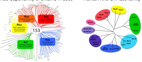
The RAS gene was originally identified in two different cancer causing virus originally discovered in rat, hence the name Ras (rat sarcoma) [1] and is the founding member of the Ras Superfamily of small GTPases containing over 150 members [2, 3]. Members of this superfamily are divided into five major branches based on sequence and functional similarities: Ras, Rho, Rab, Ran, and Arf. (Figure 1-1) However, variations in structure, post-translational modifications, subcellular locations, and effectors proteins allow GTPases to function as modulators of complex and diverse range of cellular processes [4].
Figure 1-1 Small GTPase
Left panel: Ras Superfamily Right panel: Human Rho GTPases (20 members; RhoA, Rac1, and Cdc42 are characterized the best)
Rho -Actin organization -Cell morphology -Cell movement Ran -Nuclear-cytoplasmic transport Rab -Vesicular trafficking -Protein trafficking Ras -Cell proliferation -Cell survival Arf -Vesicular trafficking -Exo-/Endocytosis
Ras Superfamily of small GTPases Human Rho GTPase family
RhoH RhoBTB2 RhoBTB1 Chp (RhoV) Wrch-1 (RhoU) TCL (RhoD) TC10 Cdc42
Rho GTPases
Figure 1-2 Rho GTPase Signaling [9]
The discovery of RhoGEFs: diverse in numbers and structure
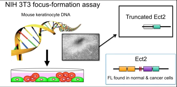
The first RhoGEF was identified initially as an oncogene in mammalian cells. Using the NIH 3T3 mouse fibroblast focus formation assay that led to the discovery of mutant Ras in human cancer, analysis of genomic DNA isolated from a human diffuse B-cell lymphoma resulted in the discovery of the DBL oncogene that encoded an N-terminally truncated protein [10]. Dbl was subsequently shown to catalyze the exchange activity of Cdc42 [11]. Additional NIH 3T3 focus formation and related assays identified Dbl-related proteins, in particular Vav and Ect2. That RhoGEFs were discovered initially as oncoproteins provided the first suggestion that Rho GTPases may also have a function in oncogenesis.
Dbl and the Dbl-related proteins all share a ~200 amino acid catalytic Dbl homology (DH; also called RhoGEF) and an immediately adjacent regulatory ~100 amino acid pleckstrin homology (PH) domain [12]. Additional RhoGEFs with this tandem DH-PH domain structure were identified by genetic and biochemical approaches and by in silico database searches. There are 72 human Dbl family RhoGEFs, with conserved orthologs found in all vertebrate species and in invertebrates, including Drosophila, C. elegans, S. cerevisiae and S. pombe. Beyond their shared DH-PH domains, Dbl RhoGEFs diverge significantly in their flanking N- and C-terminal sequences, which commonly contain a diversity protein-protein interaction domains and motifs involved in regulating intrinsic RhoGEF catalytic activity, determining subcellular localization or facilitating complex formation with other proteins. (Figure 1-3)
mammalian host Rho GTPases, in particular, proteins that mimic the function of mammalian RhoGEFs [17, 18].
Figure 1-3 Dbl family RhoGEFs
Ect2: Epithelial Cell Transforming Sequence 2
Figure 1-4 Discovery of Ect2 in NIH 3T3
The transforming Ect2 was truncated and constitutively activated protein not found in nature or cancer [19]
Ect2 in Cytokinesis
Cytokinesis is the physical division of one cell into two and is the final step in mitosis or meiosis, a cell division process that is tightly regulated by many proteins and is essential in proliferation or development respectively. (Figure 1-5) Cytokinesis requires coordinated actions of the cytoskeleton, membrane systems, and other cell cycle machinery, which are precisely controlled spatially and temporally. During late anaphase the actomyosin ring is formed and then starts to ingresses during telophase giving rise to the cleavage furrow and by late telophase the cleavage furrow is fully ingressed forming an intracellular bridge, called the midbody [20]. SNARE-dependent membrane fusion steps promote the abscission event needed to complete cytokinesis [21]. Cytoskeleton rearrangements are required during the formation of the actomyosin ring and then ingression until abscission is complete is dependent on Rho GTPase
Ect2 originally identified as an
activated oncogene
Mouse keratinocyte DNA
NIH 3T3 focus-formation assay
Truncated Ect2
Ect2
FL found in normal & cancer cells
Figure 1-5 Cytokinesis[20, 21, 23]
RacGAP1; knockdown of either RhoA or RacGAP lead to multinucleation [23, 33, 34].
Ect2 Domain Structure
Ect2 is multi-domain protein with unique features not found in other RhoGEFs. (Figure 1-6) For example, the N-terminus of Ect2 contains a pair of tandem BRCA1 C-terminal (BRCT) repeats. BRCT domains are found in a large superfamily of ~40 onorthologous proteins that have functions in cell cycle checkpoints and/or in the DNA damage response [35]. BRCT domains can promote heterodimerization with other BRCT-containing proteins or recognize phosphorylated peptides. There is evidence suggesting that the second but not the first BRCT domain of Ect2 may function as auto-inhibitor of the DH domain [31, 36]. These data support a model where protein binding or modification of Ect2 relieves BRCT auto-inhibition, leading to reversible activation of Ect2 RhoGEF function. There is evidence that BRCT domains found in tandem can cooperate to provide sequence-specific binding of phosphorylated peptides [37]. For example, the tandem BRCTs domains have a positive role to promote Ect2 localization to the midbody during cytokinesis: the BRCT domains bind to Plk1-phosphorylated MgcRacGAP31 (RacGAP1), a protein that, like Ect2, is both located to the midbody during cytokinesis and required for its successful completion [26].
dispensable for mouse development (Jeffrey Frost, personal communication). The N-terminal truncated forms of Ect2 lacking the NLS motifs are able to transform NIH 3T3 as well as full-length Ect2 containing missense mutation that abolish NLS function, suggesting that mislocalization to the cytoplasm is important for Ect2 to drive growth transformation. Furthermore, cytoplasmic Ect2 has been linked to Rac1 activation and cellular transformation [38].
Figure 1-6 Ect2 multi-domain RhoGEF
cells, all three Rho GTPases were necessary for transformation by an N-terminally truncated Ect2 (ΔN-Ect2) [36, 43]. Usually RhoGEF substrate specificity is dictated solely by the DH domain [12], however the GTPase specificity of Vav, was influenced by its flanking sequences [44]. However, for Ect2 it is unknown if the DH domain alone or flanking sequences determine GTPases specificity.
Following the tandem DH-PH domains, the C-terminus is composed of highly conserved sequence of approximately 363 amino acids that lacks any known domains or motifs. (Figure 1-7) However, this C-terminal sequence of Ect2 was found to be required for ΔN-Ect2 to cause growth transformation of NIH
Figure 1-7 Ect2 C-terminus
RhoGEFs and development
The combined number of human Dbl and DOCK RhoGEFs greatly exceed the number of Rho GTPases, suggesting apparent redundancy in RhoGEF function. This is particularly striking for RhoA, where at least 25 Dbl RhoGEFs can activate this single GTPase. With six of 20 human Rho GTPases constitutively active and not believed to be regulated by RhoGEFs [49], the apparent redundancy in RhoGEFs is even more significant.
domains and a serine/threonine kinase domain. AKAP13 is a scaffolding protein with additional activities that include association with the regulatory subunit of protein kinase A to spatially regulate protein kinase A substrate utilization, protein kinase C and D isoforms, and interaction with heterotrimeric G alpha subunits. In contrast Ect2 has no other known catalytic function aside from its RhoGEF activity suggesting that Ect2 is an essential RhoGEF among a highly redundant family of proteins. In my studies, I determined that the requirement for Ect2 in development is likely due to its critical role in cytokinesis [53].
Ect2 in development
Recently we showed that Ect2-deficient mice are not viable [53]. Whereas heterozygous Ect2+/- mice showed normal development and lifespan, no Ect2 -/-embryos were found at birth or as early as embryonic day 8.5, suggesting a requirement for Ect2 for either preimplantation or early postimplantation development. We further characterized the defect in vitro with isolated blastocysts at E3.5 and identified homozygous Ect2-/- blastocysts displayed abnormal outgrowth, indicating that Ect2 is required for peri-implantation development.
also produces a similar embryonic lethality phenotype [54].
RhoGEFs and cancer
The three Ras proteins are the founding members of the Ras superfamily of small GTPases and they comprise the most commonly mutated oncoproteins in cancer. However, with the exception of the recently described mutational activation of Rac1 in melanoma [55, 56], Rho (Ras homologs) GTPases are not frequently mutated in human cancer. Nevertheless, there is substantial experimental evidence that aberrant Rho GTPase function can contribute to cancer cell proliferation, invasion and metastasis [57]. Rather than direct mutational activation, Rho GTPases are instead activated by indirect mechanisms in cancer. These mechanisms include altered location by GDIs, increased RhoGEF and/or decreased RhoGAP activity, and altered gene expression of Rho GTPases, RhoGEFs, RhoGAPs, and RhoGDI expression. Other mechanisms include alternative gene splicing such as Rac1b [58].
D bl id en tif ie d as a tra nsf ormi ng g en e MC F 2 id en tif ie d as an o nco ge ne , l at er
found to be Dbl
Ti am1 id en tif ie d as an in va si on an d me ta st asi s gene V av ove re xp re ssi on is re qu ire d fo r pa ncre at ic ca nce r gro w th V av id en tif ie d as an on co ge ne LAR G id en tif ie d as a re arra ng ed g en e in a n AML p at ie nt So s1 mu ta tio ns
identified in Noonan Syn
dro me C ol lyb ist in lo ss-of -f un ct io n mu ta tio ns identified in pa tie nt s w ith X-lin ke d me nt al re ta rd at io n PR EX1 su pp re sse s PT EN tu mo r su pp re sso r fu nct io n PR EX1 co nt rib ut es to H ER 2-dri ve n bre ast ca nce r gro w th Act iva tio n PR EX2 mu ta tio ns
found in mela
no ma Tri o id en tif ie d do w nst re am of G apha q mu ta tio ns in uve al me la no ma p1 15 R ho G EF id en tif ie d as a on co ge ne Mu ta tio ns in ,T ia m1
found in ca
nce r Ti am1 de fici en cy imp ai rs H R AS-in du ce d ski n tu mo rs Ti am1
identified as
a R as ef fe ct or PR EX1 de fici en cy imp ai rs N R AS-dri ve n me la no ma me ta st asi s BC R id en tif ie d as tra nsl oca tio n pa rt ne r w ith Ab l i n C ML
FGD1 identified as
activated RhoGEFs were not present in the original tumor DNA and instead arose artifactually during the DNA isolation/transfection procedure [59]. Similarly, other Dbl RhoGEFs identified as oncogenes also possessed rearranged sequences, most commonly N-terminal deletion of sequences upstream of the DH domain, which arose during in vitro DNA manipulation/or the transfection process. Surprisingly, despite their potent transforming activities when assayed in NIH 3T3 mouse fibroblasts, such activated Dbl RhoGEFs have not been identified in cancers. Instead, mechanisms that lead to the deregulated expression and/or activation of full length RhoGEFs have been identified. In this section, we summarize the findings with the Dbl RhoGEFs with the strongest evidence for their involvement in cancer growth. (Figure 1-8)
Ect2 in Cancer
Ect2 gene and/or protein overexpression has been described in glioblastoma [60-62], non-small cell lung cancer (NSCLC) [38], lung and esophageal [63], pancreatic [44] and oral cancer [64].
did not result in any proliferation or multi-nucleation defects that are associated with Ect2 loss in normal cells [53]. Furthermore, this study was the first description of a mislocalization and activation of Rac1 mechanism mediated by Ect2. In my studies, I have addressed a role for altered mislocalization of Ect2 to the cytoplasm in driving CRC growth.
Another study showed that Ect2 expression correlated with poor prognosis of glioma patients and that in vitro Ect2 was important for proliferation, migration, and invasion of giloma cells [61]. However, the depletion of Ect2 expression in giloma tumor cell lines showed an increase in multinucleation, characteristic of a defect in cytokinesis. Therefore, the decrease in proliferation, migration and invasion could be cause by the inability for cells to complete mitosis. Another study found that Ect2 was overexpressed in glioblastoma and showed that Ect2 was required to mediate the invasive behavior in glioblastoma in vitro by siRNA knockdown [62].
cancer. In my dissertation research, I have evaluated the role of Ect2 in normal and neoplastic growth, with a focus on the role and mechanism by which aberrant Ect2 expression may drive CRC growth.
Colorectal Cancer (CRC)
Colorectal cancer (CRC) is a major cause of cancer morbidity and mortality, with ~150,000 U.S. residents diagnosed annually with CRC, and approximately one-third of CRC patients die from the disease [67]. The lifetime risk of CRC (from 2005-2009) in the United States is ~5%, and the average age at diagnosis is 69 years [68]. Risk factors for CRC development include, age, diet, and inherited and/or somatic mutations [69].
Figure 1-9 Genetic model of colorectal cancer
Colorectal cancer progresses through accumulation of genetic mutations [70, 73].
Specific Aims
Based on the importance of Rho GTPases activity in cellular behavior; I sought to determine Ect2 specific effect in normal and tumor biology. I used structural and functional full-length Ect2 mutants to determine critical Ect2 elements required for cytokinesis and oncogene.
The next two chapters of my dissertation address my original three aims. Aim I, determine the role of Ect2 overexpression in the invasive and metastatic growth properties of colorectal cancer. (Chapter 3) Aim II, determine the structural and functional requirements of Ect2 activity in normal cell cytokinesis. (Chapter 2 & 3) Aim III, determine the structural and functional requirements of Ect2 that confer an oncogenic phenotype. (Chapter 3) I conclude my dissertations with final thoughts on my studies and future directions. (Chapter 4)
Normal
Epithelium crypts foci Aberrant adenoma Early adenoma Intermed adenoma Late Carcinoma in situ metastasis Invasion
Chapter 2 Characterization of Ect2 in development and normal cell function1
Overview
Ect2 is a member of the human Dbl family of guanine nucleotide exchange factors (RhoGEFs) that serve as activators of Rho family small GTPases. Although Ect2 is one of at least 25 RhoGEFs that can activate the RhoA small GTPase, cell culture studies using established cell lines determined that Ect2 is essential for mammalian cell cytokinesis and proliferation. To address the function of Ect2 in normal mammalian development, we performed gene targeting to generate Ect2 knockout mice. The heterozygous Ect2+/- mice showed normal development and lifespan, indicating that Ect2 haplodeficiency was not deleterious for development or growth. In contrast, Ect2-/- embryos were not found at birth or post-implantation stages. Ect2-/- blastocysts were recovered at embryonic day 3.5 but did not give rise to viable outgrowths in culture, indicating that Ect2 is required for peri-implantation development. To further assess the importance of Ect2 in normal cell physiology, we isolated primary fibroblasts from Ect2fl/fl embryos (MEFs) and ablated Ect2 using adenoviral delivery of Cre recombinase. We observed a significant increase in multinucleated cells and accumulation of cells in G2/M phase, consistent with a role for Ect2 in
cytokinesis. Ect2 deficiency also caused enlargement of the cytoplasm and impaired cell migration. Finally, although Ect2-dependent activation of RhoA has been implicated in cytokinesis, Ect2 can also activate Rac1 and Cdc42 to cause growth transformation. Surprisingly, ectopic expression of constitutively activated RhoA, Rac1, or Cdc42, known substrates of Ect2, failed to phenocopy Ect2 and did not rescue the defect in cytokinesis cause by loss of Ect2. In summary, our results establish the unique role of Ect2 in development and normal cell proliferation.
Introduction
Rho family small GTPases are regulators of diverse cellular processes that include cell proliferation and survival, actin organization and cell shape, polarity and movement, and endocytosis and exocytosis [6, 49]. There are 20 human Rho GTPases, with RhoA, Rac1 and Cdc42 the best-characterized members. Rho GTPases function as GDP-GTP regulated binary switches that are activated in response to extracellular stimuli. Activated Rho GTPases in turn associate with effectors that then stimulate cytoplasmic signaling networks [74]. Rho-specific guanine nucleotide exchange factors (RhoGEFs) promote GDP-GTP exchange and formation of active Rho-GDP-GTP [12], whereas Rho-specific GTPase activating proteins (RhoGAPs) accelerate hydrolysis of the bound GTP to stimulate formation of inactive Rho-GDP [75].
pleckstrin homology (PH) regulatory domain. The DH domain may be highly or broadly specific for a subset of Rho GTPases. For example, Tiam1 is a specific activator of Rac, Asef is specific for Cdc42, whereas Vav is broadly active and can activate RhoA, Rac, Cdc42 and RhoG.
requirement for Ect2 in cytokinesis in vitro. No assessment of mammalian Ect2 function in normal cells in vitro or in vivo has been described.
Although Ect2 was identified originally as an activated oncoprotein that was activated by truncation and loss of N-terminal sequences that include the BRCT domains and nuclear localization signals [19], no such truncated proteins have been detected in human cancers. Instead, recent studies have identified aberrant overexpression and mislocalization of full length Ect2 to the cytoplasm in glioblastoma and lung cancer tumor tissue and cell lines [38, 60, 62]. These studies used RNA interference to suppress Ect2 expression, which caused impaired lung tumor cell anchorage-independent growth and Matrigel invasion in vitro and reduced tumorigenic growth in vivo. Interestingly, in lung tumor cells,
Ect2 suppression did not impair cytokinesis, indicating that Ect2 function in oncogenesis is distinct from that in cytokinesis [38]. Further support for this possibility was provided by the observation that constitutively activated Rac1 could rescue the loss of endogenous Ect2 and restore lung tumor cell growth [38]. This finding contrasts with previous studies that found that RhoA is the substrate critical for Ect2-dependent cytokinesis [65, 81].
Previous studies of mammalian Ect2 function have been done in established cell line studies in vitro. To evaluate the function of Ect2 in vivo, in normal cells and in the context of heterogeneous tissue, we generated both floxed (conditional) and Δfloxed (constitutive) knockout mice to assess the
late blastocyst stage. Utilizing mouse embryo fibroblasts derived from Ect2fl/fl conditional mice, we determined that loss of Ect2 completely impaired cell proliferation and migration in vitro, causing accumulation of cells in G2/M phase and formation of enlarged multi-nucleated cells. Surprisingly, expression of activated Rho GTPases failed to rescue the loss of Ect2 to restore cell proliferation or migration. Our observations provide further evidence for the highly unique function of this RhoGEF in normal cell physiology.
Methods and Materials
Vector construction: The targeting vector was based on a 5.3 kb genomic fragment from the ect2 gene encompassing exons 8 to 12 and surrounding sequences. This fragment, obtained from the C57Bl/6J RP23 BAC library, was modified by inserting the distal loxP site into intron 8 and the proximal loxP site including an FRT-flanked neomycin resistance gene into intron 7. A thymidine kinase (TK) cassette was inserted at the 3’ end of the genomic fragment.
Embryonic stem (ES) cell culture: The quality tested C57BL/6NTac ES cell line was grown on a mitotically inactivated feeder layer comprised of MEFs in Dulbecco’s modified eagle (DMEM) high glucose medium supplemented with 20% fetal bovine serum (FBS; PAN Biotech GmbH) and 1200 U/ml leukemia inhibitory factor (Millipore; ESG 1107). One x 107 ES cells and 30 µg of linearized DNA targeting vector were electroporated (Biorad Gene Pulser) at 240 V and 500
Resistant ES cell colonies with a distinct morphology were isolated on day eight after transfection and expanded in 96 well plates. Correctly recombined ES cell clones were identified by Southern blot analysis using external and internal probes and were frozen in liquid nitrogen.
Generation of mice: The animal study protocol was approved according to the German Animal Welfare Act (§ 8 (1) TierSchG) by the local authority. Mice were kept in the animal facility at TaconicArtemis GmbH in microisolator cages (Techniplast Sealsave). Feed and water were available ad libitum. Light cycles were on a 12:12 h light:dark cycle with the light phasing starting at 06:00 h. Temperature and relative humidity were maintained between 21 to 23°C and 45 to 65%, respectively. After administration of hormones, superovulated BALB/c females were mated with BALB/c males. Blastocysts were isolated from the uterus at dpc 3.5. For microinjection, blastocysts were placed in a drop of DMEM supplemented with 15% FBS under mineral oil. A flat tip, piezo actuated microinjection-pipette with an internal diameter of 12 - 15 µm was used to inject
presence of black, strain C57BL/6, offspring (G1) and creation of i) selection marker deleted conditional (floxed) knockout mice or ii) constitutive knockout mice by Flp- or Cre-mediated removal, in one breeding step. Chimeras bred to iii) C57BL/6 wild-type mice resulted in germline offspring with selection marker in the Ect2 genomic locus described as targeted mice.
Genotyping of mice by PCR: Genomic DNA was extracted from 1 to 2 mm long tail tips using the NucleoSpin Tissue kit (Macherey-Nagel). Genomic DNA (2 µl) was analyzed by PCR protocol 1 in a final volume of 50 µl in the presence of 2.0 mM MgCl2, 200 µM dNTPs, 100 nM of each primer, and 2 U Taq DNA polymerase (Invitrogen) with the following primers: 1 =
(5’-GCACTCCAATTATGAAGCCAGAATGG-3’), 2 =
(5’-CAATATGTTGGGTAGAGAGATGGC-3’) and 3 =
(5’-TCCTCCGGGTGGACCAGAG-3’) detecting the presence of the wild-type allele (335 bp), targeted (413 bp), the conditional allele (413 bp) and the constitutive allele (498 bp). Following a denaturing step at 95°C for 5 min, 35 cycles of PCR
were performed, each consisting of a denaturing step at 95°C for 30 s, followed by an annealing phase at 60°C for 30 s and an elongation step at 72°C for 1 min.
used. The outgrowths were scraped from the well with a micropipette tip and transferred to a PCR tube. The tubes were spun down and the supernatant was removed. The cells were resuspended in 5 µl of 400ng/µl Proteinase K/17 µM SDS and overlayed with mineral oil. The tubes were incubated for 1 h at 50°C, and then denatured at 99°C for 30 min. For the first PCR reaction, 45 µl of a PCR mix containing 25 pmol of external primers
(5’-CCCTCCAGGTTGAGAACTGCTACTAAG-3’ and
5’-GCAGGCTGAGAGCAAGCCAGGAGA-3’). After amplification, 1 µl of this first PCR reaction was used for a second round of PCR reactions, which used PCR protocol 1 as described above.
Blastocyst outgrowth analyses: Timed breedings of ect2 +/- breeding pairs were set up and at E3.5, blastocysts were flushed from the uterine tract from the females. Blastocysts were placed in 20 µl of ES cell medium (DMEM, 15% FCS, 0.01% β-mercaptoethanol) in Nunc dish microwells (Nalgene,Palo Alto, CA) and grown under mineral oil in 5% CO2 incubator at 37°C. After seven days, the cultures were evaluated and scored for degree of outgrowth, photographed, and then genotyped by PCR.
Mouse embryonic fibroblasts cultures: Ect2fl/fl mice were crossed and
resuspended in DMEM supplemented with 10% FCS. This solution was then filtered through a cell filter and then plated onto 150 mm tissue culture plates and grown in a 5% CO2 incubator at 37°C. After 18 h, the medium was changed to fresh growth medium. MEFs were immortalized by trypsinizing the cells every three days at replating on a 100 mm tissue culture plate at a density for 1 X 10³ cells/ dish. For knocking out Ect2 expression in the Ectfl/fl MEFs, cells were infected with recombinant adenovirus expressing Cre recombinase (Ad-Cre) with a green fluorescent protein expressing control adenovirus (Ad-GFP) used to assess Cre-specific activities (Gene Transfer Vector Core, University of Iowa).
Constructs: An expression vector for human Ect2 was generated using a human Ect2 cDNA sequence from an expression vector kindly provided by Dr. A. P. Fields (Mayo Clinic, Rochester, MN), which was subcloned into the FUGW lentiviral expression vector [84], a gift from Dr. Bryan Roth (University of North Carolina at Chapel Hill, Chapel Hill, NC). Mammalian expression vectors of wild-type human Rho GTPases [RhoA, Rac1 and Cdc42] were constructed by ligation of cDNA sequences into a pBabe-puro retrovirus expression vector [85]that introduces an N-terminal HA epitope tag. The constitutively active [RhoA(Q63L), Rac1(Q61L) and Cdc42(Q61L)] and fast-cycling Rho GTPases mutants [RhoA(F30L), Rac1(F28L) and Cdc42(F28L)] were created using QuikChange site-directed mutagenesis kit (Stratagene).
Biotechnology), anti-HA (Covance) and anti-β-actin (Sigma) as a loading control.
Additionally, we utilized a second Ect2 polyclonal antibody generated against KLH-conjugated linear peptide corresponding to C-terminal sequences in human Ect2 (Millipore).
Immunofluorescence: To visualize nuclei content, three days after Ad-Cre-infection the MEFs were plated onto coverslips. Twenty-four h after attachment, coverslips were rinsed with PBS, and fixed with 2% paraformaldehyde in phosphate-buffered saline (PBS) for 10 min. The cells were stained for 5 min with 5 ng/ml 4',6-diamidino-2-phenylindole (DAPI) in PBS, washed 2-3 times with PBS and then mounted onto slides. The cells were visually analyzed for nuclei content on an Olympus BX61 upright fluorescence microscope. Images collected using Velocity (Perkin Elmer). To visualize polymerized F-actin, cells were stained with Alexa Fluor® 647 phalloidin (Invitrogen).
Flow cytometry: Cells were trypsinized, washed in PBS and then fixed in 70% ethanol and then stored at -20°C. The day prior to analysis the cells were washed in PBS and then stained with 0.5 ml of propidium iodine staining solution (1% Triton-X-100, 1 mg/ml PI, 100 mg/ml of RNAse A) overnight at 4°C. Data were collected on Beckman Coulter CyAn analyzer and ModFit (Becton Dickinson) was used for cell cycle analysis.
Results
mouse Ect2 locus by gene targeting. Our targeting strategy focused on exon 8, which encodes the N-terminal tandem BRCT domains, by flanking it with loxP sites (Figure 2-1A). Cre-mediated deletion resulted in a Δfloxed allele lacking
exon 8 that is a null mutation based on the complete loss of Ect2 protein expression as we describe below. Figure 2-1A shows a schematic diagram of the wild-type Ect2 allele, the targeting vector, the targeted allele after homologous recombination, and the resulting floxed (conditional) and Δfloxed (conditional null)
alleles.
Figure 2-1 Targeted disruption of the mouse Ect2 gene produces a null mutation
(A) Schematic diagram of the targeting vector, the wild-type allele, and the resulting Ect2 floxed allele (fl/fl) after FLP mediated deletion and the Ect2Δfloxed (null) allele after Cre mediated
Genotypic analysis of wild-type (+/+), heterozygous Δfloxed (+/-), mutant (null; mut) and floxed
(fl/fl) DNA by PCR. Primer pair 1 and 2 produced a 334 bp wild-type fragment. Primer pair 3 and 2 produced a 498 bp mutant fragment. Primer pair 3 and 4 produced a 535 bp fragment from the wild-type allele, and a 655 bp fragment from the floxed allele. Abbreviations used are: DNA isolated from heterozygous constitutive (Δfloxed) mice, +/- DNA; DNA isolated from wild-type
mice, +/+ DNA; DNA isolated from homozygous conditional (floxed) mice, fl/fl DNA; detection of the constitutive allele, Δfloxed; detection of the conditional allele,floxed); detection of the wild-type
allele, WT. (Data provided by Kauselmann, Schoor, Kuehn, Friedman)
Peri-implantation lethality of Ect2 mutant embryos. A ubiquitous Cre driver was used to generate constitutive null heterozygotes. Intercrosses of these Ect2+/- mice failed to produce any homozygous mutants at birth among 224
offspring, suggesting that Ect2-/- is embryonic lethal (Table 2-1). Therefore, we performed Ect2+/- intercrosses as timed matings and genotyped embryos at progressively earlier stages of development. No Ect2-/- embryos were obtained at
embryonic days (E) 15.5, 13.5, or 8.5 (Table 2-1). These results suggested that Ect2 is required for either pre-implantation or early post-implantation development.
Table 2-1. Genotypes of Weanlings and Embryos from Ect2 -/+ Intercrosses
(Data provided by Solski, Bultman, Cowley, and Van Dyke)
Genotype
Stage +/+ +/- -/-
At weaning 92 132 0
Embryos (13.5-15.5) 10 16 0
Embryos (8.5)a 5 5 0
a Examination of maternal uteri exhibited empty deciduas, suggesting peri-implantation
lethality.
normal trophectoderm (TE) and inner cell mass (ICM) (Figure 2-2). We cultured 125 blastocysts for five days and then scored the resulting outgrowths and determined their genotypes. After this culture period, 84% of wild-type outgrowths and 92% of Ect2+/- outgrowths were scored as normal based on the presence of both TE and extensive ICMs (Figure 2-2, Table 2-2). In contrast, no Ect2-/- outgrowths were scored as normal. Instead, 14% of homozygous blastocysts failed to hatch from their zona pellucida and the remaining 86% were scored as abnormal based on the ICM not being viable. These results indicate Ect2 is required for peri-implantation development.
Figure 2-2 Ect2-/- blastocysts display abnormal growth in vitro
Table 2-2 Phenotypes and Genotype of Blastocyst Outgrowth Derived from Ect2
intercrosses
(Data provided by Solski, Bultman, Cowley, and Van Dyke)
+/+ +/- -/- ND
Normala 27 58 0 12
Abnormalb 3 2 6 6
Unhatched 2 3 1 5
Total 32 63 7 23
ND, not determined.
aSurvival and good outgrowth for trophectoderm and inner cell mass.
bHatched, but inner cell mass failed to grow
Normal development of Ect2+/- mice. Since Ect2-/- mice were embryonic
lethal, we monitored Ect2+/- mice for haploinsufficiency. We compared Ect2 +/-mice to their wild-type littermates at several stages of development. No changes in weight, gross appearance, or histology were found in Ect2+/- compared to
wild-type littermates at E15.5, 30 days, and one year (Figure 2-3A and data not shown). The lifespan of Ect2+/- mutant mice were then followed longitudinally. No differences in survival were observed between Ect2+/- mice and their wild-type
littermates (Figure 2-3B).
Given that we did not identify phenotypic differences between Ect2+/- mice
Ect2+/- and Ect2+/+ MEFs, suggesting that Ect2+/- mice had equivalent levels of Ect2 protein compared to their wild-type littermates.
Figure 2-3 Ect2+/- mice exhibit normal development and survival
(A) Ect2+/- mice are similar in size and appearance as Ect2+/+ mice at E15.5, 30 days, and 1 year. (B) Ect2 protein expression in wild-type and ect2+/- MEFs. Cell lysates from the indicated cells were blotted with anti-Ect2 antibody and anti-β-actin to verify equivalent total protein loading.
Densitometry scanning was used to quantitative the level of Ect2 expression, which were then normalized to the level of β-actin and then normalized to +/+ MEFs, with fold difference shown.
Ect2 deficiency results in multinucleated cells with altered
morphology and impaired migration. As Ect2 deficiency was embryonic lethal,
we isolated and immortalized MEFs from Ect2fl/fland Ect2+/+ mice. These cells
Figure 2-4 Loss of Ect2 expression results in an accumulation of multinucleated cells
no treatment (NT), adenovirus GFP (Ad-GFP), or adenovirus Cre (Ad-Cre). Cells were stained with a nuclear specific dye DAPI. Results are representative from one of three experiments. Nuclei per cell were counted for approximately 100 cells per field and five fields per condition. Standard error of the mean (SEM) is shown and were determined by the standard deviation divided by the square root of n = 5 different fields. (D) Immunofluorescence of Ect2 expression in Ect2fl/fl MEFs that were not treated (NT) or infected with Cre adenovirus (Ad-Cre). Green is endogenous Ect2 expression detected using an anti-Ect2 antibody. (E) Cell cycle profiles by flow cytometry of analysis of Ect2+/+,Ect2fl/fl vector (FUGW), Ect2fl/fl Ect2 (FUGW-Ect2). Abbreviations used are: MEFs with no treatment ,NT; control virus,Ad-GFP; Cre recombinase expressing virus, Ad-Cre.
Since Ect2 is an activator of Rho GTPases and Rho GTPases regulate actin reorganization, we also evaluated the consequences of Ect2 loss on actin organization. Phalloidin staining of control and Ad-Cre-infected MEFs showed similar low levels of actin stress fibers, but there was a reduction in membrane ruffling (Figure 2-4B). Since Rho GTPases can regulate cell attachment and migration we also utilized time-lapse microscopy to monitor cell motility. Control Ad-GFP-infected MEFs exhibited high motility, whereas Cre-mediated loss of Ect2 caused a striking near complete loss of this activity.
Rho GTPases cannot rescue the multinucleated phenotype caused
Figure 2-5 Rho GTPases did not rescue the defect cytokinesis caused by loss of Ect2
Discussion
Despite the fact that Ect2 is one of at least 25 RhoGEFs that activate RhoA, observations made in cell culture studies and in Drosophila [29] and C. elegans [88] suggest an essential and unique role for this RhoGEF in cytokinesis
and cell growth. To evaluate a function for Ect2 in normal mammalian cell physiology, we generated knockout mice. We found that an Ect2 deficiency caused early embryonic lethality and a complete loss of proliferation in vitro that was associated with impaired cytokinesis, altered cell morphology and suppressed cell migration. We conclude that Ect2 function is unique among all 83 mammalian RhoGEFs in its essential requirement for cytokinesis.
Our studies provide the first analyses of mammalian Ect2 function in normal cells in vitro and in vivo. Previously, Ect2 function in vivo was evaluated in Drosophila and C. elegans invertebrate species. In Drosophila, loss of function of
the Ect2 ortholog, pebble, resulted in embryonic lethality [29]. Homozygous pebble deficient mutant embryos at the end of embryogenesis contained fewer
Additionally, Ect2-independent cytokinesis has been described in some human cell types,[89]. Therefore, we anticipated the possibility that Ect2 may not be required for normal cell cytokinesis in vivo. However, our finding of embryonic lethality secondary to Ect2 deficiency is consistent with the essential role for this RhoGEF in normal cell cytokinesis. As Ect2 activates Rho GTPases that can be activated by other RhoGEFs, Ect2 must regulate Rho GTPases in a highly unique spatial and/or temporal pattern of activation that cannot be facilitated accurately by any other RhoGEF.
Similar to our observations with an Ect2 deficiency, a MgcRacGAP deficiency also results in pre-implantation lethality [54]. MgcRacGAP is a component of central spindlin and facilitates Ect2 recruitment to the central spindle. siRNA depletion of MgcRacGAP phenocopies Ect2 loss and results in a cytokinesis defect in vitro [23, 25, 26, 90]. At E3.0-3.5, the MgcRacGAP deficiency caused formation of binucleated blastomeres, suggesting that MgcRacGAP is required for normal mitosis and cytokinesis in the
pre-implantation embryo. All homozygous mutant blastocysts failed to grow out in vitro. The similar developmental consequences of Ect2 or MgcRacGAP loss
suggest that the Ect2 deficiency-induced embryonic lethality is a consequence of the role of Ect2 in cytokinesis.
of maternal Ect2 in the oocytes derived from Ect2-/+ intercrosses. The requirement for Ect2 in meiosis was found to be associated with RhoA and RhoA activation of its effector, the ROCK serine/threonine kinase.
studies will address the importance of Ect2 subcellular localization in supporting cytokinesis.
In addition to a cytokinesis defect, we also observed a defect in MEF cell morphology and migration. Ect2 loss in MEFs resulted in enlarged and flattened cells with increased cytoplasmic content. Whereas wild-type MEFs are highly motile, Ect2 loss resulted in a near complete loss of movement. Similar Ect2 deficiency-associated defects were seen in normal cell migration in C. elegans and Drosophila [88, 93], and in tumor cell migration and invasion [38, 62]. However, these defects were associated with Rac and not RhoA activation. We observed that the Ect2 depletion-associated loss of MEF cell motility was associated with decreased membrane ruffling activity, an activity also associated with Rac activity.
Chapter 3Aberrant Ect2 expression and subcellular localization in colorectal cancer2
Overview
Ect2 is an activator of the RhoA small GTPase, is nuclear restricted in interphase cells and essential for normal cell cytokinesis. Ect2 was identified originally as potent oncoprotein when expressed in NIH 3T3 mouse fibroblasts. However, the mechanism of activation, N-terminal truncation and cytoplasmic mislocalization (designated ΔN-Ect2) was due to in vitro DNA manipulation and
truncated Ect2 proteins have not been found in human cancer. We identified elevated full length Ect2 protein levels in CRC tumors and cell lines. Our immunohistochemistry analyses of a CRC tumor tissue microarray detected both cytoplasmic and nuclear Ect2, consistent with cytoplasmic mislocalization as a mechanism for Ect2 activation. However, unexpectedly, the ratio of cytoplasmic to nuclear expression correlated with improved patient survival. To address the importance of subcellular localization in CRC, we first determined that suppression of Ect2 expression impaired CRC cell line anchorage-independent growth and Matrigel invasion. Surprisingly, we found that ectopic expression of Ect2 variants with impaired nuclear localization did not rescue loss of endogenous Ect2 to restore anchorage-independent growth. Furthermore, the
cytoplasmic and constitutively activated ΔN-Ect2 mutant strongly impaired CRC
growth. We conclude that nuclear Ect2 is important to support CRC growth, whereas cytoplasmic Ect2 activity is deleterious for CRC growth and may explain why truncated Ect2 proteins have not been found in human cancers. Finally, we determined that Ect2 nuclear localization is also essential to support normal cell cytokinesis. In summary, our results emphasize the critical role of precise subcellular localization in dictating Ect2 function in normal and neoplastic cells.
Introduction
Rho family small GTPases function as key signaling nodes that are activated by diverse extracellular stimuli that act on receptor tyrosine kinases, G protein-coupled receptors, integrins and other cell surface receptors. Once activated Rho GTPases engage downstream effectors that regulate cytoplasmic signaling networks that control actin organization, cell cycle progression and gene expression [6]. Therefore, it is not surprising that the aberrant activation of Rho GTPases has been implicated in cancer, neurological and developmental disorders and other human diseases [57, 94, 95]. However, in contrast to the related Ras small GTPases which are mutated in cancer and developmental disorders, Rho GTPases are infrequently mutated in disease. Instead, Rho GTPase activities are disrupted by indirect mechanisms [9].
exchange and formation of active Rho-GTP. Active Rho-GTP then binds multiple effectors, stimulating a cascade of cytoplasmic signaling networks. When the stimulus is terminated, Rho-selective GTPase activating proteins (RhoGAPs) accelerate the intrinsic GTP hydrolysis activity of Rho GTPases, returning the protein to the inactive GDP-bound state. Aberrant activation of RhoGEFs or loss-of-function of RhoGAPs has been found in cancer, leading to persistent formation of the GTP-bound protein and stimulus-independent activation of effector signaling [9]. For example, we previously showed that the Rac-selective RhoGEF Tiam1 is an effector of mutationally activated Ras [96]. That mice deficient in Tiam1 or Rac1 show impaired mutant RAS-induced tumor formation demonstrates the important driver function of Rac activation in cancer development [97-99]. Similarly, we recently found that the Rac-selective RhoGEF P-Rex1 was upregulated by ERK mitogen-activated protein kinase signaling in melanoma and P-Rex1 deficient mice showed greatly reduced incidence of metastatic melanoma formation in an NRAS/INK4A-driven mouse model of melanoma [100]. A genome sequencing study identified frequent mutational activation of the related P-Rex2 protein in melanoma [101].
homology domain. However, these activation events were determined to occur as a consequence of in vitro DNA manipulation rather than bona fide genetic events in cancer cells. Rather surprisingly, despite their potent transforming activities in NIH 3T3 cells, truncated RhoGEFs have not been found in human cancers [9]. Paradoxically, when implicated in cancer growth, only full length (FL) RhoGEFs have been found. Furthermore, while Ect2 is normally sequestered in the nucleus of interphase cells, they found that Ect2 was mislocalized and found in the cytoplasm due to phosphorylation by protein kinase C iota [38]. This mechanism of activation is functionally analogous to the N-terminally truncated and activated Ect2 (designated ΔN-Ect2) discovered initially in NIH 3T3 focus
formation assays, where nuclear localization sequences (NLS) were lost in the truncated protein [19]. That disruption of the NLS motifs in FL Ect2 created a transforming protein when expressed in NIH 3T3 cells provided strong evidence that Ect2 oncogenic function is unmasked by mislocalization from the nucleus to the cytoplasm [36].
may be important for cancer development and cytoplasmic Ect2 may be deleterious for cancer growth. To address this possibility, we determined that nuclear Ect2 was important to support CRC anchorage-independent growth and conversely that cytoplasmic, constitutively activated Ect2 was detrimental to CRC growth. Finally, we determined that CRC growth dependency on Ect2 was distinct from the role of Ect2 in cytokinesis, and unexpectedly, despite the absence of the nuclear envelope in cells undergoing cytokinesis, Ect2 support of cytokinesis required intact NLS motifs. In summary, our studies demonstrate a critical role for proper Ect2 subcellular localization in normal and neoplastic cell biology.
Methods and Materials
65°C to Agilent 4×44 K whole human genome arrays (Agilent Technologies). Finally, arrays were washed and scanned using an Agilent scanner (Agilent Technologies).
Oncomine Analyses: Sample data includes age, histology, microsatellite status, TNM stage, KRAS mutation status, sex, stage, and others. This dataset is a combination of Colon Adenocarcinoma [COAD] and Rectum Adenocarcinoma [READ] data from the TCGA data portal and consists of Level 2 (processed) data. Sample data includes age, histology, microsatellite status, TNM stage, KRAS and BRAF mutation status, sex, stage, and others. This dataset is a combination of Colon Adenocarcinoma [COAD] and Rectum Adenocarcinoma [READ] data from the TCGA data portal and consists of Level 3 data (segmented using CBS). The resulting segments were mapped to RefSeq gene coordinates as provided by UCSC (UCSC refGene, July 2009; hg18, NCBI 36.1, March 2006). The samples were originally run on the Affymetrix SNP6 platform.
paraffin-embedded colorectal tissues. Each microarray block included duplicate or triplicate cores of CRC and adjacent normal tissues from each patient. 29 TMAs that included 441 patients were selected for staining.
Immunohistochemistry (IHC) for ECT2: To validate the anti-Ect2 polyclonal rabbit antibody (Millipore) for IHC, we utilized CRC cell lines and tissue. Positive controls included a colon cancer tissue and HT29 cells. HT29 cells with ECT2 gene knockdown were used as negative controls. TMAs, positive and negative control slides were stained using anti-Ect2 antibody at 1:350 dilution using Bond Antibody diluent (Leica Microsystems Inc.). Antigen was heat retrieved in citrate buffer, pH 6, for 30 min. The remainder of the staining was carried out using the Bond Polymer Refine Detection kit with the Bond Autostainer (Leica Microsystems Inc.) for the following times: primary antibody 6 h, post-primary 8 min, polymer 8 min, peroxide block 5 min, 3,3-diaminobenzidine 10 min, hematoxylin 7 min, and bluing 5 min.
with staining artifact were excluded from analysis, which yielded 146 patients for analyses. Ect2 protein expression was measured by H-scores for nuclear or cytoplasmic staining (Cytoplasm Algorithm User’s Guide from Aperio). Briefly, H-score is an intensity H-score derived from the average intensity of the staining of the corresponding area (cytoplasm or nucleus) or, in other words, cellular average. For example, there are three intensity thresholds (1+, weak, 2+, moderate and 3+, intense), and H-score equals sum of (% of cells with 1+ staining)+2*(% of cells with 2+ staining)+3*(% of cells with 3+ staining). This would yield a range of scores from minimum of 0 and maximum score of 300, where 300 would represent 100% of the cells are 3+ in intensity. In this study, H-score and Ect2 H-scores represent the same, and these words were used interchangeably.
Internationale Contre le Cancer stages 1, 2, 3, or 4), grade (well, moderate or poorly differentiated), location of CRC (proximal or distal), and chemotherapy (yes or no). All analyses were performed using SAS Version 9.2 (SAS Institute, Cary, NC).
Cell lines and plasmids: CRC cell lines were obtained from ATCC and maintained in either DMEM-H or RPMI-1640 supplemented with 10% fetal calf serum, and frozen down to maintain limited passage history. Lentivirus vectors with shRNA sequences targeting human ECT2 and an expression vector for an shRNA resistant Ect2 cDNA expression vector kindly provided by Alan Fields (Mayo Clinic, Rochester, MN). The shRNA-resistant ECT2 cDNA sequence was used as a template for introduction of missense mutations or generation of truncations by the QuikChange site-directed mutagenesis kit (Stratagene). The wild type or mutant ECT2 cDNA sequences were subcloned into the pCDH EF1 IRES puro lentiviral vector (Systems Biosciences) to encode an N-terminal hemagglutinin (HA) epitope tag. Immortalized mouse embryo fibroblasts (MEFs) derived from mouse embryos that harbor a conditional Ect2 floxed allele (Ect2fl/fl) [53]. Cre-mediated deletion results in a Δfloxed allele lacking exon 8 that is a null mutation based on the complete loss of Ect2 protein expression. Recombinant adenovirus expressing Cre recombinase (Ad-Cre) or green fluorescent protein (Ad-GFP) to control for effects due to adenovirus infection were used to induce loss of Ect2 expression (Gene Transfer Vector Core, University of Iowa).
calf serum (Colorado Serum Company). Cells were transfected using lipofectamine 2000 (Invitrogen) in 6-well plates. Approximately 14-days post-transfec- tion, the plates were stained with crystal violet, and the appearance of foci of transformed cells was quantified by visual inspection.
Matrigel invasion assays: Real-time invasion assays where performed on the xCELLigance system (Roche). Similar to traditional Boyden chamber a CIM-Plate 16 (Roche) was used where the top of the trans-well was coated with Matrigel (BD Bioscience) and Matrigel and allowed to gel at 37°C, 5% CO2 for two h. After two h, cells where plated over the Matrigel in serum-free growth medium and complete growth medium supplement with 10% fetal calf serum was added to the bottom of the transwell as a chemoattractant. Invasive cells will migrate through the Matrigel then though the micropores of the CIM-Plate 16. These migrating cells are detected by the electronic sensing microelectrodes, producing changes in impedance, reported as cell index values. The xCELLigence system was set to collect impendence data every two min for at least 40 h [107, 108].
number of viable colonies of was quantified by counting the number of colonies in three wells per condition. Results are expressed as mean ± S.D.
MTT proliferation assays: Cells were plated in 96-well plate at 1,000 cells/well and allowed to grow at 37°C, 5% CO2, for 24-120 h. To asses growth MTT (3-(4,5-dimethylthiazol-2-yl)-2,5-diphenyl tetrazolium bromide (Sigma) was dissolved in PBS at 5 mg/ml and 20 uL of MTT solution was added to each well. The plates were incubated at 37°C, 5% CO2 for two h. After two h, the medium and MTT solution is removed and then the formazan product produced by living cells is dissolved by adding 100 uL of DMSO. After a few minutes at room temperature to ensure that all crystals were dissolved, the plates were read on ELX800 universal microplate reader (Bio-Tek instruments) at 570 nm.
Flow Cytometry: Flow cytometry studies were performed on a Beckman-Coulter (Dako) CyAn ADP. The analyzer is equipped forward and side scatter and 9 colors of fluorescence using 405 nm, 488 nm and 635 nm excitations.
Western blot analyses: To evaluate protein expression cell lysates were resolved by SDS-polyacrylamide gel electorphoresis followed by western blot analysis with antibodies that recognize Ect2 (Millipore), HA (Covance), β-actin
(Sigma), lamin A/C (Cell Signaling), and HSP 90 (Upstate).
Ect2 RhoGEF activity assays: The bacterially expressed glutathione S-transferase fusion protein containing the nucleotide-free mutant of RhoA [GST-RhoA(G17A)] was used as an affinity reagent to isolate active Ect2 expressed in cells were done as we have described in detail previously [110-112].Briefly, Ect2 expressing cell lysates were normalized for equivalent total cellular protein and then incubated with 30 µg of purified GST-tagged RhoA(17A) bound to glutathione-sepharose beads for 60 min at 4°C. Samples were then washed with lysis buffer and processed for SDS-PAGE.
Immunofluorescence: Twenty-four h after attachment, coverslips fixed with 2% paraformaldehyde in phosphate-buffered saline (PBS) for 10 min. The cells were stained for 5 min with 5 ng/ml 4',6-diamidino-2-phenylindole (DAPI) in PBS, washed 2-3 times with PBS and mounted with FluorSave (Calbiochem). The cells were visually analyzed for nuclei content on an Olympus BX61 upright fluorescence microscope. Images collected using Velocity (Perkin Elmer). To visualize polymerized F-actin, cells were stained with Alexa Fluor® 647 phalloidin (Invitrogen).
Results
Increased Ect2 gene and protein expression in CRC. To identify genes
set of primary CRC and unmatched nontumor tissue. One gene found upregulated was ECT2 (Figure 3-1A). To determine whether Ect2 protein expression is elevated in CRC tumors, we performed blot analyses on a panel of matched normal, primary and metastatic CRC tissue. Increased Ect2 protein that comigrated with FL Ect2 was seen in six of eight primary CRC tumors when compared with normal (Figure 3-1B), with variable results seen with metastatic tumors. We also found high Ect2 protein expression in a majority of CRC cell lines (Figure 3-1C). Finally, adenoma tissue from the APC/Min mouse also exhibited elevated FL Ect2 protein levels (Figure 3-1D). When considered with APC mutation as the first genetic alteration in CRC tumor progression [69], and
74
Figure 3-1. Increased Ect2 RNA and protein expression in colorectal cancer
A. Box plot of Ect2 mRNA expression from a microarray of 47 normal colon samples compared to 184 tumor samples. ***P-value = 4.784417e-16 B. Ect2 protein expression in normal, primary, and metastatic tissues from 8 CRC patients. C. Elevated Ect2 protein expression in panel of CRC cell lines. D. Increased Ect2 protein in APC min mouse adenoma. E. Ect2 mRNA levels in 215 colorectal adenocarcinoma and 22 paired normal
colorectal tissue samples were analyzed F. No significant ECT2 gene amplification in CRC. Data compiled from 436 CRC adenocarcinoma and
351 paired normal blood and 94 paired normal CRC tissue samples were analyzed.
Figure 1
A
B
COLO-320-HSR LS-1034 SNU-C1 SW48 T84 HCT
-1
16
LOVO LS-174T
SW480 SW620
Ect2 Vinculin C
D
N N A
WT APCmin/+
Mouse:
Ect2 Actin
Normal Tumor
1
0
-1
-2
Relative gene expression
Ect2 GAPDH
N = normal, P = primary, M = metastases
N P M N P M N P M N P M N P M N P M N P M N P M
1 2 3 4 5 6 7 8
E
F
0. Normal (22)
1. Cecum adenocarcinoma (22) 2. Colon adenocarcinoma (101) 3. Colon mucinous adenocarcinoma (22)
log2 median-centered ratio
0 1 2 3 4 5 6 7 0.0 -0.5 -2.0 0.5 1.0 1.5 2.0 -1.0 -1.5 -2.5 -3.0 -3.5
0. Normal (445)
1. Cecum Adenocarcinoma (65) 2. Colon Adenocarcinoma (212) 3. Colon Mucinous Adenocarcinoma (37)
ECT2 gene copy number
0.0
-0.5
-1.0 0.5 1.0
log2 copy number units
0 1 2 3 4 5 6 7 4. Rectal adenocarcinoma (60) 5. Rectal mucinous adenocarcinoma (6) 6. Rectosigmoid adenocarcinoma (3) 7. Rectosigmoid mucinous adenocarcinoma (1)
suggests that increased Ect2 expression is an early event in CRC tumor
progression.
Our identification of ECT2 by gene array analyses suggested that
increased Ect2 protein expression was due to increased ECT2 gene
transcription. To further address this possibility, we utilized Oncomine analyses
and found increased ECT2 expression in both colon and rectal adenoma and
adenocarcinoma from five published gene microarray datasets [113-117] as well
as from the TGCA database (Figure 3-1E). In particular, the TGCA data set
comprised of RNA data from 215 CRC adenocarcinoma and 22 paired normal
CRC tissue and genomic DNA copy number data from 436 CRC and 351 paired
normal blood and 94 paired normal colorectal tissue samples indicate that
increased ECT2 expression is not due to gene amplification (
http://tcga-data.nci.nih.gov/tcga/) (Figure 3-1F). This result contrasts with lung cancer,
where increased ECT2 gene expression was attributed to ECT2 gene
amplification [38].
Increased ratio of Ect2 cytoplasmic-to-nuclear staining correlates
with improved CRC patient survival. In addition to overexpression, the
deregulated subcellular localization of normally nuclear Ect2 has also been
described as a mechanism for causing aberrant Ect2 function in cancer [36, 38].
To determine if cytoplasmic mislocalization is associated with Ect2 in CRC, after
validating an anti-Ect2 antibody for immunohistochemistry (IHC) analyses, we
performed Ect2 expression analyses on a CRC tumor microarray. The results are
![Figure 1-2 Rho GTPase Signaling [9]](https://thumb-us.123doks.com/thumbv2/123dok_us/8322464.2206202/18.918.255.646.122.507/figure-rho-gtpase-signaling.webp)

![Figure 1-5 Cytokinesis[20, 21, 23]](https://thumb-us.123doks.com/thumbv2/123dok_us/8322464.2206202/22.918.155.768.110.440/figure-cytokinesis.webp)


