R E S E A R C H
Open Access
Inhibition of androgen receptor can
decrease fat metabolism by decreasing
carnitine palmitoyltransferase I levels in
skeletal muscles of trained mice
Jisu Kim
1,2, Jonghoon Park
3, Nahyun Kim
3, Hun-young Park
1,2and Kiwon Lim
1,2,4*Abstract
Background:Androgen hormone levels are strongly associated with obesity in adult mammals, especially with
advanced age. We investigated androgen receptor inhibition on fat metabolism and long-chain fatty acid (LCFA) transport proteins in skeletal muscle during exercise.
Methods:Male ICR mice were randomly divided into three groups: CON (control), EX (exercise), and EXIN (exercise + androgen receptor inhibition). EX and EXIN groups were trained on a treadmill five times a week. After 4 weeks, the fat metabolism of each group was measured using open-circuit calorimetry during 1 hour of exercise. After the metabolism measurement, the expression levels of LCFA transport proteins (FAT/CD36, CPTI) were analyzed in skeletal muscle.
Results:Weight gain and final body weight were significantly lower in the EX group than in either the CON or EXIN groups. Conversely, food intake was significantly higher in the EX group than it was in the CON and EXIN groups. The total weight (CON; 2.07 ± 0.6, EX; 1.64 ± 0.2, EXIN; 1.95 ± 0.2) of the abdominal adipose tissue were significantly lower in the EX group than in the CON and EXIN groups (P< 0.05). However, there was no different between the CON and EXIN group. Oxygen uptake and fat oxidation during exercise tended to be lower (12%) in the EXIN group than in the EX group. Total fat oxidation in the EXIN group was significantly lower during the initial 20-min (P< 0.003) and 40-min (P< 0.041) phases compared to that in the EX group. In addition, the level of FAT/ CD36 protein in the EX and EXIN groups was approximately double that in the CON group (P< 0.001,P< 0.001). CPTI expression in the EX group was higher than that in the EX group (P< 0.0069) as well as in the CON group.
Conclusion:Exercise training increases the expression of LCFA transport proteins (FAT/CD36, CPTI). Blocking androgen receptors can decreases the expression of CPTI in the skeletal muscle, which reduces fat metabolism. Thus, reducing sex hormones or suppressing the sensitivity of AR receptors can inhibit energy efficiency and fat metabolism by suppressing CPTI.
Keywords:FAT/CD36, CPTI, Androgen hormone, Fat metabolism, LCFA transport protein, Androgen receptor
© The Author(s). 2019Open AccessThis article is distributed under the terms of the Creative Commons Attribution 4.0 International License (http://creativecommons.org/licenses/by/4.0/), which permits unrestricted use, distribution, and reproduction in any medium, provided you give appropriate credit to the original author(s) and the source, provide a link to the Creative Commons license, and indicate if changes were made. The Creative Commons Public Domain Dedication waiver (http://creativecommons.org/publicdomain/zero/1.0/) applies to the data made available in this article, unless otherwise stated. * Correspondence:[email protected]
1
Department of Sports Medicine and Science, Konkuk University, Gwangjin-gu, Seoul, Korea
2Physical Activity and Performance Institute (PAPI), Konkuk University, Gwangjin-gu, Seoul, Korea
associated with energy imbalance, impaired glucose control, reduced insulin sensitivity and dyslipidemia [4]. Therefore, maintaining higher levels of androgens is important to prevent obesity.
An androgen is any natural or synthetic steroid hormone in vertebrates that binds androgen receptors (AR) to regulate the development and maintenance of male characteristics [5]. ARs, members of the steroid hormone receptor family, play important roles in the physiology and pathology of many tissues [6]. AR li-gands, which include circulating testosterone and locally synthesized dihydrotestosterone, bind to and activate ARs to elicit their effects [7, 8]. The AR initiates a diverse range of biological actions that play roles in the development and maintenance of the reproductive, musculoskeletal, cardiovascular, immune, neural, and haemopoietic systems. Aberrant AR signaling may be in-volved in the development of tumors in the prostate, bladder, liver, kidney, and lung [7,9].
ARs are present in muscles and brown adipose tissues (BAT) that consume and expend energy [10]. ARs are also expressed in cultured brown adipocytes. We previ-ously reported that blocking androgen hormone produc-tion decreases fat oxidaproduc-tion during acute exercise [11]. This study observed metabolism during acute exercise but did not examine the effect of AR inhibition on a long-term exercise training program that would more accurately reflect a general health regimen. We also fo-cused on whole-body metabolism but did not investigate tissue-specific effects.
Guerrero J et al. subjected 9-week-old male CB17SCID mice to an AR inhibitor (enzalutamide; 1–50 mg/kg/day) and measured tumor volume and body weight at 2-to 3-day intervals for 4 weeks [12]. The AR inhibitor (10 and 50 mg/kg/day) treatment decreased tumor volume and increased body weight by 8.5 and 12.1% compared to baseline respectively, which indicated healthy mice. In contrast, 13-to-14 week-old C57BL/6 male mice that underwent chronic (21 days) androgen hormone treat-ment may have developed an improved metabolic profile by regulating lipolysis and various critical pathways. We therefore hypothesized that androgen hormone enhances fat oxidation and energy expenditure.
Endurance exercise increases capillary density, mito-chondrial population, and the activity of the tricarboxylic acid cycle and other oxidative enzymes
(hormone-of fatty acid oxidation in skeletal muscles (hormone-of humans and animals [14,15].
The direct effect of AR blockade is understood to be a decrease in resting metabolic rate and a concomitant in-crease in body weight [12]. We previously found that AR blockade decreased whole body fat utilization during acute exercise. However, this scenario is atypical of clinical reality. A more relevant scenario would be the effect of a chronic AR blockade on energy substrate utilization, comparing a regular exercise regimen to sed-entary behavior. We hypothesize that chronic AR block-ade in male mice would inhibit the elevation of LCFA transport protein (FAT/CD36 and CPTІ) expression that is normally induced by running training. The physio-logical effect would be a reduction of whole-body fat oxidation. Accordingly, the purpose of this study was to ascertain the effects of chronic AR blockade on the ex-pression of LCFA transport proteins in skeletal muscle, and on whole-body fat oxidation during exercise.
Materials and methods
Animals
Twenty-four male ICR mice were obtained from Orient Bio Inc. (Seongnam, Korea) and adapted to the labora-tory housing conditions for 1 week. They were given free access to water and a non-purified commercial diet (5 L79, Orient Bio Inc.) containing crude protein (180 g/ kg); crude fat (52 g/kg); crude fiber (52 g/kg); minerals (57 g/kg); and carbohydrates (368 g/kg). The protein, fat, and carbohydrate ratio (%) based on calories was 21:14: 65. The gross and metabolizable caloric contents of the diet were 4.04 and 3.21 kcal/g, respectively.
Training method
The EX and EXIN groups were adapted to treadmill training intensity of 12 m/min, 8° slope for 3 days. The mice were then trained 5 times per week for 4 weeks with the following conditions: 15 m/min, 8° slope, 50 min/day for 2 weeks; then 18 m/min, 8° slope, 50 min/ day (about 60% of maximum oxygen uptake) for 3–4 weeks [16,17].
Energy metabolism alterations during exercise
After 4 weeks of training, energy metabolism was mea-sured during 1 hour of exercise using the training condi-tions of the final week (18 m/min, 8° slope, 60% of maximum oxygen uptake). Two hours before the meas-urement, the mice were placed in exercise metabolic chambers with a volume of approximately 3 L to reduce stress. The flow rate was kept constant at 3 L/min and measured for 1 h. The energy metabolism during exer-cise was measured using an open-circuit device based on methods reported in previous studies [17].
Surgical procedure
After metabolic measurement subjects were euthanized by sodium pentobarbital overdose. Skin was removed from the hind limbs and soleus muscle was extracted by established methods [18].
Protein extraction and western blot analysis
The muscle (soleus) tissue samples (35 mg) were homogenized in 700μL EzRIPA lysis buffer (ATTO Biotechnology, Sungnam, Korea) using a mortar and TissueRuptor (QIAGEN, Germany). The muscle lysates were mixed using a rotator for 2 h at 4 °C and then cen-trifuged at 12,000 rpm at 4 °C for 15 min. The protein concentration of the supernatant was determined using a GenDEPOT protein assay plus reagent kit (Gen-Depot Laboratories, USA) using bovine serum albumin (BSA) as the standard.
Total protein (25μg/lane) was separated using 12% so-dium dodecyl sulfate (SDS)-polyacrylamide gel electro-phoresis (PAGE) at 80–110 V for 150 min and then transferred to a polyvinylidene difluoride (PVDF) mem-brane (Millipore, Billerica, MA, USA) at 100 V for 2 h. The membrane was blocked for 1 h at 25 °C with phosphate-buffered saline (HyClone Laboratories, USA) containing 5% skim milk (Difco, USA) and then washed three times (5, 5, and 15 min) with PBS plus 0.1% Tween 20 (PBS-T) buffer. After an overnight incubation at 4 °C with primary antibodies against FAT/CD36 and CPTІ (Santa Cruz Biotechnology, USA), the membranes were washed with PBS-T and incubated with an HRP-conjugated secondary antibody for 1 h at 25 °C .
Immunodetection was carried out using an enhanced chemiluminescence (ECL) detection reagent (Amersham Biosciences, Uppsala, Sweden). Quantitative analysis was carried out using the Image J program (National Insti-tutes of Health, NIH, Bethesda, MD, USA) including data from at least three independent experiments.
Blood analysis
Blood samples were collected after for 4 weeks. Plasma glucose was measured using commercial kits (Asan Pharmaceutical Co.,Hwaseong-si Gyeonggi-do, Korea), the plasma FFA level using a non-esterified fatty acid kit (Wako Pure Chemical Industries), the plasma insulin level was determined with an enzyme-linked immuno-sorbent assay kit (Morinaga Bioscience Laboratory, Yokohama, Japan), and the plasma glycerol level was determined using the colorimetric assay kit (Cayman CO., Ellsworth RD, USA) according to the instruction of the manufacturer.
Statistical analysis
Data are given as means ± standard deviation (SD). All statistical analyses were performed with SPSS version 19.0 software (SPSS, Inc., Chicago, IL, USA). Oxygen up-take, RER (respiratory exchange ratio), carbohydrate
Results
Changes in body weight, food intake and abdominal fat
Table 1 shows the changes in body weight, food intake and abdominal fat in CON, EX and EXIN groups after 4 weeks of treatment and endurance training. There were significant differences between the groups in final body weight (CON; 40.51 ± 1.8, EX; 36.14 ± 1.1, EXIN; 40.01 ± 1.3) and weight gain (6.97 ± 2.0, 3.30 ± 1.50, 6.98 ± 2.0). The EX group values were significantly lower than the CON and EXIN groups (P< 0.001, P< 0.001). However, the EXIN group underwent the same exercise intensity as the EX group but did not lose weight, gaining a simi-lar amount as the CON group (P= 0.619). Nevertheless, food intake (in g/4 weeks and g/day) was significantly higher in the EX group than in the CON and EXIN groups (P< 0.001, P< 0.002). The total weight (CON; 2.07 ± 0.6, EX; 1.64 ± 0.2, EXIN; 1.95 ± 0.2) of the ab-dominal adipose tissue were significantly lower in the EX group than in the CON and EXIN groups (P< 0.05). However, there was no different between the CON and EXIN groups. In additional, the mesentery fat was sig-nificantly higher in the EXIN group than in EX group (P< 0.05). However, there was no significant different between the EXIN and CON groups. Furthermore, The EX group tended to have less abdominal fat than other groups. On the other hand, the EXIN group showed similar fat weight as the CON group without exercise.
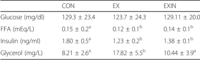
(P< 0.001) higher in EX group, respectively compared to respective CON and EXIN groups. However, no signifi-cant different between the CON and EXIN groups. Insu-lin levels were lower by 46 and 30% in EX and EXIN groups compared to CON group (P< 0.001,P< 0.01).
Energy metabolism during exercise
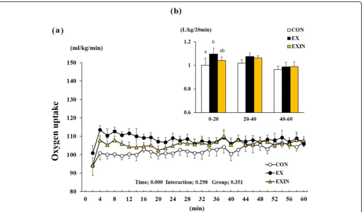
Repeated measures of oxygen uptake showed that time had a significant effect (P< 0.001), while interaction (P= 0.298) and group (P= 0.351) did not (Fig. 2a). Oxygen uptake during the initial 20 min period was elevated in the EX group compared to the CON and EXIN groups, which were nearly identical. (Fig.2b).
Repeated measures of RER showed that time had a sig-nificant effect (P< 0.001). Group-by-time interaction was also significant (P< 0.001) but group was not (P= 0.386) (Fig. 3a). The RER was significantly lower in the EX group than in the CON and EXIN groups during the ini-tial 20-min phase (Fig.3b), while there was no difference between the CON and EXIN groups.
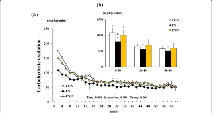
Carbohydrate oxidation was significantly affected by time (P< 0.001), interaction (P< 0.001), and group (P= 0.060) (Fig. 4a). It was significantly lower in the EX group than in the CON and EXIN groups during initial 20 min phase (Fig.4b), while there was no difference be-tween the CON and EXIN groups.
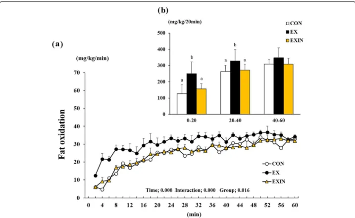
Fat oxidation was affected by time (P< 0.001), inter-action (P< 0.001), and group (P< 0.016) during the 1 h exercise (Fig.5a). The sum of fat oxidation during the 1 h period averaged 13% higher in the EX group than in the CON and EXIN groups (data not shown). Fat oxida-tion increased significantly during the initial 20 min phase in the EX groups compared to that in the CON Table 1Change of body weight, food intake and abdominal fat
for 4 weeks of experiment
CON EX EXIN
BW (g) Initial 33.53 ± 1.8 32.81 ± 1.4 32.96 ± 0.9
Final 40.51 ± 2.0a 36.14 ± 1.1b 40.01 ± 1.3a
Gain 6.97 ± 2.0a 3.30 ± 1.5b 6.98 ± 2.0a
Food intake (g) Day 5.15 ± 0.1 5.87 ± 0.1 5.60 ± 0.2
Total 144.20 ± 2.9 164.40 ± 1.5 156.83 ± 4.1
Abdominal fat (g) Epididymal 0.93 ± 0.3a 0.69 ± 0.1b 0.81 ± 0.2ab
Perirenal 0.36 ± 0.1 0.25 ± 0.0 0.33 ± 0.1
Mesentery 0.78 ± 0.1ab 0.69 ± 0.0b 0.81 ± 0.1a
Total 2.07 ± 0.6a 1.64 ± 0.2b 1.95 ± 0.4ab Note.Change of body weight (g), food intake (g/day, g/4 weeks) and abdominal fat for 4 weeks. CON (control,n= 8), EX (exercise, 60% VO2max,
n= 8), EXIN (Exercise + AR inhibitor, 60% VO2max,n= 8). Values are presented
as means ± standard deviations (n= 8). Different superscripts mean significant differences among the groups
Table 2Change in the plasma glucose, FFA, insulin and glycerol levels
CON EX EXIN
Glucose (mg/dl) 129.3 ± 23.4 123.7 ± 24.3 129.11 ± 20.0
FFA (mEq/L) 0.15 ± 0.2a 0.12 ± 0.1b 0.14 ± 0.1b
Insulin (ng/ml) 1.80 ± 0.5a 1.23 ± 0.2b 1.38 ± 0.1b
Glycerol (mg/L) 8.21 ± 2.6a 17.82 ± 5.5b 10.44 ± 3.9a
Note. Change in the plasma glucose, FFA, insulin and glycerol levels after 4 weeks of exercise. CON (control,n= 8), EX (exercise, 60% VO2max, n = 8), EXIN
(Exercise + AR inhibitor, 60% VO2max,n= 8). Values are presented as means ±
Fig. 2Change in oxygen uptake during 1 h of exercise (a), the sum of the oxygen uptake of the test group for each 20 min block of exercise (b). Energy metabolism measured during 1 h of exercise. CON (control,n= 8), EX (exercise, 60% VO 2 max,n= 8), EXIN (Exercise + AR inhibitor, 60% VO 2 max,n= 8). Values are presented as means ± standard deviations (n= 8). Different superscripts indicate significant differences between the groups (P< 0.05)
and EXIN groups (Fig. 5b) (P< 0.001, P< 0.003). In addition, the EX group showed higher fat oxidation than the CON and EXIN groups after 40 min of exercise (P< 0.020,P< 0.041). However, the EXIN group experienced the same intensity as the EX group but did not produce a high fat oxidation, like the CON group.
Expression of FAT/CD36 and CPTІin skeletal muscle
Western blot analysis was performed using protein ob-tained from the mouse skeletal muscle (soleus) samples. The FAT/CD36 protein level in the EX and EXIN groups was approximately double that of the CON group (P< 0.001) (Fig. 6a). The level of CPTІincreased in the EX group compared to the CON group (P< 0.0125) (Fig.6b). However, CPTI in the EXIN group was significantly lower than in the EX group (P< 0.0069).
Discussion
We demonstrated that a 4-week regimen of AR inhibitor treatment decreased oxygen uptake and fat oxidation relative to mice receiving placebo treatment during exer-cise in trained mice. On the protein expression level, we found that the AR inhibitor treatment decreased the ex-pression of CPTІ in trained mice. Furthermore, the AR inhibitor treatment group (EXIN) showed significantly higher body weight, abdominal fat tissue weight (total fat and mesenteric fat) compared with the placebo
treated exercise group. Our results suggest that the sup-pression of androgen hormone activity or lowering the sensitivity of AR can inhibit energy expenditure and fat oxidation by reducing CPTІin skeletal muscle.
In this study, we observed that oxygen uptake tended to be elevated during the initial exercise phase in the EX group, higher than the CON and EXIN groups. Interest-ingly, the EXIN group, which exercised with the same intensity during the same period, experienced decreased oxygen uptake during exercise. The EXIN group experi-encing AR blockage also showed a significant decrease in fat oxidation (12% lower than EX, data not shown) during the initial 0–20 min (P< 0.003) and after 20–40 min (P< 0.041). Furthermore, we found that body weight, abdominal fat (total and mesenteric fat) and plasma glycerol levels were significantly higher for the EXIN group compared to the EX group. Because both trained groups experienced elevated energy expend-iture, the AR inhibited group may indicate lower energy efficiency and dependence on carbohydrate utilization during exercise. In additional, plasma insulin was found to be reduced in both in EX and EXIN groups due to exercise effects. However, FFA showed a significant decrease only in the EX group than in the CON group. This means that the triglycerides break-down smoothly and FFA released into the blood is well used as energy source.
Fig. 5Change in fat oxidation during 1 h of exercise (a), the sum of each group’s fat oxidation for every 20-min block of exercise (b). Energy metabolism measured during 1 h of exercise. CON (control,n= 8), EX (exercise, 60% VO 2 max,n= 8), EXIN (Exercise + AR inhibitor, 60% VO 2 max,n= 8). Values are presented as means ± standard deviations (n = 8). Different superscripts indicate significant differences between the groups (P< 0.05)
mone depletion during the aging process. As in previ-ous studies, the inhibition of fat oxidation was similar. This study clearly confirms that chronic blockage of an-drogen receptors reduces energy efficiency and inhibits fat oxidation.
In this study, the FAT/CD36 and CPTІprotein levels were significantly higher in the EX group than in the CON group (P< 0.001, P< 0.0125). Continuous exercise has been reported to increase the expression of FAT / CD36 and CPTІ. These molecules transport fatty acids, mobilizing them for use as an energy source [14,19–23]. In particular, FAT / CD36 transports fatty acids from the cell membrane to the cytoplasm and mitochondria, while CPTІis present in the mitochondrial outer mem-brane and assists in translocation to the matrix [24]. The difference in CPTI expression in this study is very inter-esting. When ARs were inhibited, the expression of CPTI was significantly reduced (P< 0.0069), while FAT/ CD36 expression did not decrease even if ARs is blocked. This pattern was less pronounced in the CON (non-exercise) group. In present study, AR inhibition has not affected the expression of FAT/CD36 while de-creasing the expression of CPT1. This seems to be a gene that, unlike CPT1, is not affected by androgen hor-mones and is increased independently through exercise. Meanwhile, the decrease in CPT1 in our study seems to be due to the activation of Malonyl-CoA. Malonyl CoA is a potent inhibitor of carnitine palmitoyl transferase (CPT-1), an enzyme that controls fatty acids transport into the mitochondrion [25] (Additional file1).
According to a recently published review of ARs, an-drogens bound to the ARs to stimulate the transcription of enzymes required for de novo lipogenesis and recep-tors that mediate the uptake of fatty acids released by lipolysis from the circulation and adipocytes [26]. Previ-ous study that ARKO (androgen receptor knock out) mice were euphagic compared to the wild-type male controls, but also less dynamic and less oxygen consum-ing. Also, ARKO mice indicated that thermogenetic uncoupling protein 1 (UCP1) was lower than in wild-type group [27].
It was recently reported that androgen hormone treat-ment increased acyl-coenzyme A dehydrogenase long chain and hormone sensitive lipase [28]. Androgen treat-ment also stimulated fatty acid and triacylglycerol production, lipolysis, and cell shape reorganization [29].
is no group that only blocks AR. However, our study aimed at the effects of during exercise on fat metabolism and fat transport protein after blocking AR. Second, we did not measure the other gene expression related to fat metabolism. However, we confirmed that ARs blocking decreased CPT1 protein expression in the skeletal muscle and, therefore, we believe that the effect of ARs blocking on RER during exercise was due to the decrease fat utilization. In addition, many studies have reported that FAT / CD36 and CPT1 play a pivotal role in fatty acids transport and are highly correlated with whole body fat oxidation. Third, we know that all chemical in-hibitors are not specific, so we think that can not rule out the metabolic changes caused by other effects of inhibitors. It is also believed that additional studies will be needed to clarify the effectiveness of the inhibitor. In future investigations, it would be necessary to elucidate the effects of AR inhibition on the resting metabolism and a clear mechanism of fatty acids transport proteins.
Conclusions
We observed that chronic treatment of mice with AR in-hibitor while exercise training reduced whole-body fat utilization and energy efficiency in male mice. Further-more, AR blockade inhibited CPTІ production in skel-etal muscle. Our results suggest that a can decrease in androgen concentration or androgen receptor sensitivity affects exercise capacity by downregulating CPTІ. Re-duction of CPTI results in inhibition of fat oxidation and reduced energy efficiency by depriving skeletal muscle mitochondria of LCFA energy sources.
Supplementary information
Supplementary informationaccompanies this paper athttps://doi.org/10. 1186/s12986-019-0406-z.
Additional file 1: Figure S1.Expression levels of the FATP and FABP in skeletal muscle analyzed by western blotting. Results are expressed as relative abundance in the EXIN group (AR inhibitor with exercise training) compared with the CON (sedentary) and EX (exercise training with placebo). Values are presented as means ± standard deviations (n= 8).
Abbreviations
Acknowledgements
This paper was supported by the KU Research Professor Program of Konkuk University.
Authors’contributions
JSK1contributed to the study conception and experimental design, collected data, and performed analyses. JHP2interpreted the data and had primary responsibility for the final content. NHK2and HYP1participated in the study conception. KWL1,3provided advice on the study design and management. All authors were involved in editing the manuscript and read and approved the final manuscript.
Funding
This work was supported by the Ministry of Education of the Republic of Korea and the National Research Foundation of Korea
(NRF-2015S1A5B5A01016088).
Availability of data and materials
The data used to support the findings of this study are included within the article or available from the corresponding author upon request.
Ethics approval
This study was conducted in accordance with the ethical guidelines of the Konkuk University Institutional Animal Care and Use Committee, which incorporate the guidelines put forth in the Declaration of Helsinki (1964).
Consent for publication
All authors have read and approved the submission of the manuscript. The manuscript has not been published and is not being considered for publication elsewhere, in whole or in part, in any language.
Competing interests
The authors declare that they have no competing interests.
Author details
1Department of Sports Medicine and Science, Konkuk University, Gwangjin-gu, Seoul, Korea.2Physical Activity and Performance Institute (PAPI), Konkuk University, Gwangjin-gu, Seoul, Korea.3Department of Physical Education, Korea University, Seoul, Korea.4Department of Physical Education, Konkuk University, Gwangjin-gu, Seoul, Korea.
Received: 29 May 2019 Accepted: 29 October 2019
References
1. Fan W, Yanase T, Nomura M, Okabe T, Goto K, Sato T, Kawano H, Kato S, Nawata H. Androgen receptor null male mice develop late-onset obesity caused by decreased energy expenditure and lipolytic activity but show normal insulin sensitivity with high adiponectin secretion. Diabetes. 2005;54:1000–8.
2. Moretti C, Frajese GV, Guccione L, Wannenes F, De Martino MU, Fabbri A, Frajese G. Androgens and body composition in the aging male. J Endocrinol Investig. 2005;28:56–64.
3. Chin KY, Soelaiman IN, Naina Mohamed I, Shahar S, Teng NI, Mohd S, Ramli E, Ahmad F, Aminuddin A, Zurinah Wan Ngah W. Testosterone is associated with age-related changes in bone health status, muscle strength and body composition in men. Aging Male. 2012;15:240–5.
4. Kelly DM, Jones TH. Testosterone and obesity. Obes Rev. 2015;16:581–606. 5. Gustafson DR, Wen MJ, Koppanati BM. Androgen receptor gene repeats and
indices of obesity in older adults. Int J Obes. 2003;27:75–81. 6. Sinha-Hikim I, Taylor WE, Gonzalez-Cadavid NF, Zheng W, Bhasin S.
Androgen receptor in human skeletal muscle and cultured muscle satellite cells: up-regulation by androgen treatment. J Clin Endocrinol Metab. 2004; 89:5245–55.
7. Davey RA, Grossmann M. Androgen receptor structure, function and biology: from bench to bedside. Clin Biochem Rev. 2016;37:3–15. 8. Narayanan R, Coss CC, Dalton JT. Development of selective androgen
receptor modulators (SARMs). Mol Cell Endocrinol. 2018;465:134–42. 9. Power RF, Conneely OM, O'Malley BW. New insights into activation of
the steroid hormone receptor superfamily. Trends Pharmacol Sci. 1992; 13:318–23.
10. Rodriguez-Cuenca S, Monjo M, Frontera M, Gianotti M, Proenza AM, Roca P. Sex steroid receptor expression profile in brown adipose tissue. Effects of hormonal status. Cell Physiol Biochem. 2007;20:877–86.
11. Kim N, Kim J, Lim K, Park J. Role of dihydrotestosterone in whole-body energy utilization during acute running exercise in mice. J Exerc Nutr Biochem. 2018;22:7–11.
12. Guerrero J, Alfaro IE, Gómez F, Protter AA, Bernales S. Enzalutamide, an androgen receptor signaling inhibitor, induces tumor regression in a mouse model of castration-resistant prostate cancer. Prostate. 2013;73:1291–305. 13. Kim J, Lim K. Relationship between FAT/CD36 protein in skeletal muscle and
whole-body fat oxidation in endurance-trained mice. J Exerc Nutr Biochem. 2016;20:48–52.
14. McFarlan JT, Yoshida Y, Jain SS, Han XX, Snook LA, Lally J, Smith BK, Glatz JFC, Luiken JJFP, Sayer RA, Tupling AR, Chabowski A, Holloway GP, Bonen A. In vivo, fatty acid translocase (CD36) critically regulates skeletal muscle fuel selection, exercise performance, and training-induced adaptation of fatty acid oxidation. J Biol Chem. 2012;287:23502–16.
15. Talanian JL, Holloway GP, Snook LA, Heigenhauser GJ, Bonen A, Spriet LL. Exercise training increases sarcolemmal and mitochondrial fatty acid transport proteins in human skeletal muscle. Am J Physiol Endocrinol Metab. 2010;299:180–8.
16. Kim J, Hwang H, Park J, Yun HY, Suh H, Lim K. Silk peptide treatment can improve the exercise performance of mice. J Int Soc Sports Nutr. 2014;11:35. 17. Kim J, Park J, Kim B, Lee CH, Lim K, Suh H. Effects of different doses of silk
peptide on energy metabolism during exercise in mice. J Exerc Nutr Biochem. 2017;21:21–5.
18. Kim J, Lee KP, Lee DW, Lim K. Piperine enhances carbohydrate/fat metabolism in skeletal muscle during acute exercise in mice. Nutr Metab. 2017;14:43.
19. Kohn PG, Clausen T. The relationship between the transparent glucose and cations across cell membranes in isolated tissues.VI. The effect of insulin, ouabain, and metabolic inhibitors on the transport of 3-O-methylglucose and glucose in rat soleus muscles. Biochim Biophys Acta. 1971;225(2):277–90. 20. Bonen A, Campbell SE, Benton CR, Chabowski A, Coort SLM, Han XX,
Koonen PY, Glatz JFC, Luiken JFP. Regulation of fatty acid transport by fatty acid translocase/CD36. Proc Nutr Soc. 2004;63:245–9.
21. Koonen DP, Glatz JF, Bonen A, Luiken JJ. Long-chain fatty acid uptake and FAT/CD36 translocation in heart and skeletal muscle. Biochim Biophys Acta. 2005;1736:163–80.
22. Tunstall RJ, Mehan KA, Wadley GD, Collier GR, Bonen A, Hargreaves M, Cameron-Smith D. Exercise training increases lipid metabolism gene expression in human skeletal muscle. Am J Physiol Endocrinol Metab. 2002; 283:66–72.
23. Jeukendrup AE, Saris WHM, Wagenmakers AJM. Fat metabolism during exercise: a review--part II: regulation of metabolism and the effects of training. Int J Sports Med. 1998;19:293–302.
24. Stahl A, Gimeno RE, Tartaglia LA, Lodish HF. Fatty acid transport proteins: a current view of a growing family. Trends Endocrinol Metab. 2001;12:266–73. 25. Ussher JR, Lopaschuk GD. Targeting malonyl CoA inhibition of
mitochondrial fatty acid uptake as an approach to treat cardiac ischemia/ reperfusion. Basic Res Cardiol. 2009;104:203–10.
26. Butler LM, Centenera MM, Swinnen JV. Androgen control of lipid metabolism in prostate cancer: novel insights and future applications. Endocr Relat Cancer. 2016;23:R219–27.
27. Yanase T, Fan W, Kyoya K, Min L, Takayanagi R, Kato S, Nawata H. Androgens and metabolic syndrome: lessons from androgen receptor knock out (ARKO) mice. J Steroid Biochem Mol Biol. 2008;109:254–7. 28. Bolduc C, Larose M, Yoshioka M, Ye P, Belleau P, Labrie C, Morissette J,
Raymond V, Labrie F, St-Amand J. Effects of dihydrotestosterone on adipose tissue measured by serial analysis of gene expression. J Mol Endocrinol. 2004;33:429–44.
29. Bolduc C, Yoshioka M, St-Amand J. Transcriptomic characterization of the long-term dihydrotestosterone effects in adipose tissue. Obesity. 2007;15(5): 1107–32.
30. Sato K, Iemitsu M, Katayama K, Ishida K, Kanao Y, Saito M. Responses of sex steroid hormones to different intensities of exercise in endurance athletes. Exp Physiol. 2016;101:168–75.
Publisher’s Note