Dynamic localization of the C. elegans GoLoco proteins GPR-1/2 mediates mitotic spindle orientation in response to extrinsic signals
Adam David Werts
A dissertation submitted to the faculty of the University of North Carolina at Chapel Hill in partial fulfillment of the requirements for the degree of Doctor of Philosophy in the Department of Biology.
Chapel Hill 2011
ABSTRACT
ADAM DAVID WERTS. Dynamic localization of the C. elegans GoLoco proteins GPR-1/2 mediates mitotic spindle orientation in response to extrinsic signals
(Under the direction of Bob Goldstein)
Normal regulation of mitotic spindle orientation plays key roles in development and contributes to preventing tumorigenesis. In many cases, cell divisions are oriented by signals from neighboring cells. However, very little is known about how mitotic spindle orientation is established by signaling between cells. GoLoco-domain proteins (C. elegans GPR-1/2, Drosophila Pins, vertebrate LGN and AGS3) are good candidates for mediating spindle orientation in response to extrinsic signals, as they are key players in cell-autonomous mitotic spindle orientation, linking plasma membrane-associated Gα proteins to microtubule-associated proteins. In C. elegans, the GoLoco-domain protein pair GPR-1/2 has been implicated in orienting the spindle of an endomesodermal
suggesting that cortical GPR-1/2 localization in P2 occurred through inhibition of LET-99, a GPR-1/2 antagonist. Instead, my investigations suggested that GPR-1/2
accumulated through diffusion and preferential trapping at one cell contact, coupled with destabilization at another cell contact. Once the mitotic spindle of P2 is oriented normally, microtubule-dependent removal of GPR-1/2 prevented excess accumulation. I conclude that the dynamic localization of GPR-1/2 can serve as a key intermediate relaying positional information from one cell to the alignment of a mitotic spindle in the
ACKNOWLEDGMENTS
At the risk of sounding too clichéd, I would like to say that I would not be where I am today without the countless number of people who have helped me and/or challenged me along the way. Herein I would like to thank several people, and groups of people, who have played a particularly large part in my completion of a PhD.
equal. Steve’s style of communicating with students makes it very easy to bounce ideas off him and come up with some sweet experiments.
Next, I would like to thank my advisor, NOT boss, Dr. Bob Goldstein. Bob’s enthusiasm with all things creative, both inside and outside of the lab, has greatly affected my approach to science. From Bob, I have learned how science can be both informative and elegant. Also, while his somewhat hands-off mentoring style has left me struggling at times, it has also forced me to gain confidence and independence as a scientist. I believe I am much more well-prepared for my future career due his mentoring approach.
you for being the lab ‘Walking-Wikipedia” as Este calls you. Seriously, your knowledge base is amazing. I almost feel guilty for all the various ways you have taken time out of your day to selflessly help me. JSB, your patience and teaching ability is unbelievable. I guess that is what happens when you have two babies. Finally, to the newest Goldstein Lab members Chris Higgins and Jenny Heppert: It’s been great getting to know you, and I wish you much success in this lab and your future endeavors.
To my cohort who came to the Biology Department in 2006, as well as other friends I have made along the way: Thanks for providing an outlet from the lab in the form of athletics (volleyball, frisbee, hiking, etc) as well as time just relaxing, hanging out, and drinking some beers. This time is greatly coveted in graduate school.
Most importantly, I’d like to thank my family –Mom, Dad, Jennifer. You have supported me in all of my interests and endeavors, no matter how strange or unusual they were. While none of you have really understood what graduate school is, actually I didn’t either until about my second year here, you always supported me. Coming from a small town in the middle-of-nowhere Michigan, I know the only reason I am where I am today is because of your support and encouragement along the way.
PREFACE
People who didn’t grow up in a city like to say that they grew up in a small town -cue John Mellencamp- but this is all relative. I grew up in the small town of Onekama, Michigan. With around 500 inhabitants, two gas stations, two restaurants, and the nearest stoplight 15 miles away in the ‘city’ of Manistee (the largest township in the county with a population of 6,500), Onekama is a small town. I attended kindergarten, elementary school, and high school in the same building with the same 35 or so students each year. We had no fast food restaurants, no mall, no swim team (or pool), no tennis team, no wrestling team, no AP classes, no fancy robotics or technology labs, etc. By my senior year of high school I had taken all the upper level classes offered and thus found myself stuck in the school building with nothing to do literally: I was required to be in the building during the day, but there were no classes left to take. This is where I found my interest in art, but that is another story. So why would I start this preface talking about seemingly trivial things? Importantly, coming from a ‘Townie” beginning where
attending college is not always the expected route in life explains a lot about how I think, who I am, and how I ended up in North Carolina in graduate school.
she had two kids, my sister Jennifer and I, and spent most of her time as a stay-at-home mom and working part-time as a secretary at various locations. My father did not attend college, but my mother did take several college courses and finished an Associates degree. Despite their limited experience with postsecondary education, they always assumed that I would be going to college. None of us knew much about college, but I was going to go.
High school was pretty easy for me, possibly because the classes offered weren’t challenging in general, but regardless, I did well. Actually, quite well: I was both
Valedictorian and Athlete of the Year of Onekama High School. Upon graduation I went off to college. Since I knew little about the differences in colleges, I chose a convenient, close-by, liberal arts school called Grand Valley State University (GVSU).
Even after four years of undergrad, I still had no idea what I wanted to ‘do’. I had never really thought about a career-path, and college itself was sort of an abstract idea to me at that point. I felt like I should have some strong urge to do something specific, but that specific ‘thing’ eluded me. I half-heartedly applied to veterinary school in the fall of 2005, but never followed up with supplementary application material. Soon after, almost on a whim, I applied to graduate school, which, long story short, got me where I am today.
The summer before moving to North Carolina, I studied the website for the Biology Department at UNC and read through all the faculty profiles. Right away I became interested in the lab of Dr. Bob Goldstein: his science looked so simple but intriguing… elegant is a word I would use to describe it today. Thus, I emailed Dr. Goldstein and asked if he had space for me to rotate during the next year. A couple weeks later, I received an email from “Bob”, which was also sent to five other students, saying that he didn’t have space for everyone to rotate so only contact him if you were seriously considering joining his lab. This email worked, in part, scaring two people away from rotating, improving my chance at joining the lab. In the end, I was able to rotate with Bob in the fall of 2006.
I soon found that graduate school was very easy and that important scientific results came from nearly every experiment that I laid my hands on. Wait, that is a lie! Graduate school is hard: emotionally, intellectually, and even physically –try not having horrible back pains after nine hours of almost non-stop sitting at the microscope. On top of that, add the fact that my first two-thirds of a year in the Goldstein Lab was spent with Bob on sabbatical at the LMB in Cambridge and me having too much independence and not enough confidence, or maybe too much confidence, to know how to approach research efficiently. After two and a half years and two projects more or less falling through, it was time to get my act together.
If I have learned nothing else in graduate school, I have learned who I am and what I want to ‘do’. I have learned that persistence and dedication is required if you want to succeed at anything. I have learned how to ask pertinent scientific questions and design simple and sometimes clever experiments to address those questions. Finally, I learned that while I am good at basic bench science, my true career interests are in medicine and applying basic science discoveries to the real world. Thus, upon completion of my PhD I will start the next chapter of my life at veterinary school.
Within this dissertation you will find three chapters. Chapter 1 was written as an invited review for a special issue of Seminars in Cell and Developmental Biology
TABLE OF CONTENTS
LIST OF TABLES……….………xv
LIST OF FIGURES……….…...xvi
Chapter I. HOW SIGNALING BETWEEN CELLS CAN ORIENT A MITOTIC SPINDLE………...………..1
Abstract…...………...1
Introduction……..………...….3
Conclusions………18
Figures….………...20
References…...………...……25
II. DYNAMIC LOCALIZATION OF THE C.ELEGANS GOLOCO PROTEINS GPR-1/2 MEDIATES MITOTIC SPINDLE ORIENTATION IN RESPONSE TO EXTRINSIC SIGNALS……….………….………...33
Abstract...………...33
Introduction…..………..35
Materials and Methods………...40
Results………….………...45
Discussion..………57
Figures.………...64
LIST OF TABLES
LIST OF FIGURES
Figure 1.1: Polarity establishment by intrinsic cues, permissive external cues, and
instructive external ………...……….20
Figure 1.2: Model of the TPR-GoLoco protein complex…..……….……..…...22 Figure 1.3: Intercellular signaling functions as an instructive cue for spindle orientation mediated by the TPR-GoLoco protein Pins in Drosophila SOP cells………...23
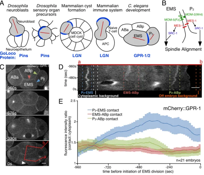
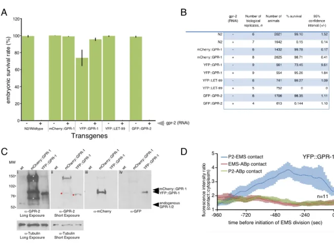
Figure 2.1: Live imaging of tagged GPR-1 accurately reports the dynamic
localization of endogenous GPR-1. ……….….………....64 Figure 2.2: mcherry::gpr-1 and yfp::gpr-1 made protein of the expected size and
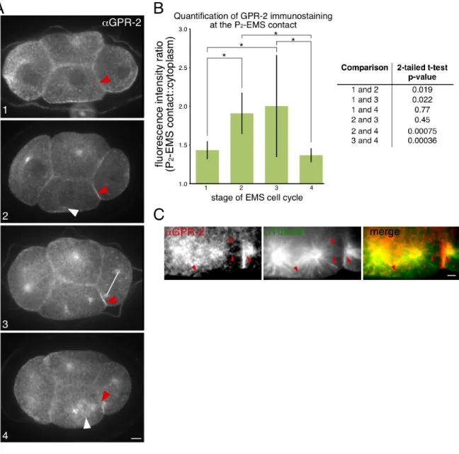
rescued knockdown of endogenous gpr-1/2.…………..………...………...……….66 Figure 2.3: Endogenous GPR-2 showed similar patterns to that of transgenic
GPR-1, with accumulation at the P2-EMS contact, then a decrease late in the cell
cycle, and accumulation on astral microtubules near the P2-EMS contact…...68 Figure 2.4: The GPR-1 transgenes recapitulated expected localization patterns of
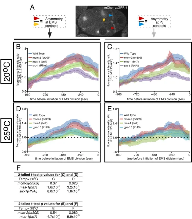
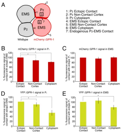
endogenous GPR-1/2 in all genetic backgrounds tested………....70 Figure 2.5: GPR-1 is enriched in P2 at the contact with EMS, positioned by cell
contact………...72 Figure 2.6: MES-1 regulates GPR-1 accumulation and spindle orientation in P2.………74 Figure 2.7: GPR-1 is asymmetric in P2 but not EMS……….………...76 Figure 2.8: Normal GPR-1 distribution in P2 plays important roles in
development…...………....77 Figure 2.9: MES-1/SRC-1 signaling localizes cortical GPR-1 independently of
LET-99………...79 Figure 2.10: mes-1(RNAi) and src-1(RNAi) disrupted accumulation of
mCherry::GPR-1, but not YFP::LET-99, at the P2-EMS cell-cell contact………..……..81 Figure 2.11: Asymmetric localization of GPR-1 in P2 is established by
destabilization at one cell-contact site, diffusion and stabilization at another
Figure 2.12: FRAP curves, maximum recovery, and corrections for expected
change in signal during FRAP acquisition………..………...85 Figure 2.13: Microtubule-dependent removal of cortical GPR-1 prevents excessive
CHAPTER 1
How signaling between cells can orient a mitotic spindle
This chapter is adapted from a solicited review written for a special issue of Seminars in
Cell and Developmental Biology, which focuses on polarized cell growth. I wrote the
first draft of the review and made all of the figures. Bob Goldstein wrote the
‘evolutionary origins and conclusions’ section, and the two of us together revised
subsequent drafts.
ABSTRACT
In multicellular animals, cell communication sometimes serves to orient the
direction in which cells divide. Control of division orientation has been proposed to be
critical for partitioning developmental determinants and for maintaining epithelial
architecture. Surprisingly, there are few cases where we understand the mechanisms by
which external cues, transmitted by intercellular signaling, specify the division
orientation of animal cells. One would predict that cytosolic molecules or complexes
exist that are capable of interpreting extrinsic cues, translating the positions of these cues
into forces on microtubules of the mitotic spindle. In recent years, a key intracellular
complex has been identified that is required for pulling forces on mitotic spindles in
localized in positions that coincide with the positions of spindle-orienting extracellular
cues. Do TPR-GoLoco proteins function as conserved, spatially-regulated mediators of
spindle orientation by intercellular signaling? Here, we review the relevant evidence
among cases from diverse animal systems where this protein complex has been found to
INTRODUCTION
1. Intercellular communication is a necessary aspect of some cell divisions in animal systems
1.1 Intercellular signals can guide cell division
Cell division orientation is an important part of development and tissue maintenance [1-6]. Abnormal placement of the division plane has been recognized to disrupt cell fate specification for over 30 years [7] and has more recently been proposed to contribute to defective morphogenesis [1, 2, 5] and cancer [8]. While cell shape has been shown to be one contributor to placement of cell division planes [9-12], recent discoveries have highlighted a role of cell signaling in spindle orientation.
Since the 1990s, it has become clear that extrinsic signals can determine the orientation in which certain cells divide. Manipulating the positions of cells and signals has revealed that the position from which an extrinsic signal is presented to a cell can determine the orientation of the cell division machinery [13-15]. These experiments make clear that extrinsic signals can function not just as simple switches that allow cells to respond to internal polarity (permissive cues), but instead can serve as important positional landmarks that determine the specific orientations of mitotic spindles (instructive cues) (Figure 1): Cells are telling their neighbors in which direction to divide.
secreted molecule, like Wnt [14], or a transmembrane or adhesive molecule, such as cadherin [17]. 2) Second, the cell needs to interpret the external cue, translating its position into internal polarity. 3) Third, the internal polarity must be translated into forces on the cytoskeleton to set up a specified axis of division. 4) Lastly, the cell needs to divide. This can result in the partitioning of cell fate determinants [18], to one daughter cell.
1.2 An example of the importance of intercellular communication for cell division orientation: the role of cadherin in the Drosophila germline
centrosome always remaining anchored to the contact with the niche [23]. The mother centrosome is likely anchored by a physical link between astral microtubules and E-cadherin-rich adherens junctions between the stem cell and the hub cells through an APC (adenomatous polyposis coli) protein [21, 24]. In this system, it has not been shown whether E-cadherin and APC function as instructive cues for spindle orientation or whether this centrosome-anchoring phenomenon merely provides a permissive external cue to orient division in response to a separate cue. In the future, it would be interesting to determine whether GSC division is oriented by instructive or permissive cues from the hub, by experimentally repositioning the adherens junctions, possibly through cell manipulations, and assaying for re-establishment of centrosome anchoring and reorientation of the mitotic spindle of the stem cell in relation to the hub.
E-cadherin and APC have also been implicated in similar processes in other systems, namely the regulation of cell polarity [17, 25], centrosome tethering [26], and mitotic division orientation [27]. This suggests that E-cadherin-mediated polarity is one key way in which cells communicate to regulate division orientation. (For a recent review on adhesion molecules regulating stem cell division, see [28]).
1.3 TPR-GoLoco proteins as candidate transducers of positional information from
intercellular signaling to spindle orientation
progenitors [41-43], mammalian T-cells [44], and gastrulating zebrafish embryos [35]. This complex was discovered largely independently in C. elegans embryos, Drosophila
neuroblasts, and cultured mammalian cells. In the mid-1990s, heterotrimeric G-proteins were implicated in cell division orientation in C. elegans embryos, as early as the one-cell stage [45]. Gα proteins were later found to be the relevant G-protein components [30, 46]. Heterotrimeric G-proteins were known to respond to extracellular signals, via seven-pass transmembrane receptors, so it was surprising to find a role for these proteins in the one-cell stage embryo, in a cell that has no neighbors from which to receive signals.
How might G-proteins function in the absence of cell-cell signaling at the one-cell stage? A possible solution came when receptor-independent activators of G-protein signaling (called AGS or RGS proteins) were identified [47-49]. Proteins resembling these were found in flies (a protein called Pins), worms (GPR-1/2) and mammals (LGN), and importantly, these proteins were shown to function in spindle positioning in each system [29, 50-53]. Each of these proteins has a Gα-binding "GoLoco" domain, which can displace Gβ and inhibit GDP dissociation from Gα [54, 55]. They also have a protein-interaction domain consisting of 34-amino acid repeats (tetratricopeptide repeats, or TPR; [56]) that include the amino acids LGN (Leu-Gly-Asn) [57].
51, 58-60]. The TPR-GoLoco protein LGN was shown to act as a conformational switch that binds Gα and NuMA simultaneously, providing a link from a plasma membrane-associated protein to the mitotic spindle [61]. Also membrane-associated is the microtubule motor dynein [62, 63], as well as Discs-large (Dlg; a membrane-associated guanylate kinase) and kinesin [64], or Inscuteable in some systems [53] (Figure 2).
Altogether, the data suggest that the TPR-GoLoco-containing complex serves as a connection between the plasma membrane and microtubules. Indeed, the complex can be demonstrated to be important for the formation of a mechanical link between the plasma membrane and astral microtubules: Laser-cutting experiments in C. elegans embryos have revealed cortical pulling forces on mitotic spindles [65, 66], and these forces are lost in the absence of members of the complex [51, 52, 65]. How then does the complex move a spindle to a specific site on the cortex, instead of providing similar pulling forces along the entire cell cortex? In many systems, one member of the complex is localized to a specific cortical site, and is absent or at lower levels elsewhere. In most cases, it is the TPR-GoLoco protein that is asymmetrically localized [36, 51-53, 67], although the NuMA/Mud/LIN-5 component can be asymmetrically localized as well in some systems [36, 58-60, 68]. TPR-GoLoco proteins are increasingly found with restricted localization in parts of the cell cortex in systems with medical relevance, including vertebrate lung epithelia [39, 69], neuroepithelia [40], neural progenitors [41-43], T-cells [44], and in a model for cyst formation [38].
However, it is not clear in all cases whether the TPR-GoLoco proteins localize downstream of extrinsic cues, or only downstream of an intrinsic polarity (Figure 1). Here, we ask a specific question: Do the protein complexes containing TPR-GoLoco proteins serve as conserved machines that can interpret positional information from extracellular cues, and translate that information into specific spindle orientations? To address this, we discuss first the most thoroughly described examples of TPR-GoLoco protein localization and function in model systems where intercellular signaling may be involved. We end with recent discoveries from vertebrate systems and discuss the evolution of TPR-GoLoco protein functions. Throughout, we point out similarities and differences between systems, in an attempt to sample the broad range of contexts to which this complex has been adapted for orienting mitotic divisions.
2. Drosophila sensory organ precursors: A clear connection between intercellular
signaling, TPR-GoLoco protein positioning, and instructive control of spindle
orientation
In the developing Drosophila peripheral nervous system, sensory organ precursor
cells (SOPs) undergo several rounds of asymmetric division, forming an external sensory
organ composed of five cell types: hair, socket, neuron, sheath, and glia [70, 71]. The
orientation of the initial division of the progenitor cell, called pI, is controlled by planar
cell polarity (PCP) proteins: the transmembrane proteins Frizzled (Fz) and
Strabismus/Van Gogh (Stbm/Vang; a four-pass transmembrane protein in Drosophila,
also called Van Gogh-like 2 or Vangl2 in mammals [72]) [15, 73]. In PCP, the
extracellular domain of Fz on neighboring cells, resulting in nonautonomous polarization
of cells, with Stbm/Vang recruited to one side of each cell [73]. This interaction may be
indirect, relying on homotypic interactions between two functional forms of Flamingo
(Fmi) that can recruit Fz and Stbm/Vang to opposite sides of a cell boundary [74] (Figure
3A). In the sensory organ precursors, PCP proteins affect the localization of the
Drosophila TPR-GoLoco protein Pins, with Pins localizing to the anterior side of the cell,
where Stbm/Vang is recruited [15, 75].
In the Drosophila SOPs, Ric-8, a guanine nucleotide-exchange factor, recruits
Pins by regulating the localization of its cortical tether Gαi [76], while Stbm/Vang
functions to refine Pins recruitment to the anterior cortex [77]. In parallel, Dsh prevents
posterior cortical accumulation of Pins [77], likely by inhibition of cortical Stbm/Vang
[15], resulting in restriction of the size of the cortical domain that attracts centrosomes
(Figure 3A). At the anterior cortex, Pins interacts with Dlg, an interaction that is
important for the anterior accumulation of both proteins [75]. Cortically localized Pins
and Dlg regulate cell polarity through localization of Bazooka (Baz; PAR-3 in other
systems), excluding Baz from the anterior cortex and allowing its accumulation at the
posterior cortex [75] (Figure 3A and 3B). Pins and Baz contribute redundantly to spindle
positioning [78]. The result of Fz-Dsh signaling and polarized Pins recruitment is an
oriented spindle which is controlled in two dimensions: along the axis of
anterior-posterior polarity of the tissue and within the plane of the epithelium, oblique to the
apical basal axis [35, 76] (Figure 3C).
Interestingly, this is the only system to date where there is a clear connection
orientation. The PCP protein Dsh acts downstream of Fz to restrict the size of the Pins
cortical domain (Figure 3A). In some Dsh mutants, the cortical domain of Pins is
expanded, and both centrosomes in the cell, rather than just one, move inappropriately
toward the Pins domain during mitosis. This results in misoriented cell division [15].
Furthermore, moving intercellular signals by generating clones of cells that lack
Stbm/Vang or that have reduced Fz results in the relocalization of cortical Pins and
reorientation of the mitotic spindle. This makes clear that Pins localization responds to
the position at which intercellular signaling occurs [15] (Figure 3D). These results
highlight the importance of intercellular signaling as an instructive cue for localizing
cortical TPR-GoLoco proteins, and they provide a template for how TPR-GoLoco
proteins might function in other systems as intermediates between instructive external
cues and spindle positioning.
3. Early C. elegans development: Role of the TPR-GoLoco protein GPR-1/2 in the
four-cell stage embryo
At the four-cell stage in the C. elegans embryo, two adjacent cells signal to each
other, affecting the division orientation of each cell. An endomesodermal cell called
EMS, and a germline precursor cell called P2, orient their divisions toward a shared
cell-cell contact [13, 79]. EMS signals to P2 via a pathway involving MES-1 (a receptor
tyrosine kinase-like transmembrane protein unique to nematodes) upstream of a Src
kinase (SRC-1). P2 signals to EMS via both the MES-1 pathway and a Wnt pathway [14,
29-31, 51, 79-84]. These signaling pathways are required for normal division orientation
demonstrated that Wnt-expressing P2 cells can provide instructive cues for spindle
orientation to the EMS cell [83]. MES-1, on the other hand, acts as a permissive cue in
EMS --required for spindle orientation toward a site at which Wnt signaling occurs [83].
What occurs downstream of these pathways that might affect division orientation?
Both MES-1 and SRC-1 have been implicated in recruiting dynactin to the P2-EMS
contact, and dynactin is required for normal EMS division orientation [31]. An
enrichment of the TPR-GoLoco protein pair GPR-1/2 is also observed at the P2-EMS
boundary [29, 30, 51]. MES-1 signaling, but not Wnt signaling, is essential for GPR-1/2
recruitment [29, 30] and enrichment of phosphotyrosine at the P2-EMS contact [82]. This
suggests that the local phosphorylation of some target(s) that might be involved in normal
spindle alignment. However, the identity of such target(s) has yet to be determined.
While GPR-1/2 appears to be a mediator of intercellular signaling and division
orientation, a direct relationship between intercellular signaling, TPR-GoLoco protein
localization, and spindle orientation has yet to be clearly demonstrated in this system.
With GPR-1/2 enrichment seen at a contact site between two cells, it is difficult to know
if GPR-1/2 is asymmetrically localized in EMS, in P2, or in both cells. However, a
consensus in the field is that GPR-1/2 cortical enrichment is involved at least in EMS
division orientation [4, 29, 30, 85, 86]. This is based in part on evidence that inactivating
one of GPR-1/2’s cortical Gα tethers, GPA-16, or inactivating a polarized, cortical
GPR-1/2 antagonist, the DEP-domain protein LET-99, both result in spindle orientation defects
in the EMS cell [30]. Since MES-1/SRC-1 signaling is required for both GPR-1/2
enrichment at the P2-EMS contact and normal P2 division [79, 81], it is possible that
to specific positions, and to image responses at high resolution, makes this system an
especially attractive one to us for addressing fundamental questions in the future.
4. Drosophila neuroblasts: The TPR-GoLoco protein Pins orients divisions
Drosophila neuroblasts, which are central nervous system progenitor cells, form
by delamination from the neuroepithelium during embryonic development. Once
delaminated, neuroblasts become polarized and undergo repeated cycles of asymmetric
divisions, resulting in self-renewal of the neuroblast and the production of ganglion
mother cells (for a recent review focusing on Drosophila neuroblast polarity see [87]).
When mechanically dissociated from the neuroepithelia, Drosophila neuroblasts retain
the ability to divide asymmetrically, albeit in random orientations with respect to
previous divisions, suggesting that they can self-polarize, but that the polarity is
randomly established from one division to the next [88]. However, when neuroblasts
remained in contact with clusters of at least two neuroepithelial cells during dissociation,
cell divisions were oriented in relation to the contact site [88]. This suggests that the
neuroblasts orient divisions by an external cue that might function either as an instructive
or permissive signal for normal spindle orientation. The identity of the relevant cell-cell
signaling molecules remains unknown to date.
In Drosophila neuroblasts, Pins functions in division orientation. Pins is recruited
to the apical cortex by two parallel pathways: the Inscuteable(Insc)/Par pathway [53, 67,
89], and the microtubule/Khc-73 (kinesin)/Dlg pathway [32, 64]. In the Insc/Par pathway,
interactions between Pins, Insc, and Baz (a homolog of Par3) are required for asymmetric
localization results in disruption of this entire protein complex, and is associated with
mitotic spindle orientation defects and loss of division asymmetry [53, 67, 89].
Interestingly, unlike in Drosophila SOPs, Pins in Drosophila neuroblasts localizes with,
rather than opposite, the cortical polarity protein Baz. This reversal of Pins polarity
between two cell types within the same organism has been attributed to the expression of
Insc, as ectopic expression of Insc in SOPs, cells in which Insc is not normally expressed,
causes Pins to colocalize with Baz and reverse the polarity of the cell [75]. This further
highlights the broad range of contexts in which the TPR-GoLoco protein module can be
applied to spindle orientation downstream of cell interactions, even within the same
organism.
In parallel to the Insc/Par pathway, the microtubule/Khc-73/Dlg pathway [32, 64]
of apical Pins recruitment appears to function at metaphase to maintain linkage between
the mitotic spindle and cortical polarity, possibly through cooperation with proteins such
as Mud and dynein [32, 64]. Pins in Drosophila neuroblasts is known to bind directly to
the microtubule-associated protein Mud, which functions similarly to C. elegans LIN-5
and mammalian NuMA, and is required for normal mitotic spindle orientation [58, 60].
Furthermore, similar to dynactin’s role in orienting divisions in the four-cell stage C.
elegans embryo ([31], discussed above), the Lis1/dynactin complex has been shown to
regulate Drosophila neuroblast spindle orientation [34], potentially downstream of Pins
and Mud [64]. Thus, conserved players may play conserved roles in different systems.
In the future, it will be interesting to identify the nature of the external cue, and
see if intercellular signaling in this system functions in an instructive or permissive
intercellular signaling orients division by similar mechanisms as in other systems, such as
the cadherin adhesions in Drosophila germline stems cells, or by novel signals, such as
functional homologs of C. elegans MES-1, to further understand the diversity of
signaling mechanisms to which TPR-GoLoco localization has become linked through
evolution.
5. Vertebrate cells use conserved TPR-GoLoco protein complex members to orient
cell divisions
In recent years, multiple studies have implicated the vertebrate TPR-GoLoco
proteins LGN or AGS3 in normal division orientation of polarized cells in various
epithelia [36-40, 69, 90, 91], neural progenitors [41-43], and possibly mammalian T-cell
divisions [44]. Vertebrate TPR-GoLoco proteins localize near certain cell-cell contacts
and orient divisions in multiple tissues [36-38, 40-43]. To our knowledge, there is little
evidence addressing whether the localization of vertebrate TPR-GoLoco proteins at
cell-cell contacts is determined by instructive, intercell-cellular signals. Alternatively, it is possible
that intrinsic polarity cues affect TPR-GoLoco protein localization, and perhaps division
alignment, independent of extrinsic signals.
The mechanisms that localize the TPR-GoLoco protein LGN in mammalian
epithelial cells has been studied in detail using an in vitro model, Madin-Darby Canine
Kidney (MDCK) cell epithelial cyst formation. When plated in a 3D matrix, MDCK cells
divide and develop into cysts of apico-basally polarized epithelial cells surrounding
hollow lumens [92]. LGN localizes near cell-cell contacts within the cyst [38]. This
morphology, as disrupting LGN’s cortical localization, disrupting interactions with
LGN’s binding partners, or artificially mistargeting LGN’s cortical Gα tether to an
ectopic membrane all result in spindle orientation defects and disorganized cysts with
multiple lumens [38]. Normal LGN localization is controlled by known cell polarity
proteins, Par3 and aPKC [37]. In this system, Par3 recruits aPKC, which can
phosphorylate LGN on serine 401, allowing a 14-3-3 protein to inhibit LGN cortical
localization by blocking phospho-LGN binding to the cortical tether, Gαi, at the apical
lumen [37]. This in turn permits LGN to be enriched near cell-cell contacts, a position to
which mitotic spindles align [37, 38]. In this system, LGN appears to localize similarly to
Pins in Drosophila SOPs: on cortical domains that have little Par3/Baz. Similarities
between the two systems suggest that intercellular communication might localize LGN to
orient divisions in MDCK cell cysts too, but this has yet to be tested.
Although LGN localization is aPKC-dependent in MDCK cells, this is not a
hallmark of LGN localization mechanisms in all vertebrate cells. In chick neuroepithelial
cells, LGN localizes to a lateral cortical belt around the cell, and is required for cell
division within the plane of the epithelium [90]. In this case, apical aPKC appears to be
neither necessary nor sufficient to exclude apical LGN localization: LGN still localizes to
the cortex when aPKC is constitutively activated and cortically localized; an aPKC
inhibitor does not prevent normal LGN localization; and mutation of serine 401 reduces,
but does not mislocalize, cortical LGN asymmetry [40]. Taken together, it appears that
similar to invertebrate systems, how LGN becomes localized in vertebrate cells is context
and organism specific, further highlighting the plasticity of the module through which
Two examples from vertebrate systems suggest that LGN localization depends on
extracellular cues. First, during asymmetric division in mouse neural progenitors, mice
deficient in the PCP mediator Vangl2 show decreased asymmetric distribution of cortical
LGN, and this is associated with altered division orientation [43]. This suggests that PCP
signaling may mediate spindle orientation through localization of TPR-GoLoco-domain
proteins as has been shown in Drosophila SOPs [15], however whether this signaling is
instructive or permissive is unknown. In a second system, mammalian skin, there is
evidence that LGN localization is mediated by extracellular signals, from both the
basement membrane, and other cells [36]. Disruption of β1 integrin, a protein essential
for focal adhesions and basement membrane assembly, or disruption of α-catenin, a
component of adherens junctions between cells, causes mislocalization of LGN and
spindle orientation defects [36]. While this suggests that the basement membrane and
cell-cell contacts may contribute to positioning LGN, whether these are instructive or
simply permissive cues for LGN localization remains untested.
6. Roles for Lin-5/Mud/NuMA in spindle orientation independent of TPR-GoLoco proteins
CONCLUSIONS
7. Evolutionary origins and conclusions
Do TPR-GoLoco proteins function as conserved, spatially-regulated mediators of
spindle orientation by intercellular signaling? Addressing the extent to which molecules
and mechanisms are conserved requires some knowledge about the molecules and
mechanisms of ancestral organisms. We would like to understand when these proteins
first arose and what functions they had at various stages of animal evolution. The
existence of proteins with TPR motifs N-terminal to GoLoco motifs in worms, flies and
vertebrates suggests an ancient origin of this protein family, in early bilateral animals. At
this stage in animal evolution, it is likely that TPR-GoLoco proteins functioned in mitotic
spindle orientation, perhaps in response to intrinsic polarity cues, since this function is
common to worms, flies and vertebrates.
We have found proteins with at least one TPR motif N-terminal to a GoLoco
motif by a CDART search [95] in more distantly related animals, such as placozoans and
cnidarians, and even outside of the animals, in a unicellular choanoflagellate,
Salpingoeca. The presence of these proteins in the broad diversity of animals plus a
choanoflagellate -- thought to be a sister group to the animals [96] -- suggests that
TPR-GoLoco proteins existed before bilateral animals arose, in the ancient, ancestral animals
present more than 500 million years ago. Whether TPR-GoLoco proteins functioned in
spindle orientation this early is not clear. Determining whether distantly related
organisms such as cnidarians or choanoflagellates use TPR-GoLoco proteins to orient
When did TPR-GoLoco proteins acquire a role in mediating instructive,
intercellular cues? To date, solid evidence that TPR-GoLoco proteins mediate instructive
extracellular cues for spindle orientation exists only in Drosophila sensory organ
precursors. In systems where there is not yet solid evidence addressing this specific point,
experimentally moving the extracellular cues to new positions will make it possible to
determine if this is the case. This protein family might have initially functioned in spindle
orientation independently of intercellular signaling, and these proteins might have been
co-opted by intercellular signaling pathways later. Work toward understanding the
mechanisms by which TPR-GoLoco proteins function with intercellular signaling, in
diverse systems, may be important for answering the more general question of how
signaling between cells can orient cell divisions in ways that can lead to normal
FIGURES
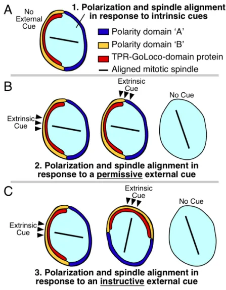
Figure 1.1. Polarity establishment by intrinsic cues, permissive external cues, and
instructive external cues
(A) Some cells align their mitotic spindles independent of external signaling cues.
Polarity domains ‘A’ and ‘B’ are nonspecific and could represent ‘Anterior’ and
‘Posterior’ polarity, ‘Apical’ and ‘Basal’ polarity, ‘Dorsal’ and ‘Ventral’ polarity, etc,
depending on the specific cell type.
(B) Permissive external cues: Some cells require an external cue (black arrowheads) for
information to cell polarity: moving the cue has no effect on cell polarity or spindle
orientation (middle). Absence of these cues leads to polarity defects and defects in
spindle orientation (right).
(C) Instructive external cues: Some cells are polarized by instructive external cues (black
arrowheads), where changing the position of the cue changes the orientation of polarity
and division. Experimentally moving the position of an extrinsic cue differentiates
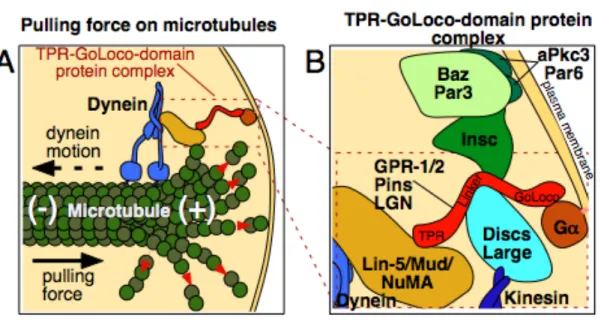
Figure 1.2. Model of the TPR-GoLoco protein complex
(A) Schematic showing how the TPR-GoLoco protein complex forms a link between
microtubules and the plasma membrane, after [62]. Red arrows show tubulin
depolymerization from the plus end of the microtubule. Proteins are depicted roughly
proportional to their relative sizes.
(B) Enlargement of part of (A), with additional associated proteins included. See text for
details. Gα is myristoylated (pink line), associating it with the plasma membrane.
Inscuteable links Par3/Baz to TPR-GoLoco proteins in some cells such as Drosophila
neuroblasts [53], but this interaction is absent in cells such as MDCK cyst cells and
Drosophila SOPs, where Par3/Baz localizes in a reciprocal cortical pattern to
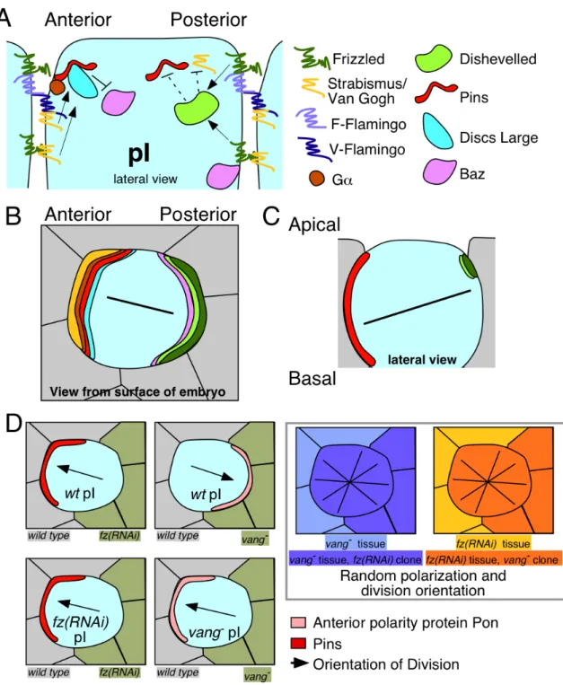
Figure 1.3. Intercellular signaling functions as an instructive cue for spindle
orientation mediated by the TPR-GoLoco protein Pins in Drosophila SOP cells.
(A) Extracellular signals from Frizzled, Strabismus, and Flamingo control Pins cortical
have been proposed: F-Flamingo and V-Flamingo [48]. F-Flamingo is proposed to
interact with and be induced by Frizzled in the same cell, while V-Flamingo is proposed
to interact with Vang in the same cell [74].
(B-C) Schematics after [35, 76]. (B) A view from the embryo’s surface of an SOP cell.
Colored crescents represent polarized localization of proteins shown in (A), using the
same colors. (C) Schematic of localization of the relevant cortical proteins controlling
division along the axis of polarity and within the plane of the epithelium. Black lines
represent the orientation of the mitotic spindle.
(D) Schematic of SOP clone border analysis from experiments described in [15]. Loss of Fz or Vang on one side of a SOP cell results in altered polarization of Pins and another anterior cortical protein called Partner of Numb (Pon), and alignment of the mitotic spindle. Black arrows represent spindle alignment, and crossed black lines on the far right panels represent randomization of the division axis in the absence of external cues. SOPs in the far right two panels still polarize and divide asymmetrically in the absence of PCP signaling, but in a random orientation.
REFERENCES
1. Baena-Lopez, L.A., Baonza, A., and Garcia-Bellido, A. (2005). The orientation of cell divisions determines the shape of Drosophila organs. Curr Biol 15, 1640-1644.
2. Fischer, E., Legue, E., Doyen, A., Nato, F., Nicolas, J.F., Torres, V., Yaniv, M., and Pontoglio, M. (2006). Defective planar cell polarity in polycystic kidney disease. Nat Genet 38, 21-23.
3. Saburi, S., Hester, I., Fischer, E., Pontoglio, M., Eremina, V., Gessler, M., Quaggin, S.E., Harrison, R., Mount, R., and McNeill, H. (2008). Loss of Fat4 disrupts PCP signaling and oriented cell division and leads to cystic kidney disease. Nat Genet 40, 1010-1015.
4. Segalen, M., and Bellaiche, Y. (2009). Cell division orientation and planar cell polarity pathways. Semin Cell Dev Biol 20, 972-977.
5. Karner, C.M., Chirumamilla, R., Aoki, S., Igarashi, P., Wallingford, J.B., and Carroll, T.J. (2009). Wnt9b signaling regulates planar cell polarity and kidney tubule morphogenesis. Nat Genet 41, 793-799.
6. Quyn, A.J., Appleton, P.L., Carey, F.A., Steele, R.J., Barker, N., Clevers, H., Ridgway, R.A., Sansom, O.J., and Nathke, I.S. (2010). Spindle orientation bias in gut epithelial stem cell compartments is lost in precancerous tissue. Cell Stem Cell 6, 175-181.
7. Whittaker, J.R. (1980). Acetylcholinesterase development in extra cells caused by changing the distribution of myoplasm in ascidian embryos. J Embryol Exp Morphol 55, 343-354.
8. Pease, J.C., and Tirnauer, J.S. (2011). Mitotic spindle misorientation in cancer - out of alignment and into the fire. J Cell Sci 124, 1007-1016.
9. Symes, K., and Weisblat, D.A. (1992). An investigation of the specification of unequal cleavages in leech embryos. Dev Biol 150, 203-218.
10. Rappaport, R., and Rappaport, B.N. (1994). Cleavage in conical sand dollar eggs. Dev Biol 164, 258-266.
11. Tsou, M.F., Ku, W., Hayashi, A., and Rose, L.S. (2003). PAR-dependent and geometry-dependent mechanisms of spindle positioning. J Cell Biol 160, 845-855. 12. Gibson, W.T., Veldhuis, J.H., Rubinstein, B., Cartwright, H.N., Perrimon, N.,
Brodland, G.W., Nagpal, R., and Gibson, M.C. (2011). Control of the mitotic cleavage plane by local epithelial topology. Cell 144, 427-438.
14. Schlesinger, A., Shelton, C.A., Maloof, J.N., Meneghini, M., and Bowerman, B. (1999). Wnt pathway components orient a mitotic spindle in the early
Caenorhabditis elegans embryo without requiring gene transcription in the responding cell. Genes Dev 13, 2028-2038.
15. Gomes, J.E., Corado, M., and Schweisguth, F. (2009). Van Gogh and Frizzled act redundantly in the Drosophila sensory organ precursor cell to orient its
asymmetric division. PLoS One 4, e4485.
16. Hertwig, O. (1893). Uber den Werth der ersten Furchungszellen fur die Organbildung des Embryos. Experimentelle Studien am Frosch und Tritonei. Archiv fur mikroscopische Anatomie 42, 662-807.
17. Dupin, I., Camand, E., and Etienne-Manneville, S. (2009). Classical cadherins control nucleus and centrosome position and cell polarity. J Cell Biol 185, 779-786.
18. Yan, B. (2010). Numb--from flies to humans. Brain Dev 32, 293-298.
19. Fuller, M.T., and Spradling, A.C. (2007). Male and female Drosophila germline stem cells: two versions of immortality. Science 316, 402-404.
20. Song, X., Zhu, C.H., Doan, C., and Xie, T. (2002). Germline stem cells anchored by adherens junctions in the Drosophila ovary niches. Science 296, 1855-1857. 21. Inaba, M., Yuan, H., Salzmann, V., Fuller, M.T., and Yamashita, Y.M. (2010).
E-cadherin is required for centrosome and spindle orientation in Drosophila male germline stem cells. PLoS One 5, e12473.
22. Voog, J., D'Alterio, C., and Jones, D.L. (2008). Multipotent somatic stem cells contribute to the stem cell niche in the Drosophila testis. Nature 454, 1132-1136. 23. Yamashita, Y.M., Mahowald, A.P., Perlin, J.R., and Fuller, M.T. (2007).
Asymmetric inheritance of mother versus daughter centrosome in stem cell division. Science 315, 518-521.
24. Yamashita, Y.M., Jones, D.L., and Fuller, M.T. (2003). Orientation of asymmetric stem cell division by the APC tumor suppressor and centrosome. Science 301, 1547-1550.
25. Nejsum, L.N., and Nelson, W.J. (2007). A molecular mechanism directly linking E-cadherin adhesion to initiation of epithelial cell surface polarity. J Cell Biol
178, 323-335.
27. Le Borgne, R., Bellaiche, Y., and Schweisguth, F. (2002). Drosophila E-cadherin regulates the orientation of asymmetric cell division in the sensory organ lineage. Curr Biol 12, 95-104.
28. Marthiens, V., Kazanis, I., Moss, L., Long, K., and Ffrench-Constant, C. (2010). Adhesion molecules in the stem cell niche--more than just staying in shape? J Cell Sci 123, 1613-1622.
29. Srinivasan, D.G., Fisk, R.M., Xu, H., and van den Heuvel, S. (2003). A complex of LIN-5 and GPR proteins regulates G protein signaling and spindle function in C elegans. Genes Dev 17, 1225-1239.
30. Tsou, M.F., Hayashi, A., and Rose, L.S. (2003). LET-99 opposes Galpha/GPR signaling to generate asymmetry for spindle positioning in response to PAR and MES-1/SRC-1 signaling. Development 130, 5717-5730.
31. Zhang, H., Skop, A.R., and White, J.G. (2008). Src and Wnt signaling regulate dynactin accumulation to the P2-EMS cell border in C. elegans embryos. J Cell Sci 121, 155-161.
32. Siegrist, S.E., and Doe, C.Q. (2005). Microtubule-induced Pins/Galphai cortical polarity in Drosophila neuroblasts. Cell 123, 1323-1335.
33. Nipper, R.W., Siller, K.H., Smith, N.R., Doe, C.Q., and Prehoda, K.E. (2007). Galphai generates multiple Pins activation states to link cortical polarity and spindle orientation in Drosophila neuroblasts. Proc Natl Acad Sci U S A 104, 14306-14311.
34. Siller, K.H., and Doe, C.Q. (2008). Lis1/dynactin regulates metaphase spindle orientation in Drosophila neuroblasts. Dev Biol 319, 1-9.
35. Segalen, M., Johnston, C.A., Martin, C.A., Dumortier, J.G., Prehoda, K.E., David, N.B., Doe, C.Q., and Bellaiche, Y. (2010). The Fz-Dsh planar cell polarity
pathway induces oriented cell division via Mud/NuMA in Drosophila and zebrafish. Dev Cell 19, 740-752.
36. Lechler, T., and Fuchs, E. (2005). Asymmetric cell divisions promote stratification and differentiation of mammalian skin. Nature 437, 275-280. 37. Hao, Y., Du, Q., Chen, X., Zheng, Z., Balsbaugh, J.L., Maitra, S., Shabanowitz,
J., Hunt, D.F., and Macara, I.G. (2010). Par3 controls epithelial spindle orientation by aPKC-mediated phosphorylation of apical Pins. Curr Biol 20, 1809-1818.
38. Zheng, Z., Zhu, H., Wan, Q., Liu, J., Xiao, Z., Siderovski, D.P., and Du, Q. (2010). LGN regulates mitotic spindle orientation during epithelial
morphogenesis. J Cell Biol 189, 275-288.
orientation, cell fate and Notch signaling in distal embryonic lung epithelium. Development 138, 1395-1407.
40. Peyre, E., Jaouen, F., Saadaoui, M., Haren, L., Merdes, A., Durbec, P., and Morin, X. (2011). A lateral belt of cortical LGN and NuMA guides mitotic spindle
movements and planar division in neuroepithelial cells. J Cell Biol.
41. Fuja, T.J., Schwartz, P.H., Darcy, D., and Bryant, P.J. (2004). Asymmetric localization of LGN but not AGS3, two homologs of Drosophila pins, in dividing human neural progenitor cells. J Neurosci Res 75, 782-793.
42. Sanada, K., and Tsai, L.H. (2005). G protein betagamma subunits and AGS3 control spindle orientation and asymmetric cell fate of cerebral cortical progenitors. Cell 122, 119-131.
43. Lake, B.B., and Sokol, S.Y. (2009). Strabismus regulates asymmetric cell divisions and cell fate determination in the mouse brain. J Cell Biol 185, 59-66. 44. Oliaro, J., Van Ham, V., Sacirbegovic, F., Pasam, A., Bomzon, Z., Pham, K.,
Ludford-Menting, M.J., Waterhouse, N.J., Bots, M., Hawkins, E.D., Watt, S.V., Cluse, L.A., Clarke, C.J., Izon, D.J., Chang, J.T., Thompson, N., Gu, M., Johnstone, R.W., Smyth, M.J., Humbert, P.O., Reiner, S.L., and Russell, S.M. (2010). Asymmetric cell division of T cells upon antigen presentation uses multiple conserved mechanisms. J Immunol 185, 367-375.
45. Zwaal, R.R., Ahringer, J., van Luenen, H.G., Rushforth, A., Anderson, P., and Plasterk, R.H. (1996). G proteins are required for spatial orientation of early cell cleavages in C. elegans embryos. Cell 86, 619-629.
46. Gotta, M., and Ahringer, J. (2001). Distinct roles for Galpha and Gbetagamma in regulating spindle position and orientation in Caenorhabditis elegans embryos. Nat Cell Biol 3, 297-300.
47. Siderovski, D.P., Hessel, A., Chung, S., Mak, T.W., and Tyers, M. (1996). A new family of regulators of G-protein-coupled receptors? Curr Biol 6, 211-212. 48. Cismowski, M.J., Takesono, A., Ma, C., Lizano, J.S., Xie, X., Fuernkranz, H.,
Lanier, S.M., and Duzic, E. (1999). Genetic screens in yeast to identify
mammalian nonreceptor modulators of G-protein signaling. Nat Biotechnol 17, 878-883.
49. Takesono, A., Cismowski, M.J., Ribas, C., Bernard, M., Chung, P., Hazard, S., 3rd, Duzic, E., and Lanier, S.M. (1999). Receptor-independent activators of heterotrimeric G-protein signaling pathways. J Biol Chem 274, 33202-33205. 50. Du, Q., Stukenberg, P.T., and Macara, I.G. (2001). A mammalian Partner of
inscuteable binds NuMA and regulates mitotic spindle organization. Nat Cell Biol
51. Gotta, M., Dong, Y., Peterson, Y.K., Lanier, S.M., and Ahringer, J. (2003). Asymmetrically distributed C. elegans homologs of AGS3/PINS control spindle position in the early embryo. Curr Biol 13, 1029-1037.
52. Colombo, K., Grill, S.W., Kimple, R.J., Willard, F.S., Siderovski, D.P., and Gonczy, P. (2003). Translation of polarity cues into asymmetric spindle positioning in Caenorhabditis elegans embryos. Science 300, 1957-1961. 53. Schaefer, M., Shevchenko, A., and Knoblich, J.A. (2000). A protein complex
containing Inscuteable and the Galpha-binding protein Pins orients asymmetric cell divisions in Drosophila. Curr Biol 10, 353-362.
54. Siderovski, D.P., Diverse-Pierluissi, M., and De Vries, L. (1999). The GoLoco motif: a Galphai/o binding motif and potential guanine-nucleotide exchange factor. Trends Biochem Sci 24, 340-341.
55. Natochin, M., Lester, B., Peterson, Y.K., Bernard, M.L., Lanier, S.M., and
Artemyev, N.O. (2000). AGS3 inhibits GDP dissociation from galpha subunits of the Gi family and rhodopsin-dependent activation of transducin. J Biol Chem 275, 40981-40985.
56. Sikorski, R.S., Boguski, M.S., Goebl, M., and Hieter, P. (1990). A repeating amino acid motif in CDC23 defines a family of proteins and a new relationship among genes required for mitosis and RNA synthesis. Cell 60, 307-317.
57. Mochizuki, N., Cho, G., Wen, B., and Insel, P.A. (1996). Identification and cDNA cloning of a novel human mosaic protein, LGN, based on interaction with G alpha i2. Gene 181, 39-43.
58. Siller, K.H., Cabernard, C., and Doe, C.Q. (2006). The NuMA-related Mud protein binds Pins and regulates spindle orientation in Drosophila neuroblasts. Nat Cell Biol 8, 594-600.
59. Izumi, Y., Ohta, N., Hisata, K., Raabe, T., and Matsuzaki, F. (2006). Drosophila Pins-binding protein Mud regulates spindle-polarity coupling and centrosome organization. Nat Cell Biol 8, 586-593.
60. Bowman, S.K., Neumuller, R.A., Novatchkova, M., Du, Q., and Knoblich, J.A. (2006). The Drosophila NuMA Homolog Mud regulates spindle orientation in asymmetric cell division. Dev Cell 10, 731-742.
61. Du, Q., and Macara, I.G. (2004). Mammalian Pins is a conformational switch that links NuMA to heterotrimeric G proteins. Cell 119, 503-516
62. Gonczy, P. (2008). Mechanisms of asymmetric cell division: flies and worms pave the way. Nat Rev Mol Cell Biol 9, 355-366.
64. Johnston, C.A., Hirono, K., Prehoda, K.E., and Doe, C.Q. (2009). Identification of an Aurora-A/PinsLINKER/Dlg spindle orientation pathway using induced cell polarity in S2 cells. Cell 138, 1150-1163.
65. Grill, S.W., Howard, J., Schaffer, E., Stelzer, E.H., and Hyman, A.A. (2003). The distribution of active force generators controls mitotic spindle position. Science
301, 518-521.
66. Labbe, J.C., McCarthy, E.K., and Goldstein, B. (2004). The forces that position a mitotic spindle asymmetrically are tethered until after the time of spindle
assembly. J Cell Biol 167, 245-256.
67. Yu, F., Morin, X., Cai, Y., Yang, X., and Chia, W. (2000). Analysis of partner of inscuteable, a novel player of Drosophila asymmetric divisions, reveals two distinct steps in inscuteable apical localization. Cell 100, 399-409.
68. Park, D.H., and Rose, L.S. (2008). Dynamic localization of LIN-5 and GPR-1/2 to cortical force generation domains during spindle positioning. Dev Biol 315, 42-54.
69. El-Hashash, A.H., and Warburton, D. (2011). Cell polarity and spindle orientation in the distal epithelium of embryonic lung. Dev Dyn 240, 441-445.
70. Schweisguth, F., Gho, M., and Lecourtois, M. (1996). Control of cell fate choices by lateral signaling in the adult peripheral nervous system of Drosophila
melanogaster. Dev Genet 18, 28-39.
71. Roegiers, F., Younger-Shepherd, S., Jan, L.Y., and Jan, Y.N. (2001). Two types of asymmetric divisions in the Drosophila sensory organ precursor cell lineage. Nat Cell Biol 3, 58-67.
72. Torban, E., Kor, C., and Gros, P. (2004). Van Gogh-like2 (Strabismus) and its role in planar cell polarity and convergent extension in vertebrates. Trends Genet
20, 570-577.
73. Wu, J., and Mlodzik, M. (2008). The frizzled extracellular domain is a ligand for Van Gogh/Stbm during nonautonomous planar cell polarity signaling. Dev Cell
15, 462-469.
74. Chen, W.S., Antic, D., Matis, M., Logan, C.Y., Povelones, M., Anderson, G.A., Nusse, R., and Axelrod, J.D. (2008). Asymmetric homotypic interactions of the atypical cadherin flamingo mediate intercellular polarity signaling. Cell 133, 1093-1105.
76. David, N.B., Martin, C.A., Segalen, M., Rosenfeld, F., Schweisguth, F., and Bellaiche, Y. (2005). Drosophila Ric-8 regulates Galphai cortical localization to promote Galphai-dependent planar orientation of the mitotic spindle during asymmetric cell division. Nat Cell Biol 7, 1083-1090.
77. Bellaiche, Y., Beaudoin-Massiani, O., Stuttem, I., and Schweisguth, F. (2004). The planar cell polarity protein Strabismus promotes Pins anterior localization during asymmetric division of sensory organ precursor cells in Drosophila. Development 131, 469-478.
78. Cai, Y., Yu, F., Lin, S., Chia, W., and Yang, X. (2003). Apical complex genes control mitotic spindle geometry and relative size of daughter cells in Drosophila neuroblast and pI asymmetric divisions. Cell 112, 51-62.
79. Arata, Y., Lee, J.Y., Goldstein, B., and Sawa, H. (2010). Extracellular control of PAR protein localization during asymmetric cell division in the C. elegans embryo. Development 137, 3337-3345.
80. Thorpe, C.J., Schlesinger, A., Carter, J.C., and Bowerman, B. (1997). Wnt signaling polarizes an early C. elegans blastomere to distinguish endoderm from mesoderm. Cell 90, 695-705.
81. Berkowitz, L.A., and Strome, S. (2000). MES-1, a protein required for unequal divisions of the germline in early C. elegans embryos, resembles receptor tyrosine kinases and is localized to the boundary between the germline and gut cells. Development 127, 4419-4431.
82. Bei, Y., Hogan, J., Berkowitz, L.A., Soto, M., Rocheleau, C.E., Pang, K.M., Collins, J., and Mello, C.C. (2002). SRC-1 and Wnt signaling act together to specify endoderm and to control cleavage orientation in early C. elegans embryos. Dev Cell 3, 113-125.
83. Goldstein, B., Takeshita, H., Mizumoto, K., and Sawa, H. (2006). Wnt signals can function as positional cues in establishing cell polarity. Dev Cell 10, 391-396. 84. Walston, T., Tuskey, C., Edgar, L., Hawkins, N., Ellis, G., Bowerman, B., Wood,
W., and Hardin, J. (2004). Multiple Wnt signaling pathways converge to orient the mitotic spindle in early C. elegans embryos. Dev Cell 7, 831-841.
85. Galli, M., and van den Heuvel, S. (2008). Determination of the cleavage plane in early C. elegans embryos. Annu Rev Genet 42, 389-411.
86. Rose, L.S., Basham, S.E. (2006). Caenorhabditis elegans embryo: establishment of asymmetry. Encyclopedia of Life Sciences.
87. Prehoda, K.E. (2009). Polarization of Drosophila neuroblasts during asymmetric division. Cold Spring Harb Perspect Biol 1, a001388.
89. Parmentier, M.L., Woods, D., Greig, S., Phan, P.G., Radovic, A., Bryant, P., and O'Kane, C.J. (2000). Rapsynoid/partner of inscuteable controls asymmetric division of larval neuroblasts in Drosophila. J Neurosci 20, RC84.
90. Morin, X., Jaouen, F., and Durbec, P. (2007). Control of planar divisions by the G-protein regulator LGN maintains progenitors in the chick neuroepithelium. Nat Neurosci 10, 1440-1448.
91. Williams, S.E., Beronja, S., Pasolli, H.A., and Fuchs, E. (2011). Asymmetric cell divisions promote Notch-dependent epidermal differentiation. Nature 470, 353-358.
92. McAteer, J.A., Evan, A.P., and Gardner, K.D. (1987). Morphogenetic clonal growth of kidney epithelial cell line MDCK. Anat Rec 217, 229-239.
93. Poulson, N.D., and Lechler, T. (2010). Robust control of mitotic spindle orientation in the developing epidermis. J Cell Biol 191, 915-922.
94. van der Voet, M., Berends, C.W., Perreault, A., Nguyen-Ngoc, T., Gonczy, P., Vidal, M., Boxem, M., and van den Heuvel, S. (2009). NuMA-related LIN-5, ASPM-1, calmodulin and dynein promote meiotic spindle rotation independently of cortical LIN-5/GPR/Galpha. Nat Cell Biol 11, 269-277.
95. Geer, L.Y., Domrachev, M., Lipman, D.J., and Bryant, S.H. (2002). CDART: protein homology by domain architecture. Genome Res 12, 1619-1623. 96. Carr, M., Leadbeater, B.S., Hassan, R., Nelson, M., and Baldauf, S.L. (2008).
Molecular phylogeny of choanoflagellates, the sister group to Metazoa. Proc Natl Acad Sci U S A 105, 16641-16646.
CHAPTER 2
Dynamic localization of the C. elegans GoLoco proteins GPR-1/2 mediates mitotic spindle orientation in response to extrinsic signals
This chapter is adapted from a manuscript that was submitted the journal Development.
Minna Roh-Johnson did the preliminary analysis of mCherry::GPR-1 dynamics using
live imaging and FRAP, initiating this project. I preformed all experiments and created
all the figures, excluding Figure 2.1A and 2.13G, presented in this section. Figure 2.1A
and 2.13G were created by Bob Goldstein. I wrote the first draft of this paper and Bob
Goldstein and I together wrote subsequent drafts and the version submitted to
Development.
ABSTRACT
INTRODUCTION
Normal division orientation of metazoan cells is essential for cell diversification, development of tissue organization, and tissue homeostasis [1]. A growing body of evidence suggests that misregulating cell division orientation contributes to cancer development [2].
We would like to understand how cell division orientation is regulated in animal cells. In diverse animal systems, a conserved protein complex has been implicated in orienting mitotic spindles and thus the mitotic division plane [3-13]. This complex is composed of a plasma membrane-anchored Gα protein (GPA-16 and GOA-1 in C.
elegans) that binds a GoLoco-domain protein (in C. elegans, two nearly identical proteins, GPR-1 and GPR-2; we use "GPR-1/2" to refer to the protein pair), which links to microtubules through a microtubule associated protein (LIN-5 in C. elegans) and dynein, and/or through the Discs large protein and a kinesin [1]. This protein complex serves as a plasma membrane-anchored microtubule-binding complex, which is necessary for generating pulling forces on astral microtubules in C. elegans, resulting in orientation of mitotic divisions toward sites where complete complexes assemble [14, 15].
spindle orientation (instructive signals) [18-20]. This suggests that extrinsic cues can transmit spatial information via intracellular molecules to orient spindles, possibly through known players such as the GoLoco-domain protein complex.
In many cases, the positions of cell-cell or cell-extracellular matrix contacts are predictors of spindle orientation, and hence might play a role in spindle orientation. In some of these cases, GoLoco-domain proteins have been identified as key regulators of normal division (Figure 2.1A). In vertebrate epithelial cell divisions in skin, lung, neuroepithelia, and developing cysts, as well as in T cells, GoLoco-domain proteins are positioned near cell-cell contacts [3-8] or away from cell contacts [9] and are required for normal spindle orientation. In mammalian cysts, mislocalization of Gα results in the mislocalization of both the associated GoLoco-domain protein, called LGN, and the mitotic spindle [21]. This demonstrates that Gα localization is required for normal spindle orientation, possibly through its effects on tethering LGN to the plasma membrane. However, none of these systems listed above distinguishes whether GoLoco protein localization is responding to extrinsic cues or whether cell contact positions merely coincide with the positions of internal polarity signals.
although GoLoco domain protein localization has not been examined after moving extrinsic cues. Thus, it is unclear if GoLoco-domain proteins function as intermediates between cell-cell signaling and spindle orientation in this system.
In Drosophila sensory organ precursor cells, extrinsic cues act through the planar cell polarity (PCP) proteins Van Gogh and Frizzled to orient the mitotic spindle, and a GoLoco-domain protein has been implicated in division orientation [18]. Interestingly, intercellular signals have been moved experimentally in this system, by generating clones of cells that lack functional Van Gogh or Frizzled. Cells at the edges of these patches localize the GoLoco-domain protein in ectopic positions that depend on the orientation of intercellular PCP signaling [18]. This result demonstrates in this system a clear, instructive effect of intercellular signaling on the positioning of a GoLoco-domain protein. Is this representative of a fundamental mechanism by which cell-cell signaling orients mitotic spindles in animal systems? Do other systems position GoLoco-domain proteins downstream of intercellular signaling? What are the mechanisms of GoLoco-domain protein movement and asymmetric localization in such systems? We are addressing these central questions in the understanding of how cell-cell signaling orients mitotic spindles, using the four-cell-cell stage C. elegans embryo as a model.
kinase, SRC-1). EMS, in turn, signals to P2 via MES-1 [27-29] (Figure 2.1B). MES-1 is required for GPR-1/2 enrichment at the P2-EMS contact [12, 30], and this is assumed to reflect an asymmetric accumulation of GPR-1/2 within at least the EMS cell [12, 30-33]. MES-1 is required in both the P2 and EMS cells for normal EMS division orientation, and it is also required for normal P2 division orientation [12, 25, 29, 30] (Figure 2.1B). The Gα-GoLoco complex has been implicated in division orientation in EMS, as shifting a temperature-sensitive allele of Gα to a nonpermissive temperature disrupts normal spindle orientation in EMS [30].
MATERIALS AND METHODS Strains
Strains (Table 2.1) were maintained at 20oC as in [34]. Sterility tests were carried out at 25oC. Temperature-sensitive alleles were shifted to 25oC during the two-to-three cell stage.
Imaging
Images were acquired as in [35], except embryos were illuminated with 488nm, 514nm, or 568nm light using a water-cooled Innova 70C Spectrum laser, and images were acquired every 15 seconds. Images were acquired using a 60x Plan Apo 1.4NA objective (Nikon), except worm images in Fig. 4D, which were acquired using DIC optics on a Nikon Eclipse E800 with a 20x Plan Fluor 0.50NA objective (Nikon) and a Spot Insight 2-Megapixel camera. FRAP was carried out on an inverted Eclipse TE2000 (Nikon) with a multi-beam confocal imaging system (VT-HAWK; VisiTech) using a 25mW solid state 491nm laser and a 16-bit cooled CCD camera (Orca R2; Hamamatsu). A 3.5x3.5µm
region of interest (ROI) was photobleached at 100% laser power for 400-650ms. Images were acquired at 1s intervals starting 183ms after photobleaching. In Fig. 7 and Fig. S6, ‘early’ FRAP is 720-540 seconds prior to initiation of EMS division, and ‘late’ FRAP is 270-90 seconds before initiation of EMS division.
Analysis and quantification of imaging