EXOSOME MEDIATED DELIVERY OF PACLITAXEL FOR THE TREATMENT OF MULTI DRUG RESISTANT PULMONARY METASTASES
Myung Soo Kim
A dissertation submitted to the faculty at the University of North Carolina at Chapel Hill in partial fulfillment of the requirements for the degree of Doctor of Philosophy in the Division
of Molecular Pharmaceutics in the Eshelman School of Pharmacy.
Chapel Hill 2016
Approved by:
Elena Batrakova
Leaf Huang
Alexander Kabanov
Robin O’Connor-Semmes
ii
iii
ABSTRACT
Myung Soo Kim: Exosome Mediated Delivery of Paclitaxel for the Treatment of Multi-Drug Resistant Pulmonary Metastases
(Under the direction of Elena Batrakova)
iv
v
TABLE OF CONTENTS
LIST OF TABLES AND FIGURES... x
LIST OF ABBREVIATIONS AND SYMBOLS ... xii
CHAPTER 1. PREPARATION OF EXOSOMAL FORMULATION OF PACLITAXEL PACLITAXEL VECTORIZED TO SIGMA RECEPTOR ... 1
OVERVIEW ... 1
Introduction ... 3
Biogenesis, Isolation, and Characterization of Exosomes ... 3
Differential Ultracentrifugation and Density Gradient Ultracentrifugation ... 5
Immunoaffinity Chromatography ... 6
Size Exclusion Chromatography ... 7
Polymer Precipitation ... 7
Natural Functions of Exosomes and Their Intrinsic Biological Activity ... 8
Immune Regulation by Exosomes ... 8
Protective and Regenerative Effects of Exosomes ... 11
Using Exosomal Carriers for Therapeutics ... 13
vi
Therapeutic Effects of Drug-Loaded Exosomes ... 17
Using Exosomal Drug Formulations in the Clinic ... 20
MATERIALS AND METHODS ... 23
Reagents ... 23
Cells ... 23
Exosome Isolation ... 23
Drug Loading into Exosomes ... 24
Quantification of Drug Loading ... 25
Synthesis of DSPE-PEG-AA ... 26
Preparation of Vectorized Exosomes ... 26
Characterization of Exosomes ... 27
RESULTS ... 30
Isolation and Characterization of Exosomes from RAW 264.7 Macrophages ... 30
Manufacture and Characterization of Exosomal Formulations of PTX (exoPTX)... 31
Manufacture and Characterization of Exosomal Formulations of PTX Vectorized to the Sigma Receptor (exoPTX-AA) ... 33
DISCUSSION ... 36
CHAPTER 2. IN VITRO CHARACTERIZATION OF exoPTX AND exoPTX-AA ... 39
OVERVIEW ... 39
MATERIALS AND METHODS ... 41
vii
Cells ... 41
Exosome Isolation ... 42
Drug Loading into Exosomes ... 43
ExoPTX Stability ... 44
Quantification of Drug Loading ... 44
Synthesis of DSPE-PEG-AA ... 45
Preparation of Exosomes Vectorized to the Sigma Receptor ... 45
Drug Release ... 46
Preparation of Liposomes ... 46
Accumulation of Exosomes and Exosome-incorporated PTX in Cancer Cells ... 47
Confocal Studies ... 48
Effect of Pgp Inhibitor, Verapamil, on the Uptake of Exosome-incorporated Drugs ... 49
In Vitro Cytotoxicity... 49
Receptor Competitive Inhibition ... 50
Effect of Proteinase K Treatment on Exosome Uptake ... 51
Intracellular Distribution of Exosomes and Exosomal Proteins and Lipids ... 52
Statistical Analysis ... 53
RESULTS ... 54
Drug Release and Stability of Exosomes Loaded with Paclitaxel ... 54
viii
Vectorized Exosomes Are Targeted to Cells Expressing Sigma Receptor ... 60
Effect of Proteinase K Treatment on Exosome Uptake ... 61
Intracellular Distribution of Exosomes ... 62
DISCUSSION ... 64
CHAPTER 3. BIODISTRIBUTION AND THERAPEUTIC EFFICACY OF EXOSOME BASED PTX FORMULATIONS IN A MOUSE MODEL OF PULMONARY METASTASES ... 67
OVERVIEW ... 67
MATERIALS AND METHODS ... 69
Reagents ... 69
Cells ... 69
Animals ... 70
Exosome Isolation ... 70
Drug Loading into Exosomes ... 71
Quantification of Drug Loading ... 72
Synthesis of DSPE-PEG-AA ... 73
Preparation of Exosomes Vectorized to Sigma-receptor ... 73
Biodistribution of Airway Delivered Exosomes in Mice with Pulmonary Metastases ... 74
Colocalization of Drug Delivered via Exosomes with Pulmonary Metastases ... 75
Biodistribution of Intravenously Injected Vectorized Exosomes in Mice with Pulmonary Metastases ... 75
ix
RESULTS ... 77
Co-localization of Airway-delivered Exosomes with Pulmonary Metastases in Lewis Lung Carcinoma (LLC) mouse model ... 77
Co-localization of Intravenously-delivered Vectorized Exosomes with Pulmonary Metastases in Lewis Lung Carcinoma (LLC) mouse model ... 79
Therapeutic Efficacy of exoPTX Against Pulmonary Metastases ... 81
DISCUSSION ... 84
x
LIST OF TABLES AND FIGURES
Figure 1.1. Schematic representation of different types of extracellular vesicles. ... 4
Figure 1.2 Different approaches for drug loading into exosomes. ... 13
Figure 1.3. The flow of the production and delivery of exosomal drug formulations to the patient. ... 21
Figure 1.4. Characterization of PTX exosomal formulations ... 30
Figure 1.5. Effect of sonication on fluidity of exosomal membranes ... 32
Figure 1.6. Overexpression of receptor in lung cancer cells ... 33
Figure 1.7 Optimization and Characterization of exoPTX-AA ... 34
Figure 2.1 Characteristics of exosomal PTX formulation ... 54
Figure 2.2 A profound accumulation of exosomes in 3LL-M27 cells in vitro ... 55
Table 2.1. Cytotoxicity of different PTX formulations in cancer cells ... 57
Figure 2.3. Effect of Pgp inhibition on Dox accumulation in resistant and sensitive cancer cells ... 58
Figure 2.4 Exosomes do not inhibit Pgp-mediated drug efflux in resistant cancer cells ... 59
Figure 2.5 R123 does not incorporate into exosomes upon incubation at RT ... 60
Figure 2.6 Receptor Competitive Inhibition Study ... 61
Figure 2.7 Exosome Uptake with/without Proteinase K Treatment ... 62
Figure 2.8 Intracellular Distribution of Exosomes ... 63
Figure 3.1. Lung metastasis model of Lewis Lung Carcinoma (3LL-M27) ... 77
Figure 3.2. Co-localization of airway-delivered exosomes with pulmonary metastases ... 78
xi
Figure 3.4. Co-localization of intravenously-delivered vectorized exosomes with
pulmonary metastases ... 80 Figure 3.5. Intravenously-delivered vectorized exosomes do not colocalize with
healthy lung cells ... 81
xii
LIST OF ABBREVIATIONS AND SYMBOLS
° Degrees
AA Anisamide
AAV Adeno-associated virus
AD Alzheimer’s Disease
AFM Atomic force microscopy
APC Antigen presenting cells
ATP Adenosine triphosphate
BCA Biochorionic acid assay
BODIPY-PC 2-decanoyl-1-(O-(11-(4,4-difluoro-5,7-dimethyl-4-bora-3a,4a-diaza--s-indacene-3-propionyl)amino)undecyl)-sn-glycero-3-phosphocholine
CAM Cell adhesion molecule
CDC Cardiosphere derived cells
CRC Colorectal cancer
DAPI 4′,6-diamidino-2-phenylindole dihydrochloride
DC Dendritic cells
DLS Dynamic light scattering
xiii
DOX Doxorubicin
DSPE 1,2-Distearoyl-sn-glycero-3-phosphoethanolamine
DSPE-PEG 1,2-Distearoyl-sn-glycero-3-phosphoethanolamine conjugated to polyethylene glycol
DSPE-PEG-AA 1,2-Distearoyl-sn-glycero-3-phosphoethanolamine conjugated to polyethylene glycol and anisamide
EPR enhanced permeability and retention effect
ER endoplasmic reticulum
EtOH ethanol
exoAA exosomes vectorized to the sigma receptor using anisamide
exoDOX exosomes loaded with doxorubicin
exoPTX exosomes loaded with paclitaxel
exoPTX-AA exosomes loaded with paclitaxel and vectorized to the sigma receptor using anisamide
FBS Fetal bovine serum
GAPDH Glyceraldehyde 3-phosphate dehydrogenase
GDNF Glial cell-derived neurotrophic factor,
GMP Good manufacturing processes
HPLC High performance liquid chromatography
xiv
HUVEC Human Umbilical Vein Endothelial Cells
i.n. Intranasal
i.v. Intravenous
IC50 The concentration of an inhibitor where the response (or binding) is reduced by half.
ISEV International Society of Extracellular Vesicles
kDa Kilo-daltons
LC Loading Capacity; (Weight of Drug)/(Weight of Formulation)
LLC Lewis Lung Carcinoma
MDR Multi-drug resistance
MHC Major histocompatibility complex
MI Myocardial infarction
MiRNA Micro RNA
MPS Mononuclear Phagocyte System
mRNA Messenger RNA
MS Multiple Sclerosis
MSC Mescenchymal stem cell
MTT Colorimetric assay for assessing cell metabolic activity
xv
NEP Neprilysin
nm Nanometers
NMR Nuclear magnetic resonance imaging
NSCLC Non-small cell lung cancer
NTA Nanoparticle Tracking Analysis
OVA Chicken egg ovalbumin
OVA-Exo Exosomes released from OVA-pulsed DCs
PD Parkinson’s Disease
pDNA Plasmid DNA
PEG Polyethylene glycol
Pep-Exo OVA peptide that was directly loaded into exosomes
Pgp P-glycoprotein, a drug efflux transporter
PTX Paclitaxel
RES Reticuloendothelial system
Rh123 Rhodamime 123
RNA Ribonucleic acid
RRI Resistance Reversal Index (IC50 of drug vs. IC50 of the formulation)
xvi
SEC Size exclusion chromatography
siRNA Small interfering RNA
SMC Smooth muscle cells
TLC Thin layer chromatography
μl Microliters
μm Micrometers
VEGF Vascular endothelial growth factor
1
CHAPTER 1. PREPARATION OF EXOSOMAL FORMULATION OF PACLITAXEL VECTORIZED TO SIGMA RECEPTOR12
OVERVIEW
The recently emerged field of nanotechnology holds great promise for developing drug delivery systems with targeting and controlled-release characteristics for cancer treatment; there have been many new advances and innovations made in this field during the past decade [1]. A large proportion of chemotherapeutic drugs have low aqueous solubility, consequently requiring the use of specialized delivery vehicles (e.g. micelle, liposome, polymeric nanoparticles, or other types of nanoparticles) for parenteral administration. These nanosized delivery vehicles are often complex and may be difficult to manufacture, cause unwanted side effects (such as the excipient Cremophor EL in the commercial formulation of PTX, Taxol), and/or are immunogenic. The most common method of reducing the immunogenicity of nanoformulated drugs is to decorate the nanoparticle in a polyethylene glycol (PEG) corona, which reduces recognition by the reticuloendothelial system (RES) and aids in avoiding clearance.
1 Some of this text previously appeared in an article in the Journal of Controlled Release. The original citation is as
follows: Using exosomes, naturally-equipped nanocarriers, for drug delivery. Elena V. Batrakova, Myung Soo Kim. Review the “Americas” Special Issue of the J Contr. Rel. 2015 Aug 1. pii: S0168-3659(15)30042-0. doi:
10.1016/j.jconrel.2015.07.030.
2 Some of this text previously appeared in an article in the journal Nanomedicine. The original citation is as
follows: Development of Exosome-encapsulated Paclitaxel to Overcome MDR in Cancer cells. Myung Soo Kim, Matthew J. Haney, Yuling Zhao, Richa Gupta, Zhijian He, Natalia L. Klyachko, Aleksandr Piroyan, Marina Sokolsky, Alexander v. Kabanov, and Elena V. Batrakova. Nanomedicine, Nov 13. pii: S1549-9634(15)00202-6. doi:
2
Exosomes are membrane-derived vesicles ~40-200 nm in diameter[1], they may be found in extracellular bodily fluids (e.g. urine, saliva, cerebrospinal fluid) and in conditioned cell culture media[2]. They are released by a variety of cell types and are formed when
multivesicular bodies inside the cells fuse with the plasma membrane and release intraluminal vesicles into the extracellular microenvironment as exosomes[2, 3]. Exosomes naturally
function as intracellular messengers, carrying RNA and proteins between the cells[4]. Recently, exosomes have begun to be explored for use as drug delivery vehicles for non-native therapeutics such as nucleic acids[5-10], gene delivery using adeno-associated virus (AAV)[11-13], and small molecule drugs, such as curcumin[2, 14, 15], and doxorubicin[16]. It has been
demonstrated that exosomes are able to deliver their intraluminal cargo into the cytosol of target cells[17]. In addition, allogenic exosomes may have an immune privileged status, which allows for decreased drug clearance compared to PEGylated nanoformulations. These unique features make exosomes an attractive option for use as a drug delivery vehicle for cancer treatment.
Lung cancer is one of the most lethal forms of cancer and has a high rate of metastasis and recurrence; non-small cell lung cancer (NSCLC) is responsible for ~85% of all lung cancers, and prognosis of metastatic NSCLC is poor, with chemotherapy providing only minimal
3
significantly increases their circulation time in mice, thus, PEGylation of exosomes may be utilized to increase the accumulation of exosomes at tumor sites or at sites of inflammation, which have enhanced vascular permeability and retention (EPR effect) [24].
Herein, we have developed a new nanoformulation consisting of exosomes loaded with PTX, a commonly used chemotherapeutic agent, and given stealth properties as well as
vectorized to target the sigma receptor by the addition of DSPE-PEG-AA.
Different methods of loading PTX into exosomes were assessed to identify the most efficient approach that provided the greatest loading capacity while minimizing any detrimental effects to the exosomal product. In this study, we show that loading PTX into exosomes under mild sonication conditions followed by a 60 min. incubation period at 37°C increased drug solubility while maintaining exosomal membrane integrity and morphology, as well as allowing for the retention of critical exosomal proteins necessary for cell adhesion and uptake. Next, the optimal formulation of exosomes vectorized to target the sigma receptor while maximizing drug loading capacity was determined. First, an introduction to exosomes, methods of isolation, natural functions, and their use in drug delivery applications is given below.
Introduction
Biogenesis, Isolation, and Characterization of Exosomes
4
epithelial cells [31]. Exosomes are also secreted by a variety of cancer cells [32]. They are defined by the size, surface proteins, and lipid composition.
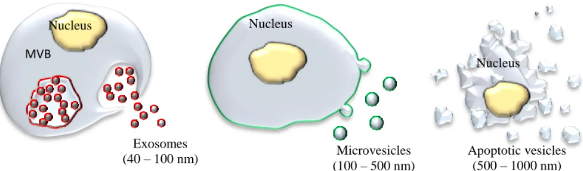
The unique properties of exosomes can be attributed to their biogenesis; they are initially produced by invagination of the endosomal membrane to create multivesicular bodies (MVB) (Figure 1.1).
Figure 1.1. Schematic representation of different types of extracellular vesicles.
In contrast, exosomes’ close relative, microvesicles, are greater in size (100 - 500 nm)
and bud directly from the plasma membrane. Consequently, exosomes and microvesicles have endosomal (red) and plasma (green) membrane origin, respectively (Figure 1.1). Larger vesicles (500 - 1000 nm) are considered to be apoptotic vesicles (Figure 1.1). Many investigations, especially in the field of drug delivery, utilize both exosomes and microvesicles, defining them as extracellular vesicles, because a complete separation and purification of each type of vesicles is extremely laborious and difficult [33].
Exosomes can be characterized by size, protein and lipid content. Different techniques were developed for the characterization of exosomes. Among them are flow cytometry, western blotting, nanoparticle tracking analysis (NTA), dynamic light scattering (DLS), mass
spectrometry (MS) and several microscopy techniques [34]. The International Society for Extracellular Vesicles (ISEV) published a position paper in 2014, in which the characterization
Nucleus
MVB
Exosomes (40 – 100 nm)
Nucleus
Microvesicles (100 – 500 nm)
Nucleus
5
of exosomes is recommended by the presence of exosome-associated surface markers, as well as the absence of proteins not associated with exosomes [35]. Exosomal surface markers include TSG101, Alix, flotillin 1, tetraspanins (CD9, CD63, CD81), integrins (ITG**), and cell adhesion molecules (CAM) [35]. Exosomes are highly enriched in cholesterol, sphingomyelin, and
hexosylceramides at the expense of phosphatidylcholine and phosphatidylethanolamine [36]. The fatty acids in exosomes are mostly saturated or monounsaturated. Together with the high
concentration of cholesterol, this may account for lateral segregation of these lipids into exosomes during their formation at MVB. Exosomes can be isolated from conditioned cell culture media or bodily fluids by differential centrifugation, filtration paired with centrifugation, immunoaffinity or size exclusion chromatography, or polymer-based precipitation.
Differential Ultracentrifugation and Density Gradient Ultracentrifugation
This method is considered the “gold standard” for isolating exosomes. It involves
applying a centrifugal force to a solution containing exosomes, e.g. conditioned cell culture media or biological fluids. First, a low speed centrifugation step (400 x g) is performed in order to remove cells and large cell debris. The supernatant is then subjected to 10,000 – 20,000 x g to remove large debris and intact organelles. Finally, the supernatant is again subjected to high speed centrifugation (100,000 - 150,000 x g) to pellet exosomes. It is worth noting that the type, quantity, and quality of exosomes isolated by this method is sensitive to the g force, rotor type, angle of rotor sedimentation, radius of centrifugal force, pelleting efficiency, and solution viscosity. One issue with differential ultracentrifugation is that it sediments exosomes as well as other vesicles, proteins, and/or protein-RNA aggregates. By including a sucrose density
6
extensive (62 – 90 h) centrifugation time [37], but provides a more uncontaminated exosome isolate than ultracentrifugation alone.
A fast and efficient method of exosome isolation was reported in [38]. Exosomes from monocyte-derived dendritic cells were rapidly purified (e.g. 4–6 h of a 2–3 L culture) based on
their unique size and density. Ultrafiltration of the clarified supernatant through a 500-kDa membrane and ultracentrifugation into a 30% sucrose/deuterium oxide (D2O) (98%) cushion (density 1.210 g/cm3) reduced the volume and protein concentration approximately 200- and
1000-fold, respectively. The percentage recovery of exosomes ranged from 40% to 50% based on the exosome MHC class II concentration of the starting clarified supernatant [38]. While differential centrifugation has the potential for higher exosome yields, this method is subject to operator-dependent variability [38].
Immunoaffinity Chromatography
Immunoaffinity chromatography is a process in which antibodies, covalently attached to beads, filters, or other matrices, bind to specific surface proteins or antigens on the target particle and non-target particles remain unbound. The unbound fraction is discarded, and the desired bound fraction may be collected by washing the stationary phase, typically with a low pH buffer. For the isolation of exosomes, antibodies to exosomal surface markers such as TSG101 or
7
Size Exclusion Chromatography
Size exclusion chromatography (SEC) is a method wherein a solution consisting of a heterogeneous population of differently sized components is separated based on their size. A column containing heteroporous beads is used in SEC; components with a smaller hydrodynamic radius are able to pass through the many small pores, akin to a maze, resulting in a longer time to elute. Components with a larger hydrodynamic radius are unable to penetrate through as many pores, and thus elute earlier from the column. In this manner, exosomes may be separated from other vesicles and contaminants of different sizes. The advantages of SEC are that it preserves the integrity and biological activity of exosomes and other molecules being separated; because SEC is typically performed using gravity flow, vesicle structure and integrity remains intact. Furthermore, SEC has excellent reproducibility and sensitivity. However, because SEC uses gravity flow, it requires a long run time which limits its scalability for high-throughput applications.
Polymer Precipitation
Polymer precipitation has been used to isolate viruses and other macromolecules for more than 50 years, typically by use of a solution containing polyethylene glycol (PEG). The most commonly used commercial polymer precipitation-based product for exosome isolation is ExoQuick-TC™ from System Biosciences. Typically, to isolate exosomes, a precipitation solution consisting of PEG with a molecular weight of 8,000 Da is used. This precipitation solution is combined with biofluid containing exosomes and is incubated overnight at 40C. The mixture is then centrifuged at low speed to form a pellet containing exosomes. The product is relatively easy to use, and does not require specialized equipment or a lengthy run time.
8
lipoproteins, as well as polymer material [40]. These issues may be addressed by pre- and post-isolation steps. Pre-post-isolation involves the removal of subcellular particles such as lipoproteins. Post-isolation involves removal of the polymer, typically by using a Sephadex G-25 column [37].
Natural Functions of Exosomes and Their Intrinsic Biological Activity
Exosomes play a significant and diverse role in intercellular communication that is an essential process for the development and function of multicellular organisms. These
extracellular vesicles were initially thought to be a mechanism for removing unneeded
membrane proteins from reticulocytes. Recent studies have shown that they are specialized in long-distance intercellular communications [41, 42] facilitating transfer of proteins [43, 44], and functional mRNAs and microRNAs for subsequent protein expression in target cells [45, 46]. This mechanism of secretion, signaling and communication is a highly efficient, robust, and economic manner of exchanging information between cells. Thus, exosomes themselves exert unique biological activity, even without any loaded drug that may be used for therapeutic purposes.
Immune Regulation by Exosomes
Tumor cells are poorly immunogenic and this has hampered the development of effective
cancer immunotherapy. By transporting ligands and receptors, exosomes can trigger an anti-tumor response by presenting anti-tumor antigens to immune cells. Initially, tumor-derived exosomes that carry antigens have been suggested as a source of specific stimulus for the immune response against tumors [47]. These exosomes were shown to induce anti-tumor responses more
9
mouse B lymphoma cells were reported to release exosomes that carry a number of heat shock
proteins (HSP) that induced significant antitumor immune responses in T cells [48].
Later, it was demonstrated that tumor-derived exosomes can also possess
immunosuppressive properties [49], promote oncogenesis, metastasis [50, 51], and drug
resistance development [52-54]. Therefore, the attention was turned to the exosomes released by activated antigen presenting cells (APCs), such as dendritic cells (DCs), macrophages, T
lymphocytes, and B cells. The presence of MHC class I and II, as well as T cell co-stimulatory molecules, on the surface of these exosomes could be an important mechanism of antigen presentation [55]. Furthermore, the immune response cells primed with antigens can package cellular components from cancer cells in exosomes that then promote immune responses [56-62]. In particular, exosomes secreted by DCs that were primed with acid-eluted tumor peptides were reported to eradicate established tumors in mice [56]. According to another study, DCs-secreted exosomes incubated with human breast adenocarcinoma cells (SK-BR-3) were able to induce tumor-sensitized T cells to secrete high levels of Interferon-γ (IFN-γ) [57]. Qazi et al. [58] reported a significant anti-cancer activity of exosomes secreted by DCs that were exposed to chicken egg ovalbumin (OVA). These exosomes elicited specific transgenic T cell proliferation
10
Along with the improving immune responses, exosomes released from T cells were shown to destroy tumor stroma, and prevent tumor invasion and metastases. In addition,
cross-talk between T lymphocytes and endothelial cells through exosomes was reported [59]. Thus, T cell-derived exosomes were shown to modulate endothelial cell responses to vascular endothelial
growth factor (VEGF) and alter tube formation and gene expression in target endothelial cells. Mechanistic studies revealed that overexpression of thrombospondin-1 and its receptor CD47 on exosomes derived from T cells allowed targeted and facilitated internalization of these
extracellular vesicles into endothelial cells. CD47 transferred to the tumor vasculature by exosomes modulated tumor angiogenesis and inhibited pro-angiogenic signaling in endothelial
cells [59]. Noteworthy, the induction of immune responses may be mediated not only by the bioactive lipids and proteins present in exosomes, but also by exosome-associated RNAs [60]. Contained inside exosomes, microRNAs (miRNAs) play a key role in mediating biological functions due to their prominent role in gene regulation. Thus, Aucher at al. [61] reported that human macrophages can transfer miRNAs to hepato-carcinoma cells and functionally inhibit proliferation of these cancerous cells. The transport of these miRNA was associated with extracellular vesicles.
Regarding infectious diseases, a succesful immunisation against difteria and Leishmania
11
the design of vaccine adjuvants and therapeutic intervention strategies to modulate immune responses.
Protective and Regenerative Effects of Exosomes
Exosomes play a vital role in regulating a broad range of physiological and pathological cellular processes [68] that may be utilized for therapeutic purposes. Mesenchymal stem cells (MSCs) derived from bone marrow, adipose tissue, cord blood, and other origins have recently received much attention as potential therapeutic agents with regenerative properties [69-76]. It was reported that MSCs-derived exosomes produced significant cardio-protective paracrine effects against myocardial ischemia/reperfusion injury in pig and mouse models [69, 70]. These exosomes may also be beneficial in pulmonary hypertension (HP). HP is a kind of malignant pulmonary vascular disease characterized by an increase in pulmonary artery pressure, which may lead to heart failure and even death. MSCs-derived exosomes directly suppressed early pulmonary inflammation and vascular remodeling [71] through the suppression of hyper-proliferative pathways, including signal transducer and activator of transcription 3 (STAT3)-mediated signaling.
Exosomes secreted from cardiosphere-derived cells (CDCs) were shown to produce a range of diverse cardio-protective effects, including inflammatory, oxidative, anti-apoptotic, anti-fibrotic, and cardiomyogenic effects [76, 77]. CDCs-released exosomes stimulate angiogenesis, promote cardiomyocyte proliferation, and decreased programmed cell death in vitro. The regenerative capacity of these exosomes was demonstrated in a model of chronic myocardial infarction (MI) in rats [78]. These effects were attributed to the ability of exosomes to reduce collagen deposition and exert anti-fibrotic efficacy via paracrine mechanisms [79]. In
12
increased viable mass after MI. The observed therapeutic effects were associated with normalized oxygen consumption, induced ATP production, and preserved mitochondrial
integrity.
Exosomes derived from endothelial cells are a promising strategy to combat
atherosclerosis [80]. Atherosclerosis, the underlying cause of myocardial infarction and stroke, occurs predominantly in predisposed spots in the large arteries. Systemic administration of exosomes released from human umbilical vein endothelial cells (HUVECs) reduced
atherosclerotic lesions in mice fed a high-fat diet. It is known that shear stress and its central transcriptional regulator KLF2 elicit atheroprotective properties to the endothelium by regulating the expression of atheroprotective genes. Exosomes secreted by shear-stress-stimulated HUVECs were enriched in multiple miRNAs, prominently miR-143 and miR-145. HUVECs-released exosomes transported these miRNAs to smooth muscle cell (SMCs) which resulted in controlled target gene expression and reduction of atherosclerotic lesion formation in the mouse aorta [80].
MSCs-derived exosomes were shown to have neuroprotective effects in stroke. The release, as well as the content, of the MSC-generated exosomes can be modified by
13
derived from human adipose tissue-derived MSCs were also reported for treatment of Alzheimer disease (AD) [72]. It was demonstrated that these exosomes carry enzymatically active
neprilysin (NEP), the most important enzyme that degrades β-amyloid (Aβ) peptide plugs in the
brain. MSCs-derived exosomes decreased both intracellular and extracellular Aβ levels in a neuroblastoma cell line N2A in vitro. Finally, Ju et al. showed protective effects of exosome-like nanoparticles isolated from crushed grapes [85]. Multivesicular bodies have been also identified in plants,and leaderless secreted proteins can be released in vesicles, as described recently [86]. Ju and colleagues observed that oral administration of exosome-like nanoparticles from grapes to mice led to the significant proliferation of the intestinal epithelium.
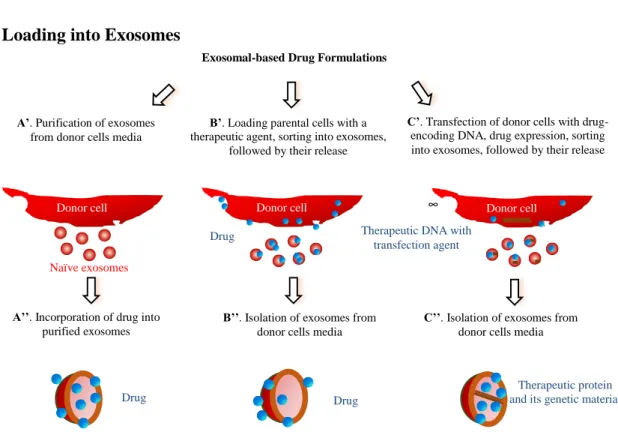
Using Exosomal Carriers for Therapeutics Drug Loading into Exosomes
Figure 1.2 Different approaches for drug loading into exosomes.
These are several distinct approaches for loading of exosomal carriers with drugs (Figure 1.2): (A) loading naïve exosomes isolated from parental cells ex vitro; (B) loading parental cells
Exosomal-based Drug Formulations
B’. Loading parental cells with a therapeutic agent, sorting into exosomes,
followed by their release
C’. Transfection of donor cells with drug-encoding DNA, drug expression, sorting into exosomes, followed by their release
A’’. Incorporation of drug into purified exosomes
Donor cell
Naïve exosomes A’. Purification of exosomes
from donor cells media
B’’. Isolation of exosomes from donor cells media
Donor cell
C’’. Isolation of exosomes from donor cells media
Donor cell ∞
Drug
Drug
Therapeutic protein and its genetic material Drug
14
with a drug, which is then released in exosomes; and finally, (C) transfecting/infecting parental cells with DNA encoding therapeutically active compounds, which are then released in
exosomes. Each of the approaches has its advantages and limitations, and may be dictated by the type of therapeutic agent.
Regarding ex vitro loading of naïve exosomes (Figure 1.2, Path A), different methods for drug incorporation were suggested. In most cases, lipophilic small molecules were passively loaded into exosomes during co-incubation with exosomes [87-93]. Thus, low molecular
antioxidant, curcumin [89, 90], anticancer agents, Doxorubicin (Dox) [92, 93] and Paclitaxel (PTX) [94], and a model drug Rhodamine 123 [94] were loaded into exosomes by the incubation at room temperature (RT). The drug loading upon this loading procedure determined by HPLC varied from 7.2% for PTX to 11.7% for Dox.
Exosomes naturally deliver mRNA, miRNA, various noncoding RNA, mitochondrial DNA, and genomic DNA [95, 96]. Therefore, they were suggested as carriers for nucleic acids transfer. Similar to the incorporation of genetic material into living cells, electroporation of purified exosomes was proposed for loading of exogenous RNA [5, 97-100]. Alvarez-Erviti et al. pioneered this method, electroporating siRNA into DC-derived exosomes [5]. The same method was used to load exosomes with miRNA to epidermal growth factor receptor (EGFR)-expressing breast cancer cells [101]. About 3,000 miRNA molecules were loaded per exosome. It should be taken into consideration that electroporation of extracellular vesicles with siRNA may be accompanied by extensive siRNA aggregate formation, which may cause overestimation of the amount of siRNA actually loaded into exosomes [102]. The authors suggested that
15
alternative methods to prepare siRNA-loaded exosomes. Exosomes are known to carry a nega-tive surface charge, hence precluding electrostatic siRNA complexation. Pre-complexation of
siRNA via cationic liposomes followed by fusion with isolated exosomes has been suggested for
their loading with siRNA by Wahlgren et al. [103]. Furthermore, elevated temperature (370C) was may be used for improved siRNA loading into exosomes [104].
Exosomes are also known to be nature’s way of delivering different proteins [105]. We
suggested harnessing this mechanism for the delivery of a potent antioxidant, catalase, in
exosomes [106]. Catalase is a large protein (MW 240 kDa that is susceptible to deactivation and rapid degradation, therefore the efficient incorporation of catalase in exosomes would be
beneficial, but is not a simple task. We evaluated several loading methods. The amount of catalase loaded into exosomes increased as follows: incubation at RT < freeze/thaw cycles < sonication extrusion permeabilization with saponin. The highest loading capacity for catalase in exosomes was in the range of 20% - 26%. We hypothesized that the extensive reformation and reshaping of exosomes upon sonication and extrusion enabled catalase diffusion across relatively tight and highly structured lipid bilayers that resulted in the high loading efficiency of exosomal carriers. Furthermore, saponin treatment also increased catalase loading into exosomes. Saponin is an efficient permeabilization agent for cellular plasma membranes [107]. It is likely that, similar to whole cells, saponin can selectively remove membrane-bound cholesterol on exosomes, creating holes/pores in the exosomal lipid bilayers and thereby promoting catalase incorporation. Notably, aside from proteins, these methods for loading into exosomes can be applied to other therapeutic and imaging agents, in particular, gold nanoparticles [106].
MSCs-16
secreted exosomes were loaded with PTX by incubating the parental cells with the drug [108]. It was reported that the murine SR4987 cells that were used as MSCs model produced a significant amount of PTX-loaded exosomes as demonstrated by HPLC [108]. A similar result was reported for HepG2 cells that were incubated with different anticancer agents: PTX, Etoposide,
Carboplatin, Irinotecan, Epirubicin, and Mitoxantrone [109]. Exosomes released from drug-treated HepG2 cells demonstrated strong anti-proliferative activity on the human pancreatic cell line CFPAC-1 and induced immunogenicity and HSPs-specific NK cell responses [109]. In another study, the breakdown of parental cells (monocytes /macrophages) loaded with anticancer agents, Dox, Gentamicin, 5-Fluoracil, or Carboplatin with subsequent isolation of exosome-like nanoparticles was also suggested [91]. An elegant approach to pack hydrophobic
photosensitizers into exosomes was developed by professor Ji-Ho Park and his team in South Korea [110]. The researchers treated parental cells with synthetic membrane fusogenic liposomes loaded with hydrophobic therapeutics. The drug-loaded liposomes were efficiently incorporated into the membrane of exosomes in the parental cells and were consequently secreted from the cells.
We developed a new approach of loading parental cells (monocytes/macrophages) with catalase followed by isolation of drug-loaded exosomes from conditioned media (Figure 1.2, Path B) [106, 111]. To preserve the therapeutic protein against degradation in host cells and increase loading capacity, catalase was incorporated into a polymer-based nanocontainer before loading. Importantly, the formulation design of this polymer-based nanocontainer was different from the common approach, where a drug nanoformulation is prepared for systemic
17
contrast, the best nanoformulation for loading into parental cells had a relatively large size (c.a.
200 nm) that resulted in improved accumulation in parental cells, and drug reshuffling into exosomes. The cross-linking of polymer-based nanoparticles with an excess of a non-biodegradable linker ensured low cytotoxicity of nanoformulation and efficient catalase protection in the parental cells [106, 112].
Finally, isolation of drug-loaded exosomes secreted from genetically-modified parental cells has been suggested as a third way of manufacturing exosome-based formulations (Figure 1.2, Path C) [33, 87, 111, 113]. As an example, OVA was loaded into exosomes when parental cells
were transfected with OVAC1C2 fusion cDNA consisting of: (i) the cargo-encoding gene
(OVA), and (ii) the gene encoding a protein known to localize to exosomes (C1C2) [113]. Our lab developed a new drug delivery system for different therapeutic proteins, where macrophages were transfected with plasmid DNA (pDNA) encoding therapeutic proteins, catalase [114], or glial cell-line derived neurotropic factor (GDNF) [111] to treat neurodegenerative disorders. An
interesting approach for incorporation of adeno-associated virus (AAV) capsids into extracellular vesicles to diminish their immunogenicity and improve gene delivery was suggested by [115]. It was reported that during production, a fraction of released AAV vectors were associated with exosomes, termed vexosomes (vector-exosomes), which outperformed conventionally purified AAV vectors in transduction efficiency in vitro.
Therapeutic Effects of Drug-Loaded Exosomes
18
characteristics, and delivering their cargo in a biologically active form. Noteworthy, exosomes possess an intrinsic ability to cross biological barriers, including the most difficult to penetrate, blood brain barrier (BBB).
Exosomes have been exploited as drug delivery vehicles for low molecular drugs in several investigations [89-94, 108, 110]. In one of the first reports, exosomes loaded with an anti-inflammatory small molecule compound, curcumin, were shown to protect mice from lipopolysaccharides-induced brain inflammation [89, 90]. The incorporation of curcumin in exosomes improved its solubility, increased circulation time, preserved drug therapeutic activity, and improved brain delivery. In another study, exosomes loaded with different
chemotherapeutics, Dox or PTX, were shown to traffic to tumor tissues and reduce tumor growth in mice without the adverse effects observed with the equipotent free drug [91-93]. Notable, the therapeutic effects of loaded exosomes were greater than the commercially available Dox-loaded liposomes, Doxil; the liposomal formulation was inefficient in reducing tumor growth in this model [91]. Pascucci et al. observed that PTX-treated MSCs mediated strong
19
Another therapeutic avenue involves the use of exosomes to deliver exogenous siRNA [5, 97-100, 103, 116-118]. Wahlgren et al. reported the efficient silencing of the target MAPK gene in monocytes and lymphocytes using peripheral blood exosomes with incorporated exogenous siRNAs [103]. In another investigation, Shtam et al. introduced two different exogenous siRNAs against RAD51 and RAD52 into exosomes derived from HeLa cells [116]. In particular, the exosome-delivered siRNA against RAD51 was functional and caused the massive reproductive cell death of recipient cancer cells. The effect of exosome-siRNA gene silencing has also been validated in [117, 118]. As an example, extracellular vesicles were used to transport siRNA targeted to miR-150 [118]. miR150 is an oncomir due to its promotional effect on VEGF. It was demonstrated that the neutralization of miR-150 down-regulated VEGF levels in mice and attenuated angiogenesis.
The genetic modification of donor cells was also used for targeting exosomes to the disease site. As an example, targeting of exosomes to the brain was achieved by engineering the parental DCs to express lysosomal-associated membrane protein 2 (Lamp2b), fused to the neuron-specific peptide derived from rabies virus glycoprotein (RVG) [5]. Systemically administered RVG-targeted exosomes delivered glyceraldehyde 3-phosphate dehydrogenase (GAPDH) siRNA specifically to neurons, microglia, oligodendrocytes in the brain, resulting in specific gene knockdown. The therapeutic potential of exosome-mediated siRNA delivery was demonstrated by the strong mRNA (60%) and protein (62%) knockdown of BACE1, a
therapeutic target in Alzheimer's disease, in wild-type mice [5].
20
genetically-modified parental cells contained the encoded therapeutic protein, as well as its genetic material (DNA and mRNA), and NF-b, a transcription factor involved in the encoded gene expression [114]. Drug-loaded exosomeswere able to efficiently transfer their contents to contiguous neurons resulting in de novo protein synthesis in target cells. Transfected brain tissues showed month-long expression of the encoded protein and prolonged attenuation of neuroinflammation (over 40 days) in mice with neuroinflammation [114]. Overall, these reports indicate that exosomes may function as exceptional gene delivery vectors that are safe, efficient, organ/cell-specific, and non-immunogenic. However, significant efforts are required to develop these therapies for clinical use.
Using Exosomal Drug Formulations in the Clinic
21
exosome-like nanoparticles allowed a 100-fold higher production yield of the drug carriers [91]. Finally, specifically designed bioreactors that resemble bioreactors for tissue engineering [122] can be utilized for exosomes scale-up. Notably, exosomes can be concentrated, lyophilized, and reconstituted in aqueous solutions, as was reported in [106].
Figure 1.3. The flow of the production and delivery of exosomal drug formulations to the patient.
As an alternative approach, MSCs may be harvested from bone marrow, propagated in culture to obtain specific cell types, or even subtypes, and then exosomes may be loaded with a therapeutic agent. Although this approach would require a more invasive procedure, a significant amount of as well as storage of well-characterized exosomal carriers would be possible [123]. Furthermore, large scale production of therapeutically efficacious exosomes can be achieved through the immortalization of donor cells; for example, mesenchymal stem cells can be
transfected by lentivirus carrying a MYC gene as reported in [124]. MYC is a regulator gene that codes for a transcription factor that plays a role in cell cycle progression. The transfection allows
Quality control
Transfection with drug-encoded pDNA or loading
cells with therapeutic agent Cell differentiation Therapeutic protein, genetic material Exosomes isolation, purification Administration
into the patient Leukapheresis
Packaging of the drug, or genetic material
22
for obtaining of immortalized cells, but does not alter the fundamental characteristics of these MSCs [124]. In this case, a library of various types of exosomal carriers for different drug formulations could be developed in the future, and stored in stock for emergency situations. Finally, exosomes may be isolated from other sources (bovine milk, crashed grapes, etc.), purified, loaded with a drug and used for oral or intranasal administration.
In fact, exosomes have already been approved for use in clinical trials, and our
experience with exosome-based therapies in humans is rapidly expanding [125]. In particular, exosomes were purified from monocyte cultures from 15 patients with advanced metastatic melanoma. The good manufacturing procedures (GMP) process allowed harvesting about 5 x 10¹⁴ exosomal MHC class II carriers. Then, the exosomes were loaded with melanoma antigen ex
23
MATERIALS AND METHODS Reagents
Paclitaxel (PTX) was purchased from LC Laboratories (Woburn, MA, USA). The stock solution was prepared in ethanol (EtOH) at a concentration of 10 mg/mL. Aliquots were stored at -20°C. Working solutions of PTX were prepared fresh according to experimental design by serial dilution in EtOH. A fluorescent dye, 2-decanoyl-1-(O-(11-(4,4-difluoro-5,7-dimethyl-4-bora-3a,4a-diaza-s-indacene-3-propionyl)amino)undecyl)-sn-glycero-3-phosphocholine (BODIPY-PC) was purchased from Molecular Probes. Cell culture medium and fetal bovine serum (FBS) were purchased from Gibco Life Technologies, Inc. (Grand Island, NY, USA). Culture flasks and dishes were purchased from Corning Inc. (Corning, NY, USA). ExoQuick-TC™ Exosome Precipitation Solution was obtained from System Biosciences (Mountain View,
CA, USA).
Cells
RAW 264.7 macrophages (purchased from ATCC, Manassas, VA, USA) were cultured in Dulbecco’s modified Eagle’s medium (DMEM) high glucose (Gibco, Grand Island, NY,
USA) supplemented with 10% fetal bovine serum (FBS; Thermo Fisher Scientific), 1% penicillin and streptomycin at 37°C and 5% CO2.
Exosome Isolation
24
cultured in exosome-depleted media for 2 days at 37°C and 5% CO2. 50 mL conditioned cell culture media were centrifuged at 300 x g for 10 min (Thermo CL-10 centrifuge with O-G26/1 rotor, Thermo Fisher Scientific, Waltham, MA, USA) in order to remove cells and cellular debris. The supernatant was then taken, filtered with a .22 µm PES filter, and ExoQuick-TC™ Exosome Precipitation Solution (System Biosciences, Mountain View, CA, USA) was added to the filtered supernatant and the mixture was vortexed and incubated overnight at 4°C. After overnight incubation, the mixture was vortexed again and subsequently centrifuged at 1500 x g for 30 min. and 5 min. to pellet exosomes. The supernatant was discarded and the exosome pellet was resuspended in PBS. Freshly-prepared exosomes or exosomes stored at -20°C were used for all experiments.
Drug Loading into Exosomes
For PTX loading into exosomes, purified exosomes (~1011 exosomes) were first mixed with PTX in 1 mL PBS. First, PTX was dissolved in ethanol (EtOH, 10 mg/mL drug in EtOH stock solution) and added to 1 mL exosomes. Different methods of drug loading were
PTX-25
loaded exosomes (exoPTX) solution was incubated at 37°C for 60 min to allow for recovery of the exosomal membrane.
Excess free PTX was separated from exoPTX, respectively, by size exclusion
chromatography using a NAP-10 Sephadex G25 column (GE Healthcare, Buckinghamshire, UK) according to the manufacturer’s recommended protocol. Briefly, 750 µL of exoPTX were added
to the NAP-10 column and the void volume was discarded. 250 µL of PBS was then added to the column and allowed to enter the gel bed completely and the eluate was discarded. 1.2 mL of PBS was then added to the column and the eluate containing purified exoPTX was collected and stored at -20°C.
Quantification of Drug Loading
The amount of PTX loaded into exosomes was measured by a high performance liquid chromatography (HPLC) method. Briefly, exoPTX or exoPTX-AA (1010 exosomes/0.1mL) in a microcentrifuge tube was placed on a heating block set to 75°C to evaporate solvent. After all solvent had evaporated, an equal volume of acetonitrile was added to the microcentrifuge tube and the mixture was vortexed, sonicated, and vortexed again. The sample was then centrifuged at 13,000 rpm (Thermo Legend Micro 21, Thermo Fisher Scientific, Waltham, MA, USA) for 10 min. Following centrifugation, the supernatant was taken and filtered through a Corning
26
concentration of standard. A calibration curve was constructed by plotting peak area versus concentrations of paclitaxel and was found to be linear within the tested concentration range (r2 = .997). Exosomal protein content was measured using the Pierce BCA Protein Assay Kit
(Thermo Fisher Scientific, Waltham, MA, USA) according to the manufacturer’s recommended
protocol. Loading capacity is expressed by µg protein of exosomes.
Synthesis of DSPE-PEG-AA
The synthesis was carried out according to the published synthetic protocol with little adjustment[23]. Briefly, to synthesize the N-(2-bromoethyl)-methoxy-benzamide,
4-methyoxybenzoyl chloride (1g, 5.8 mmol) in 50 mL of pre-warmed benzene was mixed with an aqueous solution of 2-bromoethylamine hydrobromide (1.32g, 6.4 mmol). The emulsion was shaken and cooled in running water during the dropwise addition of 5% aqueous solution of sodium hydroxide. The precipitate was solidified out of the reaction mixture within a few
minutes to an amorphous mass. The mixture was continuously stirred for 1h, after which time the sold amide was filtered with suction and washed once with benzene and air dried for 2-3h. Then, the synthesized N-(2-bromoethyl)-4-methoxy-benzamide (100 mg, 0.4 mmol) was reacted with DSPE-PEG-NH2 (100 mg, 23.3 µmol) in acetonitrile (5 mL) in the presence of DIPEA (30 uL, 0.2 mmol) at 65-70 °C for 16h. After evaporating the solvent, 5 mL of methanol was added to dissolve the pellet followed by excess ether (50mL) and it was the kept at -80 °C overnight. The precipitate was collected after centrifugation and recrystallized twice. The overall yield was 70%. The products was characterized by NMR and TLC as reported elsewhere[129].
Preparation of Vectorized Exosomes
27
then 10mg/mL DSPE-PEG-AA was added to the exosome solution. 100μL of 10mg/mL PTX in EtOH was also added to the mixture of exosomes with DSPE-PEG-AA when preparing exoPTX-AA. The mixture was then sonicated by the same method used by our lab previously [130]. Briefly, the mixture was sonicated using a Model 505 Sonic Dismembrator with .25” tip
(Thermo Fisher Scientific, USA) with the following settings: 20% amplitude, 6 cycles of 30 s on/off. After sonication, the solution was incubated at 37°C for 60 min to allow for recovery of the exosomal membrane. Excess free PTX and DSPE-PEG-AA were separated from exoPTX-AA or exoexoPTX-AA by size exclusion chromatography using a NAP-10 Sephadex G50 column (GE Healthcare, Buckinghamshire, UK) according to the manufacturer’s recommended protocol. Briefly, 750 µL of exoPTX-AA was added to the NAP-10 column packed with Sephadex G50 and the void volume was discarded. 250 µL of PBS was then added to the column and allowed to enter the gel bed completely. 1.2 mL of PBS was then added to the column and the eluate containing purified exoPTX-AA was collected and stored at -20°C. Loading capacity was determined by HPLC and formulations were evaluated to determine which provided the greatest drug loading.
Characterization of Exosomes
Nanoparticle Tracking Analysis (NTA). Exosomes were identified and characterized using NanoSight LM 10 instrument (NanoSight Ltd., Amesbury, UK). The settings were
optimized and kept constant between samples, and each video was analyzed using the Nanosight system to obtain the size and concentration of exosomes. The stability of exosomes was
monitored by measuring size over a period of time under various conditions (4°C, room
28
concentration in the region of ~108 particles/mL, in accordance with the manufacturer’s recommendations. All samples were analyzed in triplicate.
Dynamic Light Scattering (DLS). The average hydrodynamic diameter and zeta potential of exosomes was measured by DLS using a Malvern Zetasizer Nano ZS system (Malvern, Worcestershire, UK) equipped with He–Ne laser (5 mW, 633 nm) as the light source at 22°C. All samples were analyzed in triplicate.
Atomic Force Microscopy (AFM). The morphology of exoPTX was investigated by AFM imaging. ExoPTX formulations were diluted in PBS and a drop of the sample was placed on a glass slide and dried under a flow of argon gas. The AFM imaging instrumentation was operated as described earlier[131].
Western Blot Analysis. The levels of proteins constitutively expressed in exosomes, Alix and flotillin 1, as well as the lymphocyte function associated antigen-1 (LFA1, subunit CD11a), were examined by western blot (WB). Protein concentrations were determined using BCA kit (Pierce Biotechnology, Rockford, IL). The protein bands were detected with Alix, flotillin 1, and CD11a primary monoclonal antibodies, (Abcam, Cambridge, UK; 1:1000 dilution), and
secondary HRP-conjugated rabbit anti-goat IgG-HRP (Santa Cruse, CA, USA; 1:5000 dilution).
29
TSG101 in exosomes with goat polyclonal antibodies to TSG101 (Santa Cruz, SC6037; 1:200 dilution).
Membrane Fluidity Measurements. BODIPY-PC, a fluorescent dye was used as a probe to examine the fluidity properties of exosomal membranes as described earlier[133]. Briefly, 30 μL exosomes with concentration 4x10¹¹ particles/mL were mixed with 20 μL BODIPY-PC (0.03
mg/ml) and supplemented with 70 μL deionized water; the mixture was incubated for 45 min at 37°C in darkness. Unbound label was removed using a ZebaTM column (Life Technologies). BODIPY-PC is a hydrophobic fluorescent compound which incorporates into the hydrocarbon regions of lipid membranes. Transfer of BODIPY-PC from the aqueous environment into the lipid bilayers results in a drastic increase of the fluorescence emission for this probe.
30
RESULTS
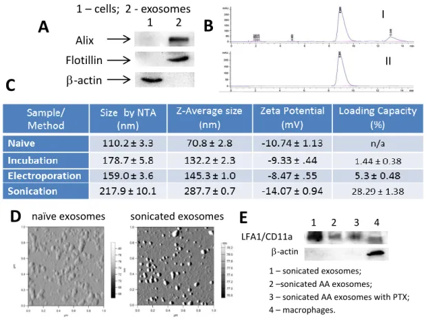
Isolation and Characterization of Exosomes from RAW 264.7 Macrophages
Exosomes were collected from the conditioned media of RAW 264.7 macrophages using a polymer purification method (ExoQuick-TC™) and were characterized by protein content, size, and morphology. Exosomes had elevated expression of exosome-associated proteins (Alix, TSG101, and Flotillin) as compared to cell lysate, which had greater levels of β-actin (Figure 1A).
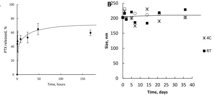
Figure 1.4. Characterization of PTX exosomal formulations.264.7 macrophages were collected from conditioned media, and the exosome protein content was confirmed by western blot (A). Exosomes were loaded with PTX by various methods: co-incubation at RT; electroporation, and sonication. The size of the exosomes with incorporated PTX was measured by NTA and DLS (C). The amount of PTX incorporated into exosomes was assessed by HPLC (B). The loading efficiency of exosomes with PTX increased in a row: incubation at RT < electroporation<< sonication (C). The morphology of drug-loaded exosomes was examined by AFM (D). Images revealed small spherical naïve exosomes, and PTX-loaded exosomes. The bar: 200 nm. LFA1/CD11a expression in exosomes as assessed by Western Blot (E).
1 – cells; 2 - exosomes
Alix Flotillin
-actin
1 2
A
B
naïve exosomes
C
sonicated exosomes
D
II I
1 2 3 4 LFA1/CD11a
-actin
1 – sonicated exosomes; 2 –sonicated AA exosomes;
3 – sonicated AA exosomes with PTX; 4 – macrophages.
31
Exosomes were also found to express the lymphocyte function associated antigen-1 (LFA1, subunit CD11a) (Figure 1E), which assists in cell uptake and may bind to endothelial cell adhesion molecules which are overexpressed on activated endothelial cells such as those found in tumors[134]. Naïve empty exosomes had a narrow size distribution, with an average particle diameter of 110.4 ± 4.2 nm and 70.8 ± 2.8 nm as revealed by NTA and DLS,
respectively (Figure 1C); and a round morphology as shown by AFM imaging (Figure 1D).
Manufacture and Characterization of Exosomal Formulations of PTX (exoPTX)
PTX was incorporated into exosomes using three different methods: a) incubation at RT, b) electroporation, and c) mild sonication, as described in the Materials and Methods section. ExoPTX formulations were purified from non-incorporated drug by size-exclusion
chromatography and analyzed by HPLC to determine the loading capacity (LC) of exosomal carriers. The typical HPLC profile for PTX extracted from exosomes (I) and PTX standards (II) are shown in Figure 1.4B. HPLC analysis revealed that the amount of PTX loaded into
32
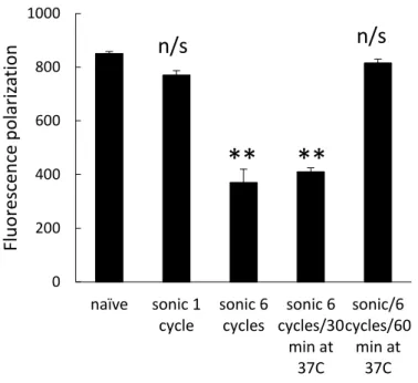
Figure 1.5. Effect of sonication on fluidity of exosomal membranes. Exosomes isolated from RAW 264.7 macrophages concomitant media were labeled with BODIPY-PC fluorescent dye as described in Materials section, and subjected to one or 6 cycles of ultrasound treatment. The fluorescence polarization was measured right after the sonication, or following 30 min or 1 hour incubation period at 37C. Values are means ± SEM (n = 4). Symbols indicate the relative level of significance compared with naive exosomes.
Interestingly, DLS studies revealed that the size of exoPTX nanoformulations increased
similarly, with the smaller being exoPTX nanoparticles obtained by electroporation or incubation at RT, and the larger being exosomes loaded with PTX by sonication (Figure 1.4C).
Noteworthy, NTA analysis confirmed these results indicating that the size of exoPTX obtained by sonication is about 1.5x greater than naïve empty exosomes (Figure 1.4C). Interestingly, exosomes sonicated in the absence of PTX (average size 326.0 ± 23.5 nm by NTA, and 356.1 ± 3.5 nm by DLS) were even larger than exosomes sonicated with PTX solution (217.9 ± 10.1 nm). We hypothesized that incorporation of PTX into exosomal membranes stabilized these
aggregates. Furthermore, all mentioned loading procedures did not significantly alter the zeta potential of the nanocarriers (Figure 1.4C). It is known that the anionic phospholipid
phosphatidylserine is abundant on cell membranes and contributes to the surface charge of
0 200 400 600 800 1000
naïve sonic 1
33
individual cellular membranes. We speculated that all of the PTX loading procedures caused no major alterations the lipid content of exosomal membranes. Thus, AFM imaging showed that exosomes retained their round morphology upon sonication (Figure 1.4D), suggesting that this procedure did not significantly disrupt the structure of exosomes.
Overall, the mild sonication procedure provided the highest amount of drug loading; the obtained LC of 28.29 ± 1.38% was much higher than the LC of commercially available
formulations of PTX, Taxol (~1% LC) or Abraxane (~10% LC). We hypothesized that the application of ultrasound creates pores in exosomes which allows for PTX to incorporate into the hydrophobic lipid bilayer of the exosome without destroying the exosome membranes. Because of the high LC, exoPTX produced by sonication was selected for further experiments.
Manufacture and Characterization of Exosomal Formulations of PTX Vectorized to the Sigma Receptor (exoPTX-AA)
The sigma receptor is overexpressed by many types of cancer, including NSCLC, making
Figure 1.6. Overexpression of receptor in lung cancer cells.(A) Western blot analysis revealed a significant amount of receptor in cancer cells, but not in normal lung fibroblasts. (B) Relative expression of target receptors to -actin.
1 - human lung fibroblasts; 2 -344SQ; 3 - 3LL-M27;
4 - H69/AR; 5 - A549
A
B
-actin
1 2 3 4 5
receptor
0 0.2 0.4 0.6 0.8 1 1.2 1.4
1 2 3 4 5
34
it an attractive target for cancer therapy [20-23]. In order to validate the sigma receptor as a target for our Lewis Lung Carcinoma 3LL-M27 cell line used in our mouse model of metastatic lung cancer, a Western Blot was performed to look for the presence of the sigma receptor in 3LL-M27 cells. Western Blot analysis revealed overexpression of the sigma receptor in target 3LL-M27 cells (Figure 1.6A), indicating that the choice of anisamide (a sigma receptor ligand) as a vector to target 3LL-M27 cells is appropriate.
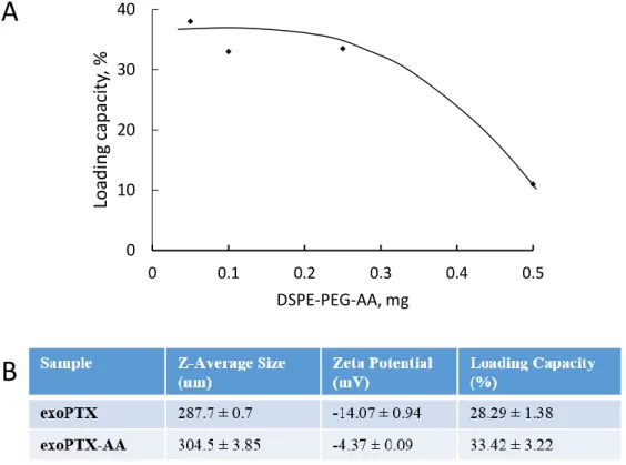
Figure 1.7 Optimization and Characterization of exoPTX-AA. (A) Varying amounts of DSPE-PEG-AA were added to exosome with 1mg paclitaxel, and the mixture was sonicated to produce vectorized and paclitaxel-loaded exosomes (exoPTX-AA). HPLC analysis of each formulation revealed the loading capacity (LC), and the formulation providing the greatest LC while maximizing the amount of DSPE-PEG-AA was chosen (.25mg DPSE-PEG-AA). (B) The exoPTX-AA formulation was characterized by DLS to obtain the Z-average size and zeta potential. LC was determined by HPLC.
We hypothesize that reorganization of exosomes caused by ultrasound treatment may result in the reorganization/reshuffling of the exosomal membrane, enabling PTX and DSPE-PEG-AA diffusion across relatively tight and highly structured lipid bilayers. Exosomes were
A
B
0 10 20 30 40
0 0.1 0.2 0.3 0.4 0.5
Loa
d
in
g
cap
acit
y,
%
35
loaded with PTX and vectorized to target the sigma receptor using the sonication method developed by our lab [29]. Varying amounts of vector were added to exosomes with paclitaxel in order to determine the optimal formulation providing the greatest loading capacity while maximizing the amount of vector. HPLC analysis of the formulations revealed that the optimal amount of DSPE-PEG-AA to add in order to achieve the greatest loading capacity was 0.25 mg, thus this formulation was chosen for further study (Figure 1.7A). HPLC analysis revealed that the loading capacity of the optimal formulation of exoPTX-AA was 33.42 ± 3.22%, similar to the loading capacity achieved by exoPTX alone without vector (28.29 ± 1.38%) (Figure 1.7B).
Further characterization by DLS showed that exoPTX had a smaller size (287.7 ± 0.7nm) than exoPTX-AA (304.5 ± 3.85 nm) (Figure 1.7B). We hypothesize that this size increase as measured by DLS is due to the presence of the long PEG chains of DSPE-PEG-AA on the surface of exoPTX-AA, as well as the insertion of DSPE lipids into the exosomal lipid
36
DISCUSSION
Although various drug nanoformulations have been developed to improve the therapeutic effect of drugs, there are still issues that significantly diminish their application in clinic, e.g.
toxicity and rapid clearance by the mononuclear phagocyte system (MPS) [135]. In fact, the safety of drug nanoformulations is the subject of substantial criticism. In this respect, exosomes, naturally-produced nanosized vesicles secreted by a variety of cells, represent an important tool for both diagnostic and therapeutic purposes. Exosomes consist of cellular membranes with a variety of adhesion proteins on their surface [136] which allow for efficient transport of
exosomal cargo into target cells. Exosomes have been reported for use as drug delivery vehicles for nucleic acids [5, 8, 10, 103, 117, 137] and small molecule drugs such as curcumin [89, 90], anticancer agents (Dox [92, 93] and PTX [94]). The low molecular-weight compounds were loaded into exosomes or exosome-like vesicles by co-incubation at RT. These studies
demonstrated the feasibility of using an exosomal formulation for cancer therapy as well as for neurodegenerative disorders. We also reported recently the development of a new exosome-based formulation of potent antioxidant, catalase, to treat Parkinson’s disease [106]. A large protein, catalase (MW 240 K), was loaded into exosomes using different procedures: sonication, extrusion, and permeabilization of exosomal membranes with saponin. The extensive
reformation and reshaping of exosomes upon sonication and extrusion, as well as the creation of holes/pores in the exosome lipid bilayer by saponin, enabled catalase to diffuse across relatively tight and highly structured lipid bilayers and resulted in high loading efficiency of exosomal carriers [106].
37
significant efforts to formulate PTX into various nanoformulations to increase solubility and improve pharmacokinetics, including polymer based nanoparticles and liposomes. However, these systems possess several undesirable properties such as the presence of toxic excipients and/or rapid clearance by the endothelial system (RES).
In the present study, we utilized various methods for PTX incorporation into exosomes: incubation at RT, electroporation, and mild sonication. Mild sonication of exosomes in the presence of PTX, provided the greatest loading capacity (28.29% ± 2.58%). PTX is a highly hydrophobic compound, therefore we hypothesized that this drug is incorporated into the
hydrophobic inner region of the phospholipid bilayer of exosomes. Indeed, exosome membranes display a high rigidity that was significantly decreased upon sonication. We hypothesized that the membrane fluidization allowed PTX incorporation into lipid bilayers resulting in the high loading capacity of exosomal carriers. The precise location of PTX in exosomes will be investigated in future studies. Importantly, drug located in the inner bilayer of exosomes may also be available for use: as the exosomal membrane fuses with the cell or endosomal membrane, its intraluminal cargo may be released into the cytosol. Next, we assessed the optimal
38
39
CHAPTER 2. IN VITRO CHARACTERIZATION OF exoPTX AND exoPTX-AA3
OVERVIEW
Multiple drug resistance (MDR) is a factor which severely limits the efficacy of a variety of chemotherapeutics, including paclitaxel. MDR may be intrinsic or acquired [1,2] and is mediated by different mechanisms, in particular, the overexpression of the drug efflux transporter
P-glycoprotein (Pgp). As a result, the response rate following treatment remains very low for many types of malignancies, including acute leukemias, malignant gliomas, metastatic breast cancer, and other cancers [1-3]. Prior treatment with antineoplastic agents is a serious adverse prognostic factor for many cancers due to the acquiring of drug resistance, resulting in low long-term
survival rates for patients with recurring cancers [3,4]. To date, there has been limited success in overcoming drug resistance in cancers through the use of novel small molecule
chemotherapeutics [7,8], or nanoformulations of existing chemotherapeutics. In addition, various Pgp inhibitors, such as quinine or cyclosporine A, have been co-administered with chemotherapeutic agents [138, 139]. These efforts did result in improved patient outcomes, however, the non-specific inhibition of Pgp frequently increased drug toxicity due to alteration of drug elimination pathways in the liver, kidney, etc. [140].
3 Some of this text previously appeared in an article in the journal Nanomedicine. The original citation is as
follows: Development of Exosome-encapsulated Paclitaxel to Overcome MDR in Cancer cells. Myung Soo Kim, Matthew J. Haney, Yuling Zhao, Richa Gupta, Zhijian He, Natalia L. Klyachko, Aleksandr Piroyan, Marina Sokolsky, Alexander v. Kabanov, and Elena V. Batrakova. Nanomedicine, Nov 13. pii: S1549-9634(15)00202-6. doi:
40
Herein, we have developed a new nanoformulation consisting of exosomes loaded with PTX (exoPTX), a commonly used chemotherapeutic agent, and vectorized to target the sigma
receptor. The main objectives of this study were: (i) to assess the feasibility of using exoPTX for MDR-related anticancer therapy, (ii) to identify mechanisms underlying inhibitory effects of exosomes on drug efflux pump in Pgp expressing cancer cells, (iii) to prepare and assess the targeting ability of exosomes vectorized to the sigma receptor, and (iv) to assess the intracellular fate of exosomes and vectorized exosomes.
41
MATERIALS AND METHODS Reagents
Paclitaxel (PTX) was purchased from LC Laboratories (Woburn, MA, USA). The stock solution was prepared in ethanol (EtOH) at a concentration of 10 mg/mL. Aliquots were stored at -20°C. Doxorubicin (DOX) was purchased from LC Laboratories (Woburn, MA, USA). The stock solution was prepared in DMSO at a concentration of 2 mg/mL and aliquots were stored at 4°C. Working solutions of PTX or DOX were prepared fresh according to experimental design by serial dilution in an appropriate medium. A lipophilic fluorescent dye, 1,1'-dioctadecyl-3,3,3',3'-tetramethylindo-carbocyanine perchlorate (DIL), was purchased from Invitrogen (Carlsbad, CA, USA). Rhodamine 123 (R123), 4′,6-diamidino-2-phenylindole dihydrochloride (DAPI), and Triton X-100 were obtained from Sigma-Aldrich (St. Louis, MO, USA). Cell culture medium and fetal bovine serum (FBS) were purchased from Gibco Life Technologies, Inc. (Grand Island, NY, USA). Culture flasks and dishes were from Corning Inc. (Corning, NY, USA). Fluorescent polystyrene nanoparticles (Fluoro-Max G100) were obtained from Thermo Fisher Scientific (Waltham, MA, USA). ExoQuick-TC™ Exosome Precipitation Solution was obtained from System Biosciences (Mountain View, CA, USA). ER Tracker Blue-White DPX, LysoTracker Green DND-26, and MItoTracker Deep Red were purchased from Thermo Fisher (Thermo Fisher Scientific, Waltham, MA, USA).
Cells
RAW 264.7 macrophages, Madin-Darby canine kidney MDCKWT and MDCKMDR1 cells (purchased from ATCC, Manassas, VA, USA) were cultured in Dulbecco’s modified Eagle’s
42
CO2. Murine Lewis lung carcinoma cell subline (3LL-M27), a highly metastatic lung clone, was a generous gift from Dr. L. Pelletier (CHUL, Laval University, QC, Canada), and were cultured in DMEM high glucose supplemented with 10% FBS, 10 mM HEPES, 1% penicillin and streptomycin at 37°C and 5% CO2.
Pgp protein levels in sensitive and resistant cancer cells were determined by western blot as previously reported [22] using monoclonal antibodies to Pgp, C219 (Dako Corp., Carpinteria, CA, USA; at dilution 1:100), and secondary horseradish peroxide donkey anti-mouse IgG antibodies (Amersham Life Sciences, Cleveland, OH, USA; at dilution 1:1500). To correct for loading differences, the Pgp levels were normalized to the constitutively expressed β-actin (stained with monoclonal antibodies to β-actin, anti-β-1-chicken integrin (Sigma Chemical Co.,
at dilution 1:200)). Specific bands were visualized using a chemiluminescence kit (Pierce, Rockford, IL, USA).
Exosome Isolation