ROLE OF ANDROGEN RECEPTOR CO-REPRESSOR SLIRP IN CELL CYCLE REGULATION
Dinuka M. De Silva
A dissertation submitted to the faculty of the University of North Carolina at Chapel Hill in partial fulfillment of the requirements for the degree of Doctor of Philosophy in the Department
of Pathology and Laboratory Medicine.
Chapel Hill Spring 2014
Approved by:
Young E. Whang
2014
ABSTRACT
Dinuka M. De Silva: Role of Androgen Receptor Co-repressor SLIRP in Cell Cycle regulation (Under the direction of Young E. Whang)
Androgen receptor (AR) reactivation is a hallmark of castration resistant prostate cancer (CRPC). One mechanism is tyrosine kinase Ack1 mediated activation phosphorylation of Tyr-267 and Tyr-363 residues on AR. Investigation into Ack1 regulation of AR identified SLIRP as a potential AR interacting protein. SLIRP acts as a co-repressor of steroid receptor signaling. We show that SLIRP interacted with AR in the absence of Ack1 activation, repressing AR signaling. In the presence of either androgen or Ack1 kinase activation, SLIRP dissociated from the
complex and AR signaling was activated. SLIRP dissociation from AR was dependent on the kinase activity of Ack1 but not the known Ack1 phosphorylation sites of Tyr-267 and Tyr-363 of AR. Furthermore, SLIRP-AR binding was dependent on RNA co-regulator SRA in AR signaling as knockdown of SRA resulted in reduced SLIRP co-immunoprecipitation with AR.
knockdown of co-repressor SHARP showed an effect on cell cycle by increased phospho-Rb protein and S-phase entry, but to a lesser extent than SLIRP. AR negative PC3 and DU145 cells did not recapitulate the results in LNCaP cells, suggesting AR dependence in the role of SLIRP in cell cycle.
In conclusion, we define a novel mechanism in which AR activity is regulated by SLIRP binding that is disrupted by Ack1 kinase or androgen. Ack1 gene amplification or AR mutations seen in CRPC may confer sensitivity of AR to low levels of androgens. Hence, our data suggests that disruption of SLIRP repression of AR-dependent transcription may be a potential
ACKNOWLEDGEMENTS
I would like to thank Dr. Young E. Whang for all the support he has shown me in my graduate studies. I am very grateful for his mentorship which allowed me to be a better graduate student. As a mentor, he was available to answer and teach any inquiries I had about the research and provided guidance when experiments generated difficult challenges. I would like to thank Dr. Shelton Earp for all his advice and our joint lab meetings which made me reflect critically about the research and become a better scientist. I would like to thank my committee members Dr. Albert Baldwin, Dr. Chris Gregory, and Dr. Bernard Weismann for their advice and encouragement. The committee members were very helpful in advancing my project.
I would like to convey my gratitude to the Department of Pathology and
(Richard), Mehmet Karaca, Dr. Yuanbo Liu, Shuli Wang, and Yu Cheng in the Whang lab for the funny stories, food, and friendship. Richard and Mehmet were a great help when I first started in the lab and also made the lab enjoyable. I would like to express my gratitude to everyone in the Earp lab for having us for their lab meeting and sharing their knowledge and encouraging us. Albert, Jing, Debra, Laura, Ling, Charlene, Anna, Francesca, Shu-mang and everyone else in the Earp lab was always there to teach me or help me when I needed a reagent. They also organized the holiday dinners and other parties which were always fun.
TABLE OF CONTENTS
LIST OF TABLES…...ix
LIST OF FIGURES……….x
LIST OF ABBREVIATIONS………...xii
CHAPTER I. INTRODUCTION 1.1 Prostate Cancer Epidemiology………...1
1.2 Natural History of Prostate Cancer………2
1.3 Prostate Cancer Therapy………5
1.4 Androgen Receptor Genomics………...8
1.5 Androgen Receptor and the Cell Cycle………...10
1.6 Tyrosine Kinase Ack1 in Prostate Cancer………...12
1.7 Co-repressors NCoR1 and SMRT………...14
1.8 Co-repressor SHARP………...16
1.9 Co-Regulator SRA and Co-Repressor SLIRP……….17
1.10 Dissertation Research Objectives………19
Figures……….21
2.2 Introduction………..29
2.3 Materials and Methods……….31
2.4 Results………..35
2.5 Discussion………39
Figures……….44
III SLIRP ACTS AS A POTENTIAL TUMOR SUPRESSOR IN PROSTATE CANCER CELLS BY REGULATING CELL CYCLE 3.1 Overview………..54
3.2 Introduction………..54
3.3 Material and Methods………..56
3.4 Results………..61
3.5 Discussion………67
Figures……….72
IV. CONCLUSIONS & FUTURE DIRECTIONS 4.1 Conclusions & Future Directions……….89
V. APPENDIX 5.1 Appendix………..96
LIST OF TABLES
5.2 List of genes used in the AR gene signature formed from LNCaP-NS
samples treated with vehicle versus 1nM DHT………97 5.3 List of genes used in the SLIRP gene signature formed from LNCaP-NS
LIST OF FIGURES
1.1 Outline of Prostate Cancer progression……….21
1.2 AR antagonists utilized in CRPC Therapy………22
1.3 Full length and truncated AR modular structure and Co-activator interacting motifs……….23
1.4 AR in the Cell cycle………...24
1.5 Ack1 modular protein structure with mutations K158R and L487F……….25
1.6 Ack1 activation of the AR pathway...26
1.7 SLIRP, SHARP, NCoR1, and SMRT co-repressor protein modular structure………….27
2.1 Ack1 and DHT impair the interaction between AR and SLIRP………....44
2.2 Effects of various growth factors, kinase ligands, AR mutants on AR-SLIRP interaction………...46
2.3 SRA is essential for AR-SLIRP interaction………...47
2.4 Validation of SLIRP knockout………...49
2.5 Effects of SLIRP on AR mediated transcriptional activity in LNCaP cells………..49
2.6 Effects of DHT and Ack1 activation on SLIRP recruitment to androgen response element (ARE)………51
2.7 Current model of Ack1 activation and DHT treatment on AR-SLIRP interaction………..53
3.1 RNA-seq Heatmap and SAM on LNCaP cells with SLIRP knockdown………...72
3.2 David and Ingenuity Pathway analysis on RNA-seq Data……….75
3.3 Gene Signatures from RNA-seq data………...76
3.4 SLIRP knockdown alters cell cycle regulators enhancing cell proliferation…………...79
3.5 SLIRP knockdown does not affect AR negative cell lines………...82
LIST OF ABBREVIATIONS
Ack1 Activated cdc42-associated tyrosine kinase 1
ADT Androgen deprivation therapy
AR Androgen receptor
ARE Androgen response element
ARR-2PB Androgen response region 2 probasin
BPH Benign prostate hyperplasia
CDK Cyclin dependent kinases that regulate cell cycle COS-7 CV1 origin SV40-7 monkey kidney cells
CRIB Cdc42/Rac interactive binding
CRPC Castration resistant prostate cancer
CTD COOH terminal domain
DHT Dihydrotestosterone
EGF Epidermal growth factor
EGFR Epidermal growth factor receptor
FL-WT-AR Full length wild type androgen receptor FL-AR-Y267F Full length wild type TYR-267F AR FL-AR-Y363F Full length wild type TYR-363F AR
HER Human epidermal growth factor receptor
HEK293 Human embryonic kidney cells
hK2 Human kallikrein2
HRG Heregulin
LBD Ligand binding domain
IL-6 Interleukin 6
MAPK Mitogen associated protein kinase
MMTV Mouse mammary tumor virus
MLS Mitochondrial localizing signal
NCoR1 Nuclear receptor co-repressor 1
NLS Nuclear localization signal
NTD N-terminal domain
PCa Prostate cancer
PIN Prostatic intraepithelial neoplasia
PSA Prostate specific antigen
PTK Protein tyrosine kinase
Rb Retinoblastoma protein
RTK Receptor tyrosine kinase
SH3 Src homology 3
SHARP SMRT/HDAC-1-associated repressor protein SLIRP SRA Stem-Loop Interacting RNA Binding Protein
SMRT/NCoR2 Silencing mediator of retinoic acid and thyroid hormone receptor/ Nuclear receptor co-repressor 2
SRA Steroid Receptor RNA Activator
SRC1 Steroid receptor co-activator 1
CHAPTER I: INTRODUCTION
1.1 Prostate Cancer Epidemiology
Cancer is a predominant public health problem around the world. For men, prostate cancer accounts for 241,740 new cases a year with 28,170 deaths in the United States [1]. Despite significant research efforts, death rates for men with prostate cancer has stayed fairly constant from 1930-2008[1]. Risk factors for prostate cancer depend on age, family history, and race or ethnicity [2]. For example, according to National Cancer Institute’s Surveillance, Epidemiology, and End Results (SEER) Program, African Americans have a higher incidence and mortality of prostate cancer than Caucasians [2, 3]. Other ethnic groups including Asian American men, American Indian and Alaskan Native men (AI/AN) and Hispanic men have a lower prostate cancer incidence and mortality rate compared to European American men (EAM) (American Cancer Society, 2007).
candidate genes HPC1 on chromosome 1q23-25, PCAP on chromosome 1q42-43, and CAPB on chromosome 1p36 [7]. In addition, trinucleotide repeats encoding polyglutamine (CAG)n and
polyglycine (GGN)n in the androgen receptor exist whose polymorphisms are associated with
increased risk of PCa [8]. Interestingly, the frequency of (CAG)n repeats were higher in African
American males compared to European and Asian but did not associate with increased PCa risk [9, 10]. A polymorphism in CYP17, an enzyme important in the synthesis of adrenal and
intratumoral androgen synthesis, may be associated with susceptibility to PCa in men of African but not European descent [11].
Diet and life style can also influence risk for prostate cancer development. The
distribution of PCa incidence and mortality rates among different geographical locations in the world demonstrates a difference in distribution. Eastern countries like China and Japan have historically lower incidence of PCa than in the United States and the Caribbean [7, 12]. An association also exists between the high consumption of red meat, saturated fat, and dairy
products and PCa pathogenesis [6, 7]. Thus, the higher rate of PCa incidence in the U.S could be related to be a higher intake of animal protein than in Asian countries [13, 14]. Hence, we must appreciate the environmental and dietary factors that play a role in the increased risk of prostate cancer, particularly in the western world.
1.2 Natural history of PC
prostate carcinoma arises in the peripheral zone [15]. The epithelial and mesenchymal interaction is important in prostate gland development as prostatic differentiation requires both components and in absence of either, the cells fail to differentiate [4]. Furthermore, androgen is required in the mesenchyme to produce signals for prostate development and growth and later in the epithelium for secretory function [4]. Secretory luminal cells, stem cell like basal cells, and the neuroendocrine cells make up the prostatic duct. Luminal cells express androgen receptor important in prostate carcinoma and are further characterized by the expression of cytokeratins 8 and 18 and CD57[4].
Initiation of progression of normal epithelium of the prostate to prostatic intraepithelial neoplasia (PIN) is marked by inflammation, oxidative stress, telomere shortening and changes in genes like NKX3.1, Myc, and TMPRSS2-ERG fusion [16]. PIN is characterized by luminal epithelial hyperplasia, a reduction in basal cells, cytoplasmic hyperchromasia, and nuclear atypia [16]. Furthermore, in high-grade PIN there is an increase in cell proliferation markers [17]. Progression from PIN to adenocarcinoma is marked by re-activation of signaling pathways including ERK/MAPK and androgen receptor (AR) pathways. Transition between
adenocarcinoma to metastasis is indicated by the development of castration resistant tumor and epithelial to mesenchymal transition (EMT) [16]. Though prostate adenocarcinoma lacks distinct subtypes, there are occurrences of ductal adenocarcinoma, mucinous carcinoma, and signet ring carcinoma although rare [18]. Metastasis of prostate cancer is primarily to bone and lymph nodes. Circulating tumor cells could be detected in the bone marrow of patients with localized disease and in metastatic disease [19].
probasin (PB) promoter is used. The construction of the promoter includes using two consensus AR binding sites within the promoter (2236 to 2223 and 2140 to 2117), known as the androgen response region (ARR) [20]. A better version of this promoter was generated by using a larger region of the probasin promoter (LPB) with 2 ARR regions called ARR2PB which maintains reliable prostate specificity but also provides very high transgene expression in transgenic mice [20]. Subcutaneous and orthotopic xenografts use immune compromised mice to facilitate tumor growth. Other models include genetically engineered mouse models (GEMMs) with germ-line knockout of important cancer genes like PTEN and NKX3.1 and SV40 transgene expression GEMMS including TRAMP and LADY. PTEN is a tumor suppressor gene whose inactivation is seen in 35% of metastatic tumors [21]. Half of heterozygotes develop multi-focal PIN and carcinogenesis [20]. Homozygosity of the null mutation results in embryonic lethality and PTEN heterozygotes fail to develop metastasis [21]. NKX3.1 is involved in early embryonic patterning and organogenesis, epithelial proliferation and is important in recognition of PIN development as noted previously. NKX3.1 null mutations disrupt normal prostate protein secretion and ductal morphogenesis and have become a model for PIN [22]. Though this model is good for early progression of prostate cancer, it fails to develop invasive carcinoma. The TRAMP (transgenic adenocarcinoma of the mouse prostate) model uses the expression of both the large and small SV40 tumor antigens regulated by the rat probasin promoter. TRAMP mice develop PIN by 18 weeks and lymphatic metastases after 28 weeks [23]. Although GEMMs rarely portray
(LPB) to drive expression of a deletion mutant of the SV40 T antigen that expresses only the large T antigen [23]. While this model can be used to study the progression from hyperplasia to adenocarcinoma, it does not model castration resistance and metastasis [23].
1.3 Prostate Cancer Therapy
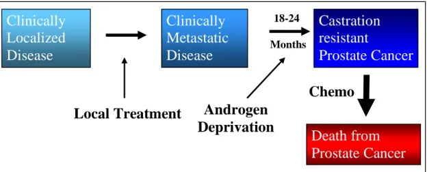
Prostate cancer is dependent on androgens for metastatic progression. Thus, androgen deprivation therapy (ADT) carried out by either surgical or chemical castration is applied to patients with metastatic disease (Fig 1.1) [24]. Chemical castration involves luteinizing
hormone-releasing hormone (LH-RH) analogs or LHRH antagonists. LHRH agonists decrease androgens generated by the testicles and include drugs leuprolide, goserelin, triptorelin, and histrelin. However, LHRH agonists initially stimulate pituitary LHRH receptors which induces an increase in LH and FSH release that increases testosterone levels[25]. This is termed a testosterone “flare” and can exacerbate bone pain in advanced patients [25]. LHRH antagonists work similarly to the agonists but do not produce the “flare” and include degarelix and abarelix. Unfortunately, within 18-24months, patients become terminal due to development of castration resistant prostate cancer (CRPC) from ADT in part due to AR reactivation (Fig 1.1).
energy photon field in precisely defined locations in the prostate. EBRT uses computer generated tomographic images of a patient’s anatomy to identify these locations. Brachytherapy (seed implant therapy) is a cancer treatment with ionizing radiation delivered via radioactive material placed near or within the tumor.
Disease progression at the CRPC stage is due to many factors including hypersensitivity to low levels of androgens, AR gene amplification, AR mutations, regulation of co-repressors and co-activators, growth factors, and activation by tyrosine kinases [26-29]. First generation anti-androgens nilutamide, flutamide, and bicalutamide have a modest affect when combined with medical or surgical castration but there is little clinical evidence of benefit with relapsed patients [30-32]. Current standard first line therapy for CRPC is docetaxel. Docetaxel induces an apoptotic affect by inhibiting microtubule dynamics leading to mitotic arrest. In the TAX327 trial, patients with metastatic CRPC (mCRPC) were treated with prednisone and randomized to docetaxel 75 mg/m2 intravenously every 3 weeks, docetaxel 30 mg/m2 weekly for 5 of 6 weeks, or mitoxantrone 12 mg/m2 intravenously every 3 weeks[33]. Patients treated with every 3-week docetaxel had better PSA response rates, pain reduction, and quality-of-life measures, and median survival of 19.2 months compared to 16.3 months with mitoxantrone [33]. Toxicities of docetaxel treatment include fatigue, hematologic toxicity, and neurotoxicity and thus require treatment breaks or discontinuation.
The new generation of therapeutics includes cabazitaxel which inhibits microtubule depolymerization and cell division, abiraterone that targets adrenal androgens, and AR
prednisone with either cabazitaxel or mitoxantrone [34]. There was improved overall survival of CRPC patients compared to mitoxantrone (15.1 months vs. 12.7months) and has been approved by the FDA [34, 35]. Adverse side effects included hematologic, infectious, and gastrointestinal toxicity. Abiraterone is an adrenal inhibitor of CYP17, a cytochrome p450 complex involved in adrenal steroidal synthesis as well as intratumoral androgen synthesis [35]. CYP17 catalyzes the conversion of pregnenolone and progesterone to dehydroepiandrosterone (DHEA) and
androstenedione. Abiraterone is given with prednisone due to the mineralocorticoid excess that results in hypertension, hypokalemia, and edema. Two phase II studies of patients pretreated with docetaxel and than abiraterone illustrated a greater than 50% decline in PSA in 36% and 51% of patients respectively [36, 37]. A phase III study investigating abiraterone plus prednisone compared with prednisone alone showed prolonged survival of CRPC patients who had disease progression after docetaxel [38]. Overall survival was greater in patients treated with abiraterone plus prednisone than with placebo plus prednisone (14.8 months vs. 10.9 months) [35, 38]. Enzalutamide (MDV3100) has greater affinity for AR, lacks agonist effects unlike bicalutamide, inhibits ligand binding to AR, and inhibits AR nuclear translocation [39]. A phase III study, the AFFIRM trial, investigated 1,199 men with docetaxel-treated CRPC that received oral
enzalutamide 160 mg once daily or placebo [40]. They demonstrated that enzalutamide improved overall survival and reduction of the risk of death by 37% compared to control group [40]. The phase III trial indicated that the median survival for patients treated with MDV3100 was 18.4 months compared with 13.6 months for placebo group [35, 40]. Since the results of this study, the FDA has approved enzalutamide for CRPC patients who have been treated with
similar populations, they may reflect differences in the stage of migration, disease management, and new emerging therapeutics [41].
1.4 AR genomics
Androgens are the main sex steroids important in male sexual differentiation, maturation during puberty, and development of prostate cancer. Androgens also play a role in
non-reproductive tissues such as skin, bone, muscle, and brain [42]. Androgens, testosterone (T) and the more potent form, 5α-dihydrotestosterone (DHT) are the ligands for AR. The human AR gene located on chromosome Xq11-12 consists of 8 known exons which encode a ~110 kDa protein. Androgens and AR may also play a role in female reproduction as AR knockout in female mice leads to ovarian dysfunction [43]. Androgen receptor plays an essential role in all aspects of prostate tumorigenesis.
Mechanisms of AR activation in CRPC consist of AR gene amplification, AR mutations, splice variants, ligand-independent activation by growth factors, kinase activation, and co-regulator changes. There is a continued reliance on AR for the progression and metastasis of prostate cancer. More than 50% of CRPC tumors have some AR alterations. AR gene
amplification occurs in 30% of CRPC tumors while 10-20% will have an AR mutation [46]. For example, in the well used LNCaP prostate cancer cell line, there is a missense mutation at codon 877 that changes the amino acid from threonine to alanine (T877A) [46]. This mutation in the ligand binding domain leads to decreased ligand specificity where other hormone like
progesterone and estrogens can bind and activate AR [46]. Another important mutation is the gene fusion of androgen dependent TMPRSS2 and Ets transcription gene ERG. TMPRSS2-ERG plays a role in early tumor development. It is expressed in pre-neoplastic lesions and 50% of primary PC as well as 30% of CRPC [47, 48]. Since the discovery of TMPRSS2-ERG fusion, many others have come to light including TMPRSS2–ETV1, NDRG1–ERG, and TMPRSS2– FKBP5–ERG which are regulated by androgen and/or androgen receptor [48].
Co-activators like TIF1/SRC1 (aka KAT13A, RIP160, bHLHe42, bHLHe74),
of the region spanning SRC-2 and 6.2% had high level amplification of the locus [50]. Taylor et al 2010 showed that overall, 8% of primary tumors and 37% of metastases had SRC-2 gain of expression or mutations [50]. On the other hand, co-repressors that inhibit transcription of AR dependent genes include NCoR1 and SMRT. These well known co-repressors can bind AR directly as well as recruit HDACs’ to promote chromatin packing and inhibit transcription.
Growth factors also play a role in aberrant AR signaling. Epidermal growth factor (EGF) is a growth factor that stimulates cell proliferation and differentiation by binding to epidermal growth factor receptor (EGFR). EGF and its receptor EGFR have increased expression in prostate metastatic disease [51]. EGFR activation can lead to activation of the MAPK pathway down-regulating AR and relieving AR inhibition of cell cycle in differentiated cells [51, 52]. Conversely, EGF stimulation of LNCaP cells induces phosphorylation of AR at Tyr-267 and Tyr-534 by Src and Ack1 kinases inducing AR activation [53]. EGF also leads to increased levels of STAT3 that results in the formation of a STAT3-AR complex and increased transcriptional activity [51].
1.5 AR and Cell cycle
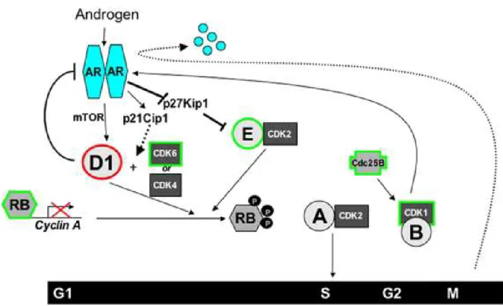
There exists a dynamic relationship between AR, androgens and cell cycle (Fig 1.4). Androgens can stimulate cell cycle and thus enhance cell proliferation by upregulating cyclins and cyclin dependent kinases (CDK) which control progression of cell cycle phases.
phosphorylation of Rb and partial inactivation. Complete inactivation of Rb is produced when other CDKs like CDK1 and CDK2 interact with cyclin B, A, and E (Fig 1.4).
Androgen is a regulator of G1-S phase transition where androgen deprivation results in G1 arrest, Cyclin D1 and D3 loss, and hypophosphorylated Rb [55, 56]. Furthermore, cyclin A, a target of Rb-mediated transcriptional repression, and CDK2 activity is reduced with androgen depletion [57]. Low p27/Kip1 expression can be a marker of short time to recurrence and androgen deprivation can induce p27Kip1 which contributes to CDK2 reduction [55, 58]. p21/Cip1 is a direct AR target and is lost with androgen deprivation in vitro [55, 59]. Conversely, AR can be regulated by the cell cycle. For instance, cyclin D1 can bind directly to N-terminus of AR and block conformational change for maximal AR activity [60, 61]. Thus, cyclin D1 acts as a
1.6 Ack1 in Prostate Cancer
There are ten families of non-receptor tyrosine kinases which involve Src, Abl, Jak, Ack1, Csk, Fak, Fes, Frk, Tec, and Syk. Ack1 or Tnk2 is a 120 kDa intracellular non-receptor tyrosine kinase comprising of an N-terminal tyrosine kinase domain, a SRC homology 3 domain, a cdc42/Rac interactive binding domain (CRIB), and a C-terminal proline rich domain (Fig 1.5) [69]. Ack1 was cloned from a human hippocampal expression library by its ability to bind activated GTP bound cdc42 and not Rac1 or RhoA [69]. Ack1 gene is located on chromosome 3q29, a region linked to prostate cancer recurrence [70]. Advanced stage primary tumors and metastatic tumors contain copy number gain of the ACK1 gene as well as overexpression of ACK1 mRNA [70]. Ack1 overexpression was found in 10 of 13 hormone refractory prostate cancer tumor samples but rarely in early stage primary tumors (1 of 53) [70]. Ack1 is expressed in various tissues with highest expression in brain, spleen, and thymus [71]. Factors that activate Ack1 include EGF, platelet derived growth factor (PDGF), and bradykinin. EGF activation of EGFR leads to Ack1 autophosphorylation which in turn can activate the guanine nucleotide exchange factor Dbl leading to actin cytoskeletal rearrangements [72]. Furthermore, EGF
stimulation also leads to interaction of Ack1 with adaptor protein Grb2 which leads to interaction with receptor tyrosine kinases Axl, leukocyte tyrosine kinase (LTK), and anaplastic lymphoma kinase (ALK) [73]. EGFR is regulated by Ack1 interaction with various partners including ubiquitin, clathrin heavy chain, and sh3px1 [74]. Ack1 degradation is mediated by the E3 ubiquitin ligase Nedd4-2 via a PPY motif [75]. EGFR activation of Ack1 drives Nedd4-2 kinase degradation [75].Ack1 co-localizes with Nedd4-2 in clathrin-rich vesicles [75].
mediates phosphorylation of p130cas which promotes cell migration [76]. A cdc42-Ack1-Cas complex is also involved in the regulation of melanoma cell spreading by melanoma chondroitin sulphate proteoglycan (MCSP), a proteoglycan that is expressed on the surface of melanoma cells [77].
Ack1 is autophosphorylated in the activation loop at tyrosine 284 [78]. Other Ack1 phosphorylation sites identified by proteomic approaches include Y826, Y856, and Y857 [74]. Phosphorylation of Y826 was identified downstream of insulin signaling and Y857 downstream of Her2 [74]. Ack1 cancer associated mutation E346K was identified in ovarian endometrioid carcinoma and destabilizes an auto-inhibited conformation of Ack1, leading to constitutively high Ack1 activity [79]. Mutations V365R in the kinase region, F820A in the Mig6 homology region (MHR) also seem to destabilize the auto-inhibition and activate Ack1 [79]. Other mutations that activate Ack1 include R34L found in lung adenocarcinoma and R99Q in ovarian carcinoma occurring in the SAM domain and M409I mutation in lung
adenocarcinoma in the SH3 domain [74]. Generated mutations W426K, that prevents binding of the SH3 domain to its ligand, produced Ack1 activation while the H464D point mutation that disrupts Cdc42 binding reduced Ack1 autophosphorylation [71]. The down-regulation of Ack1 involves an intramolecular interaction of the MHR with the kinase domain [79].
role in epithelial-to-mesenchymal transition (EMT). Ack1 has been shown to play a role in promoting prostate tumorigenesis by Wwox tumor suppressor degradation, phosphorylation and activation of AR, and phosphorylation of Akt [80-82]. Ack1 phosphorylates the pro-apoptotic WW domain containing oxidoreductase (Wwox) at tyrosine 287 which leads to the dissociation of Ack1-Wwox complex and Wwox polyubiquitination and degradation [82]. Constitutive active Ack1 with a L487F mutation, which disrupts auto-inhibition of Ack1, was shown to promote prostate xenograft tumor growth in mice (Fig 1.5) [81, 82]. In addition to the inhibition of tumor suppressor Wwox by Ack1, Ack1 enhances AR transcriptional activity, increases AR
recruitment, and promotes androgen independent tumor growth through AR tyrosine
phosphorylation at Tyr-267 and Tyr-363 (Fig 1.6) [81]. Tumor samples showed increased Ack1 and AR phosphorylation; particularly 44% of CRPC tumors had AR phosphorylation while none of 13 androgen-dependent prostate cancer (ADCaP) tumors or 18 BPH samples showed AR tyrosine phosphorylation [81]. Activation of Ack1 and subsequent phosphorylation of AR at Tyr-267 was induced by EGF, heregulin, and Gas6 and inhibited by Src kinase inhibitor dasatinib [53]. Hence, Ack1 role in tumorigenesis and particularly in the prostate is an important mechanism of cancer progression and maintenance.
1.7 Co-repressors NCoR1/SMRT
Nuclear receptor co-repressor (NCoR1) is a ~270kDa protein isolated using a
three N-terminal repressor domains (RD) which recruit additional components (Fig 1.7). The repressive ability of NCoR1/SMRT is attributed to the N-terminal RD domains which also contain motifs SANT1 and SANT2 (Swi3/Ada2/N-Cor/TFIIID) between RD1 and RD2. The SANT1 submotif plays a critical role in the interaction with HDAC3 through the deacetylase interaction domain (DAD) [86]. The SANT2 motif enhances the repression of SANT1/HDAC3 by acting as a histone interaction domain (HID) [87]. RD1 of NCoR1 binds HDAC1 and 2 via Sin3 co-repressor while RD3 binds HDAC 4, 5, and 7 [88-90].
NCoR1 knockout mice are lethal at embryonic development day E15.5 [91]. Phenotypes involve smaller livers, small body size, erythropoietic defects, and abnormal nervous system development [91]. SMRT behaves similarly to NCoR1 in terms of HDAC association with its domains. SMRT/HDAC3 activation requires chaperone TRiC-1 dissociation in an ATP
dependent manner upon complex formation [92]. SMRT knockout mice are embryonically lethal by E16.5 due to cardiogenic defects [93, 94]. Rescue of the cardiac defects of SMRT null mice were obtained with re-expression of alpha-myosin heavy chain SMRT [93]. Though the mice survived till birth, they exhibited major defects in neural development [93].
binding to ARE regions was only seen in S-phase [97]. Furthermore, it was shown that NCoR1 and AR interacted with each other in the DNA binding regions of AR target genes [97]. SMRT is involved in the ability of anti-androgen cyproterone acetate (CPA) to inhibit AR transactivation but not other anti-androgens [98]. SMRT behaves similarly to NCoR1 in that it interacts directly with AR through the RD2 domain, inhibits DHT-bound AR, and competes with p160/TIF2 co-activator for binding [99]. Additionally, analysis of over 200 prostate cancer tumors showed that 4% of primary tumors and 16% of metastatic tumors had loss of NCoR1 and 23% of primary and 21% of metastatic tumors had a loss of SMRT [50].
1.8 Co-repressor SHARP
A yeast-2-hybrid screen utilizing the SMRT LBD domain as bait identified
Notch signaling in neurogenesis of Xenopus where constitutively active Notch resulted in no lateral and no intermediate primary neurons [101]. However, when SHARP RNA was co-injected, it rescued the formation of intermediate and lateral primary neurons indicating its importance in neurogenesis [101].
1.9 Co-regulator SRA and Co-repressor SLIRP
Steroid receptor RNA activator, SRA, was the first to be identified as a co-regulator that acts as an RNA transcript and exists in a ribonucleoprotein complex with co-activator SRC-1 [103]. Using a yeast-2-hybrid assay with AF1-containing N-terminus of hPR, SRA was
identified via screening of human cDNA libraries from skeletal muscle, heart, and the HeLa S3 cell line [103]. SRA enhanced PR transactivation when induced with progestin [103]. Further analysis of other steroid receptors revealed that SRA enhanced steroid receptor mediated transactivation of GR, AR, ER, thyroid hormone (TR), retinoic acid (RAR and RXR), and peroxisome proliferator activated receptor (PPAR) [103]. Secondary structure predictions suggest that there are multiple stem-loop structures where co-activators and co-repressors bind [100, 104, 105]. SRA has been shown to associate with co-regulators SRC-1, PGC-1, SHARP, and SLIRP.
in oncogenesis [105]. SLIRP mRNA is ubiquitously expressed with the highest levels in heart, liver, skeletal muscle, and testis [105]. SLIRP was detected in breast cancer cell lines SK-BR-3, MCF-7, and MDA-MB-468 and prostate cancer cell line LNCaP [105].
SLIRP can repress SRA mediated transaction of Estrogen receptor (ER) luciferase reporter [105]. Furthermore, SLIRP can modulate other nuclear receptors including
glucocorticoid (GR), androgen (AR), thyroid (TR), and Vitamin D receptor (VDR) [105]. When SLIRP is co-transfected with SHARP and SRA, repression of ER signaling is further enhanced [105]. SLIRP competes with other co-activators like SKIP for SRA binding to regulate NR signaling. Imaging studies show that SLIRP mainly colocalizes to the mitochondria and only a small percentage is localized in the nucleus [105]. SLIRP knockout mice are viable but sub-fertile [106]. The percentage (21%) of SLIRP knockout mice born were less than what was predicted with Mendelian genetics [106]. SLIRP KO mice produced less progressively motile sperm, has interruption of the mid-piece/annulus junction, and altered mitochondrial morphology compared to the WT mice [106].
In the mitochondria, SLIRP maintains mitochondrial-localized mRNA transcripts that encode Oxidative Phosphorylation (OxPhos) protein subunits [107]. The OxPhos pathway is important in energy homeostasis by serving as the cell's producer of ATP [107]. OxPhos consists of five complexes and Complexes I through IV comprise the oxygen-dependent electron
abundance of complex IV subunit and COX2 and thus shows that SLIRP is important in OxPhos function by regulating mtRNA abundance [107]. Furthermore, SLIRP was found to
co-immunoprecipitate with LRPPRC [108]. Mutations in LRPPRC, a leucine-rich protein of the pentatricopeptide repeat family, leads to French Canadian variant of Leigh syndrome (LSFC), an early onset and fatal, neurodegenerative disorder. LRPPRC plays a role in regulating the stability and handling of mature mRNAs like cytochrome C oxidase (COX) subunits [108]. Accordingly, LRPPRC/SLIRP complex can also suppresses mRNA degradation mediated by PNPase and SUV3 and promote polyadenylation of mRNA mediated by mitochondrial poly (A) polymerase MTPAP [109]. Hence, in the mitochondria, SLIRP can form a complex with LRPPRC and together they regulate the stability of mtRNAs that are required for proper energy homeostasis.
1.10 Dissertation Research Objectives:
Castration resistant progression of prostate cancer (CRPC) is terminal and no curative treatment options exist for patients. A hallmark of CRPC is the reactivation of androgen receptor (AR) and thus identifying how AR is reactivated in the last stage of metastatic prostate cancer is crucial to treating patients. We previously demonstrated that Ack1 (activated cdc42-associated kinase) promotes growth of prostate xenograft tumors in castrated animals by activation of AR through phosphorylation of the Tyr-267 site [81]. However, the molecular mechanisms
co-repressor of AR signaling and if Ack1 is involved in SLIRP-AR interaction; (2) Characterize the loss of SLIRP in cell cycle regulation. Thus, understanding the mechanism of AR reactivation and its maintenance in CRPC is crucial in identifying novel targets and thus improving
Fig 1.1: Outline of Prostate Cancer progression.
Patients diagnosed with localized prostate cancer undergo prostatectomy; however some patients develop metastatic disease. Common therapy for metastatic disease is androgen deprivation therapy. Unfortunately, 18-24months later, all patients develop castration resistant prostate cancer which is terminal. (Adapted from Scher, 2008 [110])
Clinically
Localized
Disease
Clinically
Metastatic
Disease
Castration
resistant
Prostate Cancer
Death from
Prostate Cancer
Local Treatment
Androgen
Deprivation
Chemo
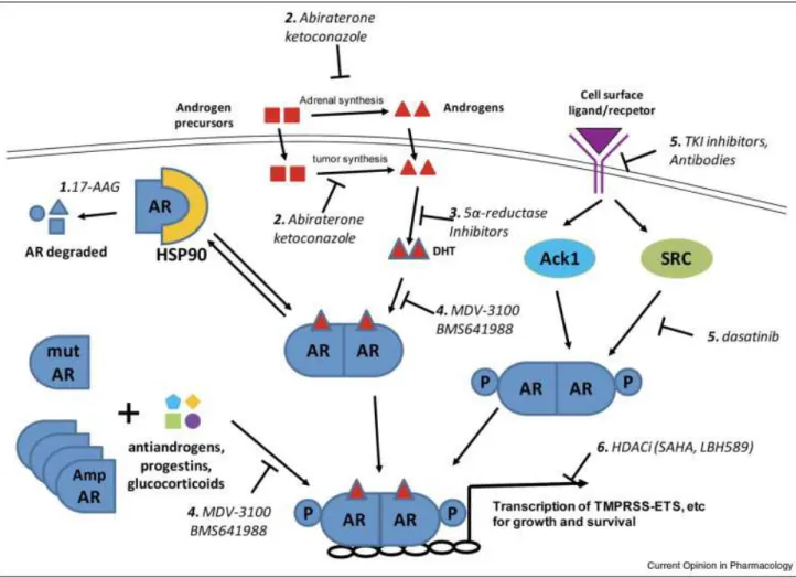
18-24Fig 1.2: AR antagonists utilized in CRPC Therapy.
Different therapeutics target multiple parts of the androgen receptor signaling cascade.
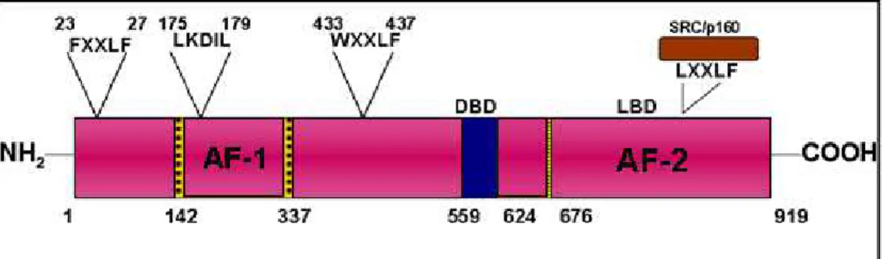
Figure 1.3: Full length and truncated AR modular structure and Co-activator interacting motifs.
Top: AF1 is the ligand independent activating function domain located in the NTD while AF-2 is the ligand dependent domain in the C-terminus. AF-1 interacts with two motifs (FXXLF and WXXLF) in the NTD. LKDIL is a core motif that mediates transcriptional activity of TAU1 and co-regulators such as SRC/p160 which interacts with AF-2 through the LXXLF motifs (Adapted from Wilson, 2007[112]). Bottom: AR consists of N-terminal region (NTD), a DNA binding domain (DBD), Hinge region (H), a ligand binding domain (LBD). Truncated AR lacks the LBD and forms a ~80kDa protein. (Adapted from Lonergan 2011, Lallous 2013 [51, 113])
NTD
DBD
H
LBD/AF-2
919aa
N
C
NTD
DBD
H
660 aa
N
Full Length AR
Fig 1.4: AR in Cell Cycle
Activated AR can stimulate Cyclin D1 accumulation by mTOR, expression of p21/Cip1, and degradation of p27/Kip1 to enhance CyclinD1/CDK4/6 and Cyclin E/CDK2 dependent
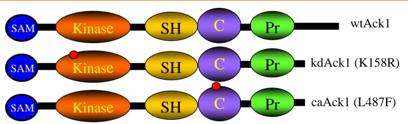
Fig 1.5: Ack1 modular protein structure with mutations K158R and L487F.
Ack1 is a non-receptor tyrosine kinase that contains a SAM (sterile-alpha motif) domain that promotes Ack1 dimerization at the plasma membrane to allow intermolecular
auto-phosphorylation. It also contains a NH2-terminal kinase domain, a Src homology domain (SH3), CRIB interacting domain, and a proline rich domain. The constitutively active (caAck1)
mutation disrupts auto-inhibitory intramolecular interaction while the kinase dead (kdAck1) mutation is in the ATP acceptor site. (Adapted from Mahajan 2005, [82]).
Kinas
e
C
R
Pr
SH
wtAck1
kdAck1 (K158R)
caAck1 (L487F)
Kinas
e
C
R
Pr
SH
Kinas
e
C
R
Pr
SH
SAM
SAM
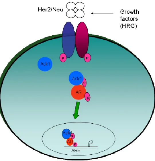
Fig 1.6: Ack1 activation of the AR pathway
Cell surface receptors are activated (by autocrine, paracrine, or mutational events) which leads to Ack1 kinase activation via auto-phosphorylation Ack1 binds and phosphorylates AR
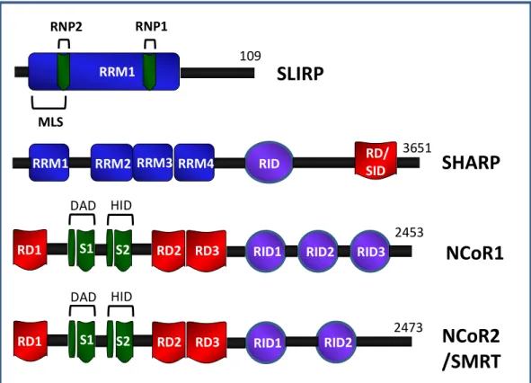
Fig 1.7: SLIRP, SHARP, NCoR1, and SMRT co-repressor protein modular structure.
SLIRP has one main RRM with submotifs RNP1 and RNP2 for SRA recognition and an N-terminal putative mitochondrial localizing signal. SHARP has multiple RRM domains which functions as a RNA recognition motif for binding RNA like SRA. SHARP can also recruit HDACs. NCoR1and SMRT contains C-terminal receptor interaction domains (RID) and three N-terminal repressor domains (RD). Submotifs SANT1 and SANT2 (Swi3/Ada2/N-Cor/TFIIID) contain deacetylase interaction domain (DAD) and histone interaction domain (HID)
respectively for HDAC recruitment. (Adapted from Hatchell 2006, Shi 2001, Huang 2000 [89, 100, 105])
RD1 S1 S2 RD2 RD3 RID1 RID2 RID3
DAD HID
NCoR1
NCoR2
/SMRT
2473 2453
RRM1 RRM2 RRM3 RID RD/
SID
SHARP
3651
RRM4 RRM1
RNP2 RNP1
SLIRP
109
MLS
RD1 S1 S2 RD2 RD3 RID1 RID2
CHAPTER II: ACK1 MEDIATED REGULATION OF CO-REPRESSOR
SLIRP IN AR SIGNALING
2.1 Overview
Prostate cancer progression into CRPC is dependent on androgen receptor (AR) reactivation. Multiple mechanisms enhance or activate AR including amplifications that sensitizes AR to low levels of androgens or mutations that confer responsiveness to other steroids besides the canonical ligands and phosphorylation of AR by kinases. In particular, we are interested in the activation of AR by the tyrosine kinase Ack1. We have previously shown that Ack1 phosphorylates AR and leads to a ligand independent activation. In order to
characterize this mechanism further, we utilized DIGE proteomics to identify components important in this pathway. SLIRP was identified as a candidate protein and further investigation into this protein revealed that SLIRP works as a co-repressor in AR signaling. Treatment of 293T cells transfected with AR and constitutively active Ack1 (caAck1) or LNCaP cells treated with DHT or caAck1 revealed that SLIRP associates with AR in the absence of AR activation. However, in the presence of DHT stimulation or Ack1 phosphorylation of AR, SLIRP
2.2 Introduction
Androgen receptor (AR) is a steroid receptor involved in prostate development and prostate cancer. Androgen deprivation therapy is used to treat patients but the cancer ultimately develops into castration resistant prostate cancer (CRPC). There is substantial evidence
indicating AR activation in low androgen environments through AR gene amplification, increased AR expression, AR point mutations occurring in the ligand binding domain that broaden ligand specificity, AR splice variants, and over-expression of AR co-activators [26-29]. When AR was over-expressed in androgen dependent xenografts, it enhanced the ability to form tumors in castrated mice while AR knockdown inhibited their tumorigenicity [114]. Prostate xenograft studies illustrate that tumors that recur after castration express AR dependent genes [115]. Furthermore, AR protein is stabilized and constitutively localized to the nucleus and becomes hypersensitive to low levels of androgen [116]. AR protein phosphorylation by non-receptor tyrosine kinases Src and Ack1 in an androgen independent manner have been indicated to play a role in the development of CRPC [81, 117, 118]. Src kinase phosphorylates AR at Tyr-534, leading to nuclear translocation and activation of AR dependent gene regulation [117, 118]. Ack1 (activated cdc42-associated kinase) expression is shown to be higher in CRPC tissue than primary prostate cancer tissue samples [70]. Specifically, Ack1 promotes prostate tumorigenesis by Wwox tumor suppressor degradation and phosphorylation of AR at Tyr-267 and Tyr-363 leading to activation of AR [81, 82]. Constitutively active Ack1 with a mutation that disrupts autoinhibition of Ack1 enhances AR transcriptional activity, increase AR recruitment, and promote androgen independent xenograft tumor growth [81].
through the RD2 domain and inhibit DHT-bound AR. NCoR1 and SMRT have also been shown to recruit HDACs to aid in receptor repression [83, 84, 86]. Another nuclear steroid receptor co-repressor, SLIRP (SRA stem-loop interacting RNA binding protein), represses receptors
including PPAR, VDR, and TR [105]. The majority of SLIRP is co-localized to the mitochondria with a small percentage in the nucleus [105]. SLIRP in the mitochondria maintains
mitochondrial-localized mRNA transcripts that encode Oxidative Phosphorylation (OxPhos) subunits [107]. SLIRP has also been shown to interact with LRPPRC in a ribonucleoprotein complex that may regulate the stability of mature mRNAs [108].
2.3 Materials and Methods
Cells and reagents- LNCaP cells and 293T cells were obtained from the American Type
Culture Collection (Manassas, VA, USA). Short tandem repeat (STR) authentication of LNCaP cell line was conducted (UNC Tissue Culture facility). DHT (Sigma-Aldrich, St Louis, MO, USA), EGF (R&D Systems, Minneapolis, MN, USA), IL-6 (R&D Systems), gas6 (R&D Systems) and bombesin (Sigma-Aldrich, St Louis, MO, USA) were purchased. Heregulin was a gift from Genentech (South San Francisco, CA, USA). Dasatinib was obtained from Bristol-Myers-Squibb (Princeton, NJ, USA). U0126 MEK inhibitor and luciferase assay kit were acquired from (Promega, Madison, WI). A mouse monoclonal antibody against AR (F39.4.1, Biogenex, San Ramon, CA, USA) was used for immunoblotting and a polyclonal antibody against AR (C-19, Santa Cruz) was used for immunoprecipitation. The antibody against total Ack1 was described previously (Mahajan et al 2005). A phospho-specific antibody against Ack1 p-Tyr-284 (# 09–142) was obtained from Millipore (Billerica, MA, USA). Antibody against SLIRP (#ab51523) was purchased from Abcam (Cambridge, MA, USA). Actin antibody and agarose A protein beads were purchased from Santa Cruz biotechnology (Santa Cruz, CA, USA). Anti-FLAG affinity gel (#A2220) and EZ view Red anti-FLAG beads were purchased from Sigma-Aldrich (St. Louis, MO, USA). Custom nonsense control (NS) siRNA
(GUUCAGGUCGAUAUGUGCA) and SMARTpool SRA siRNA (L-027192-00) was obtained from Dharmacon (Thermo Scientific Dharmacon, Pittsburgh, PA). A pool mix of SLIRP siRNA from Invitrogen (HSS130109, HSS188666, HSS188667; Life Technologies, Grand Island, NY) was used. GAPDH (Left 5’ACAGTCAGCCGCATCTTCTT3’, Right
Right 5’GCTGCCTCCTCTGAAAACAG3’) primers were used for PCR (UNC Nucleic Acid Core Facility, Chapel Hill, NC)
Plasmids-The plasmids encoding AR, truncated AR (Tr-AR), wild-type (wt) Ack1,
kinase dead (kd) Ack1, constitutively active (ca) Ack1, and the ARR2-PB-luciferase reporter were described previously (Mahajan NP, 2007) (Mahajan NP, 2005). FLAG-SLIRP and SRA expression vectors were purchased from Origene Inc. (Rockville, MD, USA). Y267F, Y363F, Y534F mutants of AR were obtained using Stratagene QuikChange™ Site-Directed Mutagenesis kit (La Jolla, CA, USA). The sequence was confirmed by direct sequencing.
Differential in gel electrophoresis – mass spectrum (DIGE-MS) assay- HEK 293T cells in
10cm culture dish were transfected with the expression vectors encoding AR (1µg), or AR (1µg) plus constitutively active Ack1(1µg) using effectene. After 24 hrs, protein extracts were
harvested and immunoprecipitated with 6ul (2ug) AR antibody plus 30ul agarose A beads overnight. The precipitant was analyzed using one-dimensional DIGE method in UNC Systems-Proteomics Core Facility, following protocols described previously (Alzate O., et al, 2004). The spot of interest was picked and analyzed by mass spectrometry at UNC Proteomics Core Facility. The resulting peptides were mixed with matrix (α-Cyano-4-Hydroxycinnamic Acid) and
analyzed using a MALDI-TOF/TOF mass spectrometer (Applied Biosystems 4800 Plus). MS spectra were obtained in reflector positive ion mode and peaks with signal-to-noise ratio above 20 were selected for MS/ MS analysis (maximum of 45 MS/MS spectra per spot). All spectra were searched using GPS Explorer Software Version 3.6 (Applied Biosystems) linked to the Mascot (Matrix Science, Inc.) search engine
Western -HEK 293T cells were co-transfected with wt-Fl-AR, wt-Tr-AR, Y267F,
cells were transfected with the Ack1 constructs. After 24hrs of transfection, cells were treated with vehicle control, DHT (10nM), heregulin (10ng/ml), EGF (100ng/mL), IL-6, or bombesin for another 24hrs. Protein extracts were quantified by Bradford assay (Bio-Rad Laboratories, Hercules, CA) and analyzed by SDS-PAGE as previously described (Steinbrecher et al., 2005). Samples were immunoprecipitated using AR antibody (C-19, Santa Cruz) plus protein A agarose beads or FLAG beads overnight. For knockdown experiments, SRA-siRNA (25nM, 50nM, 100nM) and non-sense (NS) control was transfected and RT-PCR was done to analyze SRA knockdown. Cells were transfected with control siRNA or SRA-siRNA (50nM) and transfected with Ack1 or treated with DHT. Immunoprecipitation was carried out using AR antibody and immunoblotted as indicated previously.
Immunoprecipitation RT-PCR Assay- This assay was done following the protocol
described elsewhere (Giles et al., 2003). Briefly, cell lysate from SRA-siRNA treated LNCaP cells was immunoprecipitated with SLIRP IgG, AR IgG, non-specific Rabbit IgG, or no IgG. Total RNA was isolated from immunoprecipitated fraction using tri-reagent (Molecular Research Center, Inc. Cincinnati, OH, USA). SRA RT-PCR was conducted following the protocol
described above.
Luciferase assay- LNCaP cells were plated at 6X10^4 cells/ well in a 12-well plate in
according to manufacture’s protocol. Statistical analysis was done with Graphpad prism (La Jolla, CA) using unpaired T-test.
Real-time Quantitative PCR- LNCaP cells were transfected with NS control siRNA or
pool mix of SLIRP siRNA at 40nM for 24hrs. Cells were washed once with 1x PBS and replaced with serum free media containing EtOH or 1nM/ml DHT for another 24hrs. RNA was collected using Qiagen RNeasy kit (74104) and cDNA was generated using Biorad iscript cDNA synthesis kit (Bio-Rad Laboratories, Hercules, CA). Real-time PCR was conducted using GAPDH (probe-FAM-CAGCCTCAAGATCATCAGCAATGCCTC-BHQ,
Fp-GTCATGGGTGTGAACCATGAGA, Rp-GGTCATGAGTCCTTCCACGATAC), PSA (probe-FAM-ATGACGTGTGTGCGCAAGTTCACCC-BHQ, Fp-GTTTTTGCCTGGCCCGTAG, Rp-GCATGAACTTGGTCACCTTCTG), hK2 (probe-TGACCTCATGCTGCTCCGCCTGT, Fp-GCCTTAGACCAGATGAAGACTCCA, Rp-GCCCAGGACCTTCACAACATC), and SLIRP (probe-CGGCCATGTCAGAAGGTGCA, Fp-GCGCTGCGTAGAAGTATCAA,
Rp-ACTGAACCCAACCCAAACCT) sets.
Chromatin Immunoprecipitation (ChIP) assay- Cells were lysed in lysis buffer
containing 50mmol/L Tris-HCl, 0.1% NP40, 150mmol/L NaCl, 10% glycerol, 2mmol/L EDTA, plus proteinase inhibitor (Roche Diagnostic, Indianapolis, IN, USA) and phosphatase inhibitor (St. Louis, MO, USA). Immunoprecipitation was done by incubating the mixture of 500 µg protein lysis with 2 µg IgG and 50 µL protein A agarose beads overnight at 4°C.
CA) or SLIRP (Abcam, Cambridge, MA, USA) was applied to immunoprecipitate DNA which was associated with AR and SLIRP. DNA was subjected to quantitative PCR using the primers and the probe targeting the distal ARE III enhancer sequence of the PSA gene or the primers and the probe targeting the distal enhancer of the hK2 gene as described previously.
2.4 Results
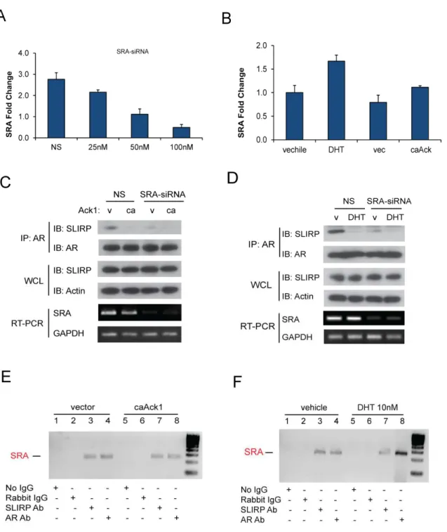
AR interaction with SLIRP is disrupted by Ack1 activation
To investigate potential interactors of AR affected by Ack1, we employed the Differential in Gel Electrophoresis (DIGE) proteomics technique to identify proteins whose expression changed in the absence and presence of Ack1. As shown in Fig 2.1A, one band demonstrated reduced binding to AR in Ack1 transfected cells compared to AR only cells. We isolated the band and analyzed it by mass spectrometry and identified SLIRP as a possible candidate interactor. In order to confirm our proteomics data, we immunoprecipitated AR and looked at SLIRP protein co-immunoprecipitation in the presence of wild-type Ack1,
constitutively active Ack1 (caAck1), and kinase dead (kdAck1) in 293T cells (Fig 2.1B). SLIRP co-immunoprecipitation with AR was demonstrated in the absence of Ack1 or in the kinase dead form. However, in the presence of Ack1 expression, SLIRP co-immunoprecipitation is reduced or lost. Furthermore, DHT treatment was also shown to disrupt interaction between SLIRP and AR (Fig 2.1C). This was confirmed with endogenous SLIRP and AR in the prostate cancer cell line LNCaP (Fig 2.1D). Immunoprecipitation of SLIRP demonstrated reduction of AR
Disruption of SLIRP-AR interaction is independent of known Ack1 phosphorylation
It has been shown that growth factors heregulin and gas6 can induce Ack1
phosphorylation of AR at Tyr-267 while Tyr-534 phosphorylation by Src kinase is induced by EGF, bombesin or- IL-6 stimulation [53]. SLIRP association was almost completely lost in the presence of heregulin treatment and thus Ack1 activation but not in the presence of other growth factors in LNCaP cells (Fig 2.2A). There is a small change in SLIRP complex with AR with both IL-6 and bombesin stimulation. Since neither of these ligands induce Ack1 mediated activation of AR, there is the possibility of another kinase leading to SLIRP dissociation. We have
where the complex between SLIRP and AR is disrupted by AR activation by either its ligand DHT or the tyrosine kinase Ack1.
SRA is necessary in SLIRP-AR interaction
It has been illustrated that RNA co-regulator SRA is important in SLIRP interaction with ER. Specifically, SLIRP is recruited to ER via binding of stem-loop 7 on SRA to mediate ER repression [105]. Therefore, we wanted to investigate the role of SRA in SLIRP-AR interaction. First, we established that SRA siRNA reduced RNA levels of SRA (Fig 2.3A). We found that DHT minimally induced SRA RNA levels while Ack1 had no affect (Fig 2.3B). SRA
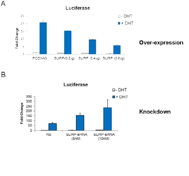
SLIRP is a co-repressor of AR signaling
Now that we have established a relationship between AR and SLIRP, we wanted to investigate the functional significance of SLIRP expression in the AR pathway. We first established our SLIRP siRNAs knockdown SLIRP in LNCaP cells (Fig 2.4A). Since SLIRP is mainly a mitochondrial protein, we also confirmed knockdown of SLIRP in the nucleus using a pool of our SLIRP siRNAs (Fig. 2.4B). Using the androgen dependent probasin promoter PB linked to two androgen response regions (ARR2PB) luciferase reporter, we looked at SLIRP effect on AR transactivation. SLIRP overexpression results in reduction in AR transactivation in a dose dependent manner (Fig 2.5A). Conversely, SLIRP knockdown results in increased AR transactivation in a dose dependent manner (Fig 2.5 B). This implicates SLIRP as a negative regulator of AR transactivation. To further investigate SLIRP’s role as a repressor, we looked at two canonical AR dependent genes, PSA and hK2. SLIRP knockdown resulted in an increase in endogenous transcriptional activity of PSA and hK2 which was enhanced by DHT treatment (Fig 2.5 C). The effect of SLIRP knockdown on hK2 transcription was more prominent than on PSA, suggesting that SLIRP repression of AR dependent genes may vary. Both DHT stimulation and Ack1 activation of AR leads to SLIRP dissociation from AR. Hence, we wanted to investigate whether recruitment of SLIRP to androgen response elements (ARE) in the absence or presence of either were affected. SLIRP was bounded to both the PSA and hK2 AREs in the absence of DHT and DHT treatment inhibited SLIRP recruitment to the AREs (Fig 2.6A and B).
Additionally, AR antagonist bicalutamide (casodex) did not affect SLIRP recruitment to PSA and hK2 AREs while Ack1 activation via heregulin treatment inhibited SLIRP recruitment (Fig 2.6 C and D). The data further substantiates the effect of Ack1 or DHT on SLIRP-AR
2.5 Discussion
We conclude with identifying a novel mechanism of AR repression that maybe perturbed in CRPC. We had identified a potential AR interactor, SLIRP, using proteomics and
immunoprecipitation techniques. We have shown that both ligand activation and Ack1 kinase activation of AR disrupts the association of SLIRP with AR. AR activation by EGF and bombesin occurs through Src kinase phosphorylation of AR at Tyr-534 [117-119]. Heregulin activates Ack1 via HER-2 receptor to phosphorylate AR at Tyr-267 and Tyr-363. We found that heregulin treatment of LNCaP cells but not EGF treatment resulted in the loss of SLIRP. IL-6 and bombesin affected SLIRP association with AR marginally. Neither bombesin nor IL-6 treatment results in AR phosphorylation of Tyr-267 and Tyr-363 by Ack1 inferring that an alternate kinase may be activated by these ligands [53]. A potential candidate that would be interesting to investigate is Src kinase as this kinase can lead to AR phosphorylation by both IL-6 and bombesin stimulation. Furthermore, dasatinib, a drug that inhibits BCR/Abl and Src kinases and used in the treatment of chronic myelogenous leukemia (CML), was found to be an Ack1 inhibitor in our work [53]. Thus, when we treated cells with dasatinib and heregulin, SLIRP was retained in the AR immunoprecipitation. Heregulin treatment can induce EGFR phosphorylation and hetrodimerzation with Her2 or Her 3 which can lead to AR phosphorylation [120]. Using MEK inhibitor, U0126, with heregulin demonstrated that Ack1 could still lead to SLIRP loss indicating that the EGFR pathway does not play a role.
Previous investigation into Ack1 mediated phosphorylation sites of AR using
phosphoprotemomics and mass spectrometry has been challenging. Therefore, we could hypothesize that an unidentified AR tyrosine target site of Ack1 could be important in the mechanism of dissociation of SLIRP from AR.
Furthermore, SLIRP dissociation from AR could be the result of Ack1 phosphorylation of another corepressor/component of the AR-SLIRP complex that has yet to be identified. NCoR1 and SMRT are well known co-repressors that can bind AR directly and inhibit AR signaling. However, NCoR1 and SMRT have weak interaction with unliganded AR but are recruited when AR is bound by antagonists such as cyproterone acetate, hydroxyflutamide or bicalutamide or agonists like DHT [95, 96, 99]. Other cofactors include those that recruit HDACs like androgen receptor corepressor-19 kDa (ARR19) which directly associates with AR and represses AR via recruitment of HDAC4 and FBI-1 which represses AR activity and can recruit HDACs [121, 122]. One candidate for the SLIRP co-repressor complex is SHARP, which like SLIRP can interact with RNA co-regulator SRA and repress steroid receptor signaling [100, 105]. It was also shown that co-transfection of SLIRP and SHARP can enhance repression of ER signaling more than SLIRP alone [105].
When we explored chromatin recruitment of SLIRP to multiple AR gene enhancers, we found that SLIRP recruitment is reduced by DHT or heregulin. However, SLIRP recruitment is restored with the addition of AR antagonist bicalutamide. It would be more informative to conduct ChIP-Seq experiments where we would be able to map all binding sites for SLIRP. Our model (Fig 2.7) illustrates a mechanism where SLIRP represses AR thereby controlling AR signaling but with the activation of AR by Ack1 or DHT, leads to the dissociation of SLIRP from AR, recruitment of co-activators possibly by SRA, and activation of AR dependent genes.
Identification of interacting proteins in this complex would help in determining the mechanism of action of Ack1 in the interaction between SLIRP and AR. One approach to identifying interactors is to use a combination of cross-linking agents with immunoprecipitation followed by mass spectrometry analysis. Methods developed have incorporated antibody cross-linking, Quantitative SILAC proteomics, and lysate cross-linking. For instance, Akt interactors were identified by using a two-step chemical cross-linking using DSS and DSP followed by Co-IP and MS [123]. Furthermore, a study trying to profile post-translational modification (PTM) dependent protein-protein interaction like phosphorylation or methylation, combined photo-cross linking using benophenone with SILAC labeled cells to identify H3K4Me3 specific proteins [124]. Thus, focusing on identification and optimization of cross-linking agents like
formaldehyde or NHS esters (amine) followed by in-gel or bead digestion and MS analysis could identify important SLIRP interactors that are apart of the AR-SLIRP co-repressor complex. Unraveling the complex is important in understanding how SLIRP works in the cell and eventually how SLIRP co-repressor function is lost in the progression of prostate cancer.
enhancement of endogenous transcription of AR dependent genes PSA and hK2. For that reason SLIRP acts as a corepressor and negatively regulates AR transactivation. However, in the mitochondria, where SLIRP is mainly localized, it functions in mitochondrial RNA stability (mtRNA) of Oxidative phosphorylation (Oxphos) proteins and knockdown can reduce
components of the Oxphos pathway [107, 108]. Oxidative phosphorylation is important as the main regulator of ATP. However, cancer cells rely more on glycolysis for energy production which is termed the “Warburg effect.” We had analyzed the Oxphos pathway through oxygen consumption rate (OCR) and the glycolysis pathway by extracellular acidification rates (ECAR) measurements using XF24 Extracellular Flux Analyzer (Seahorse Bioscience, Massachusetts, US). We found that when SLIRP was knocked down by siRNA in LNCaP cells, there was increased glycolysis as measured by increases in ECAR compared to our control (data not shown). We would expect that SLIRP knockdown decreased OCAR or had no effect has high levels of SLIRP are associated with the mitochondria. Interestingly, we found that SLIRP
knockdown enhanced OCAR levels and hence Oxphos (data not shown). Hence, there may be an alternative role of SLIRP in the mitochondrial besides maintaining mtRNA levels that would account for the OCAR increase.
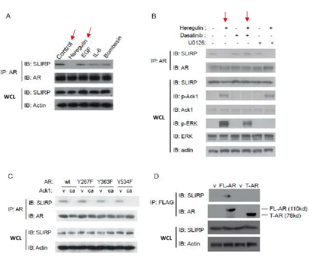
Figure 2.1: Ack1 and DHT impair the interaction between AR and SLIRP. (A) DIGE
analysis in 293T cells. 293T cells were transfected with AR (1µg) only (Cy3) or AR (1µg) plus constitutively active Ack1 (1µg) (Cy5). Band indicated was isolated and analyzed through MS. (B) Ack1 activation (wild-type (wt) or constitutively active (ca)) interrupts the interaction of AR and SLIRP in 293T cells. The interruption is dependent on the kinase function of Ack1. (C) DHT treatment impairs the interaction between AR and SLIRP in 293T cells. (D) DHT and Ack1 activation impairs the interaction between AR and SLIRP in LNCaP cells. (E) Association between AR and SLIRP is disrupted by DHT and Ack1 in 293T cells as demonstrated by SLIRP immunoprecipitation. These experiments were done by Dr. Zhentao (Richard) Zhang, in
Figure 2.2: Effects of various growth factors, kinase ligands, AR mutants on AR-SLIRP interaction. (A) Effects of heregulin (10 ng/ml), EGF (100 ng/ml), gas6 (100 ng/ml), IL-6 (10
ng/ml), and bombesin (1nM) on AR-SLIRP interaction in LNCaP cells. LNCaP cells were treated with the growth factors for 60mins in serum-free medium before harvest. (B) Dasatinib (10ng/ml) inhibits Ack1 kinase activity and maintains AR-SLRIP association with heregulin (10 ng/ml) treatment in LNCaP cells. (C) AR tyrosine phosphorylation at Y267F, Y363F or Y534F does not contribute to the Ack1-induced AR-SLIRP dissociation in 293T cells. (D) Lack of the LBD (Tr-AR, aa 1-660) impairs AR-SLIRP interaction in LNCaP cells. These experiments were done by Dr. Zhentao (Richard) Zhang, in collaboration.
WCL
WCL
Figure 2.3: SRA is essential for AR-SLIRP interaction. (A) SRA knockdown by SRA-siRNA
in 293T cells with non-sense (NS) control. Values present the means ± SEM (N=3). (B) SRA expression with DHT (10nM) treatment or caAck1 activation in LNCaP cells. RNA was isolated and real-time qRT-PCR was done to analyze SRA RNA levels. Values present the means ± SEM (N=3). (C and D) SRA knockdown abrogates the interaction between AR and SLIRP. (C)
Figure 2.4: Validation of SLIRP knockdown by siRNA
Figure 2.5: Effects of SLIRP on AR mediated transcriptional activity in LNCaP cells.
Figure 2.6: Effects of DHT and Ack1 activation on SLIRP recruitment to androgen
response element (ARE). (A) DHT treatment impairs the recruitment of SLIRP to ARE on PSA
Figure 2.7: Current model of Ack1 activation and DHT treatment on AR-SLIRP
interaction. In the absence of Ack1 activation and DHT, the co-repressor SLIRP binds to AR
and is recruited to ARE of AR target to suppress gene expression. With the activation of Ack1 or DHT treatment, the association between AR and SLIRP is abrogated and AR dependent
CHAPTER III: SLIRP ACTS AS A POTENTIAL TUMOR SUPRESSOR IN
PROSTATE CANCER CELLS BY REGULATING CELL CYCLE
3.1 Overview
Mechanisms underlying AR reactivation in CRPC is not well understood. We have demonstrated that SLIRP forms a co-repressor complex with AR and controls AR dependent transcription. In addition, Ack1 can dissociate this co-repressor complex and activate AR signaling. Thus, SLIRP repression of the AR signaling pathway may be a mechanism that is undermined in late stage prostate cancer. We wanted to understand the effect of the loss of SLIRP on prostate cancer biology. We preformed RNA-seq analysis to examine gene expression changes with the loss of SLIRP. Interesting, we saw a broad global change in gene expression with SLIRP knockdown. When we used a published AR gene signature set to compare our knockdown samples to control, we identify a clear partitioning of our samples validating the significance of SLIRP in prostate cancer. Using our RNA-seq data, we generated a 176 AR gene signature which separated genes into three distinct classes. A closer look into the pathways affected by SLIRP knockdown led us to study cell cycle regulation, a hallmark of cancer, as many proteins in this pathway were changed. Androgens increase cell proliferation by advancing cells through the cell cycle. We find that SLIRP knockdown affect cell cycle regulators including phospho-Rb, E2F1, E2F2, CDK4, cyclin B1, and cyclin A2. This phenomenon is further
illustrated with an increase of LNCaP cells in S-phase with SLIRP knockdown. Consequently, this supports the role of SLIRP as a tumor suppressor that can regulate cell cycle progression in part through regulation of AR signaling.
3.2 Introduction
resurgence of androgen receptor (AR) activity. AR gene amplification, AR mutations,
deregulation of co-activators/co-repressors, and alterations in upstream AR signaling pathways all may contribute to the re-emergence of AR activity in CRPC. Furthermore, other signaling mechanisms like transcription factors, oncogenes, and tumor suppressors are altered in prostate cancer and contribute to the development of CRPC. Thus understanding the interplay between AR and other signaling networks and their involvement in metastatic CRPC is important in developing proper therapeutic agents.
One hallmark of cancer cells is the ability to bypass cell cycle checkpoints and induce uncontrolled cell division. Androgens and the androgen receptor have also been shown to be important in cell cycle in androgen dependent cells. Cells in resting phase, G0, are regulated by retinoblastoma protein (Rb) to prevent unplanned cell cycle progression. Androgens can
Thus a dynamic interplay exists between androgens, AR, and the cell cycle in prostate cancer. We have shown that SLIRP acts as a co-repressor of AR signaling and it dissociates when AR is activated. Gene expression analysis using RNA-seq with SLIRP knockdown demonstrated a change in the global gene expression profile compared to control. Multiple AR dependent genes showed an increase with SLIRP knockdown. Furthermore, we generated an AR gene signature that was used to cluster our samples and illustrated generation of three classes of genes. These three classes represent one set that is more SLIRP dependent and SLIRP loss reduces expression of these genes, a group where SLIRP knockdown has a greater impact on the up-regulation of the genes, and a group that is more androgen dependent than SLIRP dependent. Analysis of pathways using gene list created by significance analysis of microarray (SAM) on our RNA-seq data, directed us to a role of SLIRP in cell cycle regulation that is AR dependent. SLIRP knockdown stimulates an increase in cyclin and CDK levels with hyperphosphorylation of Rb. Flow cytometry analysis showed an increase of cells in S-phase with SLIRP knockdown compared to control. Furthermore, the role of SLIRP in cell cycle seems to be AR dependent as AR negative cells did not demonstrate this feature. The effect of AR co-repressors NCoR1 and SMRT knockdown on cell cycle did not demonstrate the enhanced cell cycle progression seen with SLIRP knockdown. SHARP co-repressor knockdown showed modest enhancement of cell cycle but not to the degree of SLIRP knockdown. Hence, the loss of SLIRP in conjunction with AR signaling may provide a mechanism by which cell cycle progression is enhanced and regulation bypassed.
3.1Materials and Methods
Cells and reagents -LNCaP cells, PC3, DU145, and HEK 293T cells were obtained from
Charles Sawyers lab. DHT was acquired from Sigma (Sigma-Aldrich, St Louis, MO, USA). Short tandem repeat (STR) authentication of LNCaP and LAPC4 cell lines were conducted (UNC Tissue Culture facility). A mouse monoclonal antibody against AR (F39.4.1, Biogenex, San Ramon, CA, USA) was used for immunoblotting. Antibody against SLIRP (#ab51523) was purchased from Abcam (Cambridge, MA, USA). Antibodies against total ERK (#9102),
phospho-specific ERK (#9101), and phospho-Rb (Ser 807/811, #9308P) were obtained from Cell Signaling Technology (Beverly, MA, USA). Rb (C-15), CDK2 (M2), and CDK4 (DCS-35) antibodies were purchased from Santa Cruz (Santa Cruz Biotechnology Inc., Santa Cruz, CA). Pan-Actin Ab-5 (ACTN05) was acquired from Neomarkers (Fremont, CA). Custom nonsense control (NS) siRNA (GUUCAGGUCGAUAUGUGCA), SMARTpool NCoR1 siRNA (L-003518-00), SMARTpool NCoR2/SMRT siRNA (L-020145-01) were purchased from Dharmacon (Thermo Scientific Dharmacon, Pittsburg, PA). Pool mix of SLIRP siRNA
(HSS130109, HSS188666, HSS188667) and SHARP/SPEN siRNA (HSS118024, HSS118025, HSS118026) were obtained from Invitrogen (Life Technologies, Grand Island, NY) and AR siRNA sequences were custom designed (Mol Cancer Ther April 2005 4:505-515)
Plasmids-The plasmids encoding Fl-WT-AR were described before (Mahajan NP, Proc
Natl Acad Sci U S A, 2007. 104(20): p. 8438-43), (Mahajan NP, Cancer Res, 2005. 65(22): p. 10514-23). FLAG-SLIRP and SRA expressing vector were purchased from Origene Inc. (Rockville, MD, USA).
siRNA Transfection- Cell lines were transfected with custom nonsense control siRNA