FLUORESCENCE AND BACTERIAL MICROBIOME OF
DENTIN IN DEEP CARIOUS LESIONS
Leslie H. Trippe
A thesis submitted to the Faculty of The University of North Carolina at Chapel Hill in partial
fulfillment of the requirements for the degree of Master of Science in the Department of
Operative Dentistry of the School of Dentistry
Chapel Hill
2017
Approved by:
Andrea Ferreira Zandona
Andrea Azcarate-Peril
Lee W. Boushell
John S. Preisser
iii
ABSTRACTLeslie H. Trippe: Fluorescence and bacterial microbiome of dentin in deep carious lesions (Under the direction of Andrea Ferreira Zandona)
Purpose: To determine if the presence or absence of fluorescence as clinically
detected by fluorescence–aided caries excavation (FACE) correlates with dentin bacterial
microbiome diversity, as assessed by 16S rRNA amplicon sequencing and to compare and
contrast traditional tactile dentin caries detection with fluorescence. Materials and
Methods: Unidentified, extracted human, carious teeth considered discarded waste were
obtained from a dental facility. Providers unrelated to the research study supplied the teeth
- NHSR (Not Human Subjects Research). Included teeth had a carious lesion two-thirds
into the dentin, verified by a radiograph (post extraction), and fluoresce red using FACE
technology (SIROInspect,Sirona, Bensheim Germany). Fluorescing carious dentin was
assessed using the traditional visual-tactile method, and comparisons made before and
after cavity preparation. Red fluorescing (RF) sites were sampled with a sterile spoon
excavator, and dentin characteristics evaluated clinically via traditional visual/tactile
methods. Once RF dentin was removed, a second sample of carious dentin with pink
fluorescing (PF) dentin was obtained with a sterile spoon excavator. After excavation with
sterile round burr to non-fluorescing (NF) dentin, the dentin characteristics were evaluated
again by visual-tactile method, and a third sample was collected with a new sterile slow
speed round burr. Samples were transported on dry ice for analysis of bacterial microbiome
to the UNC Microbiome Core Facility. Results: Out of 134 extracted teeth collected, 21 fit
iv
had a higher number of observed OTUs (Operational Taxonomic Units), at 154, followed by
PF 109 and (NF) 100. Regarding tactile assessments: RF carious dentin was primarily
‘soft’, but also had readings of ‘leathery’ or ‘hard’ tactically, and the NF dentin was assessed
as ‘hard’ 100% of the time by both examiners. The rank correlation chi-squared statistic for
the association of fluorescence and tactile was highly statistically significant (p < 0.001) for
each examiner strongly suggesting an association between fluorescence and tactile
assessment. However, approximately one-third of the tactile assessments of hard dentin
still displayed some level of fluorescence, either PF or RF. Conclusion: The sampled
fluorescing (RF, PF) and NF carious dentin layers displayed diverse bacterial taxa that
varied in the proportion of bacterial species. Tactile assessments correlated with RF, PF
and NF approximately two-thirds of the time, as one-third of the time hard dentin
v
To my wonderfully loving and supportive family – Kevin, Tessa and Kara.
The three of you have been my true and faithful followers as I journeyed down this road of
continuing my education! I know deep in my heart that I could not have accomplished my
goals without each of you, and your unconditional love. Thank you.
To my sisters, Donna and Nancy and my father, Don Hobbs – who along the way provided
me with continual support, love and encouragement. I love you all so much!
To my mentor, Dr. Andrea Ferreira Zandona – words cannot not describe the respect I have
for you! Understanding what you have already achieved, and knowing what more you have
to accomplish overwhelms me. I am thrilled to say that you are my mentor, and have truly
vi
ACKNOWLEDGEMENTS
I would like to express my sincere appreciation to my mentor, Dr. Andrea Zandona
and my committee members, Dr. Andrea Azcarate, Dr. John Preisser, Dr. Lee Boushell and
Dr. Apoena Ribeiro for their invaluable guidance, patience and knowledge.
Special thanks to Dr. Terry Donovan who taught me everything there is to know,
and much more, about dental literature. I will forever look at our copious dental journals
with a new, and yet more critical view.
I also would like to thank all of our awesome Operative faculty and leadership: Dr.
Andre Ritter, Dr. Harald Heymann, Dr. John Sturdevant, Dr. Rick Walter, Dr. Gustavo
Olivera, Dr. Scott Eidson, Dr. Patricia Miguez, Dr. Taiseer Sulaiman, Dr. Ken May and Dr.
Al Wilder.
Thank you Shannon Tate, Kim Schoen and Barbara Walton for taking such good
care of all of the residents – day in and day out.
Thank you to my co-residents who have supported me throughout this process and
made the past 3 years truly wonderful: Caroline Nguyen-Ngoc, Eduard Epure, Islam Abd
Alraheam, Mohammad Atieh, Elizabeth Griffis, Sama Suliman, Awab Abdulmajeed,
Vilhelm Olafsson, Clayton Rau and Upoma Guha.
Lastly, I would like to extend a special thank you to Sirona for supplying us with 2
vii
TABLE OF CONTENTS
LIST OF TABLES ...ix
LIST OF FIGURES ...x
LIST OF ABREVIATIONS... xi
1. CHAPTER 1: LITERATURE REVIEW ...1
1.1 Introduction ………..…...1
1.2 Sound Dentin...2
1.2.1 Dentin Caries Development and Process...3
1.2.2 Histopathology of Carious Dentin...4
1.3 Complete Caries Removal ……….6
1.4 Incomplete Caries Removal ……….……….8
1.5 Caries Detection and Removal Techniques...10
1.5.1 Caries Detection Dye …...11
1.5.2 Fluorescence Based Technology...13
1.5.3 Caries Removal by Chemical Means...14
1.6 Microbiome of Caries ……….……….….…...…..16
2. CHAPTER 2: The Use of FACE Technology for Detecting Infected Dentin In Deep Dentinal Lesions, an In-Vitro Study 2.1 Introduction ………..………..……..18
2.2 Materials and Methods ………..……...…….20
viii
2.4 Results ………..……….….24
2.5 Discussion ………..27
2.6 Limitations ………....32
2.7 Conclusion ……….32
3. TABLES …..……….……….34
4. FIGURES ……….……….……….…………..40
REFERENCES ……….……….………..46
ix
LIST OF TABLES
Table 1: Flowchart of sampling methods ………..34
Table 2: Microbiome diversity within each sample regarding
fluorescence (Alpha Diversity) ………...……….35
Table 3: Predominant species in RF ………..35
Table 4: Predominant species in PF ………..36
Table 5: Predominant species in NF ……….………..……..36
Table 6: Microbiome diversity within each sample regarding tactile ……….…..37
Table 7: Tactile assessments based on Fluorescence, Examiner 1 and 2 ………..…….….37
Table 8: Tactile Assessment Agreement *Kappa 0.79 and Weighted Kappa 0.86 …..…..38
Table 9: Principal Coordinate Analysis (PCoA) Beta Diversity, between samples……....38
x
LIST OF FIGURES
Figure 1: Histological layers and clinical manifestations;………40
Figure 2: Fluorescence ratio - Red: Green Lennon 2006 ………..………..40
Figure 3. Fluorescence images from Lennon article, 2006 ……….……….41
Figure 4: Layers sampled during our in-vitro study, RF, PF and NF ………..41
Figure 5: SIROInspect, Sirona (Bensheim, Germany) ……….………....42
Figure 6: Tooth fluorescing red through the use of SIROInspect ………..42
Figure 7: Radiograph of tooth revealing deep dentin caries ………..43
Figure 8: Lab packet for sample collection ...…...43
Figure 9: Sample A, pre-excavation ………..44
Figure 10: Post-excavation tactile assessment ……….……….44
Figure 11: Sample C, Post-excavation ……….44
Figure 12: Microbiome Lab sequences ………..……….45
xi
LIST OF ABREVIATIONS
CCR Complete Caries Removal
CDA California Dental Association
CDD Caries Detection Dye
CFU Colony Forming Units
CMCR Chemomechanical Caries Removal
DEJ Dentin Enamel Junction
DNA Deoxyribonucleic Acid
FACE Fluorescent Aided Caries Excavation
KHN Knoop Hardness Number
LIF Laser Induced Fluorescence
NF Non Fluorescing
OTU Operational Taxonomic Unit
PCR Partial Caries Removal
PEARL Practitioners Engaged in Applied Research and Learning
PF Pink Fluorescing
RDT Remaining Dentin Thickness
RF Red Fluorescing
1
CHAPTER 1: LITERATURE REVIEW
1.1 Introduction
Dental Caries is defined as the progressive demineralization of susceptible tooth
surfaces due to the metabolic activities that occur in dental plaque (i.e. the biofilm). The
four basic ingredients required for caries lesion formation are: susceptible tooth surface,
bacteria, fermentable carbohydrates and time. The term dental caries refers to the process
while a dental caries lesion describes the ‘result of this process’ of actual dissolution of the
hard tissues (enamel, dentin, cementum), which occurs due to acidic bacterial byproducts of
carbohydrate fermentation, followed by the loss of the protein matrix. This dynamic process
can be acute or chronic, causing significant mineral loss with or without sensitivity or pain
recognition from an individual.[1, 2] Once there is significant mineral loss, a cavitation
may become apparent. Management of a cavitated caries lesion typically involves
excavation of the involved tooth structure and preparation of a cavity to receive a
restorative material. This dental caries excavation process is very subjective as it usually
involves removing all presumably infected and affected dentin until tactile sensation
indicates that firm and intact tooth structure is reached. Traditionally only hard dentin
was considered clean, healthy dentin, acceptable as supporting tissue underneath a
restoration. This process is referred to as complete caries removal (CCR).
The evolving understanding of the dental caries process and of the histopathology of
caries lesions has led to a paradigm shift in dental caries management as it relates to caries
2
as ‘inner carious dentin’, and towards disease management following tooth preserving
principles. [3, 4] [5, 6] New restorative materials allow for more conservative cavity
preparations and can rely on enamel bonding rather than the compact packing of
restorative materials for a seal. Compounding evidence indicates that when a good seal is
present the caries lesion arrests, even if bacteria are still present. [7] [8] [9] [10] Recent
data with reverse-capture checkerboard analysis indicate that excavating to hard dentin
does not assure a ‘sterile’ dentin, as bacteria are present in all dentinal layers in deep caries
lesions.[11] Thus, it is essential that the correlation between the biofilm microbiome
content and dentin characteristics are better understood. This understanding has the
potential to translate to a more accurate and clinically applicable method to differentiate
between infected, now referred to as ‘outer carious dentin’ and inner carious dentin, a
critical need in dentistry.
1.2 Sound Dentin:
Dentin is a bone-like matrix that is porous, has a yellow-hue and is made of 70%
inorganic materials, 20% organic materials and 10% water by weight. Due to its elastic
properties, it functions as a supporting tissue for enamel and aids in prevention of enamel
fracture. It comprises most of the tooth structure and, in teeth with a viable pulp, it
continues to form after tooth eruption and throughout its life via the activity of
odontoblasts. [12] Dentin structure encompasses microtubules (dentinal tubules), which
house the odontoblast processes and dentinal fluid. The microtubules are surrounded by a
highly mineralized peri-tubular zone and intertubular dentin between the microtubules. In
the coronal dentin the dentinal tubules extend from the dentinal enamel junction (DEJ) to
the outer wall of the pulp, while in the root portion they extend from the cemento-enamel
3
(fibrillary Type 1) and non-collagenous proteins. The inorganic phase of dentin consists of
apatite crystals, mainly hydroxyapatite and non-crystalline amorphous calcium
phosphate.[13] Because the microtubules follow the tooth anatomy, their diameter is
smaller in the outer dentin compared to the inner dentin, which results in more compacted
dentinal tubules. Therefore, in the inner dentin, the microhardness has been reported as
72.53 (Knoop Harndess Number- KHN) in outer (superficial) dentin and 65.05 KHN in deep
dentin.[14]
1.2.1 Dentin Caries Development and Process:
Dental caries is a diet regulated and biofilm modulated multifactorial disease. There
is strong evidence indicating that a diet high in fermentable carbohydrates will modify the
microbial biofilm, favoring acidogenic and aciduric bacteria. Organic acids, such as acetic,
lactic and propionic acid are produced through a carbohydrate fermentation process, [15]
causing a drop in the oral biofilm pH. Breakdown of enamel is reported to occur when the
pH drops to ~5.5, while for dentin breakdown occurs when pH drops to ~6.4, and depends
on the level of fluorapatite present. However, the critical pH below which enamel and
dentin dissolves is not constant but rather a range which is inversely proportional to the
concentrations of calcium and phosphate in the saliva and plaque fluid.[16] Fejerskov et al,
refers to caries as the ‘result’, which occurs after a shift in the ecology and metabolic
activity of the biofilm. The consequence of this shift is an imbalance of enamel and dentin
mineral content which can create tooth structure dissolution.[1] Carious lesions may
initiate in the enamel and progress into dentin [17, 18], or may initiate in exposed dentin.
The initiation and progression of dental caries is attributed to not only the type of
microorganisms present, but also the quantity of acid producing bacteria. Streptococcus
4
highly aciduric, feeds off a high sucrose diet and produces an insoluble glucan, which
promotes further bacterial adhesion. Simon-Soro, et al reported that particular bacteria are
abundantly present during the initial stages of the enamel carious process; however, at the
dentin level more proteolytic bacteria are involved. Lactobacillus, Prevotella and
Streptococcus are reported to increase in number in the deeper dentin carious lesions,
whereas Neisseria, Capnocytophaga and Fusobacterium significantly decrease in
numbers.[19].
1.2.2 Histopathology of Carious Dentin:
A carious lesion in dentin was described by Fusayama to have three layers: outer,
inner and sound dentin. The outer layer, or the most superficial part of the carious lesion is
characterized as being non-remineralizable in contrast to the inner carious dentin layer
which can be remineralized. These layers may further be described and categorized into
‘zones’. The outer carious dentin layer is also called the zone of destruction and contains
dentin which has been decomposed due to the action of acid (which results in mineral loss)
and proteolytic enzymes (which result in loss of the dentin collagen matrix). This zone has
also been described as the ‘necrotic’ or ‘contaminated’ zone, and relates to soft
non-remineralizable dentin that is heavily infected with bacteria. (Figure 1) Collagen in this
zone has been irreversibly denatured and thus, can no longer function as a frame work for
remineralization. The inner carious dentin comprises the zone of bacterial penetration and
the translucent zone. The zone of bacterial penetration can be identified by the destruction
of odontoblastic processes, further leaving the tubules empty with the ability of bacteria to
invade. In a slowly developing lesion tubular sclerosis may be observed. Tubular sclerosis
can be defined as one of the initial defense mechanisms of the pulp-dentin complex, by
which there is deposition of minerals from the intertubular dentin into the dentin tubules,
5
dentin and when visualized under a microscope it appears translucent. This translucent
zone is a zone of demineralization which has also been described as the ‘transparent’ zone,
and is argued that this area may tactically feel either firm or leathery and can ultimately
remineralize.[20, 21] In some slowly developing lesions the tubular occlusion and further
mineralization of the intratubular dentin is able to form a highly mineralized layer that
appears dark clinically and transparent under the microscope. Sound hard dentin and
tertiary dentin may also be present adjacent to the pulp chamber.
Shovelton in 1968 was already reporting that demineralization precedes bacterial
invasion. He stated that in ‘hard’ dentin post caries excavation, only 64% was technically
free of bacteria. Ultimately he concluded that softening dentin generally precedes the
invading organism, however even in the deepest parts some infected dentinal tubules may
remain.[22] Angker, et al. correlated the mechanical properties and mineral content of
carious dentin. Their study concluded that in a progressing dentin carious lesion, when
there is a reduction in mineral content, evidence of deterioration of mechanical properties
exists. Hardness and elastic modulus were discovered to be an exponential function of its
mineral content.[23]
Recommendations have been made to leave the inner carious dentin intact, and only
remove the outer carious dentin layer of greatly softened and discolored dentin, as this
layer is considered non-remineralizable where the collagen is irreversibly denatured. [24,
25] This has been referred to as partial caries excavation or more recently, selective caries
excavation. The goal of these procedure is to remove only the irreversibly demineralized
dentin (outer dentin caries) which is in contrast to the traditional notion of complete caries
6
1.3 Complete Caries RemovalTraditionally, when managing a carious lesion at any depth, the dental
provider was to remove all of the inner and outer carious dentin to not only prevent further
tooth destruction by the cariogenic activity, but also to provide a sound base of dentin for a
restoration.[26] Dentists have been taught to remove all of the softened dentin in order to
eliminate the inner carious dentin tissue, assuming that both the biofilm and the respective
microorganisms within this layer of dentin is what drives the carious process. Many are
still taught to not only remove all soft outer and inner carious dentin, but also to remove
stain at the dentin-enamel junction (DEJ).[17] Current approaches recognize that, as the
caries removal process approaches the pulp, the potential for an unnecessary pulpal
exposure becomes a concern. The dental provider must then decide if they wish to attempt
an indirect/direct pulp capping procedure, or perform a pulpotomy or pulpectomy. Every
dental procedure has inherent risks, which may negatively impact the pulpo-dentinal
complex, and potentially lead to further treatment such as a root canal, or even extraction.
In light of this clinical reality, Schwendicke has described the caries removal process as
‘initiating a cascade of re-interventions’, which ultimately compromises long term pulp
vitality.[27]
It has long been suggested that pulp exposures were a result of ‘too vigorous removal
of affected dentin’, or inner carious dentin. If a tooth is symptomless, and responds well to
vital pulp testing, stained dentin (inner carious dentin) can be left over the pulp as long as
it is ‘reasonably hard’.[6] Furthermore, Kidd concludes that biologically, it may be
potentially harmful to even attempt to remove all of the inner carious dentin, and
emphasizes the placement of a well-sealed restoration.[6] As Tomes stated back in 1859 “It
is better that a layer of discolored dentine should be allowed to remain for the protection of
7
removal process (i.e. when is the carious dentin clinically hard enough?) is very subjective
and varies widely according to where the dentist received training, and that individual’s
interpretation of when softened, inner carious dentin tissue may remain directly adjacent to
the pulp.
Regardless of what type of caries excavation method is utilized, CCR or a type of
selective caries removal process, the dentist clinician still must determine at what point to
cease the removal of dentin. Currently, the most common criterion used to guide the caries
excavation is by means of visual/tactile perception. The visual determination of the
presence of carious tooth structure is often led by variations in the color of the dentin, and
the tactile sensation is accomplished through the use of a dental probe or explorer where
the assessments are based on hardness or texture. These methods are inherently
subjective; vary between dentists in clinical practice as well as dental researchers and
dental educators. These variations inevitably produce different sizes of cavities, and
potentially carry significant clinical implications. [29] The pulpal health may be jeopardized
if too much dentin is removed, and the overall strength of the tooth is compromised
structurally, leaving minimal remaining dentinal thickness (RDT).
Ricketts et al., through a Cochran Oral Health Group review, analyzed current
operative management techniques, and concluded that both stepwise (SW) and partial
caries removal (PCR) procedures reduced the incidence of pulp exposure in symptomless,
vital carious primary and permanent teeth. The authors further stated that complete caries
removal techniques have clear disadvantages, including damage to the pulp and weakening
of tooth structure.[30] However, in clinical practice as evidenced by a recent PEARL
(Practitioners Engaged in Applied Research and Learning) network study, the preferred
8
the PEARL network using case studies, 62% percent of those responding (85/92) indicated
that, in a case where pulp exposure may be expected, they would remove all outer and inner
carious dentin (CCR) and only 18% would consider selective caries removal.[31]
Financial consequences of dental caries is estimated to be the fourth-most expensive
disease to treat by the World Health Organization (WHO). In the United States, as reported
by the American Association of Endodontics, there are over 41 thousand root canals
performed each day at a cost of US$34 million annually. It is likely that many pulp
exposures and subsequent endodontic treatments may be avoided if a more conservative
approach of caries removal is chosen.[27, 32]
1.4 Incomplete Caries Removal
Kidd and Ferjerskov ask the question, “what drives this caries process?”, and
wondering if it is exclusive to the biofilm entering from the cavity surface or possibly from
the bacteria which are present within the lesion? They suggest that the progression of the
lesion can be arrested by the sealing of any remaining bacteria, which may exist in the
cavity, by restoring the tooth properly, thus allowing the ongoing removal of the cariogenic
biofilm by routine daily hygiene. [20] The sealing of any remaining bacteria, which may
exist in the inner carious dentin, is referred to as Incomplete Caries Removal, and may be
approached in two different ways: 1) Stepwise Excavation (SW), or 2) Selective Caries
Removal (SCR). SW is performed by removing the outer carious dentin layer or the zone of
destruction/contamination of the caries lesion, leaving soft residual caries, placing a
medicated liner/base and then a provisional therapeutic restoration, which has to be
removed and replaced with a permanent restoration in 6 weeks to 3 months. SCR,
9
of necrotic, disorganized dentin to tactile ‘hardness’ along the peripheral walls of the cavity
preparation, leaving soft dentin over the pulp, placement of an indirect pulp cap and a
permanent, well-sealed restoration. In both protocols only the outer carious dentin is
removed, while the inner carious dentin (zones of bacterial penetration and translucent
zone) are preserved prior to placement of a permanent restoration. The outer carious
dentin is characterized by necrotic, disorganized and irreversibly demineralized dentin
tissue, while the inner carious dentin is characterized by vital, organized, and
remineralizable dentin. Ample evidence shows that sealing inner carious dentin will lead
to lesion arrest, [7] [8] [9] and preservation of pulp vitality, thus decreasing the need for
endodontic treatment [32-34].
This conservative approach preserves tooth structure, which limits the need for
more complex, costly procedures. High caries risk patients particularly reap great benefits
from the resulting health-gains and cost-savings. [8] [27] Selective caries excavation relies
on accurate differentiation between outer and inner carious dentin, which is not
straightforward clinically. Clinicians rely on tactile sensation to determine if characteristics
of the dentin indicate inner carious dentin (hard, firm or leathery) or outer carious dentin
(very soft, disorganized). This uncertain diagnosis, compounded with the concern that
leaving bacteria behind would allow its proliferation and cause lesion progression, has led
to low acceptance of selective caries excavation. [31] [35] [36] However, studies have shown
that the total colony forming unit (CFU) counts of common caries pathogens were gradually
reduced during the dentin partial excavation. [7] [37] [38] Maltz et al conducted a
multicenter randomized clinical trial (RCT) to determine the primary outcome of pulp
vitality utilizing SW and SCR with a 3 year follow up. At 3 years, the success rates were
91% and 69% for SCR and SW respectively. The types of failures were similar, as both
10
attributed part of the success rate of SCR over SW was due to the low completion rate of
SW treatments.[8] The fact that SW is a two-stage procedure is alone a risk for failure, as
the dentist must rely on patient compliance to return for the second stage procedure.
Additionally the risk of pulp exposure may be increased by a re-entry. In regards to cost
effectiveness, Schwendicke found that SCR, especially in high risks groups was associated
with lower costs than other caries removal strategies. He concluded that the SCR
technique retained teeth and their vitality longer with lower out of pocket costs to the
patient.[27] In a systematic review, Kidd concluded that procedures which utilize selective
caries removal in symptomless primary and permanent teeth, reduce the risk of pulpal
exposure. Furthermore, Kidd also suggested that SCR would be the preferable treatment
over the traditional CCR to reduce the risk of carious exposure, and that any
microorganisms which remained were insignificant.[28]
1.5 Caries Detection and Removal Techniques
Banerjee et al. observed that at present there are no clear, objective and
reproducible histological markers available to differentiate the outer carious dentin that
should be removed, from the remineralizable inner carious dentin which can remain.[39]
Iain Pretty describes our current visual and tactile assessment evaluation techniques as
‘the most ubiquitous caries detection system’, using features such as color and texture,
which are qualitative in nature. [40]
Presently, in clinical practice, when intervention is necessary to restore a carious
tooth, the lesion is excavated removing soft outer carious dentin until the remaining dentin
is hard to touch and stain free. The question to the dental practitioner is ‘how much dentin
must be removed from the cavity to ensure the success of a restoration without unnecessary
removal of hard tooth structure?’[5] A systematic review and network meta-analysis was
11
criteria during caries removal. Their systematic search sought to identify research studies
which evaluated the excavation of cavitated primary or permanent teeth, and described the
methods utilized to assess caries removal endpoints in different groups. Only 28 of 375
studies met the inclusion criteria. Color and moisture were not included in the review.
Categories included: tactile hard, tactile soft, chemo-mechanical caries removal (CMCR),
Er-YAG laser and non-stainable. ‘Tactile hard’ was described as: dentin remaining on the
pulpal floor was not sticky to probing and did not exert any tug-back. ‘Tactile soft’ was
described as: remaining dentin was not hard, but leathery or sticky to probing and did exert
tug-back. With regards to risk of complications, they determined that the removal of all
softened biomass until only hard or non-stainable dentin remained was clinically
ineffective. Furthermore, in the studies that were reviewed, it was found that performing
CCR had no advantage over removing only soft outer carious dentin. From all the assessed
strategies, leaving softened (sticky/leathery) dentin proximal to the pulp seemed the most
beneficial, reducing post-operative risks and treatment time. They concluded by stating
that the number of remaining bacteria was insignificant relative to long term clinical
success of a restoration, post-operative complications and patient comfort.[41]
1.5.1 – Caries Detection Dye
There have been several efforts to develop strategies which would assist the dental
provider in the differentiation of the layers found in dentinal caries lesions, and to aid in
the removal of only the superficial, non-remineralizable or “heavily infected” layer of
carious dentin. One of the first attempts was the caries detection dye (CDD). Caries
detection dyes (CDD) were first described by Stao and Fusayama in 1976. They sought to
discover a caries removal process which would guide the dental clinician by using 0.5%
12
unstainable dentin which could be remineralized. Their ultimate goal was to assist the
practitioner in removing only the infected, superficial layer which could not be
remineralized, and thus producing a smaller cavity preparation. The results of their study
indicated that bacteria was only present in the fuchsin-stained areas, and not present in
the excavated side of the extracted teeth they evaluated. They concluded that CDD
staining was considered to be a reliable clinical guide for the complete removal of
deteriorated dentin, and bacteria without causing failure to remove the ‘infection’
completely.[42] Although this dye was developed with the intention of linking the
fuchsin-stainable first layer with the presence of bacteria, it is not specific for bacteria. Anderson
and Charbeneau in 1985 performed a bacteriologic study of a caries detection dye which
revealed that a 0.5% solution of basic fuchsin in propylene glycol showed no statistically
significant effect on the viability of the bacteria most commonly associated with carious
dentin. Their study contradicts Fusayama where he described the fuchsin dye as
preceding bacterial invasion, as they were still able to identify colony forming units(CFU)
in the non-stained dentin.[43] In 1994 Yip et al discovered that the dyes appear to neither
stain the bacteria, nor to delineate the bacterial front, but actually in fact stain
demineralized organic matrix. His study concluded that the circumpulpal dentin and the
dentin comprising the DEJ may be more susceptible to the dye staining due to the higher
proportion of organic matrix present at these sites, and that the dyes actually lack the
specificity for accurate detection of carious dentin.[44] An in-vitro study which was
conducted by Banerjee and Kidd sought to compare and contrast different methods of caries
excavation. The results of their study confirmed that the dye will actually stain clinically
sound as well as carious dentin, and thus lead to over-preparation of the cavities when
13
1.5.2 Fluorescence Based TechnologyFluorescence is a phenomenon by which an object is excited by a particular
wavelength of light and the fluorescent (reflected) light is of a larger wavelength.
Fluorescence occurs as a result of the interaction of the wavelength illuminating the object
and the molecules in the object. Natural tooth structure has long been described as having
natural fluorescence. However, demineralization will result in the loss of
‘auto-fluorescence’ which may be quantified using caries detection methods based on the
differences in fluorescence between sound and carious tooth structure. Optical caries
detection methods are based on the observation of the interaction of energy that is applied
to the tooth, or that which is emitted from the tooth. The demineralized tooth structure
will be filled with mainly bacteria and water, leading to distinct optical changes that may
be measured and quantified with advanced detection methods.[40, 46] Konig and
Schneckenburger in 1994 reported that several microorganisms were producing orange-red
fluorophores or bacterial porphyrins, as by-products of their metabolism. Therefore,
determining the orange-red fluorescence in dental hard tissues may be a good way to mark
the ‘zone of bacterial invasion’. [47] DIAGNOdent (Kavo, Biberach, Germany) utilizes Laser
Induced Fluorescence (LIF) technology by means of a semiconductor laser (655nm
wavelength) for caries detection and reportedly is able to detect bacterial porphyrins.
However, since residual dentin, post caries excavation is often stained, DIAGNOdent is not
useful to determine the end point of caries removal.[48]
Lennon et al obtained cultures of varying species of microorganisms, which are
commonly associated with dental caries, and subjected them to fluorescence imaging to
determine the ability of the microorganism to fluoresce red. Their results indicated that
Lactobacillus, Actinomyces and Prevotella all fluoresced red, whereas F. nucleatum and the
14
2). They concluded that under optimal excitation and emission conditions, the use of
fluorescing technology and the presence of red fluorescence would be a good indicator of the
presence and activity of bacteria in dentin caries.[49] (Figure 3) In another study, Lennon
et al compared the specificity and sensitivity of a new Visible Fluorescence (VF) method to
detect residual caries, post excavation. This in-vitro study utilized three different dentin
caries evaluation techniques (tactile, DIAGNOdent, and CDD) to evaluate the residual
caries after use of the VF, a fluorescent aided caries removal device. Visible Fluorescence
(a type of Fluorescence Aided Caries Excavation - FACE) was carried out with a xenon
charged lamp, and a blue band-pass filter to generate excitation of the samples. This
research used extracted, carious teeth and evaluated them histologically post-caries
excavation utilizing confocal microscopy and imaging analysis. Visual Fluorescence had the
highest sensitivity, specificity and percent correct score over the methods evaluated, and
was found to be significantly more sensitive and specific than CDD or tactile assessment.
[50]
1.5.3 – Caries Removal by Chemical Means
Two different types of chemomechanical caries removal (CMCR) systems which will
be reviewed are: Carisolv™ (Medi Team Dental AB, Sweden), and Papacarie® (Brazil).
Carisolv™, which is a 5% gel comprised of NaOCL, is marketed and sold by Troll Dental,
and claims to assist the dental provider in removing “necrotic, decayed, dentin leaving
healthy tooth structure unaffected”. Using conventional caries removal as a control, Azrak
performed an in-vivo study using CMCR, Carisolv™ on primary teeth with brown, softened
carious dentin. Forty-two teeth were treated, 21 with traditional methods of excavation
and 21 using Carisolv™. Two samples were acquired per tooth, one pre-excavation and one
post-excavation, then transferred to agar plates for determination of the total viable
15
of caries excavation showed a statistically significant reduction in microorganisms, however
at the level of ‘sound’ dentin there were still viable organisms present. Pretreatment colony
forming units (CFU) were in the range of 106, and post caries removal, the CFU dropped to
103 or 102 in 90-95% of the samples.[51] Hamama et al conducted a meta-analysis of mean
caries excavation time, which reviewed different caries excavation methods focusing on
CMCR. Their analysis of caries excavation time revealed that use of the Carisolv™,
Papacarie®, or hand excavation methods ( 8.12, 6.36 and 6.98 minutes respectively)
required more than double the time that of the rotary instrumentation (2.99 minutes).[52]
Papacarie® means ‘eating caries’, and is a product that is produced in Brazil for use as a
CMCR. Papain is a proteolytic enzyme, which has bacteriostatic and anti-inflammatory
characteristics and, according to manufacturer claims, is an agent which will not damage
healthy tissue. Motta et al performed a very similar split-mouth design study, n-40
samples of carious dentin before and after caries excavation using low-speed bur as the
control, and Papacarie® in the study group. Although their results were not statistically
significant between the control and test group, there was a notable reduction of
Streptococcus, Lactobacilli and overall total bacterial counts when comparing all groups
(rotary and Papacarie®) in the pre-excavation to the post-excavation samples.
1.6 Microbiome of Caries
The ‘specific plaque hypothesis’ was originally formulated due to the observation, by
Loesche, that specific microorganisms were cultured in association with dental caries.[53]
Streptococcus mutans, Lactobacillus and Actinomyces species were the microorganisms
that dominated the caries process, and subsequently chemotherapeutic agents and
strategies began to emerge which would eliminate or suppress these particular bacteria.[54]
16
bacteria’s ability to produce acid, which causes demineralization of the outer layer of the
tooth. Takahashi and Nyvad reported that, although Sreptococcus mutans is identified, the
main oral microbiota which are significantly evident in the initiation of caries are S.
sanguinis, S. oralis and S. mitis. This group represents the ‘non-mutans streptococci’, and
are genetically different from S. mutans. They further state that caries lesion development
is dominated by the non-mutans streptococci and Actinomyces and, with further
progression of the lesion, the biofilm changes to become dominated by Lactobacillus,
Prevotella, and Bifidobacterium.[55] In multiple studies, it was discovered that there is an
overall ‘lack of involvement’ of Streptococcus mutans in many carious lesions, and that
caries has been found to occur in the absence of S. mutans. These studies support a
‘mixed/non-specific microbial hypothesis’ revealing a diverse population of bacteria to
include Bifidobacterium, Propionibacterium, Veillonella and Selenomonas.[56-58] In 1985
Hoshino evaluated carious dentin lesions from both shallow and deep layers and was able
to isolate the predominant microorganisms in each group. He discovered the majority of
the bacteria from both layers were obligate anaerobes, with the predominant genera:
Propionibacterium, Eubacterium, Arachnia, Lactobacillus, Bifidobacterium and
Actinomyces.[59] Previously, the microbiome which was identified in carious dentin was
acquired through means of bacterial culturing processes. However, current technologies
allow for the identification of bacteria in mixed populations. Sequencing of 16s rRNA
amplicon have allowed us to understand that the oral biofilm is truly a ‘complex ecosystem’,
composed of over 1200 microorganisms including Actinomyces, Lactobacillus, Veillonella
and Bifidobacterium. [19, 20, 55] Kianoush et al isolated DNA from 110 dentin samples
according to layers, with layer 1 being the most superficial to layer 5 which was the deepest
part of the lesion to investigate the ‘core’ microbial population which is associated with
17
Firmicutes, Actinobacteria and Bacteroidetes, which accounted for 95% of the
sequences.[57] In a recent California Dental Association (CDA) article, Tanner et al
compiled a visual reference, which displays the microbiome associated with initial enamel
caries and dentinal caries. The predominant species in the initial enamel lesion were:
Actinomyces, Veillonella and non-mutans streptococci. And the microorganisms associated
18
CHAPTER 2: MANUSCRIPT
The Use of FACE Technology for Detecting Infected Dentin In Deep Dentinal Lesions, an In-Vitro Study
2.1 Introduction
For many decades, prior to placement of a restoration, all carious dentin tissue had
to be removed until hard dentin was present. This meant that only hard dentin was
considered clean, healthy dentin, which was acceptable as supporting tissue underneath a
restoration. In deep lesions, caries removal to hard dentin often led to pulp exposures,
frequently with poor prognosis.[61, 62] The evolving understanding of the dental caries
process and the histopathology of caries lesions has led to a paradigm shift on dental caries
management as it relates to caries excavation. The focus has moved away from removing all
of the inner carious dentin and towards disease management following tooth-preserving
principles.[4, 63] In selective caries excavation (a.k.a. stepwise excavation, or partial caries
removal), only the outer carious dentin or zone of destruction is removed, while the inner
carious dentin is preserved prior to placement of a permanent restoration. The outer
carious dentin is characterized by necrotic, disorganized and irreversibly demineralized
dentin tissue, while the inner carious dentin is characterized by vital, organized, and
remineralizable dentin. Selective caries excavation minimizes the risk of pulp exposure,
allows the pulp to react and produce tertiary dentin, and therefore improves the prognosis
19
arrest [7, 9, 32], and preservation of pulp vitality, thus decreasing the need for endodontic
treatment. [32-34] This conservative approach preserves tooth structure, while also being
cost effective by preventing more complex procedures. Patients with high caries risk
particularly reap great benefits from the resulting health-gains and cost-savings. [8, 27]
Selective caries excavation relies on accurate differentiation between outer and inner
carious dentin, which is not straightforward clinically. Clinicians rely on tactile sensation to
determine if dentin characteristics indicate inner carious dentin (hard, firm or leathery
dentin) or outer carious dentin (very soft, disorganized dentin). This uncertain diagnosis,
compounded with the concern that leaving bacteria behind would allow its proliferation and
cause lesion progression, has led to low acceptance of selective caries excavation. [31, 35,
36] However, studies have shown that the total colony-forming units (CFU) counts of
common caries pathogens were gradually reduced when dentin was partially excavated.
[37, 38, 64] Yet, there is no clear clinical definition of a threshold where dentin is no longer
considered outer carious dentin and inner carious dentin. Therefore, a more accurate and
clinically applicable method to differentiate outer carious and inner carious dentin is
greatly needed in dentistry.
Fluorescence-aided caries excavation (FACE) has been proposed to allow removal of
outer carious dentin without removing inner carious dentin.[5, 65, 66] However, there is
very limited data available to support this concept. Therefore, the first aim of this study
was to determine if the presence or absence of fluorescence, as clinically detected by FACE,
correlates with dentin bacterial microbiome diversity. The secondary aim was to determine
if the presence or absence of fluorescence, as clinically detected by FACE, correlates with
the traditional tactile clinical assessment of dentin characteristics. The hypothesis was
20
to the non-fluorescing dentin, and that dentin which probes leathery or hard will have no
fluorescence as detected by FACE.
2.2 Materials and Methods
Unidentified teeth extracted for clinical reasons (N=134), considered discarded
waste, by providers who are unrelated to the research study were collected for this project-
NHSR (Not Human Subjects Research – IRB#15-3183). Freshly extracted teeth were
rinsed with deionized water and immediately placed in individually numbered containers
and subsequently on dry ice for storage. The teeth were then transported to -80o Celsius
refrigeration to maintain the stability of microorganisms’ DNA, through collection,
transport and storage processes. Samples were collected at three levels of fluorescing
dentin which further represented layers of carious dentin; superficial Red Fluorescing (RF)
prior to any excavation, middle Pink Fluorescing (PF) after removing RF only, and inner No
Fluorescing (NF). (Figure 4)
Inclusion criteria – Selected extracted carious permanent human teeth met the following
inclusion criteria: evidence of caries that extended approximately 2/3 into the dentin, and at
least 1mm of dentin between the caries and pulp, verified radiographically post extraction.
Caries had to fluoresce red with the use of SIROInspect (Sirona, Bensheim, Germany).
(Figure 5 and Figure 6)
Exclusion criteria – Teeth with root caries, secondary caries and/or caries which was less
than 1mm from the pulp were excluded. Teeth that had pulp exposures during the
21
Radiographs were made of the extracted teeth to evaluate, measure and record the
proximity of the caries to the pulp (>1mm)(Figure 7). Two calibrated examiners
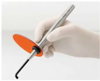
participated in the data collection. Sirona’s SIROInspect which utilizes FACE technology
was used to assess fluorescence. Lab collection packets which were used for dentin caries
sampling, (Figure 8) were put together and contained the following:
Sterile High-speed/Low-speed Handpieces 3 sterile explorers
2
sterile spoon excavators
2
sterile high-speed round burs
1 sterile low-speed round bur Air/water syringe tip*Air/water syringe plastic cover 3 patient bibs
Plastic bonnet for water collection bowl
*used deionized water in the air/water syringe and high speed handpiece
(Table 1)
Experimental procedures were as follows:
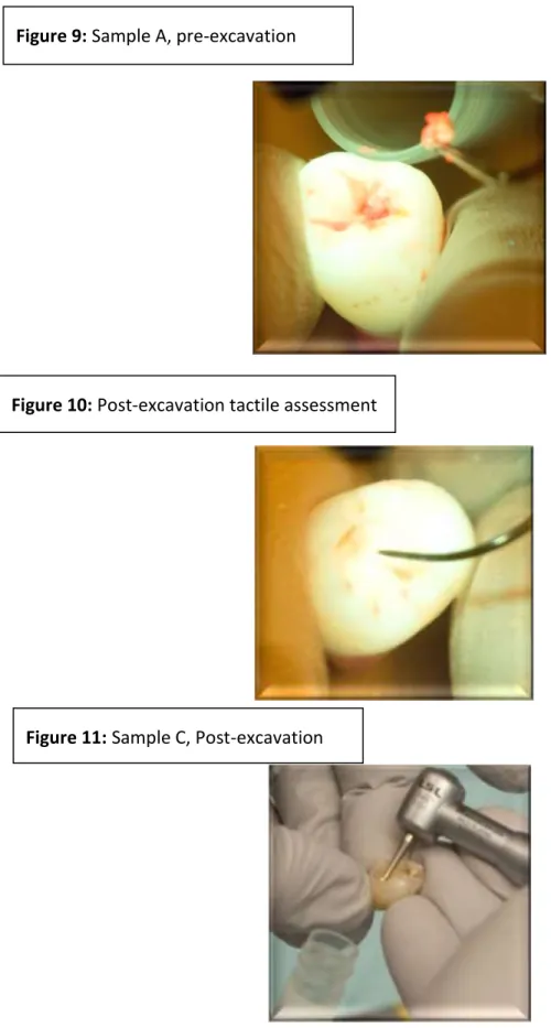
Step 1 - Visual and tactile assessment with a sterile explorer of the carious lesion was
independently recorded by both examiners.
Step 2 – Second examiner used SIROInspect with FACE technology to collect dentin from a
red fluorescing axial wall. An area which was fluorescing red was sampled using a sterile
22
Step 3 – Excavation of caries: Only dentin which was fluorescing red utilizing the
SIROInspect was removed with high speed hand-piece and sterile round bur. Excavation
was terminated when there was no red fluorescing dentin in the prepared cavity.
Step 4 – After the first layer of red fluorescing carious dentin was removed, excavated
dentin was reassessed by both examiners to determine and record visual-tactile evaluation.
The layer of pink fluorescing carious dentin present was sampled with a sterile spoon
excavator, (mid-excavation – Sample B)
Step 5 – All remaining pink fluorescing dentin was excavated with a high speed handpiece
and sterile round bur. Both examiners reassessed and recorded tactile sensation using
sterile explorer. (Figure 10)
Step 6 – Second examiner used the SIROInspect to verify that no remaining pink
fluorescing dentin was present and sampled an area post caries excavation using a sterile
slow speed handpiece and sterile round bur, post-excavation – Sample C, Figure 11)
Three samples per tooth specimen were obtained, and placed in individual sample
containers. The individual samples after being stored in -80o Celsius were subsequently
transported on dry ice to the UNC Microbiome Core Facility for microbiome analysis.
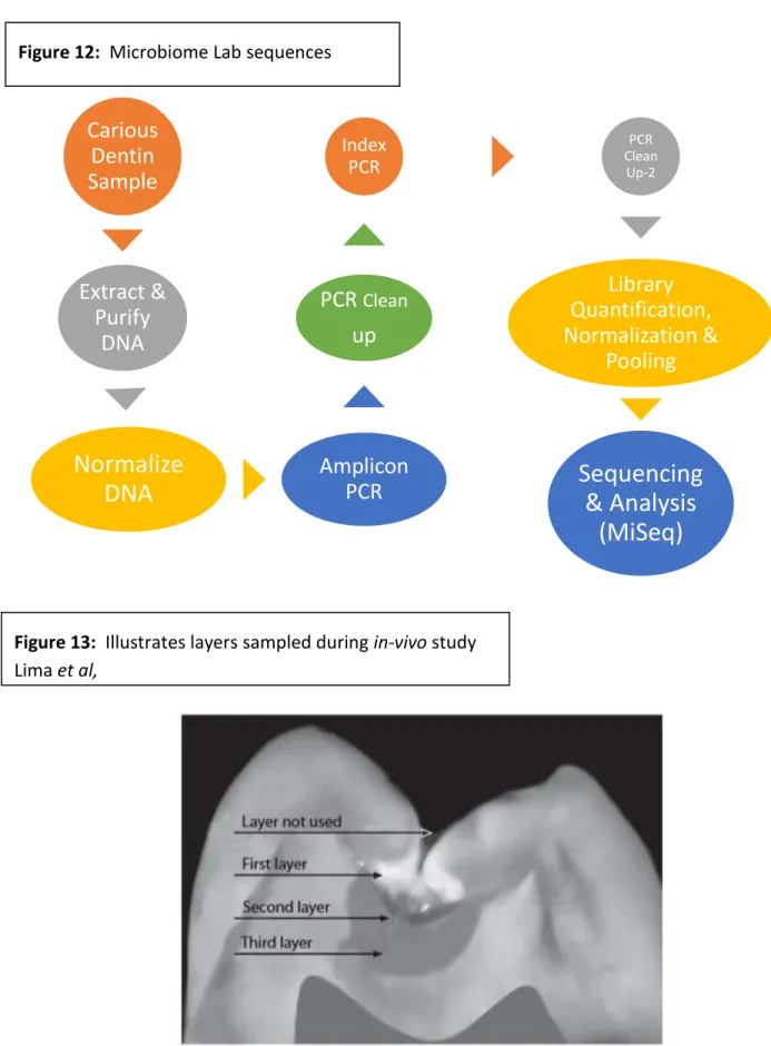
16S rRNA amplicon Illumina sequencing + HOMINGS:
Bacterial DNA extraction was performed using QIAmp DNA extraction kit
(QIAGEN) and total dsDNA content was quantitated using Quan-iT PicoGreen dsDNA
Quantitation kit (Invitrogen). Sequencing libraries were prepared by PCR amplification
23
and primers (Integrated DNA Technologies)directed against the 16S rRNA V3– V4 region
[67, 68] was designed to incorporate Illumina compatible sequencing adaptors. PCR
products were purified using AMPure XP reagent (Beckman Coulter, Indianapolis, IN), and
quantified by Quanti-IT Picogreen dsDNA 1 kit (Invitrogen). For MiSeq library
preparation, the Nextera XT Index kit was used. Illumina sequencing was performed in a
MiSeq instrument (Illumina, San Diego, CA) operating Real Time Analysis software (RTA)
version 1.17.28. Raw sequencing data files were processed using the open-source software
pipeline Quantitative Insights into Microbial Ecology (QIIME) version 1.8 [69] and
Operational taxonomic units (OTUs) were clustered using QIIME implementation of
UCLUST and compared with HOMINGS data at 98.5% sequence similarity. Phylogenetic
and non-phylogenetic alpha and beta diversity metrics was recorded and compared at the
10,000 rarefaction depth (Figure 12).
2.3 Statistical Analysis
Regarding our first aim, to determine whether fluorescence was correlated with the
dentin microbiome within genera, statistical analysis was performed on aggregated totals of
expression counts. Expression counts obtained from all samples of different extracted
teeth, producing 18,453 Operational Taxonomic Units (OTUs) with known taxonomy.
Fluorescence was treated as categorical and analyses were conducted on genus instead of
OTUs. In particular, we summed the counts relating to expression over the OTUs within
taxa defined at the genus level for each sample. Analysis was provided by the
Mantel-Haensel (MH) mean score Chi-square statistic using standardized modified rank scores for
the stratified tables of fluorescing by genus expression with teeth as strata. These tests
were based on the within-tooth ranks (1, 2 and 3 in the absence of ties, with mid-ranks for
24
ranks were computed over the 21 teeth to allow comparison and reporting of the relative
expression levels according to fluorescence. Finally, false discovery rate (FDR; Benjamin
and Hochberg, 1995) and family-wise error rate (FWER; Hochberg, 1988) methods, as
described in Preisser et al. (2011), were applied to the p-values of the stratified MH mean
score statistics for exploratory and confirmatory testing, respectively, to identify any
statistically significant differences among genera. An overall error rate of 0.05 was used in
each procedure.
In regards to the second aim, the unweighted and weighted kappa statistics were
computed to assess agreement between the two examiners with respect to their tactile
evaluations. Kappa statistics were computed for the agreement between fluorescence status
and tactile score for each examiner; this statistic is of interest to the extent that correlation
is expected between the two measures in the sense that layer A (RF) has soft dentin, layer
B (PF) has leathery dentin, and layer C (NF) has hard dentin. A complimentary analysis is
provided by the Mantel-Haensel Correlation Chi-Squared Statistic using standardized
modified rank scores (SAS PROC FREQ option “scores=modrifit”) for the stratified tables of
fluorescence by tactile (for each examiner) with tooth as strata. Specifically, the rank
correlation statistics test the null hypothesis of no association between fluorescence and
tactile score.
2.4 Results
Out of 134 extracted carious teeth that were collected, 21 fit into the inclusion
criteria and were subsequently used in our study. The 113 teeth that were excluded
presented with root caries, secondary caries, caries which was <1mm from the pulp, or
25
each, with 2 teeth only yielding 2 samples – giving a total of 61 samples which were
analyzed by the UNC Microbiome Core Facility. Two of the teeth did not present with a
‘Sample B’ due to not revealing an intermediate layer of PF, therefore - once the RF was
removed, NF was evident and sampled. Therefore only Sample A, and Sample C were
obtained for these two teeth.
The 16S rRNA amplicon sequencing analysis revealed 18,453 Observational
Taxonomic Units (OTUs). Total number of reads was 10,268,174 with a median of 120,223
and a mean of 151,002 per sample. The microbiota discovered in the various layers of
fluorescing dentin varied greatly in composition. The red fluorescent portion showed a
higher number of observed OTUs (154); followed by the pink fluorescing (109) and the no
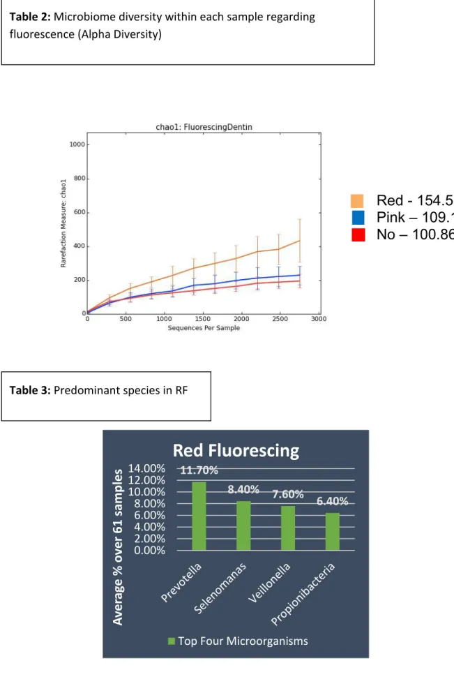
fluorescing (100) (Table 2). The t-statistical analysis confirmed a significant difference
between RF and NF (p-=0.003), as well as between RF and PF (p=0.003). No statistically
significant difference was detected between PF and NF (p=0.843) (Table 2). Red
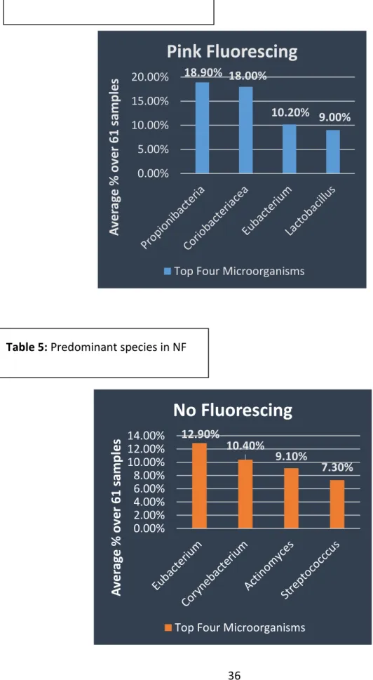
fluorescing (RF) carious dentin had a predominance of: Prevotella (11.7%), Selenomanas
(8.4%), Veillonella (7.6%) and Propionibacteria (6.4%)(Table 3). The Pink fluorescing (PF)
dentin had a predominance of: Propionibacterium (18.9%), Coriobacteriacea (18.0%)
Eubacterium (10.2%) and Lactobacillus (9.0%)(Table 4). The Non fluorescing (NF) dentin
had the predominant microorganisms: Eubacterium (15.9%), Corynebacterium (10.4%),
Actinomyces (9.1%) and Streptocuccus (7.3%) (Table 5).
There were 230 genera identified after collapsing data within genera and 229 among
them are informative (one non-informative genus had only zero counts in all 61 tooth sites).
A total of 62 among 229 genera were detected as statistically significant by FDR error
26
significance level of 0.05. Microorganisms which were predominant amongst the three
layers (RF, PF, and NF) are highlighted in Table 11. .These 29 genera were
differentially expressed among Red, Pink and No fluorescing. Among these, 20 genera had
increased counts for RF, 2 for PF and 7 were most pronounced under NF. Thus, in the
context of these significant genera that were differentially expressed across fluorescence
(table 11), the hypothesis seems to be supported with greater “diversity” among RF.
Regarding our secondary aim, which was to determine if the presence or absence of
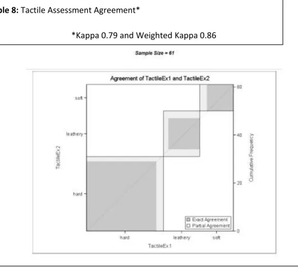
fluorescence as detected by FACE correlates with the clinical assessment of dentin
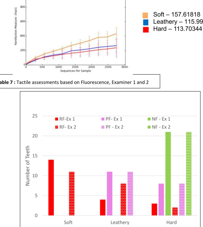
characteristics, we assessed the following among the 61 samples: Hard – 32, Leathery –
15, Soft – 14 – assessments obtained by examiner one; Hard – 31, Leathery – 19, Soft – 11
– assessments obtained by examiner two. Alpha diversity values found in the tactile
assessments were remarkably similar to the values noted in the RF, PF, and NF samples.
The mean number of OTUs identified were as follows: soft dentin – 157, leathery- 115 and
hard – 113. The t-statistical analysis also confirmed a significant difference between soft
and leathery dentin (p= 0.006) and soft and hard dentin (p= 0.003). Whereas, differences
between hard and leathery dentin in regards to tactile assessments were not significant (p=
1.00) (Table 6). The rank correlation chi-squared statistic for the association of fluorescence
and tactile was highly statistically significant (p < 0.001) for each examiner, strongly
suggesting an association between fluorescence and tactile assessment. The agreement
between examiners’ tactile scores is “Substantial” with a kappa of 0.78. While the
agreement between fluorescence and Examiner 1’s tactile score was “Substantial” with
Kappa of 0.62, agreement between fluorescence and Examiner 2’s tactile score was
“Moderate” with Kappa of 0.56 (Tables 4 and 5). The tactile assessment of soft, leathery
27
of inter-examiner agreement. RF carious dentin was primarily ‘soft’, but also had readings
of ‘leathery’ or ‘hard’ tactically. Approximately one-third of the tactile assessments of hard
dentin still displayed some level of fluorescence, either pink or red. Similar findings were
found with pink fluorescing carious dentin, but the non-fluorescing was assessed as ‘hard’
100% of the time by both examiners.
Principal Coordinate Analysis (PCoA) of Unifrac distance matrices is a method often
applied to visualize similarities or dissimilarities of research findings, by trying to find a
main axis through the data. Each component in the PCA (Principal Component Analysis)
represented one of the 61 samples, and were placed in three different axis’, PC1, PC2 and
PC3. The points along each axis were also separated by RF, PF and NF characteristics and
identified by the colors Red, Pink and Yellow. This PCoA analysis which was used to
observe the Beta diversity, assisted in further describing the differences between the
samples (Table 6). The Beta diversity plot showed each sample, and the clustering of the
groups according to RF, PF, and NF with distinct separation among them in relation to
bacterial content.
2.5 Discussion:
One question which has been repeatedly researched over the decades is whether or
not bacteria remains in ‘hard’ dentin, post-caries removal? The result of our study supports
that yes, bacterial DNA remains in the hard dentin, however the diversity is compromised.
Controversy has surrounded the subjectivity of assessing outer and inner carious dentin,
and how far the clinician should excavate prior to placement of a restoration. Fusayama
28
which proved to stain dentin which could actually be remineralized, thus creating
unnecessarily larger preparations.[6, 25, 70]
The manufacturer of the SIROInspect, Sirona, reports in their brochure that FACE
technology can tell the end user where the bacteria is present: “With FACE, users can see
where bacterially infected dentine is located and where not”. However, they also state that
“red-fluorescing dentin must be completely excavated so that as little bacterially infected
dentin as possible is left behind”.[71] Our present research showed remaining DNA from
multiple bacterial species even at the hard and non-fluorescing level to include
Eubacterium, Corynebacterium, Actinomyces and even Streptococcus. However, bearing in
mind, only the genera that are expressed significantly differentl at RF, PF and NF dentin,
there is less diversity in the hard NF dentin. Although there is some overlap on the
bacterial species across the different levels of fluorescence even if considering only the top
predominant bacteria on RF and PF (Propionobactirium) and PF and NF (Eubacterium) the
different layers (outer dentin, inner dentin and sound dentin) was able to be identified by
both fluorescence and tactile sensation.
The International Caries Consensus Collaboration meeting, held in Leuven,
Belgium, in February 2015, developed guidelines for the dental practitioner based on lesion
depth into the dentin. These guidelines indicate that shallow or moderate lesions, which do
not reach the inner third or ¼ of dentin, should be selectively excavated to firm dentin,
defined by a ‘feeling of resistance’ to a hand excavator. In regards to the deep lesion, where
the caries extends into the pulpal third or ¼ of dentin, it is recommended to perform
29
peripheral enamel and dentin are prepared to ‘hard’ dentin prior to placement of a
restoration so as to create an effectively sealed restoration.[72]
Kidd points out, however, in reviewing multiple studies on Stepwise (SW)
excavation, that suggest that remaining microorganisms had become altered to a ‘less
cariogenic flora’, and upon re-entry the dentin was dryer, harder and darker. Thus
indicating a positive correlation to bacterial degradation when the nutrient supply is
removed by a well-sealed restoration.[28] She also mentions that the remaining
microorganism which have been ‘entombed’ become irrelevant due to the lack of
permeability of the reparative and tertiary dentin and ultimately states that there is no
clear evidence suggesting that it is deleterious to leave infected dentin.[73] Furthermore,
clinical studies have found that there are no detrimental effects to the pulp by ‘sealing’
remaining bacteria with a proper restoration which is cleansable by the patient.[74] It is
possible that there are changes in the metagenomics of the bacterial species due to the
conditions of the environment and/or the interaction within species at each of the different
layers that may explain the fluorescence expression (or lack of expression).
As early as the 1980’s there was a change in identifying and classifying bacteria
from using culturing methods to the use of molecular methods. These molecular methods
are based on gene sequences which give the ability to identify bacteria in any environment
examined.[60] The use of 16S rRNA sequencing methodology in evaluating species present
in the oral cavity was first introduced by Paster Laboratories in 2001 where they looked at
diversity among the bacteria in periodontally involved patients.[75]
Simon-Soro, et al., in an in-vivo study, obtained dentin caries samples from 6 teeth
30
from 6 other teeth with ‘hidden’ caries diagnosed through radiographs and exposed after
drilling through the enamel with a water cooled high-speed handpiece. RNA was extracted
and cDNA was constructed and used to amplify the 16S rRNA gene. The sequences
obtained through this process were used to determine the bacterial composition. Their
findings revealed Lactobacillus, Shlegelella, Pseudoramibacter Eubacterium, and
Atopobium to be ‘clearly associated with dentin lesions’, however this was the combined
conclusion of all 12 dentin samples obtained, from both open dentin lesions and hidden
dentin lesions.[76] These results were similar to ours, however Atopobium was negligible
at 0.4% in the presumed heavily infected red fluorescing dentin caries, and, Shlegelella was
not represented and/or detected in our taxonomic results.
In another in-vivo study from Japan, Obata et al. acquired deep dentin caries
samples from 32 teeth after removal of plaque, enamel and a shallow layer of carious
dentin. 16S rRNA sequencing technology was utilized for microbiome classification and
analysis. The results were classified into ‘clusters’, varying according to the abundance of
Lactobacillus; Cluster I, II, III with high, medium and low levels of Lactobacillus
respectively. The mean distributions among the clusters identified the genera that followed
in proportion to Lactobacillus were: Propionibacterium, Prevotella, Atopobium,
Streptococcus and Actinomyces. The authors suggest that the abundance of Atopobium
were possibly considered unique to the Japanese population, further stating that ‘no
previous reports have reported Atopobium as a dominant microbiota associated with
dentinal caries’.[77] Differences noted between their findings and our research study: 1)
in-vivo versus in-vitro, 2) our carious lesions were exposed, and they removed layers of
31
into ‘clusters’ according to abundance of Lactobacillus, and 4) Atopobium was not a
predominate microorganism discovered in our study.
In a study which was conducted similarly to ours, Lima et al. sought to analyze and
identify the microbiota in the different layers of dentinal caries (superficial, middle and
deep) through the use of a reverse-capture checker-board hybridization assay. Twenty
Seven (27) patients yielded 81 samples. Their reported tooth crown conditions were: intact
11 (40.7 %) microcavitated 8 (29.6 %) cavitated 8 (29.6 %), with a mean age of 13.5. Use of
the reverse-capture checker-board allowed the author to survey specific bacteria, which
they selected 28 to probe with the 16s rRNA analysis. Bacteria which were prevalent in
the superficial layer are as follows: Fusobacterium, Lactobacillus, Atopobium, and
Veillonella. Middle layer: Atopobium, Lactobacillus, Fusobacterium. And, the deep layer:
Fusobacterium, Atopobium, Lactobacillus, Bifidobacterium and Streptococcus. Their
research study concluded that bacteria which dominates in the deep dentinal lesions may
further be involved with pulpal damage, stating that they are in the ‘front line’ of the
lesion. It was also suggested that the superficial bacterial layers of caries could represent
historical information of the disease process, and they may provide a nutrient source as
well as protection to the inner layers of the carious lesion.[10] The major differences from
our study: 1) our population age was unknown; 2) our lesions were already through the
enamel with exposed soft carious dentin; 3) we sampled the first very soft layer, and they
discarded the first layer (Figure 13). Interesting to note that they did not probe for certain
bacteria which were predominant in our study, such as: Selenomonas, Coriobacteriacea and
Eubacterium.
32
The collection of extracted carious teeth was not conducted by the same individual
throughout the study process, therefore handling of the tooth specimens may have been
inconsistent. Post-extraction, the carious teeth were to be rinsed with deionized water to
attempt to remove any blood and/or debris. Any bioburden which may have been left on the
tooth could have been represented in the microbiome analysis. Furthermore, although
steps were taken to attempt to maintain a clean field, by the use of individually
sterilized/packaged instruments and burs, which were changed during each step of the
sampling process (A, B, C), the protocol we used to acquire the samples from the carious
extracted teeth was not in a totally ‘sterile’ environment. Tooth to tooth variation in
regards to the depth of the caries, type of tooth (incisor, premolar, molar), cariogenic history
of the patient, use of fluoride, diet and hygiene practices may have impacted the
microbiome results. The drawback of using a non-culturing technique, such as this
DNA-based study is that the PCR step may amplify DNA from inactive or even dead
microorganisms. Thus, it would become necessary to determine the viability of the bacteria
which may be present in the deepest layer, by performing an additional step through
culturing.[76]
2.7 Conclusion:
Within the limitations of this study, the presented protocol sought to evaluate the
diversity of microorganisms at varying layers of carious dentin using FACE technology, as
well as determine if fluorescence correlated with the traditional tactile method of caries
assessment. It became evident that a vast array of microorganisms were present
throughout the layers of carious dentin from soft to leathery to hard, as well as from RF, to
PF to NF. In addition, the carious dentin layers sampled displayed a variation in proportion
33
accepted, considering only the genera which were significantly differentially expressed in
RF, PF and NF dentin, there was a higher bacterial species diversity in RF dentin as
compared to PF and NF dentin. When comparing fluorescence (RF, PF, NF) with tactile
assessments, approximately one-third of the hard dentin was noted as Pink Fluorescing
(PF) or Red Fluorescing (RF), thus providing strong evidence to reject the second hypothesis