ii
iii ABSTRACT
Tyann Blessington: Leptospirosis Frequency in Animals and their Habitats in the Southern United States: A Systematic Review
(Under the direction of Dr. Anna Schenck)
iv
EXECUTIVE SUMMARY
Background: Leptospirosis is considered an emerging bacterial zoonotic disease and is found worldwide. Symptoms in infected humans and animals can range from subclinical and
nonspecific to serious and fatal. Infection has been historically associated with flooding and tropical environments, and direct contact with animals, sewage, or contaminated freshwater or soil. The majority of the southern region of United States has a subtropical climate, which may be favorable for transmission and spread of Leptospira. Human disease is underdiagnosed and there is a lack of epidemiological data on the incidence of disease. Understanding the frequency and dynamics of the disease in animals and the environment can aid in outbreak responses, and contribute to the development of prevention measures for both humans and animals.
Methods: A systematic review of animal leptospirosis measured in 17 states and jurisdictions covering the southern region of the continental U.S. was performed. Articles containing Leptospira disease frequency in animals within the prescribed geographic location were reviewed for suitability. Studies were excluded if the animals were associated with a human illness outbreak or were selected based on symptomology. Disease frequency measures for studies with 50 or more animals were plotted per decade.
v
lower than the seroprevalence. There were wide ranges of disease frequencies reported among the studies; however, some studies were case reports focused on one animal. For studies that consisted of 50 or more animals, the disease frequency for Muridae (seroprevalence: 46-65%; isolation frequency: 7-33%) and Mephitidae (seroprevalence: 62%; isolation frequency: 14-57%) were consistently high. Bovidae, Cervidae, Mephitidae, and Suidae had wide ranges of disease frequency. When plotting disease measures by decade, there was a slight increase in disease for animals in wild habitats (0.18% seroprevalence and 0.09% isolation frequency per year).
vi
TABLE OF CONTENTS
List of Tables ... vii
List of Figures ... vvii
INTRODUCTION ... 1
METHODS ... 7
Geographic Specificity... 7
Literature Search and Data Sources ... 7
Article Review and Selection Criteria ... 8
Data Collection ... 8
Data Analysis ... 9
RESULTS ... 10
Evaluation of Literature and Article Selection ... 10
Characteristics of Studies ... 12
Seroprevalence and Bacterial Isolation Frequency (Incidence) ... 13
Seroprevalence per Animal Type... 16
Bacterial Isolation (Incidence) per Animal Type... 16
Serovar and Serogroup ... 17
Environment, Habitat, and Social Factors ... 18
Trend Analysis ... 21
DISCUSSION ... 22
Published Literature ... 22
Diversity in Disease Measures ... 22
Disease Dynamics ... 24
Trend Assessment ... 26
Is Leptospirosis Increasing in the Southern United States? ... 27
Climate Change ... 30
Diagnostic Needs ... 30
Leadership in Promoting One Health Surveillance ... 30
CONCLUSION ... 32
TABLES & FIGURES ... 33
vii
LIST OF TABLES
Table 1. Human leptospirosis outbreaks (or illness clusters) in the U.S.
southern states and jurisdictions……….33 Table 2. Search terms for systematic review article search...………...34 Table 3. Selected articles for the systematic review analysis………...35 Table 4. Leptospira frequency seroprevalence and isolation measures
in animals from systematic review selected articles……….…..……….. 38 Table 5. Leptospira frequency seroprevalence and isolation measures
consisting of 50 or greater animals among animal families from
viii
LIST OF FIGURES
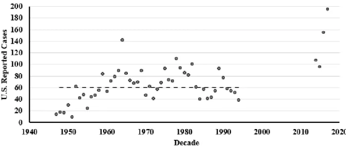
Figure 1. Number of human leptospirosis cases reported to the U.S. Centers for Disease Control and Prevention from 1947 to 1994 and 2014
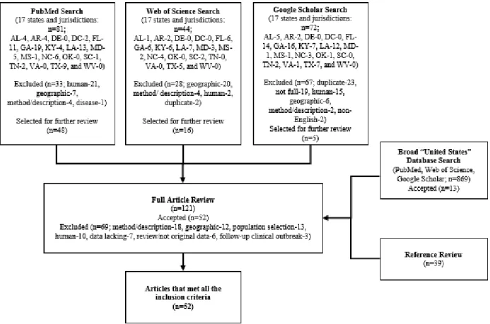
to 2017……….……….47 Figure 2. Flow diagram for the evaluation and selection of relevant
publications describing Leptospira disease frequency in animals residing in the southern United States between the years 1905
and 2019 (present day)……….……….48 Figure 3. Frequency of a) publishing date of selected articles and b)
state or jurisdiction of animal collections for seroprevalence and c) isolation frequency described in the systematic review
selected articles……….…….49 Figure 4. Scatterplots of seroprevalence by decade for animals in a)
all habitats, b) wild habitats, and c) non-wild habitats, and isolation frequency by decade for animals in d) all habitats
1 INTRODUCTION
Leptospirosis is an emerging disease caused by a pathogenic spirochete (Evangelista and Coburn, 2010). The disease is considered the most wide spread zoonosis in the world and affects humans, wildlife, domestic and agricultural animals (Adler and De La Peña Moctezuma, 2010). Leptospirosis was first described by Adolf Weil in 1886 in Germany. Although the disease has been recognized for more than a century, today the disease, transmission, and environmental risk factors are understudied and neglected. Rodents have long been considered a reservoir host for the bacteria (Li and Davis, 1952). The role of other animals has not been fully explained, but agricultural animals including cattle and pigs, companion animals such as dogs, and wildlife species have been identified as carriers (Guernier et al, 2018).
Infection manifestations can be subclinical to severe for both humans and animals. For humans, the disease is often underdiagnosed and symptoms are often mistaken for other
2
consider leptospirosis if a dog exhibits signs of renal or hepatic failure, pulmonary hemorrhage, uveitis, acute febrile illness, or abortion (Sykes et al., 2011). For livestock, infection is usually associated with reproduction problems such as infertility, abortion, stillbirth, and decreased milk production (Petrakovsky et al, 2014). Animals can become chronically infected when the
bacteria colonizes the proximal renal tubules; afterwards, the bacteria is then periodically shed in the urine (Adler and De La Peña Moctezuma, 2010). The pathogen is spread in the environment through infected urine.
The bacteria has a unique ability to survive in a variety of environmental matrices including water, soil, and manure for extended periods of time, in some cases several months if conditions remain warm and moist (Diesch, 1971; Smith, and Turner, 1961). The main
transmission routes are believed to be through direct contact with contaminated water or soil through mucosa or small skin abrasions or by drinking contaminated water (Mwachui et al, 2015; Cacciapuoti et al, 1987).
Leptospira bacteria have traditionally been classified by phenotypic serological
presentations, using the microscopic agglutination test (MAT). Species were grouped within the saprophytic bifexa complex (Leptospira biflexa sensu lato) or were grouped within the
3
humans and animals; however, the impact of antibiotics in severe cases is understudied and debated (Charan et al, 2013).
The spirochete bacteria was first described in the United States in 1905 when it was isolated from the kidney of a patient in New Orleans, who was believed to have died of Yellow Fever (Heath et al, 1965). Some researchers believe outbreaks of human leptospirosis occurred in the U.S. during the Civil War and even as far back as the 1600s (Neill, 1918; Marr and Cathey, 2010). There is a lack of reliable epidemiological data on the incidence of human leptospirosis in the U.S., especially in the continental U.S. Leptospirosis was a nationally reportable disease from 1947 to 1994 and has only recently, in 2014, became a national reportable disease again. From 1947 to 1994 the median number of cases per year was 60, the lowest yearly reported value was 9 cases in 1951, and the highest yearly reported value was 142 cases in 1964. Since re-instating leptospirosis as a nationally reportable disease, the reported values were 96 cases in 2015, 155 cases in 2016, and 195 cases in 2017 (Figure 1; CDC, 2019a). As of 2019, 9 states (Arkansas, Colorado, Iowa, Kansas, Mississippi, New York, North Dakota, Tennessee, and Texas) do not report illnesses (CDC, 2019b). Of note, four of these states (Arkansas, Mississippi, Tennessee, and Texas) are southern states.
4
becoming ill, patients reported swimming in fresh water or contacting floodwater, contacting ill animals, working at a dairy, serving in the military or participating in a recreational adventure sport race.
Leptospirosis is not listed in the U.S. Department of Agriculture (USDA)’s National List of Reportable Animal Diseases (NLRAD) and confirmed livestock Leptospira illnesses are not included in the National Animal Health Reporting System (NAHRS) (USDA APHIS, 2019). Despite this, animal infections in the continental U.S. have been documented in wildlife,
agricultural, and domestic animals. Animal health surveys, both wildlife and livestock, have been initiated by researchers in the southern U.S. since the early 1950s, largely for the purpose of agricultural and livestock health needs. The reported presence of the disease is also supported by pharmaceutical demand. In the U.S., commercial Leptospira vaccines are available for cattle, dogs, and pigs; however, their protection is limited to a handful of serovars included in each animal-specific vaccine (Adler and De La Peña Moctezuma, 2010). Vaccination is generally recommended for cattle used for breeding; however, the vaccine is generally not part of a standard vaccine routine for other animals. Additionally, protection requires periodic
revaccination. There are limited publications on the extent of the vaccine use and its efficiency. There are no leptospirosis vaccines available for human use in the U.S.
5
wide range of climates and consists of a diverse grouping of ecosystems. Based on the Köppen Climate Zones, the majority of the south is classified as having a Moist Subtropical Mid-Latitude climate, but also contains areas of Tropical, Moist Continental Mid-Latitude, and Dry climates (NWS, 2019). Ecosystem communities range from cropland, desert shrub land, forest (deciduous or pine), grassland, marsh, and wetlands (Wickham, 1995). Southern states have also been periodically plagued by flooding from heavy rains, hurricanes, and storm surge. The intensity of these climate-sensitive hazards in these states have outnumbered other areas in the U.S. in both scale and magnitude by a ratio of 4:1 since the early 2000s (Emrich and Cutter, 2011).
Additionally, flooding conditions are expected to increase in the southern part of the east coast as the climate changes (Schwartz and Randall, 2003). These environmental and climatic changes may result in favorable conditions for leptospirosis transmission in the southern U.S.
The “One Health” concept is a strategy for developing and expanding interdisciplinary collaborations for the improvement of human, animal, and environmental health. This approach is recognized as essential for addressing antibiotic resistance and the development of novel bacterial infection therapies that are robust under a complex environment setting where resistance can evolve (Destoumieux-Garzón et al., 2018). Similarly, this interdisciplinary approach is needed to address the gaps in leptospirosis disease dynamics. Understanding the frequency and dynamics of the disease in animals and the environmental conditions that
6
7 METHODS
Geographic Specificity
For this analysis, the southern U.S. is defined by the U.S. Census Bureau’s definition of the South. This includes the following districts: South Atlantic (Delaware, Florida, Georgia, Maryland, North Carolina, South Carolina, Virginia, District of Columbia, and West Virginia), East South Central (Alabama, Kentucky, Mississippi, and Tennessee), and West South Central (Arkansas, Louisiana, Oklahoma, and Texas) (U.S. Census Bureau, 2019).
Literature Search and Data Sources
8 Article Review and Selection Criteria
An article’s title and abstract were first reviewed for key suitability criteria including data on Leptospira disease frequency, to include a prevalence or bacterial isolation frequency
(incidence), in animals within the prescribed geographic locations. Articles meeting the criteria, were obtained as full-text and the reported methodology and data were evaluated. Articles were excluded if they 1) described experimental infections, 2) did not contain original research data, 3) were not retrievable as a full text article, or 4) were written in a language other than English. Articles were also excluded if the geographic location of the habitat or residency of animals was not defined, if the animals selected had symptoms of the disease or were involved in a human outbreak investigation. Articles were not restricted by publication date; articles published between 1905, which was when the pathogen was first described in the U.S., and 2019 (present day) were considered for further evaluation. All articles that met the criteria were included in the review.
Data Collection
9
serogroup/ serovar; 6) environmental changes, including recent changes in weather, land use, and population dynamics, and 7) pertinent disease route and environmental maintenance factors were documented. Microsoft Excel was used to collect extracted data.
Data Analysis
Collected data were assessed for commonalities and trends. Measures of disease
10 RESULTS
Evaluation of Literature and Article Selection
The initial search, involving 17 states and jurisdictions, identified 197 studies, including 81 through PubMed, 44 through Web of Science, and 72 through Google Scholar (Figure 2). Titles and abstracts were reviewed for inclusion. Thirty-eight articles described human infections; 33 were studies performed or involved animals in geographic locations outside the defined criteria; 25 were duplicates; 19 were citations for studies where full-articles were not available including conference descriptions, 10 were reviews or laboratory diagnostics and methodologies, two were written in languages other than English, and one did not describe the criteria pathogen, Leptospira. Broad-database searches, using the geographic identifier, “United States” were utilized to identify national studies involving the states and jurisdictions within the criteria; 869 articles, including 562 through PubMed, 235 through Web of Science, and 72 through Google Scholar. Majority of these articles were excluded based on the geographic criteria and duplicative search processes. Reference sections were reviewed for historical relevant articles.
11
outbreaks and the animals selected for infection measurements were association with human illnesses (Barkin and Glosser, 1973; White et al., 1981; Vinetz et al., 1996). Studies that
calculated infection measurements based on a review of Leptospira diagnostic submissions were excluded because of potential population bias of those animals that may have received a
diagnostic test. Diagnostics may have only been performed on animals exhibiting clinical symptoms and residing in regions with previous confirmed illnesses. Bias may have occurred in the preparation of some reports if veterinarians did not consider leptospirosis as a potential cause of general clinical signs (e.g. loss of appetite). Indeed, this bias may have been heightened due to a paucity, of information about geographic zoonotic transmission in the region. Diagnostic-based measures were used to calculate canine and equine leptospirosis rates (Gautam et al., 2010; Lee et al., 2014; Siza, 2016; Ward, 2002a; Ward, 2002b; Ward et al., 2002, Ward et al., 2004; White et al., 2017). These values are critical for veterinary services but may not adequately estimate the general population disease rate and the population at risk. Reproductive problems can be a symptom of leptospirosis infection; therefore, articles that calculated Leptospira infection frequencies among animal fetal losses, where previous diagnoses were not identified, were also excluded (Donahue et al., 1991; Donahue et al., 1992; Donahue et al., 1995; Erol et al, 2015). Additionally, assessing cattle herds with known reproductive problems or unknown vaccine status were excluded from this analysis (Hussain et al., 1978; Talpada et al., 2003). The
12 Characteristics of Studies
Animal type
Eleven articles included infection data on more than one animal type. Fourteen articles included data on rodents (scientific order Rodentia), which covered six families. Data on non-rodents covered 16 scientific family types, including nine articles on pigs (Family Suidae), eight on canines (Family Canidae), seven on cervids (Family Cervidae), six on cattle (Family
Bovidae), and five each on cats (Family Felidae) and skunks (Family Mephitidae) (Table 4). The majority of selected studies included wild animals (42 articles). Agricultural animals were included in nine articles and domestic pets were included in two articles. One article included both wildlife and agricultural animals.
Publication year
13
Jena et al., 2004; Peters et al., 2017; Schneider et al., 2012; Sutter and Sosa Pascual, 2018; Victoriano et al., 2009), which, further supports the notion that the disease is an emerging threat. This designation may have rekindled research interest in this field, since publications have increased in the last decade.
Geographic distribution
Early publications were dominated by a few researchers at Schools of Veterinary Medicine, including Louisiana State University (E.E. Roth, K. Newman, and M. Moore) and University of Georgia (G.W. Gorman and E.B. Shotts). In more recent years, studies have
diversified to other geographic areas; the first focused publications in Arkansas, Kentucky, North Carolina, and South Carolina have occurred since the 1980s. Selected studies produced 202 disease measures in animals evaluated for leptospirosis in 14 states (Figure 3b and 3c). There were no collection locations in Delaware, District of Columbia, and West Virginia. Georgia accounts for more than 35% of the total disease rate collection locations and nearly 60 percent of the isolation disease rate collection locations, largely because of multi-species design analyses performed by University of Georgia researchers (Brown et al., 1960; Gorman et al., 1962; McKeever et al., 1958; Shotts et al., 1975). Collection locations in Florida and Texas account for more than 16 and 12% of the total disease measures, respectively.
Seroprevalence and Bacterial Isolation Frequency (Incidence)
14
Microscopy (DFM) in combination with culture-based techniques, culture-based techniques, and Polymerase Chain Reaction (PCR) DNA amplification. Each presents its own diagnostic
challenges.
The MAT-based serology technique measures infection based on immune responses. The MAT is a technique used to measure of seroprevalence, since immune responses may be reactive after an infection resolves. Immune response may not be sensitive during early stages of an infection and for some asymptomatic carriers an immune response may never occur or is short-lasting and limited. Consequently, seroprevalence may not reflect a true prevalence for infection. Animals may not develop an immune response to an infection. Successfully assessing for
Leptospira reactions using MAT techniques requires incubating serum with various laboratory maintained Leptospira serovars. Agglutination reactions are often specific to a single serovar. Variability with this technique can occur if different agglutination dilutions define a Leptospira positive result. There is no uniformly accepted standardized assessment criteria for concluding that a titer response is positive. Reported positive-defining titers ranged from 1:25 to 1:250 among the selected publications.
15
the bacteria, may need to be extracted to obtain the necessary tissue quantity for culture protocols. The PCR detection methods pose less hazards to laboratory workers, since culture-based methods and MAT techniques require the maintenance of pure cultures and the
manipulation of cultured samples with potentially high-levels of pathogens. However, PCR detection methods may not be sensitive if an animal is infected with small numbers of
leptospires, since this method require large amounts of DNA for positive detections (Budihal and Perwez, 2014). Among the selected articles, DFM techniques were only utilized prior to 1950 and in conjunction with culture techniques. Most studies that employed culture-based techniques, collected tissues from extracted kidneys or kidneys and other tissues. A single study aseptically extracted urine for the culture and identification of leptospires (White et al., 1961). Additionally, one article, Pedersen and coworkers (2017a), identified leptospires through PCR techniques, where Leptospira DNA was directly extracted and amplified from kidney tissues.
Some authors have used the bacterial detection methods, to calculate a bacterial incidence measure based on frequency results; however, in most cases, specific time dimensions for this measure were not defined. Therefore, in some cases the isolation frequency may represent a prevalence rather than an incidence or a cumulative incidence. Additionally, since kidneys may harbor leptospires for prolonged periods; detection and isolation may not represent a new illness. Bacterial isolation frequency will be referred as an incidence for the remainder of this
assessment; however, this measure may not represent a true incidence.
In studies where both techniques, seroprevalence and isolation frequency, were
16
(1964) observed serum from a bacteriologically positive deer failed to agglutinate with specific tested antigens.
Seroprevalence per Animal Type
Seroprevalence was measured in 19 animal family types, including five families within the scientific order Rodentia (Table 4). A wide range (0-100%) of seroprevalence values were reported among the animal types; however sample sizes for some animal types were as low as one animal. Non-rodent taxonomic families with sample sizes of 50 or more include: Bovidae, Canidae, Cervidae, Dasypodidae, Didelphidae, Equidae, Mephitidae, Procyonidae, Suidae, and Ursidae. The only rodent family with a large sample size was Muridae (Table 5). The range values for Bovidae (4-71%), Cercidae (1-31%), Mephitidae (25-62%), Muridae (46-65%), and Suidae (1-44%) span more than 25 percent points. The range values of Mephitidae and Muridae, and the single measure for Equidae (46%) indicate that a large population (over 25%) of the studied animals were exposed and developed an immune response to Leptospira.
Bacterial Isolation (Incidence) per Animal Type
17
analyzed, isolation frequency measures for the total rodent population were calculated, resulting in a range of 4-19% (Brown et al. 1960, Gorman et al. 1962, and Redetzke and McCann 1980). The range of values for Bovidae (1-36%), Mephitidae (14-57%), and Muridae (7-33%) span more than 25 percent points. The range or single measures for Dasypodidae (4%), Leporidae (0-<1%), and Sciuridae (0%) were less than the ranges for Dipelphidae (6-16%), Mephitidae (14-57%), and Muridae (7-33%) and the single value for Heteromyidae (19%). In general, the isolation frequency value ranges were lower than the seroprevalence ranges; however, the range of isolation frequency for Didelphidae and Procyonidae were greater than their single measures of seroprevalence.
Serovar and Serogroup
There was wide serovar diversity among animals in the south; 26 serovars were identified (Andamana, Australis, Autumnalis, Balcanica, Ballum, Bataviae, Borincana, Bratslava,
Canicola, Djasiman, Grippotyphosa, Hardjo, Hebdo, Hebdomadis, Hyos, Icterohaemorrhagiae, Javanica, Mini Georgia, Mycocastorius, Paidjan, Pomona, Pyrogenes, Sejroe, Sentot, Shermani, and Tarassovi) (Table 6). Grippotyphosa was identified in the order Rodentia and every non-rodent family, except Equidae. Icterohaemorrhagiae was identified in the order Rodentia and every non-rodent family, except Felidae. Canicola was identified in the order Rodentia and every non-rodent family, except Dipelphidae, Equidae, and Felidae.
18
these reservoir hosts were also well distributed among the other animals. Bratislava was not identified in horses, but was identified in four other animal families.
Serovar identification was dependent on methodology of the studies; researchers who had access to diverse laboratory maintained isolates for serotyping may have identified a more
diverse range of serovars because they tested for more serovars. Certain serovars may have been evaluated in certain regional laboratories where other serovars may have been evaluated in other regions and this geographic specificity may not reflect the true environmental circulating
diversity of serovars.
Environment, Habitat, and Social Factors
Leptospires have been identified in diverse environments and habitats of the southern states. The majority of habitats where animals have been studied are identified as wildlife environments (26 articles). Eight studies each were performed in agricultural settings and in intersections between agriculture and wildlife. Nine studies were performed in urban settings and two studies were performed in settings where suburban and wildlife areas intersect. One study was performed in both urban and agricultural settings (Table 4). Some of the authors observed geographic, environmental, and characteristic differences in Leptospira rates among the animals studied. Multiple factors are likely contributing to Leptospira transmission and disease
maintenance in the southern states.
Seasonal and weather conditions
19
to those in northern regions (60%). Miller and coauthors (1991) also noted higher seroprevalence rates in cattle in the southeastern and south central regions of the country when compared to northeastern, north central, northern plains, and Rocky Mountain regions. The difference was attributed to higher temperatures in southern regions. Redetzke and McCann (1980) observed seasonal changes in bacterial isolation frequency among rodents of West Texas; isolation frequency increased progressively through May and additional increases were observed in
August, and November. The authors partly attributed the late spring isolation frequency increases to breeding behaviors of the rodents. Li and coworkers (1952) also noted slight but
nonsignificant increases in seroprevalence rates in urban rats in the spring and fall and attributed the differences to capturing more mature rats during those times. The mature animals had greater opportunities for exposure to leptospires through social animal behaviors within the rat colony. Approximately 50 years later, Easterbrook and coworkers (2007) did not identify any seasonal trends with prevalence rates among rats in the same region.
Land use and animal interactions
20
hay and lick the salt blocks left out for cattle. Jenkins and coworkers (1979) noted swine farmers in Alabama often have stray dogs on their property.
Animal density
Increasing animal density can also encourage pathogen transmission. Byrne and
Chambers (1959) observed higher percentages of Leptospira positive animals within herds with more cattle. Brown and coworkers (1960)’s demonstrated an association between house mouse population levels and Leptospira isolation frequencies among all rodent types within a habitat. Higher wild pig seroprevalence rates were observed in some southeast and southcentral states including Arkansas, Mississippi, Oklahoma, and Texas (Pedersen et al, 2015). One possible reason for these higher rates may be that these states have large numbers of commercial swine facilities and wild pig populations that may occasionally interact.
Sociodemographic factors
21 Trend Analysis
22 DISCUSSION
Published Literature
The systematic review analysis demonstrates that Leptospira are widespread throughout the southern U.S. and are not limited to regions with tropical climates. Infected animals were identified in all except one study (Sherrill et al., 2012). Infected animals were identified in wild and rural environments, including woodlands, grasslands, and desert locations; additionally, infected animals were found in peri-urban, and urban environments. Despite this, there are significant data gaps in understanding Leptospira disease dynamics in the south.
Only ten articles calculated a population-based measure of infection for agricultural animals and domestic pets (nine articles included measures for agricultural animals and one included a measure for domestic pets), the animals humans interact most with. Only nine studies were performed in urban or suburban environments or the intersection of these environments and wilderness habitats. Among those, only limited locations were evaluated, including Asheville, NC; Bald Head Island, NC; Baltimore, MD; Galveston, TX; and Nashville, TN. There are few recent assessments in urban, suburban, or agricultural environments, with only four studies published since the year 2000.
Diversity in Disease Measures
A diverse range of seroprevalence and incident values among agricultural, domestic, and wildlife were reported. For studies that consisted of 50 or more animals, the disease frequency for Muridae (seroprevalence: 46-65%; isolation frequency: 7-33%) and Mephitidae
23
Suidae (seroprevalence: 44%; isolation frequency: 2-26%), and Cervidae (seroprevalence: 1-31%; isolation frequency: studies included too few animals). In some incidences, the disease values were surprisingly high, including a 71% seroreactive response in Florida cattle (White et al., 1982), 49% seroreactive response in Texas wild pigs (Pederson et al., 2017b), 46%
seroreactive response in horses (Siza, 2016), and 65% seroreactive response in in Baltimore, Maryland Norway rats (Easterbrook et al., 2007). High seroprevalence measures were similarly observed in national studies, including 39% to 53% seroreactive responses for cattle (Miller et al., 1991; Pederson et al., 2017a; Talpada et al., 2000) and 37% seroprevalence for multiple wildlife animals, including 29% for coyotes, 41% for raccoons, and 45% for white-tailed deer (Pedersen et al., 2018). The two domestic dog studies in the analysis resulted in a 24-35% seroreactive response (Alexander et al., 1957; White et al., 1961) and this was considerably higher than the seroreactive positive results of national commercial lab veterinary samples, 4-14% (Gautam et al., 2010; Lee et al., 2014; White et al., 2017) and national diagnostic testing results <1% to 9% (Ward et al., 2002; Ward, 2002a; Moore et al., 2006). Differences may be due to the limited number of studies assessed, the methodology of sample collection, or the half a century time difference between the publishing dates of the studies.
Of note, two human Leptospira antibody prevalence measures were identified for humans among the assessed articles. Sixteen percent of inner-city Baltimore residents visiting a sexually transmitted disease clinic between 1987 and 1988 (n=1,150) and 17% of West Texas residents (n=905) were seroreactive to Leptospira antibodies (Childs et al., 1992; Newman and Pool, 1977). These two studies produced very similar disease measures in two very different
24
a 2006 national conference (n=511) and there was evidence of previous infection in 2.5% of veterinarians. The range in prevalence in these studies was within the values assessed in the analysis. Additional studies are needed to determine if these measures reflect wider human population measures.
Disease Dynamics
Interspecies interactions and animal density were highlighted as possible major sources of disease introduction and transmission among the articles assessed. Historically certain animals including, rodents, pigs, dogs, and cattle, were considered reservoir hosts for specific serovars while other animals, including horses, sheep, and goats, were considered accidental or secondary hosts. Given the wide diversity of serovars identified in the animals, additional studies are needed to clarify the role of each species in pathogen amplification and maintenance within specific geographic locations and habitats. Brown and coworkers (1960) hypothesized that among the 30 Georgia habitats studied, Leptospira was almost exclusively identified in locations where there were sizable house mouse populations. The bacteria could have been maintained in those environments due to unique characteristics of the house mouse, the high density of
25
residential areas, domestic cat ownership is protective for human residents of Baltimore, Maryland.
Leptospira can survive independently in manure, soil, and water for weeks to months (Diesch, 1971; Smith, and Turner, 1961). The role of non-host environments in causing sporadic illnesses and outbreaks have not been clearly been defined, however; environmental risks have been proposed (Thibeaux et al, 2017). Additionally, some researchers have identified specific ecological, geologic, and geographic characteristics, such as flooding, living below the median altitude of a community and the proximity of specific land-use areas (agricultural, suburban, built-up areas, and deciduous forest regions), access and service of modern-sanitation, and having clay soil as environmental predictors in leptospirosis risk analyses (Lau et al., 2012; Mwachui et al., 2015; Rood et al., 2017; White et al., 2017). Further research on assessing non-host environmental factors that are associated with Leptospira transmission is warranted. This research would support our ability to predict and prevent disease based on identifying and monitoring Leptospira environmental hazards.
26 Trend Assessment
Leptospirosis spread and transmission have historically been assumed seasonal, with a peak in rainy seasons in warm climates and during summer and fall months for temperate climates (Levett, 2001). Limited seasonal trends were noted among the studies; however, Redetzke and McCann (1980) noted that El Paso, Texas rodent Leptospira isolation frequency progressively increased till late spring and then again increased in fall. This area rarely receives freezing temperatures and rain usually occurs from June to October (U.S. National Weather Service, 2019). El Paso is a desert climate and not known for severe floods. Additionally, the typical rainy season would not explain the progressive increase in disease isolation frequency during the spring season. Redetzke and McCann (1980) proposed that since rodents burrow the microclimate in the rodent tunnels may not necessarily reflect the climate on the desert floor. Favorable microclimates may also explain why Leptospira was so prevalent in other rodent populations, especially Norway and brown rats in urban environments. Easterbrook and coworkers (2007) measured seroprevalence rates as high as 65% in Baltimore, Maryland. Additional studies should be performed to determine the impact of recent rat control and city revitalization efforts on rodent Leptospira rates (Duncan and Wenger, 2017; Milligan, 2018).
27
correlated the increase to rain events within three months prior to diagnosis. Siza (2016) also noted a spike in equine leptospirosis diagnoses during the winter months in Kentucky, but suspected that this spike may have been a byproduct of the thoroughbred breeding season. Although these studies were not selected for the systematic review, they provide insight into canine diagnostic regional spatial differences. Some of the illness clusters were identified in southern states (Ward 2002a, Gautam et al., 2010; White et al. 2017). Gautam and coworkers (2010) identified clusters in Central Texas and White and coworkers (2017) developed a model based on diagnostic results and noted specific counties in the Appalachia region (West Virginia, Eastern Kentucky, and West Virginia) yielded the highest overall predicted risk for canine leptospirosis diagnoses.
Is Leptospirosis Increasing in the Southern United States?
Researchers have noted that animal leptospirosis diagnoses have increased in dogs in U.S., Canada, and Switzerland (Major et al., 2014; Ward et al., 2002; White et al., 2017), and horses in Kentucky (Siza, 2016). Similarly, there are an increasing number of identified outbreaks and sporadic illnesses of leptospirosis in humans within specific regions, including Nicaragua, Northern India, and Thailand (Schneider et al., 2012; Sethi et al., 2010; Tangkanakul et al., 2005). Vijayachari et al (2008) assessed sporadic and outbreak human illness reports and collectively concluded that Leptospira incidence is increasing in both developing and developed countries. Enhanced awareness of the disease can result in additional diagnoses (Kritikos et al., 2017). Other factors that may be influencing recent human diagnoses include land use
28
The distribution and geographic risk factors for leptospirosis are not fully understood. While the disease may be becoming more prominent in certain geographic areas; it may not be uniformly increasing worldwide. Data compiled in this analysis were compared to the
seroprevalence measures identified in the systematic review of animals in the Caribbean and Latin America. Seroprevalence measures were similarly diverse across assessed studies. Caribbean animal measures were similar to those identified in the U.S. for canines (2-63%), cattle (5-92%), pigs (5-35%), and black, brown, and Norway rats (16-43%) (Pratt and Rajeev, 2018). Seroprevalence ranges for many of the animals assessed in Latin America span from 0 to 100%; median measures were similar to those assessed in this current study for armadillo (28%), opossum (7%), rodentia (27%), and white-tailed deer (18%) (Vieira et al., 2018). Limited studies in the southern U.S. and Caribbean included horses; however, the measures in the Caribbean were slightly higher (64-76%) than in a single southern U.S. study (46%) (Pratt and Rajeev, 2018).
A time trend analysis was performed on the data evaluated in this study. Seroprevalence and isolation frequency trends for animals across all habitats (total measures) and in non-wild habitats were inconclusive; the analysis estimated both slight increases and slight decreases in seroprevalence and isolation frequency. Both disease frequency measure trends were in agreement for wild animals and estimated a slight increase in disease over time (0.18%
29
frequencies. The large ranges of disease measures among state assessments are likely to have influenced the wild animal trend assessment. It is possible that the disease is becoming more prevalent across certain populations within the southern U.S.; however, additional studies are needed to assess this hypothesis.
Climate Change
30 Diagnostic Needs
Worldwide diagnostic services are often underfunded and lacking resources. There is need for improved differential diagnostics that are sensitive and specific and can be performed in low resource settings (Musso and La Scola, 2013). The diagnostics should adequately measure current disease state, should not require an animal to be killed, and should be able to definitively measure infection despite periodic and infrequent pathogen shedding in urine. The MAT is the most commonly used diagnostic technique; however, its use should be delegated to screening protocols rather than as a diagnostic for an individual animal (Otaka et al., 2012; Pinto et al., 2017). The use of PCR techniques on urinary samples has been promoted as a more accurate measure for agricultural animals; however, multiple samples are needed to make an accurate diagnosis (Hamond et al, 2014). After an initial extraction procedure, the use of diagnostic molecular techniques, do not require laboratory personnel to handle and process pure or enriched cultures of infectious leptospires. Molecular techniques can reduce laboratory risk of accidental exposure. These techniques should be further evaluated for routine human and animal diagnostic use.
Leadership in Promoting One Health Surveillance
31
(LERG) to support research on accurate estimates of the disease (WHO, 2010). In 2015, Costa and coworkers estimated the annually burden of leptospirosis to be 1.03 million cases and 58,900 deaths worldwide.
These efforts have contributed to the broader understanding of the disease; however, surveillance is still lacking worldwide. A “One Health” approach is needed to understand the animal and human disease dynamics and develop effective mitigation measures for prevention and control. The “One Health” concept is a strategy for developing and expanding
32
burden of this disease; however, even limited data can synergistically enhance disease understanding across fields.
In 2012, the Council of State and Territorial Epidemiologists approved to reinstate leptospirosis as a Nationally Notifiable Condition and in 2014 states and territories began
reporting the disease nationally (CDC, 2019b; Guerra, 2013). States elect to report illnesses and, as of 2019, four southern states, Arkansas, Mississippi, Tennessee, and Texas, have opted to not report illnesses nationally. Publicly reporting leptospirosis illnesses will require additional laboratory capacity and will require officials to resolve challenges in diagnosing; however, the value of enhanced transparency will advance our understanding of the disease and its future risk potential. Based on data compiled in this analysis, public health leaders across both human and animal fields should enhanced surveillance efforts for leptospirosis in the southern states and jurisdictions.
CONCLUSION
33 TABLES & FIGURES
Table 1. Human leptospirosis outbreaks (or illness clusters) in the U.S. southern states and jurisdictions.
Year State Cases Likely location and exposure notes Reference
1940 GA 35 suspect Population in Wrens, GA Bowdoin, 1942
1942-1944 NC Around 120 suspect
U.S. military troops in NC who developed
“Fort Bragg Fever”; exposures known Gochenour et al., 1952
1950 AL About 50 (18 confirmed)
Swimming in a creek near Geneva, AL;
reported dead pigs Schaeffer, 1951
1952 GA 26 (24 confirmed; 2 suspect)
Swimming hole in Aldridge’s Quarters near Columbus, GA; reports of infected swine, cattle, and dogs
Williams et al., 1956
1952 AL 3 (confirmed)
Illnesses admitted to U.S. Army Hospital at Fort McClellan, AL; two illnesses had infected dogsa
Dvoskin & Hook, 1956
1958 FL 9 (7 confirmed; 2 suspect)
Homes adjacent to dairy in Madison County,
FL Coggins, 1962
1971 TX 7 (confirmed) Children played in puddles in Baytown, TX; neighborhood households had ill dogs Barkin & Glosser, 1973
1975 TN 7 (confirmed) Swimming in a small stream near Dover, TN Anderson et al., 1978
1978 FL 11a All illnesses were employees at three dairy
farms in Lafayette County, FL White et al., 1981
2005 FL 44 (14 confirmed;
30 suspect) Adventure sport race outside Tampa, FL Stern et al., 2010
2016 LA 2 (confirmed) Exposed to floodwater in south-central LAb Frawley et al., 2017
aStudy did not define the criteria for a confirmed illness.
34
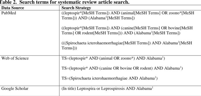
Table 2. Search terms for systematic review article search.
Data Source Search Strategy
PubMed ((leptospir*[MeSH Terms]) AND (animal[MeSH Terms] OR zoono*[MeSH Terms])) AND (Alabama1[MeSH Terms])
((leptospir*[MeSH Terms]) AND (canine[MeSH Terms] OR bovine[MeSH Terms] OR rodent[MeSH Terms])) AND (Alabama1[MeSH Terms]) (((Spirochaeta icterohaemorrhagiae[MeSH Terms]) AND Alabama1[MeSH Terms]))
Web of Science TS=(leptospir* AND (animal OR zoono*) AND Alabama1)
TS=(leptospir* AND (canine OR bovine OR rodent) AND Alabama1) TS=(Spirochaeta icterohaemorrhagiae AND Alabama1)
Google Scholar (In title) Leptospira or Leprospirosis AND Alabama1
1Assessments included all southern states and jurisdictions including: Alabama, Arkansas,
35
Table 3. Selected articles for the systematic review analysis.
# Article Reference
1
Alexander, A.D., Flyger, V., Herman, Y.F., McConnell, S.J., Rothstein, N., & Yager, R.H. (1972). Survey of wild mammals in a Chesapeake Bay area for selected zoonoses. J Wildl Dis, 8(2), 119-126.
2 Alexander, A.D., Gleiser, C.A., Malnati, P., & Yoder, H. (1957). Observations on the prevalence of leptospirosis in canine populations of the United States. Am J Hyg, 65(1), 43-56.
3 Bronson, E., Spiker, H., & Driscoll, C.P. (2014). Serosurvey for selected pathogens in free-ranging American black bears (Ursus americanus) in Maryland, USA. J Wildl Dis, 50(4), 829-836.
4 Brown, R.Z., & Gorman, G.W. (1960). The occurrence of leptospiral infections in feral rodents in southwestern Georgia. Am J Public Health Nations Health, 50, 682-688.
5 Byrne, R.J., & Chambers, C.F., Jr. (1959). A serological survey for leptospiral antibodies in Maryland cattle. J Am Vet Med Assoc, 134(11), 498-502.
6 Chatfield, J., Milleson, M., Stoddard, R., Bui, D.M., & Galloway, R. (2013). Serosurvey of leptospirosis in feral hogs (Sus scrofa) in Florida. J Zoo Wildl Med, 44(2), 404-407.
7
Chitwood, M.C., Swingen, M.B., Lashley, M.A., Flowers, J.R., Palamar, M.B., Apperson, C.S., Olfenbutte, C., Moorman, C.E., & DePerno, C.S. (2015). Parasitology and serology of free-ranging coyotes (Canis latrans) in North Carolina, USA. J Wildl Dis, 51(3), 664-669.
8 Cole, J.R., Jr., & Pursell, A.R. (1973). Serologic incidence of leptospirosis in Georgia horses. Meet U S Anim Health Assoc (77), 632-637. Proc Annu
9
Corn, J.L., Cartwright, M.E., Alexy, K.J., Cornish, T.E., Manning, E.J., Cartoceti, A.N., & Fischer, J.R. (2010). Surveys for disease agents in introduced elk in Arkansas and Kentucky. J Wildl Dis, 46(1), 186-194.
10 Corn, J.L., Swiderek, P.K., Blackburn, B.O., Erickson, G.A., Thiermann, A.B., & Nettles, V.F. (1986). Survey of selected diseases in wild swine in Texas. J Am Vet Med Assoc, 189(9), 1029-1032.
11
Davidson, W.R., Crum, J.M., Blue, J.L., Sharp, D.W., & Phillips, J.H. (1985). Parasites, diseases, and health status of sympatric populations of fallow deer and white-tailed deer in Kentucky. J Wildl Dis, 21(2), 153-159.
12
Easterbrook, J.D., Kaplan, J.B., Vanasco, N.B., Reeves, W.K., Purcell, R.H., Kosoy, M.Y., Glass, G.E., Watson, J., & Klein, S.L. (2007). A survey of zoonotic pathogens carried by Norway rats in Baltimore, Maryland, USA. Epidemiol Infect, 135(7), 1192-1199.
13
Evans, L. B., Wood, G. E., Flyger, V., Alexander, A. D., Yager, R. H., & Rubin, H. L. (1962). Natural Occurrence of Leptospira icterohaemorrhagiae in an Opossum. Proc Soc Exp Biol Med, 110(1), 113-115.
14
Ferguson, D.V., Heidt, G.A. (1981). Survey for rabies, leptospirosis, toxoplasmosis and tularemia in striped skunks (Mephitis mephitis) from three public use areas in northwestern Arkansas. J Wildl Dis. Oct;17(4):515-519.
15 Gorman G.W., Mckeever S, Grimes R.D. (1962). Leptospirosis in wild mammals from southwestern Georgia. Am J Trop Med Hyg. Jul;11:518-524.
16 Hampy, B., Pence, D.B., Simpson, C.D. (1979). Serological studies on sympatric barbary sheep and mule deer from Palo Duro Canyon, Texas. J Wildl Dis. Jul;15(3):443-446.
17
36
# Article Reference
18
Holzman, S., Conroy, M.J., & Davidson, W.R. (1992). Diseases, parasites and survival of coyotes in south-central Georgia. J Wildl Dis, 28(4), 572-580.
19 Howerth, E.W., Reeves, A.J., McElveen, M.R., & Austin, F.W. (1994). Survey for selected diseases in nutria (Myocastor coypus) from Louisiana. J Wildl Dis, 30(3), 450-453.
20
Jenkins, E.M., Harrington, R., Jr., Gbadamosi, S.G., & Braye, E.T. (1979). Survey of leptospiral agglutinins in the sera of swine of southeastern Alabama. Am J Vet Res, 40(7), 1019-1021.
21 Jobling, J.W., & Eggstein, A.A. (1917). The wild rats of the southern states as carriers of icterohemorrhagiae. J Am Med Assoc, LXIX(21), 1787-1787. Spirochaeta
22
Li, H.Y., & Davis, D.E. (1952). The prevalence of carriers of Leptospira and Salmonella in Norway rats of Baltimore. Am J Hyg, 56(1), 90-91.
23
McKeever, K.S., Gorman, G.W., Chapman, J.F., Galton, M.M., & Powers, D.K. (1958). Incidence of leptospirosis in wild mammals from Southwestern Georgia, with a report of new hosts for six serotypes of leptospires. Am J Trop Med Hyg, 7(6), 646-655.
24
Miller, D.A., Wilson, M.A., & Beran, G.W. (1991). Relationships between prevalence of Leptospira interrogans in cattle, and regional, climatic, and seasonal factors. Am J Vet Res, 52(11), 1766-1768.
25
Miller, D.L., Schrecengost, J., Merrill, A., Kilgo, J., Ray, H.S., Miller, K.V., & Baldwin, C.A. (2009). Hematology, parasitology, and serology of free-ranging coyotes (Canis latrans) from South Carolina. J Wildl Dis, 45(3), 863-869.
26 Motie, A., Myers, D. M., & Storrs, E. E. (1986). A serologic survey for leptospires in nine-banded armadillos (Dasypus novemcinctus L.) in Florida. J Wildl Dis, 22(3), 423-424.
27
New, J.C., Jr., Delozier, K., Barton, C.E., Morris, P.J., & Potgieter, L.N. (1994). A serologic survey of selected viral and bacterial diseases of European wild hogs, Great Smoky Mountains National Park, USA. J Wildl Dis, 30(1), 103-106.
28
New, J.C., Jr., Wathen, W.G., & Dlutkowski, S. (1993). Prevalence of Leptospira antibodies in white-tailed deer, Cades Cove, Great Smoky Mountains National Park, Tennessee, USA. J Wildl Dis, 29(4), 561-567.
29 Packchanian, A, & Sonnier A, B. (1948) Incidence of leptospirosis in man and rodents in Galveston. Rep Biol Med. 6(4), 453-460. Tex
30
Pedersen, K., Anderson, T.D., Bevins, S.N., Pabilonia, K.L., Whitley, P.N., Virchow, D.R., & Gidlewski, T. (2017). Evidence of leptospirosis in the kidneys and serum of feral swine (Sus scrofa) in the United States. Epidemiol Infect, 145(1), 87-94.
31
Pedersen, K., Bauer, N.E., Rodgers, S., Bazan, L.R., Mesenbrink, B.T., & Gidlewski, T. (2017). Antibodies to various zoonotic pathogens detected in feral swine (Sus scrofa) at abattoirs in Texas, USA. J Food Prot, 80(8), 1239-1242.
32
Pedersen, K., Pabilonia, K.L., Anderson, T.D., Bevins, S.N., Hicks, C.R., Kloft, J.M., & Deliberto, T.J. (2015). Widespread detection of antibodies to Leptospira in feral swine in the United States.
Epidemiol Infect, 143(10), 2131-2136.
33 Redetzke, K.A., & McCann, M.J. (1980). Isolation of Wildl Dis, 16(3), 333-337. Leptospira from desert rodents of West Texas. J
34
Robinson, G.H. 1924. Occurrence of Leptospira icterohemorrhagiae in wild rats of Baltimore. Am J Hyg. 4(4), 327-329.
35
37
# Article Reference
36
Roth, E.E., Adams, W.V., Sanford, G.E, Greer, B., & Mayeux, P. (1962). Leptospira paidjan (bataviae
serogroup) isolated from nutria in Louisiana. Public Health Rep, 77, 583-587.
37 Roth, E. E., Adams, W.V., Sanford, G.E., Newman, K., Moore, M., & Greer, B. (1964). Isolation of Leptospira pomona from white-tailed deer in Louisiana. Am J Vet Res, 25, 259-261.
38
Roth, E.E., & Galton, M.M. (1960). Isolation and identification of Leptospira hardjo from cattle in Louisiana. Am J Vet Res, 21, 422-427.
39 Rubin, H.L. (1977). Serological incidence of leptospirosis in Florida cattle. Health Assoc, (81), 197-200. Proc Annu Meet U S Anim
40
Saliki, J. T., Rodgers, S. J., & Eskew, G. (1998). Serosurvey of selected viral and bacterial diseases in wild swine from Oklahoma. J Wildl Dis, 34(4), 834-838.
41
Sasmal, I., Gould, N.P., Schuler, K.L., Chang, Y.F., Thachil, A., Strules, J., Olfenbuttel, C., Datta, S., & DePerno, C. S. (2018). Leptospirosis in urban and suburban American black bears in western North Carolina. J Wildl Dis. doi:10.7589/2017-10-263.
42
Sherrill, B.L., Snider, A.G., Kennedy-Stoskopf, S., & DePerno, C. S. (2012). Survey of zoonotic pathogens in white-tailed deer on Bald Head Island, North Carolina. Southeast Nat, 11(3), 529-533.
43 Shotts, E.B., Jr., Andrews, C.L., & Harvey, T.W. (1975). Leptospirosis in selected wild mammals of the Florida panhandle and southwestern Georgia. J Am Vet Med Assoc, 167(7), 587-589.
44 Shotts, E.B., Jr., Andrews, C.L., Sulzer, C., & Greene, E. (1971). Leptospirosis in cottontail and swamp rabbits of the Mississippi Delta. J Wildl Dis, 7(2), 115-117.
45
Shotts, E.B., Greer, W.E., & Hayes, F.A. (1958). A preliminary survey of the incidence of brucellosis and leptospirosis among white-tailed deer (Odocoileus virginianus) of the Southeast. J Am Vet Med Assoc, 133(7), 359-361.
46 Shotts, E.B., Jr., & Hayes, F.A. (1970). Leptospiral antibodies in white-tailed deer of the southeastern United States. J Wildl Dis, 6(4), 295-298.
47 Stuart, B. P., Crowell, W. A., Adams, W. V., & Carlisle, J. C. (1977). Spontaneous renal disease in Louisiana armadillos (Dasypus novemcinctus). J Wildl Dis, 13(3), 240-244.
48 Stuart, B. P., Crowell, W. A., Adams, W. V., & Morrow, D. T. (1978). Spontaneous renal disease in beaver in Louisiana. J Wildl Dis, 14(2), 250-253.
49 Trainer, D.O., & F. Knowlton, F. (1968). Serologic evidence of diseases in Texas coyotes. Manage, 32(4), 981-983. J Wildl
50 Walch, E.W., and Walch-Sorgdrager, G.B. 1927. Observations on the wild rats of Baltimore. Am J Hyg. 7, 393-406. Leptospira icterohaemorrhagiae in
51 White, F.H., Stoliker, H.E., & Galton, M.M. (1961). Detection of leptospires in naturally infected dogs, using fluorescein-labeled antibody. Am J Vet Res, 22(89), 650-654.
38
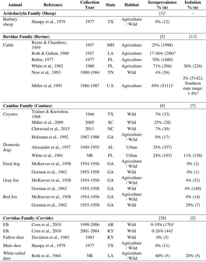
Table 4. Leptospira frequency seroprevalence and isolation measures in animals from systematic review selected articles.
Animal Reference Collection
Year State Habitat
Seroprevalence % (n)
Isolation % (n)
Articdactyla Family (Sheep) [1]a --
Barbary
sheep Hampy et al., 1979 1977 TX
Agriculture
/ Wild 0% (12)
Bovidae Family (Bovine) [5] [11]
Cattle Byrne & Chambers,
1959 1957 MD Agriculture 25% (1998) Roth & Galton, 1960 1957 LA Agriculture 17-36% (288)b Rubin, 1977 1977 FL Agriculture 70% (1660)
White et al., 1982 1980 FL Agriculture 71% (306) 36% (226) New et al., 1993 1980-1984 TN Wild 4% (56)
Miller et al, 1991 1986-1987 U.S. Agriculture 49% (5111)c
2% (5142); Southern state range:
1-4%d
Canidae Family (Canines) [6] [7]
Coyotes Trainer & Knowlton,
1968 1966 TX Wild 3% (33)
Miller et al., 2009 2005 SC Wild 25% (28) Chitwood et al., 2015 2011 NC Wild 7% (30)
Holzman et al., 1992 1987-1988 GA Agriculture
/ Wild 0% (17) Domestic
dogs Alexander et al., 1957 1949-1955 AL Urban 35% (357)
White et al., 1961 NR FL Urban 24% (193) 11% (156)
Feral dog McKeever et al., 1958 1954-1956 GA Agriculture
/ Wild 0% (2)
Gorman et al., 1962 1955-1958 GA Wild 0% (1)
Gray fox McKeever et al., 1958 1954-1956 GA Agriculture
/ Wild 4% (52)
Gorman et al., 1962 1955-1958 GA Wild 4% (140)
Red fox McKeever et al., 1958 1954-1956 GA Agriculture
/ Wild 0% (14)
Gorman et al., 1962 1955-1958 GA Wild 29% (7)
Cervidae Family (Cervids) [28] [2]
Elk Corn et al., 2010 1998-2006 AR Wild 0-10%(170)e Elk Corn et al., 2010 2001-2004 KY Wild 0-26% (44)f Fallow deer Davidson et al., 1985 1983 KY Wild 0% (5)
Mule deer Hampy et al., 1979 1977 TX Agriculture
/ Wild 0% (11) White-tailed
deer Roth et al., 1964 NR LA
Agriculture
39
Animal Reference Collection
Year State Habitat
Seroprevalence % (n)
Isolation % (n)
White-tailed
deer Shotts et al., 1958 NR
South east U.S. Wild 2% (403); southern state
range: 0-4%g Shotts et al., 1970 1958-1959
South east U.S. Wild 19% (1544); southern state range: 8-31%h Shotts et al., 1975 NR FL &
GA
Agriculture
/ Wild 0% (6) 0% (6) Davidson et al., 1985 1983 KY Wild 20% (5)
New et al., 1993 1980-1984 TN Wild 23% (463)
Sherrill et al., 2012 2008 NC Peri-Urban/
Wild 0% (8)
Dasypodidae Family (Armadillos) [3] [1]
Non-banded
armadillo Motie et al., 1986 1980-1983 FL Wild 11% (286) Motie et al., 1986 1980-1983 LA Wild 25% (4)
Stuart et al., 1977 NR LA Agriculture
/ Wild 38% (50) 4% (50)
Didelphidae Family (Opossums) [3] [4]
Opossum McKeever et al., 1958 1954-1956 GA Agriculture
/ Wild 6% (213)
Gorman et al., 1962 1955-1958 GA Wild 16% (821) Evans et al., 1962 1961 MD Wild 14% (37) 5% (37) Alexander et al., 1972 1961-1962 MD Wild 5% (64)
Shotts et al., 1975 NR FL & GA
Agriculture
/ Wild 29% (17) 41% (17)
Equidae Family (Horses) [1] --
Horses Cole & Pursell, 1973 1972-1973 GA Agriculture 46% (1,606)
Felidae Family (Felines) [3] [5]
Bobcat Shotts et al., 1975 NR FL & GA
Agriculture
/ Wild 100% (1) 100% (1) Heidt et al., 1988 1982-1984 AR Wild 25% (8)
Feral house
cats McKeever et al., 1958 1954-1956 GA
Agriculture
/ Wild 0% (24)
Gorman et al., 1962 1955-1958 GA Wild 0% (69) Alexander et al., 1972 1961-1962 MD Wild 0% (5)
Wildcat McKeever et al., 1958 1954-1956 GA Agriculture
/ Wild 8% (40)
Gorman et al., 1962 1955-1958 GA Wild 8% (66)
Leporidae Family (Rabbits) [3] [6]
Cottontail
rabbit McKeever et al., 1958 1954-1956 GA
Agriculture
/ Wild 0% (100)
40
Animal Reference Collection
Year State Habitat
Seroprevalence % (n)
Isolation % (n) Cotton-tailed
rabbit Shotts et al, 1971 1969 MS Wild 79% (43) 73% (44)
Shotts et al., 1975 NR FL & GA
Agriculture
/ Wild 0% (25) 4% (25)
Marsh rabbit McKeever et al., 1958 1954-1956 GA Agriculture
/ Wild 0% (2)
Swamp
rabbit Shotts et al, 1971 1969 MS Wild 60% (5) 33% (6)
Mephitidae Family (Skunks) [3] [5]
Skunks Ferguson & Heidt,
1981 1979 AR Wild 47% (53)
Spotted
Skunk McKeever et al., 1958 1954-1956 GA
Agriculture
/ Wild 0% (1)
Gorman et al., 1962 1955-1958 GA Wild 29% (7) Striped
Skunk McKeever et al., 1958 1954-1956 GA
Agriculture
/ Wild 14% (132)
Gorman et al., 1962 1955-1958 GA Wild 16% (430)
Roth et al., 1963 1959-1960 LA Wild
62% (277) (subsection of study) 57% (650); 59% (277) (subsection of study) Alexander et al., 1972 1961-1962 MD Wild 25% (104)
Molossidae Family (Bats) -- [1]
Mexican free
tail bat Gorman et al., 1962 1955-1958 GA Wild 0% (10)
Mustelidae Family (Otters) -- [2]
Otter McKeever et al., 1958 1954-1956 GA Agriculture
/ Wild 0% (1)
Gorman et al., 1962 1955-1958 GA Wild 0% (1)
Procyonidae Family (Raccoons) [2] [3]
Raccoon Alexander et al., 1972 1961-1962 MD Wild 1% (97)
McKeever et al., 1958 1954-1956 GA Agriculture
/ Wild 4% (200)
Gorman et al., 1962 1955-1958 GA Wild 8% (715)
Shotts et al., 1975 NR FL & GA
Agriculture
/ Wild 59% (17) 38% (21)
Suidae Family (Pigs) [24] [7]
Domestic
swine Jenkins et al., 1979 1974-1976 AL Agriculture 19% (627)
Wild swine Gorman et al., 1962 1955-1958 GA Wild 20% (5) Corn et al., 1986 1983 TX Wild 43% (100)
New et al., 1994 1990 TN &
41
Animal Reference Collection
Year State Habitat
Seroprevalence % (n)
Isolation % (n)
Wild swine Saliki et al., 1998 1996 OK Agriculture
/ Wild 44% (117) Chatfield et al., 2013 NR FL Wild 33% (324)
Pedersen et al., 2015 2007-2011 U.S. Wild
13% (2055); southern state range (0-36%)i
Pedersen et al., 2017a 2012-2014 U.S. Wild
53% (642); southern state range 27-48%j
3% (677); southern state range
2-26%k Pederson et al., 2017b 2015 TX Agriculture 49% (376)
Soricidae Family (Shrews) [1] [1]
Least shrew Shotts et al., 1975 NR FL & GA
Agriculture
/ Wild 0% (1) 100% (1)
Ursidae Family (Bears) [2] --
Black bear Bronson et al., 2014 1999-2011 MD Wild 23% (61)
Sasmal et al., 2018 2014-2015 NC Peri-Urban/
Wild 9% (96)
Order Rodentia (Rodents)
Castoridae Family [1] [1]
Beaver Stuart et al., 1978 1961-1967 LA Wild 4% (25) 4% (25)
Cricetidae Family [2] [19]
Cotton
mouse Brown et al., 1960 1953-1955 GA
Agriculture
/ Wild 0% (90)
Gorman et al., 1962 1955-1958 GA Wild 0% (50)
Cotton rat Brown et al., 1960 1953-1955 GA Agriculture
/ Wild 2% (104)
Gorman et al., 1962 1955-1958 GA Wild 8% (52)
Shotts et al., 1975 NR FL & GA
Agriculture
/ Wild 0% (8) 33% (9) Redetzke & McCann,
1980 1975-1976 TX Wild 0% (1)
Deer mouse Redetzke & McCann,
1980 1975-1976 TX Wild 26% (31)
Golden
mouse Brown et al., 1960 1953-1955 GA
Agriculture
/ Wild 0% (2)
Gorman et al., 1962 1955-1958 GA Wild 0% (3) Grasshopper
mouse
Redetzke & McCann,
1980 1975-1976 TX Wild 17% (46)
Marsh rice
rat Brown et al., 1960 1953-1955 GA
Agriculture
/ Wild 0% (10)
Muskrat Alexander et al., 1972 1961-1962 MD Wild 0% (1)
Pine vole Brown et al., 1960 1953-1955 GA Agriculture
/ Wild 0% (1)
42
Animal Reference Collection
Year State Habitat
Seroprevalence % (n) Isolation % (n) Plains packrat
Redetzke & McCann,
1980 1975-1976 TX Wild 0% (2)
Scorpion mouse
Redetzke & McCann,
1980 1975-1976 TX Wild 25% (8)
White throated packrat
Redetzke & McCann,
1980 1975-1976 TX Wild 18% (159)
Whitefooted
mouse Brown et al., 1960 1953-1955 GA
Agriculture
/ Wild 1% (390)
Whitefooted
mouse Gorman et al., 1962 1955-1958 GA Wild 0% (115) Wood rat Gorman et al., 1962 1955-1958 GA Wild 0% (9)
Echimyidae Family [2] [1]
Nutria Roth et al., 1962 NR LA Wild 42% (24) 31% (26) Howerth et al., 1994 1988-1989 LA Wild 6% (32)
Heteromyidae Family -- [4]
Desert pocket mouse
Redetzke & McCann,
1980 1975-1976 TX Wild 19% (21)
Merriam kangaroo rat
Redetzke & McCann,
1980 1975-1976 TX Wild 19% (16)
Ord
kangaroo rat
Redetzke & McCann,
1980 1975-1976 TX Wild 19% (68)
Silky pocket mouse
Redetzke & McCann,
1980 1975-1976 TX Wild 0% (3)
Muridae Family [4] [13]
Bush mouse Redetzke & McCann,
1980 1975-1976 TX Wild 0% (1)
Harvest
mouse Brown et al., 1960 1953-1955 GA
Agriculture
/ Wild 0% (22)
Gorman et al., 1962 1955-1958 GA Wild 0% (15) House
mouse Brown et al., 1960 1953-1955 GA
Agriculture
/ Wild 21% (284)
Gorman et al., 1962 1955-1958 GA Wild 17% (97)
Shotts et al., 1975 NR FL & GA
Agriculture
/ Wild 0% (1) 100% (1) Redetzke & McCann,
1980 1975-1976 TX Wild 40% (5)
Norway and brown rats
Walch &
Walch-Sorgdrager, 1927 1924-1925 MD Urban 33% (51) Norway rat Robinson, 1924 1923-1924 MD Urban 7% (100)
Packchanian & Sonnier,
1948 NR TX Urban 6% (18) 23% (93)
Li et al., 1952 1947-1949 MD Urban/
Agriculture 46% (1643) l Brown et al., 1960 1953-1955 GA Agriculture
43
Animal Reference Collection
Year State Habitat
Seroprevalence % (n)
Isolation % (n) Norway rat Gorman et al., 1962 1955-1958 GA Wild 0% (42)
Easterbrook et al., 2007 2005-2006 MD Urban 65% (190)
“Wild Rats” Jobling & Eggstein,
1917 1917 TN Urban
>10% (>100)
Sciuridae Family [4] [8]
Flying
squirrel Shotts et al., 1975 NR
FL & GA
Agriculture
/ Wild 0% (1) 0% (1)
Fox squirrel McKeever et al., 1958 1954-1956 GA Agriculture
/ Wild 0% (28)
Gorman et al., 1962 1955-1958 GA Wild 0% (67)
Shotts et al., 1975 NR FL & GA
Agriculture
/ Wild 0% (24) 4% (27)
Gray squirrel McKeever et al., 1958 1954-1956 GA Agriculture
/ Wild 0% (2)
Gorman et al., 1962 1955-1958 GA Wild 0% (2)
Shotts et al., 1975 NR FL & GA
Agriculture
/ Wild 0% (21) 4% (26) Spotted
ground squirrel
Redetzke & McCann,
1980 1975-1976 TX Wild 12% (8)
Woodchuck Alexander et al., 1972 1961-1962 MD Wild 0% (10)
Collective rodents -- [3]
Total rodents Brown et al., 1960 1953-1955 GA Agriculture
/ Wild 7% (933)
Total rodents Gorman et al., 1962 1955-1958 GA Wild 4% (453)
Total rodents Redetzke & McCann,
1980 1975-1976 TX Wild 19% (369)
Total
measures [98] [104]
aBracketed values indicate the number of measures identified in the assessment. bData presented prevalence per serovar: Pomona 36%, Sejroe 17%.
cNational seroprevelence was reported in an earlier article: Miller, D. A., Wilson, M. A., &
Beran, G. W. (1991). Survey to estimate prevalence of Leptospira interrogans infection in mature cattle in the United States. Am J Vet Res, 52(11), 1761-1765.
dSouthern state isolation percentage (n): AL: 3% (69), FL: 1% (70), GA: 3% (75), KY: 3%
(127), LA: 3% (68), MS: 1% (93), OK: 4% (185), TN: 3% (119), TX: 3% (572), and VA: 3% (62).
eData presented prevalence per serovar: Bratislava 10%, Canicola 1%, Grippotyphosa 4%,
Icterohaemorrhagiae 5%, and Pomona 1%.
fData presented prevalence per serovar: Bratislava 3%, Canicola 0%, Grippotyphosa 26%,
Hardjo 3%, Icterohaemorrhagiae 3%, and Pomona 3%.
gPreliminary data; complete data included in data row for Shotts et al., 1970; southern state
seroprevalence (n): AL: 3% (68), FL: 0% (9), GA: 1% (105), KY: 2% (53), LA: 4% (69), MD: 0% (23), MS: 0% (27), NC: 0% (7), SC: 0% (2), and VA: 0% (40).
hSouthern state seroprevalence (n): AL: 14% (152), AR: 9% (117), FL: 12% (69), GA: 12%
44
iSouthern state seroprevalence (n):AL: 16% (77), AR: 25% (181), FL: 8% (49), GA: 9% (145),
KY: 18% (11), LA: 15% (116), MS: 36% (162), NC: 1% (81), OK: 25% (183), SC: 3% (40), TN: 0% (11), and TX: 21% (429).
jSouthern state seroprevalence range for the serovars Bratislava, Canicola, Grippotyphosa,
Hardjo, Licterhaemorrahagiae, and Pomona (n): AL: 0-39% (31), AR: 4-48% (25), LA: 0-27% (41), MS, 6-47% (47), OK: 0-30% (53), and TX: 2-40% (58).
kPCR detection; southern state isolation percentage (n): AL: 3% (31), AR: 8% (25), LA: 2%
(46), MS: 26% (53), OK: 2% (61), and 3% (60).
lAuthors evaluated seroprevalance in three habitats (n): urban/residential: 46% (1220), poultry


![Crystal structure of [tris(pyridin 2 ylmethyl)amine κ4N]copper(II) bromide](data:image/gif;base64,R0lGODlhAQABAIAAAP///wAAACH5BAEAAAAALAAAAAABAAEAAAICRAEAOw==)