NUCLEOTIDE SECOND MESSENGERS SIGNAL THROUGH TRANSCRIPTIONAL AND POSTTRANSCRIPTIONAL REGULATORS TO CONTROL THE PRODUCTION OF A
COLONIZATION FACTOR IN VIBRIO CHOLERAE
Ankunda Therese Kariisa
A dissertation submitted to the faculty at The University of North Carolina at Chapel Hill in partial fulfillment of the requirements for the degree of Doctor in Philosophy in the Department of
Microbiology and Immunology.
Chapel Hill 2015
© 2015
ABSTRACT
Ankunda Therese Kariisa: Nucleotide Second Messengers Signal Through Transcriptional and Posttranscriptional Regulators to Control the Production of a
Colonization Factor in Vibrio cholerae (Under the direction of Rita Tamayo)
The diarrheal human pathogen Vibrio cholerae causes millions of cases of
severe cholera disease every year, resulting in substantial morbidity and mortality.
Although V. cholerae is primarily an aquatic organism, it is also adept at colonizing and
flourishing in the human small intestine. To persist and transition between the aquatic
environment and the host, V. cholerae controls the production of colonization factors
and virulence determinants. V. cholerae alters this through the production of intracellular
second messengers, such as cyclic diguanylate (c-di-GMP) and cyclic adenosine
monophosphate (cAMP), that relay information about the extracellular environment to
intracellular effectors. Within the cell, c-di-GMP interacts with a variety of effector
molecules, such as RNA riboswitches and proteins, to achieve transcriptional,
post-transcriptional and post-translational control of regulated processes. cAMP signals
primarily through its receptor, the cAMP Receptor Protein (CRP), and regulates
processes at the level of transcription. In this study, we examine the regulation of gbpA
by c-di-GMP and cAMP; GbpA is a colonization factor that contributes to attachment of
Vc1 can bind c-di-GMP in vitro and that mutations that reduce binding abrogate GbpA
production. Thus, c-di-GMP positively regulates gbpA expression via Vc1. In addition,
we defined an additional mechanism of regulation of gbpA in which c-di-GMP negatively
impacts activation gbpA transcription initiation by acting through cAMP-CRP. These
studies identify two distinct signals, c-di-GMP and cAMP, that contribute to the
regulation of gbpA. Our findings highlight the complex contribution of nucleotide second
ACKNOWLEDGMENTS
I would like to thank Rita Tamayo for her interest in my development as a
scientist and in the advancement of my career. Her patience, commitment, and
dedication to my training was tremendous.
I would also like to thank the members of the Tamayo lab for constant support
and continuous and helpful feedback throughout my studies. For contributions to
Chapter 1, I would like to thank Alyssa Grube, and for contributions to Chapter 2, I
would like to thank Kevin Weeks. Last but not least, I am thankful for the support of my
DEDICATIONS
This dissertation is dedicated to my late father Dr. G.M.B Kariisa. He lived a
TABLE OF CONTENTS
LIST OF TABLES ... ix
LIST OF FIGURES... x
LIST OF ABBREVIATIONS AND SYMBOLS ... xii
CHAPTER 1: INTRODUCTION ... 1
CHOLERA DISEASE... 1
CHOLERA HISTORY ... 3
COLONIZATION FACTORS IN V. cholerae... 8
NUCLEOTIDE SECOND MESSENGERS... 11
c-di-GMP SIGNALING ... 13
c-di-GMP SIGNALING VIA PROTEIN AND RNA BASED RECEPTORS ... 15
PILZ DOMAIN EFFECTORS ... 15
c-di-GMP SENSING TRANSCRIPTION FACTORS... 17
CLASS I AND CLASS II c-di-GMP SENSING RIBOSWITCHES ... 19
cAMP AND THE PHOSPHOENOLPYRUVATE TRANSPORT SYSTEM ... 21
cAMP SIGNALING VIA THE cAMP RECEPTOR PROTEIN ... 23
REGULATION OF gbpA BY c-di-GMP AND cAMP... 26
REFERENCES….………..31
Introduction………..55
Results... 59
Discussion ... 66
REFERENCES ... 86
CHAPTER 3: TWO NUCLEOTIDE SECOND MESSENGERS REGULATE THE PRODUCTION OF THE VIBRIO CHOLERAE COLONIZATION FACTOR GBPA... 91
Introduction... 92
Results... 102
Discussion ... 111
REFERENCES ... 130
CHAPTER 4: DISCUSSION ... 137
SUMMARY OF RESULTS... 137
BIOLOGICAL IMPACT OF gbpA REGULATION ... 142
CAVEATS AND FUTURE DIRECTIONS... 144
REFERENCES.………148
LIST OF TABLES
Table 2.1. Strains and plasmids used in this study. ... 80
Table 2.2. Primers used in this study. ... 83
Table 3.1. Strains used in this study. ... 126
Table 3.2. Plasmids used in this study... 127
LIST OF FIGURES
Figure 1.1. Structures of nucleotide second messengers. ... 28
Figure 1.2. c-di-GMP regulated processes in bacteria. ... 28
Figure 1.2. cAMP-CRP regulated processes in bacteria. ... 28
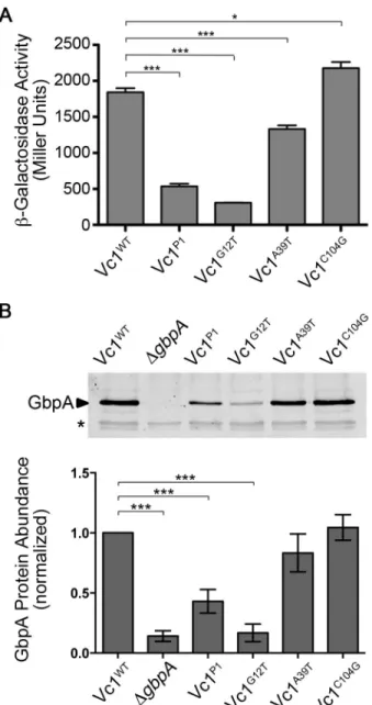
Figure 2.1. Vc1 secondary structure and putative contact residues for c-di-GMP. ... 71
Figure 2.2. Vc1 influences downstream gene expression. ... 72
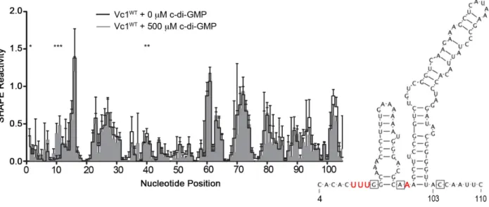
Figure 2.3. c-di-GMP impacts the reactivity of specific regions in Vc1 RNA... 73
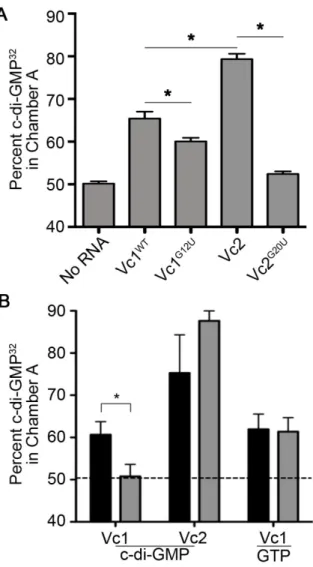
Figure 2.4. Vc1 directly and specifically interacts with c-di-GMP. ... 74
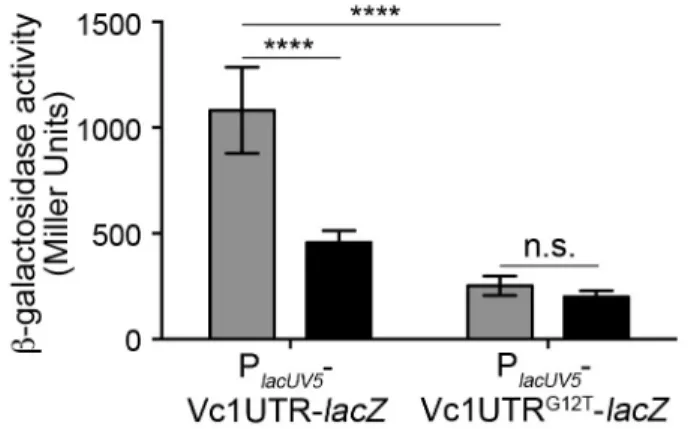
Figure 2.5. Lowering intracellular c-di-GMP reduces Vc1-dependent gene expression. ... 75
Figure 2.6. Vc1 can respond to dose dependent changes in intracellular c-di-GMP levels. ... 76
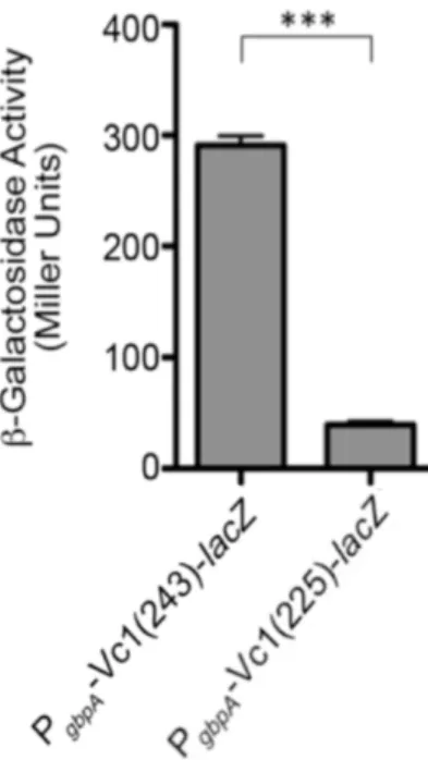
Figure 2.7. The translation start site of gbpA is at position +243. ... 77
Figure 2.8. GTP does not induce structural changes in Vc1 RNA. ... 78
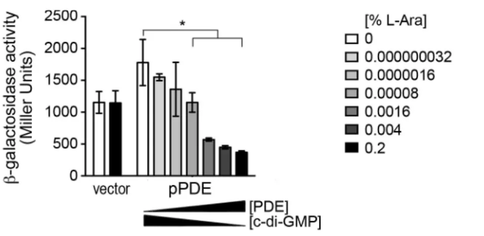
Figure 2.9. The effect of PDE (VieA) and DGC (VCA0956) gene expression on PlacUV5-UTR-lacZ reporter activity is specifically due to reduction of c-di-GMP. ... 79
Figure 3.1. c-di-GMP inhibits gbpA expression independently of the Vc1 riboswitch. ... 118
Figure 3.2. Known c-di-GMP effectors FlrA, VpsT and VpsR do not regulate gbpA in response to c-di-GMP. ... 119
Figure 3.3. c-di-GMP inhibition of gbpA expression is influenced by carbon source availability. ... 120
Figure 3.4. Reduction of c-di-GMP induces gbpA expression in a CRP dependent manner. ... 121
Figure 3.5. Inactivation of the cAMP-CRP signaling pathway prevents c-di-GMP inhibition of gbpA expression. ... 122
Figure 3.7. cAMP-CRP specifically impacts gbpA expression in
response to c-di-GMP ... 124
Figure 3.8. Model of c-di-GMP and cAMP regulation of GbpA production... 125
LIST OF ABBREVIATIONS AND SYMBOLS
V. Vibrio
E. Escherichia
CT Cholera Toxin
TCP Toxin co-regulated pilus
CTXΦ Lysogenic bacteriophage
VPI Vibrio pathogenicity island
LPS Lipopolysaccharide
cAMP Cyclic adenosine 3’,5’-monophosphate
c-di-GMP Cyclic dimeric guanosine 3’,5’-monophosphate
CRP cAMP receptor protein
PTS phosphoenolpyruvate carbohydrate phosphotransferase transport system
DGC Diguanylate cyclase
PDE Phosphodiesterase
CpdA cAMP phosphodiesterase
AC Adenylate cyclase
UTR 5’ untranslated region
SHAPE Selective 2’-hydroxyl acylation analyzed by primer extension
CHAPTER 1: INTRODUCTION
CHOLERA DISEASE
Cholera is a gastrointestinal disease characterized by profuse watery “rice water”
diarrhea (1-3). The voluminous stool can reach volumes of up to 1 L per hour, and if left
untreated, cholera can result in death due to dehydration within hours of onset of
symptoms (4). The World Health Organization (WHO) estimates that approximately 1.4
billion people worldwide are at risk of contracting cholera, predominantly people living in
developing countries (5). The highest burden of cholera is observed in areas where it is
endemic, about 2.8 million cases and 91,000 deaths occur annually (5). Within these
at-risk populations in cholera-endemic areas, roughly 50% of disease and death occurs in
children under the age of 5 (5), likely because young children are more susceptible to
cholera due to a naïve immune system and poor protective immunity (5). In contrast, in
areas where cholera is not endemic, the number of cholera cases and deaths annually
is 87,000 and 2,500, respectively (5). Furthermore, in non-endemic areas, children and
adults are equally susceptible to cholera due to lack of previous exposure (5,6).
Currently, over 99% of the reported incidence and burden of cholera occurs in
Bangladesh, India and Sub-Saharan Africa (5,7). In the developed world, cholera has
been completely eradicated, and isolated outbreaks are rapidly contained and resolved.
health care infrastructure, high population density and environmental factors.
Environmental factors can include increasing rainfall, rising water temperatures and
natural disasters, such as earthquakes and floods. The contribution of these factors to
cholera transmission was most recently observed in Haiti during the outbreak that
started in October 2010. Following the earthquake on January 12th 2010, Haiti’s already
frail infrastructure incurred significant damages that had detrimental effects on the
health care and sanitation sectors. To aid with relief efforts, seemingly healthy UN
Nepalese health care workers, among others, were recruited to Haiti. One or more of
the workers were asymptomatically infected with pathogenic Vibrio cholerae, a
bacterium indigenous to marine and brackish waters and the causative agent of cholera.
Using genomic approaches, it was determined from independent stool samples that the
outbreak was caused by a single pathogenic strain of V. cholerae (8). Furthermore,
comparative genomics revealed that the Haitian strain was nearly identical to
contemporary Nepalese V. cholerae strains. Thus, it was concluded that the first strain
of pathogenic V. cholerae in Haiti was introduced into water sources by Nepalese UN
workers (9). The aquatic environment, ambient temperature and stagnant fresh water
promoted V. cholerae outgrowth and further contamination of water sources. Due to the
large number of people displaced from their homes, many were obliged to live in very
dense and often overcrowded camps. Favorable V. cholerae growth conditions and high
population densities, coupled with the lack of access to clean water, led to the rapid
transmission of cholera within a week of the arrival of the UN Nepalese health care
2014, the Haitian officials reported 697,256 cases and 8,534 deaths due to cholera (10).
Notably, this occurred in a region in which cholera had not previously been reported.
CHOLERA HISTORY
Cholera has been endemic in the Ganges delta region of the Indian subcontinent
for many centuries. However, the first reports of cholera were not until 1817, when it
crossed into Russia. The transmission event was likely due to the rise in trade and
travel. In addition to spreading to Russia, the outbreak extended to parts of China and
the Middle East. This outbreak resulted in the first cholera pandemic, the Asiatic cholera
pandemic, which lasted until 1824. In the second pandemic, from 1829-1851, cholera
reached the Americas and Europe for the first time. The third and fourth pandemics,
which spanned 1852-1875, introduced cholera into parts of South America and North
Africa and Sub-Saharan Africa, where it remains a significant health concern. During the
fifth and sixth pandemics, from 1881-1923, cholera outbreaks were the most
widespread in history affecting parts of Africa, the Middle East, South East Asia and
Europe. However, the deaths from cholera were lower during the later pandemics due
to greater understanding of cholera transmission and increased surveillance.
Collectively, over the last 200 years, there have been seven cholera pandemics. The
current and ongoing seventh pandemic, which includes the outbreak in Haiti, began in
1961 in Indonesia. In the last 50 years, the seventh has spread rapidly to other
countries in Asia, Europe, Africa and Latin America (3).
Before transmission of cholera was understood, the miasma theory, which
transmission during a cholera outbreak in London, that the oral-fecal route of
transmission gained support. Snow traced the source of the outbreak to the broad street
pump in London and urged authorities to remove the handle of the pump to curb the
outbreak. As a result of Snow’s investigation, monitoring of cholera transmission
founded the field of epidemiology and cholera became the first reportable disease (11).
Vibrio cholerae AND PATHOGENESIS
The causative agent of the disease cholera is the Gram-negative, curved rod,
facultative bacterium called Vibrio cholerae (12). V. cholerae has two circular
chromosomes composed of four million base pairs that encode roughly 4000 genes. V.
cholerae was first isolated in 1854 by an Italian anatomist named Filippo Pacini (13).
Pacini went on to publish extensively on V. cholerae, describing its pathogenesis and
proposing methods for cholera treatment. In 1883, Robert Koch independently identified
V. cholerae as the causative agent of cholera disease, following research in Egypt and
Calcutta (12).
V. cholerae is primarily an aquatic bacterium, commonly found in the fresh water
reservoirs, where it can grow planktonically or associate in a biofilm with various
organisms such as zooplankton and phytoplankton (14,15). In the environment, V.
cholerae is often associated with crustaceans, such as copepods, where it survives by
feeding on their chitinous exoskeleton, which provides a carbon and nitrogen source
(16-18). V. cholerae species can also survive on larger crustaceans such as arthropods,
on chironomid egg masses and within amoebae (19-21). Environmental factors that
promote the bloom and distribution of zooplankton and increase the spread of cholera in
susceptible areas (22). As a species, V. cholerae exhibits genetic diversity, and in
aquatic reservoirs over 200 serogroups have been identified (1). Serogroups are
differentiated by O antigen variations in the lipopolysaccharide (LPS). Serogroups can
be subdivided into biotypes, and biotypes can be subdivided into serotypes. Only
serogroups O1 and O139 have been associated with cholera disease, suggesting that
V. cholerae has primarily evolved to survive in the aquatic environment (3,23).
Pathogenic O1 and O139 serogroups are distinguished from non-pathogenic strains by
the presence of the Vibrio pathogenicity island (VPI) genomic island and the lysogenic
bacteriophage CTXΦ. CTXΦ and VPI encode two key players in V. cholerae
pathogenesis, cholera toxin (CT) and toxin co-regulated pili (TCP), respectively (24, 25).
The O1 serogroup is composed of two biotypes, El Tor and classical, and each biotype
contains the Inaba and Ogawa serotypes (23). The classical and El Tor biotypes differ
in the type of CTXΦ and their number of VPIs; the classical biotypes encode one
(VPI-1) and the El Tor biotypes encode two (VPI-1 and VPI-2) (26). The classical biotype was
responsible for the first six pandemics but it has been largely replaced by the El Tor
biotype, which was responsible for the seventh pandemic and most of the ongoing
outbreaks (23). The O139 serogroup was first identified in 1992 and has since caused
severe epidemics in South Asia (23, 27, 28). In addition to its role in pathogenesis, TCP
acts as the receptor for CTXΦ (29). Therefore, evolution of pathogenic V. cholerae
relies on the lateral transfer of VPI, which contains the gene encoding TCP, followed by
disease through horizontal gene transfer. This finding, along with the recent emergence
of the serogroup O139, draws concerns that new strains of pathogenic V. cholerae may
continue arise (30). In addition, many O1 and O139 variants isolated in the last few
decades possess the SXT genetic element, which confers resistance streptomycin and
co-trimoxazole (31).
V. cholerae is normally transmitted to a human host through contaminated food
or water. Upon ingestion, pathogenic V. cholerae can colonize the small intestine (2, 3).
Colonization can be asymptomatic and result in shedding for only a few days, or
symptomatic and result in diarrhea and shedding for up to 2 weeks (32, 33).
Symptomatic patients can experience either mild or severe diarrhea, and if left
untreated, severe diarrhea is fatal in over 50% of cases due to rapid dehydration (34).
Human infections may play an important role in the V. cholerae life cycle as bacterial
shedding can lead to the introduction or reintroduction of large numbers of V. cholerae
into aquatic reservoirs. For instance, human carriers coming from regions where cholera
is endemic can seed new outbreaks of cholera in naïve areas (see Haiti outbreak
above). In addition, pathogenic strains shed by a host are hyperinfectious for up to 5
hours post-infection. Hyperinfectious bacteria, which have an infectious dose 1/10th of
environmentally derived strains, may accelerate the transmission of cholera during
outbreaks (35). Indeed, mathematical models that incorporate hyperinfectivity into their
calculations can reproduce the explosive rates of transmission often observed during
outbreaks (36).
The main driver of cholera symptoms is the enterotoxin cholera toxin (CT). CT is
Production and secretion of A1B5 by V. cholerae during infection leads to the
engagement of B5 with the GM1 ganglioside receptor on intestinal epithelial cells and
the translocation of A1 into the host cell cytosol (38). Once cytoplasmic, A1 becomes
enzymatically active and ADP-ribosylates the Gsα subunit of host adenylate cyclase,
locking it into a constitutively active GDP bound conformation. This irreversibly activates
the adenylate cyclase leading to uncontrolled synthesis of cAMP and activation of the
Cystic Fibrosis transmembrane receptor (CFTR) (39, 40). The consequence of
activating this signaling cascade is the massive efflux of ions and water into the
intestinal lumen and the hallmark profuse and watery diarrhea. Massive fluid losses can
reach volumes of 1 L per hour and result in death due to dehydration within hours of
onset of disease (4).
In response to severe diarrheal symptoms from cholera disease, aggressive
rehydration therapy can reduce mortality from over 50% to 0.2% (3). The composition of
this therapy is critical to restoring normal electrolyte homeostasis, and depending on the
severity of the patient’s condition it is administered via oral or intravenous routes (41,
42). Additionally, in response to diarrhea, antibiotic treatment can be administered to
help reduce symptoms (in about 50% of cases) and shorten the duration of disease (34,
43). In rare incidences, tetracycline, erythromycin and ciprofloxacin resistant strains
have been isolated (44, 45). Therefore, the emergence of antibiotic resistant strains
may make the treatment of cholera more challenging and costly. Nevertheless,
antibiotics are secondary treatments to rehydration therapy since dehydration is the
of a major vaccine program. One major obstacle that remain is limited long-term
efficacy; only 50% of patients retain protective immunity when challenged 2-3 years
following vaccination (46, 47). Although long-term protection is poor, vaccinating a
proportion of a population at-risk can reduce the overall incidence of cholera
transmission even in unprotected individuals (48). This phenomenon, termed “herd
immunity”, may be an important consideration when implementing future vaccine
programs to combat cholera.
COLONIZATION FACTORS IN V. cholerae
To cause disease in a mammalian host and persist in the aquatic environment,
V. cholerae must produce colonization factors that mediate attachment to the various
surfaces it encounters (49). One of the best-studied colonization factors is TCP, which
is encoded within VPI in epidemic and pandemic strains (25, 29). Along with CT, the
second essential virulence factor is TCP, which plays an important role in V. cholerae
colonization of the small intestine and is coregulated with CT (50, 51). Infections models
using mice and humans have confirmed that TCP is absolutely required for colonization
and subsequent disease (50, 51). TCP is a type IV pilus composed of TcpA subunits
that make up length of the pilus structure, and help mediate microcolony formation
through pilus-pilus interactions (52). Modifications to the pili that abrogate these
interactions, but preserve pilus structure, reveal that microcolony formation is required
for colonization of the mammalian intestine and infection by CTXΦ (52). Furthermore,
physical barrier (53). In recent studies, TCP was shown to directly play a role in
attachment to Caco-2 human intestinal epithelial cells (53).
V. cholerae in the aquatic environment, including those recently shed from an
infected human, must establish interactions with biotic and abiotic surfaces in the water
(14, 15). In the environment, V. cholerae will typically engage these surfaces as a
biofilm, to resist environmental stresses (54). When biofilm formation on the chitin shell
of the squid pen was measured, TCP was identified as an important factor in late stage
maturation biofilm, but not initial attachment to chitin (55). Interestingly, using a test-tube
to measure biofilm production, TCP was shown to be dispensable during all stages of
biofilm formation (56). Therefore, although TCP does not function as an adhesin for
chitin, it is important for biofilm maturation and potentially bacterial fitness. Overall, TCP
appears to play a role in colonization in both the mammalian host and in the
environment.
Two other pili have been implicated in colonization of chitin and biofilm
production: mannose-sensitive hemagglutinin-like pili (MSHA) and chitin regulated pili
(ChiRP). MSHA was shown to participate in biofilm formation on abiotic surfaces, but
not on chitin (56). However, despite the negligible contribution of MSHA to biofilm
production on chitin, MSHA appears to be important for the initial attachment to the
chitinous exoskeletons of zooplankton such as copepods (57, 58). ChiRP is another
chitin binding pilus that is produced during growth with chitin and/or chitin
oligosaccharides. Studies to identify factors required for chitin utilization, found that a
Two nonpilus adhesins have been found to play a dual role in colonizing the host
and environment: FrhA and GbpA. In a transcriptome analysis of flagellar regulatory
mutants, frhA was identified as a positively regulated target of the flagellar regulatory
hierarchy (59). FrhA is a hemagglutinin involved in binding to and agglutination of
erythrocytes and attachment to human epithelial cells (59). Consistent with these roles,
frhA mutants showed a colonization defect in the infant mouse model of infection.
Interestingly, FrhA was also shown to be important for the early stages of biofilm
production on abiotic surfaces and chitin beads (59). Collectively, these studies suggest
that FrhA plays a role in the host and environmental phases of the life cycle of
pathogenic V. cholerae.
The second nonpilus adhesin, GbpA, is a N-acetylglucosamine (GlcNAc)-binding
protein that is encoded by locus VCA0811 (60). GbpA is secreted by the type two
secretion system onto the bacterial cell surface, where it recognizes GlcNAc
modifications found on some proteins, and GlcNAc derived carbohydrates such as chitin
(60). GbpA is composed of four domains (1-4), and it exists as a monomer in solution.
Domains 1 and 4 bind chitin oligosaccharides and GlcNAc modified glycans, whereas
domains 3 and 4 are dispensable for chtin binding but are important for GbpA binding to
the bacterial cell surface (61). GbpA production is positively regulated by its substrate,
GlcNAc (17). When grown on chitin, V. cholerae secretes chitinases that release chitin
oligosaccharides and GlcNAc monomers. GlcNAc is transported into V. cholerae and
phosphorylated by specific phosphoenolpyruvate transport system (PTS) components,
and serves as a carbon and nitrogen source (62). In addition, phospho-GlcNAc can bind
GlcNAc sensing and metabolism, including gbpA (62). Interactions of V. cholerae with
mucin, which also contains GlcNAc moieties, can also result in increased gbpA
expression through NagC depression, suggesting that during V. cholerae colonization of
the small intestine GbpA production might be elevated (63). GbpA levels also are
affected post-translationally through the activities of the quorum-sensing regulated
proteases, HapA and PrtV (64). HapA and PrtV are produced at high cell density and
are believed to aid in detachment of V. cholerae from surfaces so that the bacterium
can transition to a new surface, e.g., from the host back into the aquatic environment or
between surfaces in the environment (64).
V. cholerae strains deficient in gbpA are significantly attenuated in binding mucin
and intestinal epithelial cells, due to failed interactions with GlcNAc containing
glycoprotein modifications (60, 63). Consistent with these results, GbpA is important for
colonization and disease in the infant mouse (60). In addition, GbpA is important for
colonizing the chitinous exoskeleton of the crustacean Daphna magna, suggesting that
it also plays a role in V. cholerae attachment to similar environmental organisms (58,
60). Therefore, along with TCP and FrhA, GbpA adds to the repertoire of V. cholerae
colonization factors that possess dual functions in host pathogenesis and environmental
persistence (49).
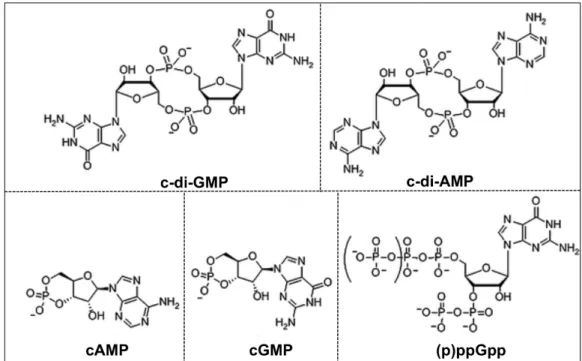
NUCLEOTIDE SECOND MESSENGERS
The production of a particular colonization factor under the right environmental
condition can be critical to bacterial survival. As bacteria transition between different
molecule second messengers, which relay information about the extracellular milieu to
intracellular effectors. In this section we will focus on nucleotide second messengers
implicated in bacterial signaling. Production of these intracellular nucleotides, which
include c-di-GMP, c-di-AMP, c-GMP-AMP, cGMP, cAMP, and (p)ppGpp, has been
shown to regulate biofilm formation, motility, virulence, nutrient acquisition, stress
responses and sporulation (reviewed in 65-68) (Figure 1.1). Accordingly, dysregulation
of these signals likely impairs the ability of bacteria to transition between disparate
environments.
Cyclic adenosine 3’,5’-monophosphate (cAMP) was first identified as a second
messenger in liver cells in 1957 by Earl Sutherland, and as a signaling molecule
regulating carbon metabolism in Escherichia coli in 1969 (69, 70). In bacteria,
intracellular cAMP levels are heavily influenced by the availability of extracellular
nutrient sources, and cAMP along with cAMP Receptor Protein (CRP) govern the
utilization of nutrient sources. Cyclic guanosine 3’,5’-monophosphate (cGMP) was first
identified in 1963 in rat urine and levels correlated with the hormonal state in the animal
(71). A role for cGMP is largely restricted to eukaryotic cells, where it is associated with
transmembrane signal transduction, protein kinase activity and many other important
processes (72). In bacteria, production of cGMP was observed as early as 1974, but
only recently has a function for cGMP in intracellular signaling been ascribed (73). In
studies using Rhodospirillum centenum, a cGMP specific synthase was indentified and
cGMP production was linked to cyst formation (74). The linear nucleotides guanosine
3’,5’-bispyrophosphate (ppGpp) and guanosine 3’-diphosphate, 5’-triphosphate
coli was first indentified in 1970 and later classified as an “alarmone” produced in
response to nutrient starvation and other stresses (75, 76).
It was not until several decades following the discovery of cAMP, cGMP and
(p)ppGpp that cyclic dinucleotides were identified. Perhaps the most broadly studied is
cyclic dimeric guanosine 3’,5’-monophosphate (c-di-GMP), identified in 1989 as an
allosteric regulator of bacterial cellulose synthase in Gluconacetobacter xylinum (77).
The increased interest in c-di-GMP as a ubiquitous bacterial second messenger in the
last decade, led to the identification of its role in virulence, motility, and biofilm formation
in a vast number of organisms. About 20 years later, in 2008, cyclic dimeric adenosine
3’,5’- monophosphate (c-di-AMP) was identified as second messenger in Gram-positive
bacteria (78). Since the discovery of c-di-AMP, studies have shown that it regulates
bacterial cell growth, sporulation, stress responses, antimicrobial resistance and
virulence (79). Lastly, cyclic guanosine monophosphate adenosine monophospate
(c-GMP-AMP) was identified in 2012. A function for c-GMP-AMP has been ascribed to few
organisms; in V. cholerae c-GMP-AMP production during infection promotes chemotaxis
and in numerous Deltaproteobacteria, including Geobacter species, it is predicted to
function in extracellular electron transfer (80-82).
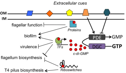
c-di-GMP SIGNALING
The second messenger c-di-GMP is widely recognized to play an important role in
bacterial adaptation to changing environmental conditions (66, 83). Numerous studies
have demonstrated that c-di-GMP modulates biological processes including motility,
cholerae modulates intracellular c-di-GMP levels as it transitions from its native aquatic
environment into the host intestine. Reduction of c-di-GMP is required to promote
bacterial motility and increase expression of virulence factors, and there is evidence
suggesting that V. cholerae may downregulate c-di-GMP production during early stages
of infection (84-88). Specifically, the production of VieSA, a two-component system
composed of the response regulator c-di-GMP phophodiesterase VieA, is required for
virulence in the mouse and CT production in vitro. Conversely, production of c-di-GMP
promotes biofilm formation, which may enhance V. cholerae survival on chitin and other
aquatic surfaces (89-91). Accordingly, inactivation vieA results in increased biofilm
production, and similarly overexpression of a diguanylate cyclase, VC0956, increases
biofilm production (91, 92). The molecular basis of c-di-GMP-dependent regulation in V.
cholerae involves impacts on transcriptional, posttranscriptional and posttranslational
mechanisms.
In response to extracellular cues, which are largely unknown, bacterial cells adjust
the intracellular level of c-di-GMP through the opposing activities of diguanylate
cyclases (DGCs) and phosphodiesterases (PDEs), which synthesize and hydrolyze
c-di-GMP, respectively. The enzymatic activity of DGCs is supplied by a GGDEF domain,
named for conserved residues in the catalytic site (93-96) Two distinct domains, EAL
and HD-GYP, also named for conserved residues, confer c-di-GMP hydrolytic activity to
PDEs (97-99). The PDE and DGC enzymatic domains are often present in proteins in
conjunction with sensory and regulatory domains such as PAS, HAMP, blue light
sensing (BLUF), haemerythrin, GAF, CHASE, MASE and/or receiver domains linked to
enzymatic domains with these additional modules provides a post-translational means
for a variety of stimuli to impact DGC and PDE activity and thus the level of c-di-GMP in
the cell. Transcriptional and post-transcriptional regulation of DGC and PDE gene
expression also influences intracellular c-di-GMP levels.
c-di-GMP SIGNALING VIA PROTEIN AND RNA BASED RECEPTORS
To date there are several known intracellular c-di-GMP receptors, which include
protein and riboswitch sensors. Protein sensors include transcription factors that directly
sense c-di-GMP to modulate gene expression, or proteins that contain PilZ domains,
degenerate EAL domains, I-site domains or GIL domains (GGDEF I-site like domains)
(100). The specific interactions of these receptors with c-di-GMP mediate the
physiological changes observed in response to changes in the level of the second
messenger. In the following section, we discuss the various types of c-di-GMP effectors,
how the effectors recognize c-di-GMP, and how c-di-GMP sensing contributes to the
regulation of biological processes. For comprehensive reviews of c-di-GMP signaling
and effectors, we direct readers to other excellent reviews, details to follow will focus on
receptors found in V. cholerae (101, 102).
PILZ DOMAIN EFFECTORS
In an early study of the regulation of the cellulose synthase in G. xylinus,
Benziman and co-workers observed that c-di-GMP stimulates the activity of the
the discovery of this domain in a variety of bacteria and suggested that the PilZ domain
may play an important regulatory role. Genes encoding a PilZ domain are broadly
distributed in bacterial genomes (103). As with GGDEF, EAL and HD-GYP domains,
bacterial genomes often encode multiple proteins containing PilZ domains in
combination with other domains, suggesting broad regulatory roles for PilZ proteins.
Numerous PilZ domain c-di-GMP receptors with diverse signaling functions and precise
modes of action have been identified.
As in many bacterial species, in V. cholerae c-di-GMP inhibits swimming motility
and favors biofilm formation; in addition, c-di-GMP negatively regulates virulence gene
expression (87, 91, 92, 104, 105). V. cholerae encodes 5 proteins containing PilZ
domains, PlzA, PlzB, PlzC, PlzD, and PlzE. In vitro, PlzC and PlzD bind c-di-GMP via
their PilZ domains (106); it is unclear whether PlzA, PlzB and PlzE bind c-di-GMP under
conditions not tested. In vivo, mutation of plzB or plzC results in decreased motility,
mutation of plzB or plzD results in decreased biofilm formation, and mutation of plzB or
both plzC and plzD results in reduced colonization of the infant mouse small intestine
through unknown mechanisms (106). PlzD contains the RxxxR motif that is conserved
among PilZ domains (and among all 5 PilZ domain proteins of V. cholerae). The
arginines in this motif are essential for binding of c-di-GMP in PlzD and PilZ domains in
other species (106, 111). Structural studies show that PlzD (VCA0042) exists as a
dimer in both apo- and ligand-bound forms (107) (PBD ID 1YLN; R Zhang, M Zhou, S
Moy, F Collart, and A Joachimiak, 2005). The PilZ and the other domain in the protein,
the YcgR-N domain, are connected by a short seven-residue loop that contains the
c-di-GMP ligand and functions in the conformational change induced by c-di-GMP
binding (107). In the apo structure, the C-terminal PilZ domain is located far from the
two-fold axis and it makes no contact with the YcgR-N domain in the same monomer or
in another monomer. In contrast, in the c-di-GMP bound structure of PlzD, the PilZ and
YcgR-N domains in a single monomer are found in close proximity, with one c-di-GMP
molecule packed tightly in their mutual interface (107). Thus, c-di-GMP binding changes
the extended apo-PlzD structure into a more compact ligand-bound structure in which
the PilZ domains make new contacts across the dimer interface.
To date, PilZ domain proteins have been identified in a variety of organisms and
associated with functions that include flagellar motor activity, type IV pilus assembly,
alginate and cellulose production and virulence (106, 108-111). The mechanisms of
action of PilZ domain containing proteins are not always apparent from sequence and
structural information; even PilZ domain proteins with very similar domain architectures
can display distinct binding stoichiometries and mechanistic properties. It is likely that
there are additional modes of action employed by this class of proteins, and that this
diversity contributes to generating the broad regulatory effects observed.
c-di-GMP SENSING TRANSCRIPTION FACTORS
Transcription factors that sense c-di-GMP have been reported in several
bacterial species. These regulators interact with c-di-GMP via various motifs, and the
DNA binding region of the transcription factor lends specificity, allowing targeted
regulation of gene expression. Thus, c-di-GMP can be broadly employed to control a
have been identified to date, each of which was previously characterized for their roles
in motility and/or biofilm development.
The master regulator of flagellar biosynthesis in V. cholerae is FlrA. FlrA is a
σ54-dependent enhancer binding protein containing AAA+ ATPase and DNA-binding
domains. FlrA binds c-di-GMP, which prevents FlrA from interacting with the flrBC
promoter and activating flagellar gene expression (112). Regulation of FlrA activity by
c-di-GMP defines the mechanism for the previously observed phenomenon that flagellar
motility is inhibited by c-di-GMP in V. cholerae. In addition, transcriptome analysis
revealed that over 300 genes are predicted to be regulated by FlrA, only 50 of which fall
within the flagellar/chemotaxis operon (59).
The LuxR family transcription factor VpsT promotes V. cholerae biofilm formation
by activating the expression of vps genes required for exopolysaccharide production
(113). VpsT consists of an N-terminal REC domain and a C-terminal HTH domain
involved in DNA binding. VpsT does not appear to be a response regulator, as no
cognate sensor kinase has been identified and residues for phosphor-transfer in the
REC domain are poorly conserved. Instead, VpsT binds c-di-GMP (KD ~ 3.2 µM), an
interaction that promotes VpsT binding to target promoters (89). Structural analysis of
VpsT with c-di-GMP revealed that two intercalated c-di-GMP molecules interact with
VpsT and stabilize the REC dimerization interface. c-di-GMP interacts with VpsT via a
four residue W[F/L/M][T/S]R sequence within the REC domain (89). Importantly,
mutation of residues in this motif abolish binding of c-di-GMP and target promoters in
The transcriptional regulator VpsR is an NtrC-family σ54-dependent enhancer
binding protein that activates biofilm genes in V. cholerae (114, 115). VpsR contains an
AAA+ ATP binding domain and a HTH DNA binding domain, and VpsR binds c-di-GMP
in vitro (KD ~ 1.6 µM), possibly via its Walker A box (116). However, binding of c-di-GMP
does not affect the ability of VpsR to interact with the target promoters tested in vitro
(116). It is possible that binding of c-di-GMP does not affect the structure of VpsR and
thus does not impact VpsR promoter-binding function. Alternatively, c-di-GMP binding to
VpsR may affect transcription of only a subset of VpsR-regulated genes, or may affect
the subcellular localization or stability of VpsR.
CLASS I AND CLASS II c-di-GMP SENSING RIBOSWITCHES
In addition to the many types of protein sensors of di-GMP, RNA sensors of
c-di-GMP have also been identified and are widely distributed in bacterial genomes.
Riboswitches, cis-acting regulatory elements found in the 5’ untranslated region (UTR)
of some mRNA, consist of an aptamer that binds a specific ligand and an expression
platform that regulates downstream gene expression. Regulation typically occurs as a
result of conformational changes in the RNA structure in response to ligand binding.
The two most common modes of gene regulation by riboswitches involve control of
transcription read-through or translation initiation. The aptamer and expression
platforms are modular in that a given aptamer can work in conjunction with different
expression platforms depending on the specific riboswitch (117-120).
Two classes of c-di-GMP sensing riboswitches have been identified, class I and
GMP riboswitches contain a GEMM motif, so named because they often reside in the 5’
UTR of genes with functions predicted to relate to the environment, membrane or
motility (124). Several studies indicate that the GEMM motif represents the aptamer of
the c-di-GMP riboswitch, however the regions anticipated to correspond to the
expression platform appear to be highly divergent among putative class I riboswitches.
The class II c-di-GMP riboswitch shares no structure or sequence homology with the
class I riboswitch. class II riboswitches are less abundant than class I riboswitches, with
45 putative class II riboswitches identified to date (125).
Due to the lack of sequence and structural similarity, the two riboswitch classes
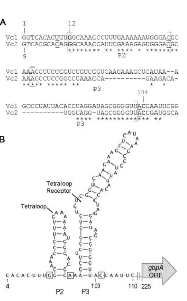
interact with c-di-GMP via distinct mechanisms. In the class I riboswitch Vc2 from V.
cholerae, c-di-GMP binds at the junction of three helices, P1, P2, and P3 (123, 126).
Vc2 makes contacts with c-di-GMP via three nucleotides, G20, C92, and A47. One
guanine base from c-di-GMP forms a Hoogsteen pair with G20, while the other forms a
Watson–Crick pair with C92. A47 bridges the P2 and P3 helices of Vc2 and also
contacts c-di-GMP by stacking between the guanine bases, producing extensive base
stacking interactions between the RNA and ligand. P2 and P3, as well as c-di-GMP
contact residues, are highly conserved among the class I riboswitches, suggesting that
these features are critical for c-di-GMP recognition by these riboswitches (123, 126). In
the class II riboswitch, no canonical base pairings are made with c-di-GMP, but stacking
interactions are observed. Three conserved adenosines, A13, A70, and A61, intercalate
below, between, and above the two guanine bases of c-di-GMP, respectively. In
addition, one guanine base of c-di-GMP is recognized as part of a base triple with A69
by hydrogen bonding). The other guanine base of c-di-GMP forms a single hydrogen
bond with G73 and contacts the 2′-OH of A70 and a fully hydrated magnesium ion (122,
125). Furthermore, several of the nucleotides directly involved in recognizing c-di-GMP,
A69, A70 and G73, are conserved in at least 90% of class II sequences identified to
date (122).
While predictions can be made as to how sensing of c-di-GMP by a riboswitch
impacts the biology of an organism based on the identity of the downstream gene, little
has been demonstrated experimentally. The class I riboswitch Vc2 in V. cholerae, for
instance, has been well characterized in vitro and has been shown to regulate a
reporter gene in response to c-di-GMP in vivo using the heterologous bacterial host E.
coli (124). The exact mechanism of action for Vc2 remains unknown; however,
c-di-GMP has no effect on transcript length, therefore the mechanism of action is likely
post-transcriptional. Vc2 lies upstream of VC1722, which is predicted to encode a protein
with homology to TfoX, a factor involved in uptake of exogenous DNA (17, 124, 127).
However, the consequence of c-di-GMP binding to Vc2 and consequent regulation of
VC1722 in V. cholerae is unknown. The second class I riboswitch in V. cholerae is
called Vc1 and lies upstream of gbpA, which encodes the GlcNAc binding protein
involved in the interaction of V. cholerae with environmental chitin and with the host
intestine. No studies of the Vc1 riboswitch or its relevance to V. cholerae biology have
been reported. The in vivo role of Vc1 will be discussed in Chapter 2.
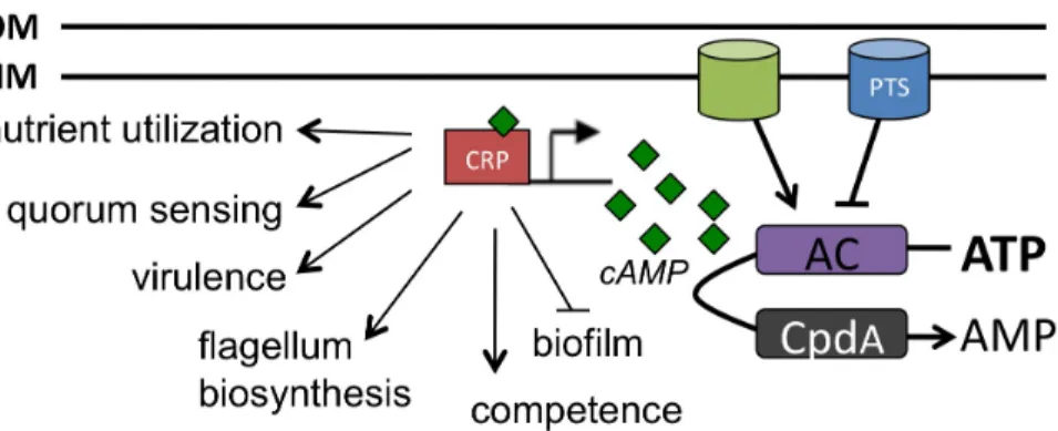
cAMP AND THE PHOSPHOENOLPYRUVATE TRANSPORT SYSTEM
the availability of extracellular nutrient sources (128). In brief, for Gram-negative
bacteria, uptake of a preferred carbon source by the phosphoenolpyruvate
(PEP)-carbohydrate phosphotransferase transport system (PTS) is dependent on key
components of the pathway and the intracellular ratio of PEP to pyruvate. When PEP to
pyruvate levels are high, a phosphoryl group from PEP is transferred to the PTS
component enzyme I (EI), which initiates a phosphorylation cascade important for
utilization of PTS dependent sugars. EI transfers the phosphoryl group to histidine
protein (HPr), HPr transfers the phosphoryl group to EIIA, EIIA transfers the phosphoryl
group to EIIBC and EIIBC transfers the phosphoryl group to the incoming sugar. PTS
dependent sugars are transported into the cell by their cognate EIIC specific
transporter. The EII components of PTS are usually membrane bound and form a
complex to facilitate uptake and phosphorylation of the incoming sugar. Once
phosphorylated, the sugar is metabolized through glycolysis, which in turn influences
the intracellular pool of PEP and the rate of EI phosphorylation. Whereas some PTS
components are specific for a particular sugar, for instance EIIBC, some PTS
components can act on multiple different incoming sugars, for instance EIIA, HPr and
EI. Accordingly, V. cholerae deficient in EIIA is unable to grow on the PTS sugars
glucose, GlcNAc, sucrose or tetrahalose (129). During growth on non-PTS carbon
sources, the phosphoryl group on EIIA is not transferred to the incoming nutrients.
Phosphorylated EIIA can interact with the enzyme adenylate cyclase (AC) to stimulate
its activity, leading to increased intracellular cAMP production. Therefore, the
phosphorylation state of PTS components, namely EIIA, and the levels of intracellular
rate of glycolysis and PEP to pyruvate ratios, which are heavily influenced by the type of
carbon source available, will also influence the PTS pathway and cAMP production.
V. cholerae has 25 genes that encode proteins with known or predicted roles as
PTS components; these include two EI homologs, three HPr homologs and nine EIIA
homologs (130). In V. cholerae, sugars dependent on PTS for uptake include sucrose,
mannitol and fructose (129). Interestingly, unlike in E. coli in which glucose uptake is
strictly PTS-dependent, V. cholerae can also utilize glucose in a PTS independent
manner; genetic deletion of several PTS components still allows for glucose utilization.
Similarly, a V. cholerae strain deficient for the EIIBGlcNAc transporter, can still grow in
media with GlcNAc as the sole carbon source, suggesting non-PTS utilization of
GlcNAc may occur (62).
The role of PTS systems in V. cholerae has proven to be very complicated and
extends beyond utilization of carbon sources. For instance, several components of PTS,
such as EI, HPr, EIIA and EIIBC have been shown to influence biofilm production and
colonization of the mammalian small intestine (129-134).
cAMP SIGNALING VIA THE cAMP RECEPTOR PROTEIN
The phosphorylation state of PTS components heavily influences intracellular
cAMP production. To date, the only known sensor of cAMP is the cAMP receptor
protein (CRP) (128). cAMP-CRP act as a regulatory complex that promotes the
expression of genes required for growth on non-PTS carbon sources. In numerous
organisms, including V. cholerae, the cAMP-CRP complex can also regulate the
incorporates information about extracellular nutrient availability to accordingly regulate
factors involved in these processes.
Early studies looking at the role of cAMP-CRP in V. cholerae showed that strains
deficient in cya, the gene encoding AC, have increased biofilm production, and
increased vps expression (136). The same was observed for a crp mutant, implicating
the cAMP-CRP complex in inhibiting biofilm production. cAMP-CRP was also shown to
repress the expression of vpsT, one of the master regulators of biofilm production in V.
cholerae (137). Interestingly, cAMP-CRP controls the expression of ten genes predicted
to encoded enzymes with DGC and PDE activity, suggesting that cAMP-CRP signaling
can influence intracellular c-di-GMP turnover (136). Deletion of one of the predicted
DGCs, cdgA, eliminated the elevated biofilm production observed in a crp mutant,
suggesting cAMP-CRP controls biofilm production by influencing c-di-GMP production
(136).
A role for cAMP-CRP in inducing competence was also observed. V. cholerae
can become competent for natural transformation in the presence of chitin (127).
Interestingly, cAMP-CRP can enhance the chitin induced competence pathway at
several junctures (16). cAMP-CRP promotes attachment to chitin, promotes utilization of
chitin as a carbon source, and enhances the expression of competence genes pilA, pilM
and comEA (16, 141). These data suggest that cAMP-CRP promotes DNA uptake by
activating important genes in the competence pathway.
Colonization and virulence factors are also regulated by cAMP-CRP. V. cholerae
deficient in cya and/or crp produces elevated levels of CT and TCP, suggesting that
attenuated for colonization in a mouse model despite elevated levels of CT and TCP,
suggesting the cAMP-CRP pathway promotes the expression of other genes required
for colonization and growth in a host (137, 140). Indeed, cAMP-CRP is a positive
regulator of the outer membrane proteins OmpT, OmpU and OmpW, as well as genes
involved in chemotaxis and metabolism (136, 137). cAMP-CRP also positively regulates
motility, however, the importance of motility in V. cholerae pathogenesis remains
unclear (137). Quorum sensing is another process controlled by the cAMP-CRP
pathway in V. cholerae. HapR, the master regulator of quorum sensing is produced at
high-cell density when levels of the autoinducer, CAI-1, are high. Strains deficient in
csqA, the gene that encodes the CAI-1 synthase, have reduced hapR expression and
increased production of CT and TCP (142). cAMP-CRP acts as a positive regulator of
cqsA expression, therefore, the effect of cAMP-CRP on CT and TCP may be linked to
its regulation of csqA (138).
Although phenotypic assays have established an important role for cAMP-CRP in
numerous processes, the cAMP-CRP regulon is likely much broader. In a screen to
identify the transcriptome profile of cya and crp mutants, up to 20% of the V. cholerae
genome were differentially regulated compared to wild type (136). Regulation by
cAMP-CRP can occur by direct interactions with a target promoter, or indirectly through its
effect on other regulators. cAMP-CRP can also act as a transcriptional regulator in a
co-regulatory or antagonistic fashion. For instance, cAMP-CRP can inhibit TcpH, an
activator of virulence gene expression, by competing with AphAB for binding at the
cAMP-CRP-dependent activation of the ompT , encoding an outer membrane protein, by
competing with the cAMP-CRP for binding at the ompT promoter (144).
REGULATION OF gbpA BY c-di-GMP AND cAMP
c-di-GMP has pleiotropic effects on bacterial physiology and broadly impacts
gene expression. With the exception of a handful of transcription factors that have been
shown to regulate gene expression in response to c-di-GMP, the molecular basis of
gene regulation by c-di-GMP is poorly understood. The identification of c-di-GMP
specific riboswitches distributed widely among bacterial genomes positions these
regulatory RNA domains to serve as important c-di-GMP effectors modulating
downstream gene expression. Elegant studies have biochemically characterized a
representative GEMM riboswitch, Vc2 from V. cholerae, but the functionality of
c-di-GMP riboswitches in their native genetic contexts has been largely unexplored (123,
126, 145). In this thesis, we examine Vc1, a putative c-di-GMP riboswitch in V.
cholerae. Vc1 is located in the 5’ untranslated region of gbpA, which encodes a
colonization factor. We hypothesize that c-di-GMP positively regulates gbpA through
Vc1.
Another major signaling network common to many bacteria is the
nutrient-responsive transcriptional regulator CRP. In V. cholerae, a recent transcriptome
analysis revealed that CRP represses the production c-di-GMP by repressing the
expression of a DGC, cdgA (136). These findings suggest that the c-di-GMP and CRP
regulatory pathways interact, and that CRP signaling can inhibit c-di-GMP signaling.
binds the gbpA promoter in a cAMP-dependent manner. These findings suggest that
c-di-GMP directly interferes with the interaction of cAMP-CRP and the gbpA promoter via
an unidentified regulator. The use of two distinct second messenger signaling
mechanisms to regulate gbpA transcription may allow V. cholerae finely modulate GbpA
production, and therefore colonization of aquatic and host surfaces, in response to
Figure 1.3. cAMP regulated processes in bacteria. In response to nutrient availability, intracellular levels of cAMP (green diamonds) are modulated by the activity of AC and CpdA enzymes. CRP is the receptor for cAMP and together they regulate the
REFERENCES
1. Morris, JG,Jr. 2003. Cholera and other types of vibriosis: a story of human pandemics and oysters on the half shell. Clin. Infect. Dis. 37:272-280.
2. Nelson, EJ, Harris, JB, Morris, JG,Jr, Calderwood, SB, Camilli, A. 2009. Cholera transmission: the host, pathogen and bacteriophage dynamic. Nat. Rev. Microbiol.
7:693-702.
3. Sack, DA, Sack, RB, Nair, GB, Siddique, AK. 2004. Cholera. Lancet. 363:223-233.
4. Phillips, RA. 1964. Water and Electrolyte Losses in Cholera. Fed. Proc. 23:705-712.
5. Ali, M, Lopez, AL, You, YA, Kim, YE, Sah, B, Maskery, B, Clemens, J. 2012. The global burden of cholera. Bull. World Health Organ. 90:209-218A.
6. Harris, JB, LaRocque, RC, Chowdhury, F, Khan, AI, Logvinenko, T, Faruque, AS, Ryan, ET, Qadri, F, Calderwood, SB. 2008. Susceptibility to Vibrio cholerae
infection in a cohort of household contacts of patients with cholera in Bangladesh. PLoS Negl Trop. Dis. 2:e221.
7. Sack, DA, Sack, RB, Chaignat, CL. 2006. Getting serious about cholera. N. Engl. J. Med. 355:649-651.
8. Centers for Disease Control and Prevention (CDC). 2010. Update: cholera outbreak --- Haiti, 2010. MMWR Morb. Mortal. Wkly. Rep. 59:1473-1479.
9. Hendriksen, RS, Price, LB, Schupp, JM, Gillece, JD, Kaas, RS, Engelthaler, DM, Bortolaia, V, Pearson, T, Waters, AE, Upadhyay, BP, Shrestha, SD, Adhikari, S, Shakya, G, Keim, PS, Aarestrup, FM. 2011. Population genetics of Vibrio cholerae from Nepal in 2010: evidence on the origin of the Haitian outbreak. MBio. 2:e00157-11.
10. Orata, FD, Keim, PS, Boucher, Y. 2014. The 2010 cholera outbreak in Haiti: how science solved a controversy. PLoS Pathog. 10:e1003967.
11. Hempel, S. 2013. John Snow. Lancet.381:1269-1270.
12. Koch, R. 1884. An Address on Cholera and its Bacillus. Br. Med. J. 2:453-459.
13. Barcat, JA. 2014. Filippo Pacini and cholera, 1854. Medicina (B. Aires). 74:77-79.
15. Tamplin, ML, Gauzens, AL, Huq, A, Sack, DA, Colwell, RR. 1990. Attachment of Vibrio cholerae serogroup O1 to zooplankton and phytoplankton of Bangladesh waters. Appl. Environ. Microbiol. 56:1977-1980.
16. Blokesch, M. 2012. Chitin colonization, chitin degradation and chitin-induced natural competence of Vibrio cholerae are subject to catabolite repression. Environ. Microbiol. 14:1898-1912.
17. Meibom, KL, Li, XB, Nielsen, AT, Wu, CY, Roseman, S, Schoolnik, GK. 2004. The Vibrio cholerae chitin utilization program. Proc. Natl. Acad. Sci. U. S. A.101: 2524-2529.
18. Nalin, DR, Daya, V, Reid, A, Levine, MM, Cisneros, L. 1979. Adsorption and growth of Vibrio cholerae on chitin. Infect. Immun. 25:768-770.
19. Abd, H, Weintraub, A, Sandstrom, G. 2005. Intracellular survival and replication of Vibrio cholerae O139 in aquatic free-living amoebae. Environ. Microbiol. 7:1003-1008.
20. Halpern, M, Broza, YB, Mittler, S, Arakawa, E, Broza, M. 2004. Chironomid egg masses as a natural reservoir of Vibrio cholerae non-O1 and non-O139 in freshwater habitats. Microb. Ecol. 47:341-349.
21. Shukla, BN, Singh, DV, Sanyal, SC. 1995. Attachment of non-culturable toxigenic Vibrio cholerae O1 and non-O1 and Aeromonas spp. to the aquatic arthropod Gerris spinolae and plants in the River Ganga, Varanasi. FEMS Immunol. Med. Microbiol.
12:113-120.
22. Lipp, EK, Huq, A, Colwell, RR. 2002. Effects of global climate on infectious disease: the cholera model. Clin. Microbiol. Rev. 15:757-770.
23. Longini, IM,Jr, Yunus, M, Zaman, K, Siddique, AK, Sack, RB, Nizam, A. 2002. Epidemic and endemic cholera trends over a 33-year period in Bangladesh. J. Infect. Dis.186:246-251.
24. Waldor, MK, Mekalanos, JJ. 1996. Lysogenic conversion by a filamentous phage encoding cholera toxin. Science. 272:1910-1914.
25. Karaolis, DK, Johnson, JA, Bailey, CC, Boedeker, EC, Kaper, JB, Reeves, PR.
1998. A Vibrio cholerae pathogenicity island associated with epidemic and pandemic strains. Proc. Natl. Acad. Sci. U. S. A. 95:3134-3139.
26. Dziejman, M, Balon, E, Boyd, D, Fraser, CM, Heidelberg, JF, Mekalanos, JJ.
27. Waldor, MK, Colwell, R, Mekalanos, JJ. 1994. The Vibrio cholerae O139 serogroup antigen includes an O-antigen capsule and lipopolysaccharide virulence determinants. Proc. Natl. Acad. Sci. U. S. A.91:11388-11392.
28. Sack, RB, Siddique, AK, Longini, IM,Jr, Nizam, A, Yunus, M, Islam, MS, Morris, JG,Jr, Ali, A, Huq, A, Nair, GB, Qadri, F, Faruque, SM, Sack, DA, Colwell, RR. 2003. A 4-year study of the epidemiology of Vibrio cholerae in four rural areas of Bangladesh. J. Infect. Dis. 187:96-101.
29. Karaolis, DK, Somara, S, Maneval, DR,Jr, Johnson, JA, Kaper, JB. 1999. A bacteriophage encoding a pathogenicity island, a type-IV pilus and a phage receptor in cholera bacteria. Nature. 399:375-379.
30. Bik, EM, Bunschoten, AE, Gouw, RD, Mooi, FR. 1995. Genesis of the novel epidemic Vibrio cholerae O139 strain: evidence for horizontal transfer of genes involved in polysaccharide synthesis. EMBO J. 14:209-216.
31. Waldor, MK, Tschape, H, Mekalanos, JJ. 1996. A new type of conjugative
transposon encodes resistance to sulfamethoxazole, trimethoprim, and streptomycin in Vibrio cholerae O139. J. Bacteriol. 178:4157-4165.
32. Cash, RA, Music, SI, Libonati, JP, Snyder, MJ, Wenzel, RP, Hornick, RB. 1974. Response of man to infection with Vibrio cholerae. I. Clinical, serologic, and
bacteriologic responses to a known inoculum. J. Infect. Dis. 129:45-52.
33. Mosley, WH, Ahmad, S, Benenson, AS, Ahmed, A. 1968. The relationship of vibriocidal antibody titre to susceptibility to cholera in family contacts of cholera patients. Bull. World Health Organ. 38:777-785.
34. Lindenbaum, J, Greenough, WB, Islam, MR. 1967. Antibiotic therapy of cholera in children. Bull. World Health Organ. 37:529-538.
35. Merrell, DS, Butler, SM, Qadri, F, Dolganov, NA, Alam, A, Cohen, MB,
Calderwood, SB, Schoolnik, GK, Camilli, A. 2002. Host-induced epidemic spread of the cholera bacterium. Nature. 417:642-645.
36. Hartley, DM, Morris, JG,Jr, Smith, DL. 2006. Hyperinfectivity: a critical element in the ability of V. cholerae to cause epidemics? PLoS Med. 3:e7.
37. Gill, DM. 1976. The arrangement of subunits in cholera toxin. Biochemistry.
15:1242-1248.
39. Cassel, D, Pfeuffer, T. 1978. Mechanism of cholera toxin action: covalent
modification of the guanyl nucleotide-binding protein of the adenylate cyclase system. Proc. Natl. Acad. Sci. U. S. A. 75:2669-2673.
40. Gill, DM, Meren, R. 1978. ADP-ribosylation of membrane proteins catalyzed by cholera toxin: basis of the activation of adenylate cyclase. Proc. Natl. Acad. Sci. U. S. A.
75:3050-3054.
41. Guerrant, RL, Carneiro-Filho, BA, Dillingham, RA. 2003. Cholera, diarrhea, and oral rehydration therapy: triumph and indictment. Clin. Infect. Dis. 37:398-405.
42. Nalin, DR, Cash, RA, Islam, R, Molla, M, Phillips, RA. 1968. Oral maintenance therapy for cholera in adults. Lancet.2:370-373.
43. Greenough, WB,3rd, Gordon, RS,Jr, Rosenberg, IS, Davies, BI, Beneson, AS.
1964. Tetracycline in the Treatment of Cholera. Lancet.1:355-357.
44. Burrus, V, Marrero, J, Waldor, MK. 2006. The current ICE age: biology and evolution of SXT-related integrating conjugative elements. Plasmid.55:173-183.
45. Faruque, SM, Islam, MJ, Ahmad, QS, Biswas, K, Faruque, AS, Nair, GB, Sack, RB, Sack, DA, Mekalanos, JJ. 2006. An improved technique for isolation of
environmental Vibrio cholerae with epidemic potential: monitoring the emergence of a multiple-antibiotic-resistant epidemic strain in Bangladesh. J. Infect. Dis. 193: 1029-1036.
46. Clemens, JD, Sack, DA, Harris, JR, Van Loon, F, Chakraborty, J, Ahmed, F, Rao, MR, Khan, MR, Yunus, M, Huda, N. 1990. Field trial of oral cholera vaccines in Bangladesh: results from three-year follow-up. Lancet. 335:270-273.
47. Clemens, JD, Sack, DA, Harris, JR, Chakraborty, J, Khan, MR, Stanton, BF, Kay, BA, Khan, MU, Yunus, M, Atkinson, W. 1986. Field trial of oral cholera vaccines in Bangladesh. Lancet.2:124-127.
48. Ali, M, Emch, M, von Seidlein, L, Yunus, M, Sack, DA, Rao, M, Holmgren, J, Clemens, JD. 2005. Herd immunity conferred by killed oral cholera vaccines in Bangladesh: a reanalysis. Lancet. 366:44-49.
49. Vezzulli, L, Guzman, CA, Colwell, RR, Pruzzo, C. 2008. Dual role colonization factors connecting Vibrio cholerae's lifestyles in human and aquatic environments open new perspectives for combating infectious diseases. Curr. Opin. Biotechnol. 19: 254-259.
50. Herrington, DA, Hall, RH, Losonsky, G, Mekalanos, JJ, Taylor, RK, Levine, MM.
51. Taylor, RK, Miller, VL, Furlong, DB, Mekalanos, JJ. 1987. Use of phoA gene fusions to identify a pilus colonization factor coordinately regulated with cholera toxin. Proc. Natl. Acad. Sci. U. S. A. 84:2833-2837.
52. Kirn, TJ, Lafferty, MJ, Sandoe, CM, Taylor, RK. 2000. Delineation of pilin domains required for bacterial association into microcolonies and intestinal colonization by Vibrio cholerae. Mol. Microbiol. 35:896-910.
53. Krebs, SJ, Taylor, RK. 2011. Protection and attachment of Vibrio cholerae mediated by the toxin-coregulated pilus in the infant mouse model. J. Bacteriol.
193:5260-5270.
54. Pruzzo, C, Vezzulli, L, Colwell, RR. 2008. Global impact of Vibrio cholerae interactions with chitin. Environ. Microbiol. 10:1400-1410.
55. Reguera, G, Kolter, R. 2005. Virulence and the environment: a novel role for Vibrio cholerae toxin-coregulated pili in biofilm formation on chitin. J. Bacteriol. 187: 3551-3555.
56. Watnick, PI, Fullner, KJ, Kolter, R. 1999. A role for the mannose-sensitive hemagglutinin in biofilm formation by Vibrio cholerae El Tor. J. Bacteriol. 181: 3606-3609.
57. Chiavelli, DA, Marsh, JW, Taylor, RK. 2001. The mannose-sensitive
hemagglutinin of Vibrio cholerae promotes adherence to zooplankton. Appl. Environ. Microbiol.67:3220-3225.
58. Stauder, M, Vezzulli, L, Pezzati, E, Repetto, B, Pruzzo, C. 2010. Temperature affects Vibrio cholerae O1 El Tor persistence in the aquatic environment via an enhanced expression of GbpA and MSHA adhesins. Environ. Microbiol. Rep. 2: 140-144.
59. Syed, KA, Beyhan, S, Correa, N, Queen, J, Liu, J, Peng, F, Satchell, KJ, Yildiz, F, Klose, KE. 2009. The Vibrio cholerae flagellar regulatory hierarchy controls
expression of virulence factors. J. Bacteriol. 191:6555-6570.
60. Kirn, TJ, Jude, BA, Taylor, RK. 2005. A colonization factor links Vibrio cholerae environmental survival and human infection. Nature.438:863-866.
62. Ghosh, S, Rao, KH, Sengupta, M, Bhattacharya, SK, Datta, A. 2011. Two gene clusters co-ordinate for a functional N-acetylglucosamine catabolic pathway in Vibrio cholerae. Mol. Microbiol. 80:1549-1560.
63. Bhowmick, R, Ghosal, A, Das, B, Koley, H, Saha, DR, Ganguly, S, Nandy, RK, Bhadra, RK, Chatterjee, NS. 2008. Intestinal adherence of Vibrio cholerae involves a coordinated interaction between colonization factor GbpA and mucin. Infect. Immun.
76:4968-4977.
64. Jude, BA, Martinez, RM, Skorupski, K, Taylor, RK. 2009. Levels of the secreted Vibrio cholerae attachment factor GbpA are modulated by quorum-sensing-induced proteolysis. J. Bacteriol. 191:6911-6917.
65. Cotter, PA, Stibitz, S. 2007. c-di-GMP-mediated regulation of virulence and biofilm formation. Curr. Opin. Microbiol. 10:17-23.
66. Hengge, R. 2009. Principles of c-di-GMP signalling in bacteria. Nat. Rev. Microbiol.
7:263-273.
67. Kalia, D, Merey, G, Nakayama, S, Zheng, Y, Zhou, J, Luo, Y, Guo, M, Roembke, BT, Sintim, HO. 2013. Nucleotide, c-di-GMP, c-di-AMP, cGMP, cAMP, (p)ppGpp signaling in bacteria and implications in pathogenesis. Chem. Soc. Rev. 42:305-341.
68. Tamayo, R, Pratt, JT, Camilli, A. 2007. Roles of cyclic diguanylate in the regulation of bacterial pathogenesis. Annu. Rev. Microbiol. 61:131-148.
69. Berthet, J, Rall, TW, Sutherland, EW. 1957. The relationship of epinephrine and glucagon to liver phosphorylase. IV. Effect of epinephrine and glucagon on the
reactivation of phosphorylase in liver homogenates. J. Biol. Chem. 224:463-475.
70. Perlman, RL, De Crombrugghe, B, Pastan, I. 1969. Cyclic AMP regulates catabolite and transient repression in E. coli. Nature. 223:810-812.
71. Ashman, DF, Lipton, R, Melicow, MM, Price, TD. 1963. Isolation of adenosine 3', 5'-monophosphate and guanosine 3', 5'-monophosphate from rat urine. Biochem. Biophys. Res. Commun.11:330-334.
72. Beavo, JA, Brunton, LL. 2002. Cyclic nucleotide research -- still expanding after half a century. Nat. Rev. Mol. Cell Biol. 3:710-718.
73. Bernlohr, RW, Haddox, MK, Goldberg, ND. 1974. Cyclic guanosine