EXAMINATION OF DIESEL AND BIODIESEL EXHAUST EXPOSURE INDUCED DISRUPTION OF LIPID ARACHIDONIC ACID METABOLISM.
Laya R. Bhavaraju
A dissertation submitted to the faculty at the University of North Carolina at Chapel Hill in partial fulfillment of the requirements for the degree of Doctor of Philosophy in the Curriculum of Toxicology in
the School of Medicine.
Chapel Hill 2014
Approved by: Frank Church
Michael C. Madden
Urmila P. Kodavanti
Ilona Jaspers
ii
iii
ABSTRACT
Laya R. Bhavaraju: Examination of diesel and biodiesel exhaust exposure induced disruption of
arachidonic acid metabolism. (Under the direction of Drs. Michael C. Madden and Urmila P. Kodavanti)
Diesel combustion emissions contributes a large amount of particulate matter found in air
pollution collected in high-traffic urban areas. Current epidemiology studies indicate a strong association
between traffic and increased susceptibility to adverse respiratory and cardiovascular events. Current air
quality initiatives have introduced biodiesel combustion as an alternative for diesel fuel to reduce
particulate emissions. Incomplete biodiesel (BD) combustion emissions analysis has detected the
presence of unique emissions components, such as fatty acid methyl esters. However, the biological
activity of BD exhaust emissions have yet to be studied unlike diesel exhaust. Particle extraction based
cellular-response studies have challenges with collection bias and sample loss. In this research, an
improved extraction method was established with both diesel and BD exhaust particle for use in cell
culture exposure assessments. Critical comparisons of BD and diesel particle extractions allowed for
assessment of composition based response alterations of arachidonic acid (AA) metabolism. Multiple
cellular functions are regulated by AA metabolites including inflammation and vascular tone. Endothelial
cells regulate vascular tone by releasing lipid signaling molecules (PGI2 and PGF2α) to signal relaxation of
smooth muscle cells. Alveolar macrophages (AM) release prostaglandins (PGE2) indicating cellular
inflammation response. The work in this dissertation compared in vitro responses of diesel and BD
exposure with alterations to AA metabolism. We initially observed the AA metabolism changes with
response to BD and diesel emission particles in AM. The significant increase in prostaglandin production
and release from BD relative to diesel led to further assessment of AA metabolite production alterations in
endothelial cells. The mechanistic response of BD extract was addressed with human umbilical cord
iv
relative to diesel exposure. The increased incorporation of AA back into the phospholipid membranes by
acyltransferase was associated with BD exposure resulting in reduction of prostacyclin and
prostaglandins. Cumulatively, these studies increase the depth of knowledge of diesel and BD induced
disruption of AA metabolism. Thus, the composition of exhaust can induce differential cellular responses
v
ACKNOWLEDGEMENTS
I have been honored with excellent guidance and mentorship, throughout my doctoral studies. I
would not be where I am without the support and encouragement of many of my colleagues, professors,
family members and friends. My sincerest gratitude to Dr. Michael C. Madden for his guidance and
patience with me and my work for the past four years. I am equally indebted to Dr. Urmila P. Kodavanti
for her continued support and mentorship and the rest of my doctoral committee. I am in admiration of
the expanse of their knowledge and their determination for consistent communication.
I could not have navigated my way around cell culture without the help of Lisa Dailey and Joleen
Soukup. I am extremely grateful for their technical expertise, trade secrets and insight on which reagent
companies recently merged. I would also like to thank Dr. Marila Cordeiro-Stone, Dr. Andy Ghio, Dr.
Michelle Hernandez, Dr. Prasad Kodavanti, Ms. Emma Rose, Dr. Tzippi Kormos, Dr. Linebel Santiago,
Jennifer Griggs, Mark Strynar and Dr. Joachim Pleil for their professional expertise.
Finally, I could not have done any of this without the constant support and encouragement from
my family and friends. I would not have pursued this endeavor without the initial nudge from my
undergrad mentor, Dr. Shannon Hinsa. A big thank you to my dear friends back in Madison and for the
lovely ladies I’ve had the privilege of meeting at Yakeley. I knew I could always count on you to keep
me smiling and laughing!
Thank you to my wonderful supportive family, who always had the best advice and maintained
faith in me. A gigantic thank you to my brother, for all your endearing help explaining coefficients and
for always knowing what to say to encourage me. Dad, thank you for your guidance and expertise. Thank
you for always finding a way to make me laugh. Mom, there are no words for the amount of gratitude I
vi
PREFACE
The experiments detailed in chapter two compare differences of prostaglandin release in
macrophages from exposure to diesel and biodiesel exhaust particles. The entirety of this chapter has
been published in Chemosphere 2014. The alveolar macrophages were harvested fresh from Wistar Kyoto
rats and a sample of spontaneously hypertensive rats. Bronchiolar lavage was used to collect the cells
from the lung environment prior to culturing the cells in media, as preparation for in vitro particle
exposure. The animal handling, injections of anesthesia and lung lavages were conducted by the author,
after initial assistance from Urmila Kodavanti and Jonathan Shannahan. Post exposure extraction of
mRNA and conversion into cDNA followed with RT-PCR quantification was conducted by the author. The
particle calculations of metals which were generated from ICP-OES, in adherence to EPA protocol, was
conducted by John McGee. The data conducted on particle sequestration of lipid mediator PGE2 was
performed by Michael Madden. The data generated from in vitro exposures of macrophages to diesel
particles and the resulting analysis of prostaglandins in media with ELISA and HPLC detection, were done
by the author. Additional experiments involving AM exposure with pre-treated particles and post exposure
challenges were also conducted by the author. Microscopy of trypan blue and the H& E stained images
were generated by the author.
Chapter three characterization experiments utilized multiple methods and new uses of existing
equipment. The endotoxin particle test was conducted by CapeCod Inc. The calculations of elemental and
organic carbon content using a thermo-optical based method of sequential pyrolytic vaporization, was
conducted at Sunset Labs. The initial adaptation of fatty acids detection from particles was done by Dr.
Sheila Flack and slightly modified by the author. Assistance to run Malvern DLS zetasizer for
vii
TABLE OF CONTENTS
List of Tables ... ix
List of Figures ... x
List of abbreviations ... xii
Chapter 1. ... 1
1.0. Air pollution and human health effects. ... 1
2.0. Proposed particle exposure induced mechanisms of cardiovascular injury. ... 6
3.0. Diesel and Biodiesel exhaust composition and response comparisons. ... 9
4.0. Engine conditions and fuel composition alter exhaust emissions composition potentially changing biological responses. ... 14
5.0. Clinical and Toxicological studies involving Diesel and Biodiesel exhaust emissions. ... 16
6.0. Pathway specific diesel and biodiesel exhaust responses. ... 19
7.0. Importance of Arachidonic acid metabolism in maintaining cellular homeostasis. ... 26
8.0. Scope of dissertation. ... 29
Chapter 2 ... 32
2.0 Overview ... 32
2.1. Introduction ... 33
viii
2.3. Results ... 38
2.4. Discussion... 41
Chapter 3 ... 52
3.0. Overview ... 52
3.1. Introduction ... 53
3.2. Materials and Methods ... 55
3.3. Results ... 59
3.4. Discussion... 63
Chapter 4 ... 77
4.0. Overview ... 77
4.1 Introduction ... 78
4.2. Material and Methods ... 81
4.3. Results ... 85
4.4. Discussion... 89
Chapter 5 ... 102
5.0. Review of Global Hypothesis. ... 102
5.2. Significant Biological Effect findings from this dissertation research ... 105
5.3. Significance discoveries from this dissertation research ... 108
5.4. Lipids are potential markers for biosensing and effects biomarkers. ... 110
5.5. Significance of Research and Future Implications. ... 114
Appendix 1.0. Chelex Treated Extract Exposure Responses and 6-keto-PGF1α Production. ... 115
ix
LIST OF TABLES
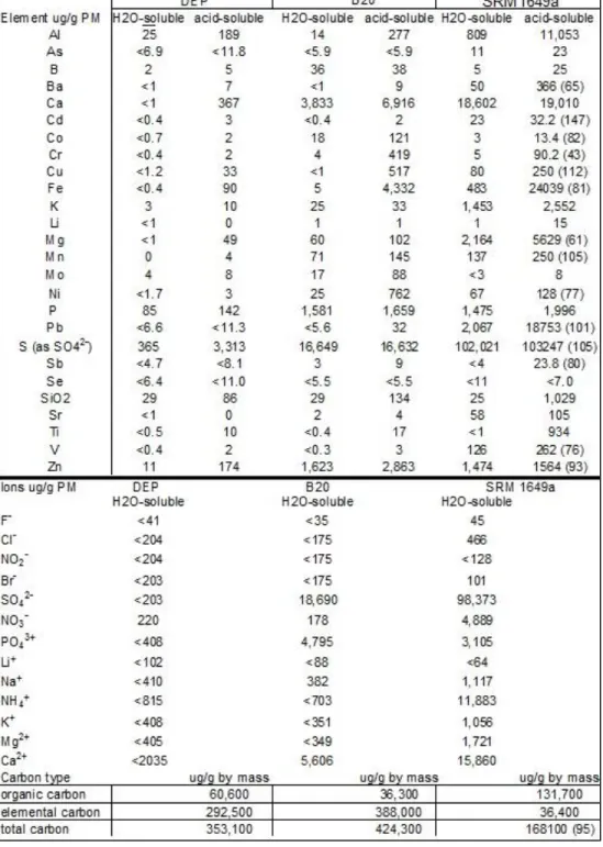
Table 2.1. Characterization of B20 and Diesel by ICP- Plasma OES. ... 46
Table 3.1. Diesel particle composition varies from both B20 and B100. ... 67
Table 3.2. Particle bound fatty acids totals elevated with B100 combustion. ... 68
Table 3.3. Total particle carbonyls increased with biodiesel and biodiesel blend. ... 69
x
LIST OF FIGURES
Figure 1.1. Proposed mechanism of cardiovascular injury with PM exposure . ... 5
Figure 1.3. Percent change of typical combustion emissions of Diesel, soy B50 and B100... 15
Figure 1.4. Arachidonic Acid Metabolism Pathway and Metabolites ... 25
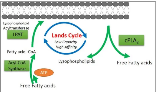
Figure 1.5. Land’s Cycle: Deacylation and reacylation cycle regulating membrane phospholipids/lysophospholipid ... 28
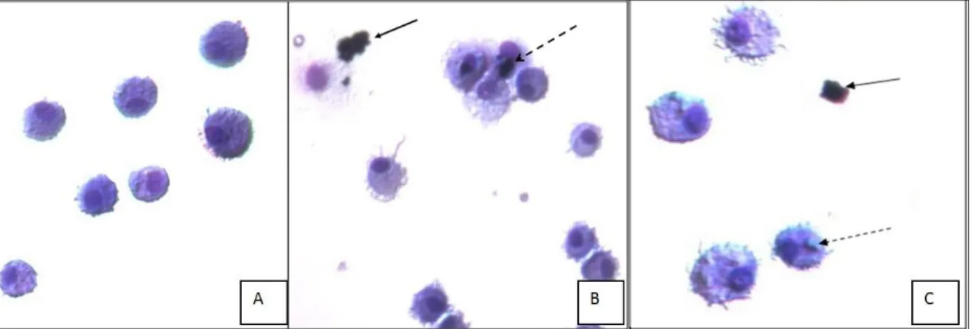
Figure 2.1. DEP and B20 particle suspension with AM. H& E stain of WKY AM with increasing B20 particle concentrations. ... 47
Figure 2.2. Cell cytotoxicity of WKY AM after 24h exposure to DEP and B20 is dose dependent ... 48
Figure 2.3. Gene expression of inflammation markers, COX-2 and MIP-2,
increased with 24hr exposure to DEP and B20 in WKY AMs ... 49
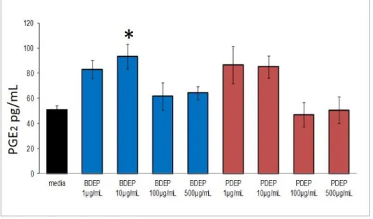
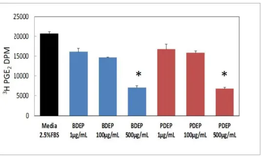
Figure 2.4. Increased PGE2 release with lower concentrations of B20 but not diesel ... 50
Figure 2.5. Diesel and B20 particle sequestering of PGE2 is equivalent. ... 51
Figure 3.1. Variable peaks detected from DEP, B20 and B100 Tricaprylin
extractions with abundant species detected from B100 ... 71
Figure 3.2. DMSO extractions with DEP, B20 and B100 found increased nonpolar species with B100 from PDA- detection of peaks in chromatogram ... 72
Figure 3.3. EA.hy926 cell cytotoxicity from exposure to different solvent extractions and particle types ... 73
Figure 3.4. Water extractions of DEP, B20 and B100 had poor non-polar species
detected in suspensions ... 74
Figure 3.5. Hexane extractions of DEP, B20 and B100 detected identical species and quantities among all samples ... 75
Figure 3.6. Acetonitrile extractions of DEP, B20 and B100 detected similar number and type of species in all samples. ... 76
Figure 4.1. Cell viability and toxicity of DEP exposures of increasing dose and time do not vary from B100 exposures ... 95
Figure 4.2. B100 exposure produced significant decrease in cytokine production with 4,6 & 8hr exposure. ... 97
xi
Figure 4.4. B100 exposure induced prostacyclin production is independent of COX-2
expression and activity. ... 99
Figure 4.5. Acyltransferase function increased with B100 exposure but not with DEP. ... 100
Figure 4.6. HUVECs exposure to stearic acid and oleic acid finds increased
acyltransferase transcripts and increased COX-2 ... 101
Appendix Figure 1. Chelex 100 Resin-treated extract exposure for 6hr to HUVECs ... 115
Appendix Figure 2. Schematic of intracellular responses to stimulus and the time it takes for detection. ... 116
xii
LIST OF ABBREVIATIONS
A549 Human Lung Adenocarcinoma Epithelial Cell Line
ANOVA Analysis of Variance
AP-1 Activating Protein-1
AA arachidonic acid
AHR acryl hydrocarbon receptor
AM Alveolar macrophages
B100 biodiesel exhaust particle
B20 biodiesel blend 20%
β-actin Beta-Actin
BSA Bovine Serum Albumin
cDNA Complementary Deoxyribonucleic Acid
CRE calcium response element
CV Cardiovascular
DEP Diesel Exhaust Extract
DMEM Dulbecco’s Modified Eagle’s Medium
DMSO Dimethyl Sulfoxide
DNA Deoxyribonucleic Acid
DPI Diphenyleneiodonium
xiii
EA.hy926 Hybrid Epithelial and Endothelial Cell Line
ECL Enhanced Chemiluminescence
EDTA Ethylene Diamine Tetraacetic Acid
EGM-2 Endothelial Growth Medium
ELISA Enzyme linked immuno staining assay
EPA Environmental Protection Agency
EC/OC elemental carbon /organic carbon ratio
GC-MS gas chromatography and mass spectrometry
HPLC High pressure liquid chromatography
HUVECs Human Umbilical vein endothelial cells
FBS Fetal Bovine Serum
HCAEC Human Coronary Artery Endothelial Cells
HCL Hydrochloric Acid
HO-1 Heme Oxygenase-1
H2O2 Hydrogen Peroxide
HRP Horseradish Peroxidase
HUVECs Human Umbilical Vein Endothelial Cells
ICAM-1 Intercellular Adhesion Molecule-1
IgG Immunoglobulin G
xiv
IL-6 Interleukin-6
IL-8 Interleukin-8
LD 50 lethal dose fifty percent
LDH Lactate Dehydrogenase
LPCAT Lysophosphatidylcholine acyltransferase
mRNA Messenger Ribonucleic Acid
NaCl Sodium Cloride
Nrf2 Nuclear Factor (Erythroid-Derived 2)-Like 2
NF-κB Nuclear Factor-Kappa B
NOx Nitric Oxide species
PAH polycyclic aromatic hydrocarbon
PBS Phosphate Buffer Saline
PCR Polymerase Chain Reaction
PGH2 Endoperoxide prostaglandin h2
PLA2 Phospholipase A2
PLC phospholipase C
PM Particulate Matter
PM10 Coarse Particulate Matter
PM2.5 Fine Particulate Matter
xv
ROFA – residual oil fly ash
ROS Reactive Oxygen Species
RT-PCR Reverse Transcriptase Polymerase Chain Reaction
SDS Sodium Dodecyl Sulfate
SHR Spontaneously hypertensive rats
SOD Superoxide Dismutase
TF Tissue Factor
TNF-α Tumor Necrosis Factor-Alpha
TM Thrombomodulin
UF Ultrafine Particle
UPLC Ultra high performance liquid chromatography
VCAM-1 Vascular Adhesion Molecule-1
1
Chapter 1.
Introduction
1.0. Air pollution and human health effects.
Re-examining historical air pollution events illustrates the evolving impact of air pollution on
human health. Adverse human health effects due to air pollution were recognized as early as the dawn of
industrialization, around the 1860s. However, only after several catastrophic historical events during the
post-industrial revolution in the 1950s was air pollution finally measured and stringently assessed.
Increased urban air pollution, combined with stagnant-winter air, led to the “Great London Smog” in
December 1952. For five days, an impenetrable smog settled on the city of London, during which the
death toll dramatically increased, which established a recognizable pattern of air pollution
inhalation-induced mortality. The smog accumulation was a mixture of sulfur dioxide and other persistent chemical
air pollutants produced by burning coal and wood. The five-day period of smog resulted not only in
fatalities, but also increased hospitalizations associated with fatal pulmonary infections [1]. This was one
of many global historical events that was used as a documented case study to illustrate the human health
outcomes of unregulated air pollutants in urban industrial settings.
An example of a national historical event, occurred in 1948 in Donora, Pennsylvania, where air
pollutants from a zinc smelting factory were linked to around 6000 hospitalizations and hundreds of local
deaths [2]. This was a national case study showing clear association of the health dangers with air
pollution from industry and manufacturing. Although the zinc factory was shut down due directly to the
health impact of poor air quality, no immediate national action was undertaken regarding air pollutants.
Decades later, in the 1960s, the federal government decided to address air quality by setting limits on air
pollutants. The Clean Air Act of 1963 was the first federal legislation to regulate air pollution in the United
States [3]. The Act was expanded in 1967 to include the right to monitor and control pollution levels.
2
(NAAQS), which set emission levels for six “criteria” air pollutants. Criteria air pollutants were defined as
air pollutants found in high concentrations in smog. These are ozone, lead, sulfur dioxide, nitrogen
oxides, carbon monoxide and particulate matter (PM). The NAAQS regulate the levels of these six criteria
pollutants, but there is a larger collection of pollutants listed as Hazardous Air Pollutants (HAPs) which
have established federal guidelines and standards. Together, the HAPs and NAAQS, nationally regulate air
pollutants with careful consideration of new toxicology evidence to minimize harmful health effects.
Air Pollution PM Classifications by size.
The six criteria air pollutants have been evaluated regarding acceptable levels of exposure.
Particulate matter (PM) specifically, was further regulated based on size. There are three size
classifications, of which two are individually regulated. The first is PM10, or coarse particulate matter, less
than or equal to 10µm in aerodynamic diameter. The current NAAQS standard is 150 µg/m3 over a
24-hour period [4]. The second regulated class of PM, is PM2.5, or fine particulate matter, less than or equal
to 2.5 µm in aerodynamic diameter. Its current NAAQS standard recently changed from 15µg/m3 to
12µg/m3 because new evidence indicated acute exposure was found to be strongly associated with
adverse cardiovascular events (NAAQS US EPA 2013). The third is ultrafine particles (UFPs), defined as
particles less than or equal to 0.1 µm in diameter, which makes them susceptible to agglomeration [5].
Currently, there is no ambient air standard-regulated level for UFPs. The standards set by the NAAQS are
federal limits, but some states have their own standards that are lower than the federal limits. In addition
to size, PM is also classified as primary or secondary. Primary PM is defined as the fraction of PM that is
emitted directly into the atmosphere; secondary PM forms from photo oxidation in the atmosphere when
PM reacts with pre-existing gases such as SOx and NOx. Ambient air pollution is also classified by the
source because PM measurements have previously found that anthropogenic sources generate smaller
PM (<PM2.5) than natural sources, such as lightning [4]. Densely populated urban regions are considered
to emit the largest volume of anthropogenic PM [4]. PM classification allows for increased accuracy when
3
Epidemiological studies establishing ambient PM induced adverse health effects including
cardiovascular events
An extensive air pollution epidemiology study called the “Harvard Six Cities Study,” which began
in 1974, was critical in establishing an association with ambient PM and adverse health effects. The study
correlated air pollution effects with mortality in approximately 8,000 adults in six U.S. cities classified as
either polluted or less polluted. The most polluted urban cities had 89.0µg/m3 PM for a 24hr sampling
period and the least polluted had one third the PM concentration of the most polluted. The study lead by
Dockery et al [6] found that cities with a higher concentration of ambient PM, such as Steubenville, Ohio,
had an increased rate of mortality compared to cities with lower ambient PM levels (e.g., Portage,
Wisconsin) [7-9]. Data from this pioneering study along with several hundred additional epidemiology
studies, since the early 1990s, indicate that increased concentrations of PM are clearly linked to a large
number of PM-associated deaths [4].
The direct association between air pollution PM exposure and cardiovascular disease is critical to
understand, since heart disease is now the number-one cause of death worldwide [10]. Air pollution
epidemiology research has long established a strong correlation between increasing PM exposure and
adverse cardiovascular health effects. Field epidemiology studies indicated a positive correlation between
increasing ambient PM (i.e., PM10) and an increase in hospital admissions associated with respiratory and
cardiovascular injury [2, 11]. This study noted not only the increase in hospital admissions but also found
1.4% increase in cardiovascular-related deaths (e.g. myocardial infractions or stroke) with 10µg/m3 mean
daily average of PM10 suggesting that there are links between increased PM and adverse cardiovascular
events [7]. Study participants involved in the Multi-Ethnic Study of Atherosclerosis (MESA) cohort,
exhibited changes in blood pressure from exposure to an average of 10µg/m3 of PM2.5 daily for 30 days
[12]. In this study, the significant increase in systolic blood pressure (SBP) was strongly associated with
subjects who lived within 300m of a highway, yet subjects who lived more than 400m did not show
significant blood pressure changes [12]. This study suggests that highway generated PM is crucial to
4
PM collected in close proximity to Los Angeles highways. This study of Los Angeles residents showed
significantly increased carotid intima-media thickness (CIMT), an atherosclerosis marker, in healthy
subjects with prolonged daily exposure to elevated PM concentrations (e.g. PM2.5 approximately 10µg/m3
collected near a highway) [13]. Based on these studies, epidemiology evidence supports PM exposure
induced cardiovascular injury. These include both physiological changes (e.g., blood pressure and heart
rate) and pathological changes (e.g., intima-media thickness). In addition, these studies find that the
source of PM (e.g., highway PM) can impact the intensity of cardiac biomarkers.
Study of PM near roadways and highways drew focus to the traffic–generated PM effects
originating from cars and trucks. Epidemiology studies focused on cardiovascular events associated with
traffic-generated PM identified increased cardiac injury associated with pathological (e.g., plaque buildup)
and physiological (e.g. heart rate variability) alterations. A field study conducted in the Netherlands [14],
indicated residents living within 50m from a major roadway have increased risk of cardiopulmonary linked
mortality. In this study the close proximity to a roadway correlated with 95% greater risk of mortality
after normalizing for factors like age, smoking habits, diet and regional poverty [14]. Another study, with
subjects in occupations requiring large amounts of time in or near highways (e.g., taxi drivers), found
increased intima-media thickness of the carotid artery [15]. This study further supports the role of PM
emissions from vehicles as a source of adverse cardiovascular effects.
In addition to measuring the intima-media thickness, one study measured coronary artery
calcification (CAC). This was assessed by electron beam-computed tomography, which measured the
plaque size. The study found of 4,814 subjects living near a major roadway (i.e., <200m from roadway)
were associated with increased CAC after normalization for extraneous factors (e.g., income, age) [16].
Based on occupational exposures and field studies, traffic PM significantly increased plaque buildup which
can potentiate atherosclerosis or disrupt vascular tone. These studies support the hypothesis that
traffic-generated PM can contribute significantly to mortality and cardiac injury markers.
The traffic-generated PM exposures mediated both pathological and physiological responses. The
composition of traffic-generated PM may play a critical role in certain biological responses. Traffic-
5
it also includes non-combustion -derived compounds (e.g., dust gathered by wearing down of
components from automobile brake linings and tires) and environmental bio aerosols entrained from the
ground (e.g., mold, pollen or bacteria) [17]. Air pollution researchers now have data associating PM with
critical mechanistic factors involved in potential adverse cardiovascular health outcomes.
6
2.0. Proposed particle exposure induced mechanisms of cardiovascular injury.
PM exposure may induce biological responses by a combination of several modes of action.
Inhalation of PM may induce adverse cardiovascular injury from particle and also from soluble compounds
on the surface of particle, as illustrated in Figure 1.1[5]. Particle inhalation results in deposition of
particles within the lung alveolar regions. Deposited particles and soluble compounds can generate
adverse cardiovascular events via three critical routes (Figure 1.1), which are not mutually exclusive and
can act concurrently. The first proposed PM-induced route of injury generates autonomic nervous system
imbalance, that results in adverse cardiovascular events including arrhythmias and sympathetic nervous
system mediated disruption of vascular tone. A second proposed PM-induced route is initiated with
respiratory and peripheral cellular responses resulting in transport of inflammation mediator into
circulation, that results in endothelial cell disruption and potentiating cardiovascular injury. The last
proposed route relies on soluble compounds from PM dissolution, thus translocating into circulation to
initiate endothelial cell injury potentiating into adverse cardiovascular events. All three routes initiate a
cascade of precursory events resulting in disruption of vascular tone, endothelial dysfunction, and plaque
buildup. These precursory events, along with PM-induced systemic inflammation, lead to a complex
multi-layer cardiovascular response. PM-induced cardiac injury is mediated by several critical steps and
exploration by air pollution researchers has established PM- soluble compounds induced both
physiological and pathological responses.
PM deposition and translocation.
Inhaled-PM deposition in the lung is a critical initiating factor for PM translocation and component
solubilization. PM deposition into the lung is primarily determined by a particle’s aerodynamic diameter.
Empirical data, along with predictive modeling data, can help determine the location of deposition of PM
with variable diameters. Volunteers inhaling, 5µm diameter particles had the largest deposition fraction
(40%) in the alveolar region [18]. The smaller 3µm particles had less (i.e. 50%) total lung deposition, but
with similar total percent (42%) deposition in the alveolar region, suggesting particle sizes resulted in
similar total deposition of the alveolar region. However with even smaller diameter particles, in a separate
study, there was a larger percent total deposition in the same region [19]. This study was also conducted
7
alveolar deposition of particles from inhalation exposure [19]. These studies found that total deposition at
the alveolar region varies with particle diameters, suggesting that there is a higher concentration of
0.04µm PM retained near the alveolar region of lung, in humans.
Recent predictive modeling programs have generated reliable accurate data regarding PM
deposition and translocation. The modeling data indicated particles of larger diameter measured between
2 to 10µm deposit primarily in the nose and upper airways [20] and smaller particles (e.g., fine, ultrafine
PM) deposit deeper in the alveolar regions [20]. The combined experimental and predictive modeling
data found PM total and regional deposition is based in part on size, but the translocation from the site of
deposition might be influenced by other factors such as PM solubility, size, phagocytosis of PM by alveolar
macrophages and clearance.
PM translocation is a topic of great debate since it is unclear if the whole particle, fragments of
particle or soluble compounds from particle diffuse into peripheral tissue, such as the vasculature, after
initial inhalation exposure. PM potential translocation of ultrafine particles was addressed in an in vivo
rodent study. In this study, inert particles, TiO2 nanoparticles (~20nm), were initially intratracheally
instilled and recovered after 1hr exposure from broncholalveolar lavage fluid (BALF). A little less than
20% of the total deposited PM amount was estimated to be recovered, based on measurement of
amounts in broncholalveolar lavage fluid (BALF), suggesting that majority translocated into pulmonary
tissue or extrapulmonary tissue or remained in AM after phagocytosis [21]. This study also found TiO2
present predominantly in the alveoli indicating that nanoparticles can likely translocate into the periphery
or they would be recovered in BALF. A second study with both inhalation and instillation of plain carbon
particles into animal lungs demonstrated carbon particles (20-29nm in diameter) were also taken up by
the epithelial cells and were re-distributed into lipophilic layers [22]. In addition, dog autopsies found
inhaled aerosolized ultrafine particles may translocate into circulation through the pulmonary capillaries
and lymphatics to deposit in the lung and peripheral organs [23]. Several studies observed particle
inhalation resulted in detection of PM, not only in lungs, but within the vascular lining, liver and olfactory
8
PM may result in translocation of whole particle into the peripheral tissues and organs, thus possibly
validating the proposed mechanism of action of PM in Figure 1.1.
Experimental evidence of soluble PM constituent – induced responses.
As previously stated in the Figure 1.1, cardiac injury could result from soluble constituents of
inhaled PM. Evidence that soluble constituents from PM induce cellular effects was identified in an in vivo
study comparing pure carbon particles with diesel exhaust particles, which contain several soluble
compounds. This study identified significant response differences from pure carbon particles with diesel
exhaust particles. The 4 hr rodent inhalation exposure to diesel exhaust particle and carbon black
particles at a relatively high ambient concentration (500µg/m3) resulted in elevated levels of endothelin-1
(ET-1) and endothelin -3 (ET-3) in plasma that is typically associated with constriction of the vascular
tone response [26]. Diesel particles, but not carbon black particles, altered hemodynamics. Also
evaluated in this study were water washed ambient Ottawa particles that had polar organic compounds
removed but other compounds remained [26]. These treated particles also caused initial ET-1 and ET-3
increase in plasma after exposure, but changes in blood pressure or heart rate were not found. This
study indicated that both particles and the non-water soluble compounds adsorbed to the particle are
likely responsible for hemodynamic plasma signal changes in rodent exposures [26]. Similarly, in a study
using rodent models of hypertension, particles with and without metals were evaluated for an
inflammation response. In this study, hypertensive rats had exposure to two different PM-with and
without metals, and also polycyclic aromatic hydrocarbons (PAHs). Exposure via intra-tracheal instillation
resulted in increased inflammation response that was significantly correlated only with particles
containing metals [27]. These findings highlight the importance of the soluble PM bound chemicals in
generating robust cellular responses.
Evidence from inhalation exposure of systemic responses and release of mediators.
Also shown in Figure 1.1, Brook et al hypothesized that soluble mediators that result from local
inflammation spread to induce a systemic response which subsequently leads to adverse cardiovascular
9
significant however the collection of soluble mediators can be evaluated for biological significance by
exposing the plasma to cell cultures in vitro. Plasma was collected from a human exposure study to dilute
diesel exhaust. The dilute plasma contained markers that were capable of activating an inflammatory
response in culture (i.e., human coronary artery endothelial cells). Incubation with the dilute plasma
resulted in increased cellular adhesion molecules and interleukin -8 (IL-8) [28]. Evaluation of results from
this study possibly suggest there are mediators of inflammation that were released into the periphery
post-diesel exposure, resulting in a systemic inflammatory response. There is a possibility that
compounds from the diesel exhaust translocated into plasma and the response is linked to the
compounds, but no measurements were made of metals or other organic substances in plasma.
Additionally, the probability is relatively low for soluble mediators, considering that the inhalation
exposure was 106µg/m3, which is three-fold lower than common diesel exhaust exposure concentrations
that previously measured inflammation responses. Measurement of PAHs in urine and exhaled breath
condensate from these diesel exhaust-exposed subjects showed that the diesel exposure did not result in
PAH increase relative to the air control exposure suggesting a low inhaled dose [29]. There is greater
probability that the increased adhesion molecules and IL-8 are a result of inflammatory mediators
released from the lung or peripheral vascular tissue, validating a hypothesis of systemic response. For
further examination of the theory presented in Figure 1.1, air pollution researchers are focused on
characterizing the PM-associated compounds and finding specific cellular responses that may lead to
cardiovascular injury.
3.0. Diesel and Biodiesel exhaust composition and response comparisons.
Anthropogenic PM2.5 concentrations across the U.S, in a 24hr period, were concentrated in
high-traffic urban settings [4] and calculated to originate from diesel emissions. In a 2012 study conducted in
Hong Kong, 43% of total PM2.5 were traced back to idling diesel-powered naval vessels in the shipyard
[30], demonstrating that globally diesel-combustion generates the most PM2.5. Recently reduced PM
emission standards for mobile diesel engines (i.e., 0.01 g per brake horsepower-hour [g/bhp-h]) forced
introduction of new emission-control technologies such as diesel particle filters and improved fuel
10
species [4]. Historically urban traffic-generated pollution was composed of exhaust emissions from diesel
and gasoline [4]. However in 2012, the U.S government passed the Energy policy Act expanding the
renewable fuels initiative that increased production of biodiesel (BD). As of 2014 the national biodiesel
production reached a record high of 1.8 billion gallons, exceeding the proposed levels stated in the 2014
Renewable Fuel Standard Rule. The global volume of all feedstock biodiesel produced and consumed is
steadily increasing in billions of liters every year [31]. The most common commercially available biodiesel
is however a blend - B20, which is 20% soy biodiesel and 80% petroleum diesel [32], which has been
successfully utilized in the U.S transportation industry. Biodiesel powered trucks are increasing in
popularity yet exhaust composition analysis remains as complicated as diesel and requires further
research regarding emissions chemistry and potential health effects. Diesel combustion exhaust is
composed of particles and gases consisting of
hundreds of different chemical components. In
general, the gas-phase volatiles in diesel exhaust
include olefins (1, 3-butadiene), aromatics
(benzene, ethylbenzene, toluene, and xylenes),
PAHs (including nitro-PAHs and oxy-PAHs),
alkanes, oxides of nitrogen, carbon, and sulfur
and carbonyls. Common PAHs found in diesel
exhaust are phenanthrene, fluorenes,
naphthalenes, pyrenes, fluoranthrenes [33, 34]. The particles emitted from diesel combustion typically
consist of a carbon-rich core with adsorbed chemicals on the surface as illustrated in Figure 1.2 [35].
Common surface compounds include metals, ions, organics and incomplete-combustion species
[36]. Transition metals such as Fe and Zn are abundant in both diesel and biodiesel exhaust particles
[37]. PAH are more abundant in diesel exhaust compared to biodiesel exhaust emissions [38]. However,
unique to biodiesel combustion are whole (C18) and fragmented (C3- C4) fatty acid methyl esters
(FAMEs). Biodiesel emissions release an abundant amount of FAMEs even though biodiesel emits fewer
11
GC-MS analyses of biodiesel combustion exhaust identified several FAME species [34, 40]. These
species are likely incomplete combustion products from the original fuel. Biodiesels are typically created
by a chemical reaction between plant oils or animal fat and an alcohol, typically methanol, in the
presence of catalyst, which generates FAMEs. An alternative for methanol is to use enzymes to generate
FAMEs [41]. Inefficiencies in the biodiesel-refining processes after transesterification result in a fuel with
impurities consisting of plant oil triglycerides, glycerol and intermediate-reaction products (e.g., fatty
acids) that can be subsequently emitted in exhaust. The fuel is the source of the unique emission
compounds found with biodiesel PM exhaust. There are hundreds of chemicals that are also released in
the exhaust with known biological or health effects that are not linked directly to the unburned fuel. In a
U.S. Environmental Protection Agency (EPA) summary report, comparison of biodiesel and diesel exhaust
emissions shows that hydrocarbons (HC), PM and CO decreased over 10% compared to diesel neat, as
displayed in Table 1.1 [4]. The differences in the exhaust emissions between diesel and biodiesel are
important to understand because health effects can be directly associated with exhaust composition.
Current research on PM-induced health effects generally has examined singular chemical compounds and
cellular responses to assess potential risks with human inhalation exposure, and the responses to some
inorganic (e.g., transition metals) and organic (e.g., PAHs) chemicals are discussed below.
12
Metals in emissions linked to oxidative stress response.Specific cellular responses have previously been associated with individual chemical species
emitted in exhaust, such as oxidative stress with metals in the emissions. Metals are typically emitted
from the combustion of petroleum diesel and biodiesel, and have been predicted to originate from both
the fuel and engine lubricants [42]. Several studies have detected from particle analysis both petroleum
diesel and biodiesel exhaust contain metals [27, 43]. The most common metals found include Zn, Fe, Mn
and Cr [44] from both diesel and biodiesel, however, Zn was noticeably elevated in biodiesel. Respirable
Zn particles were previously identified to deposit within the lung and disperse into the cardiac tissue. A
rodent exposure study, found stable Zn isotopes in murine extrapulmonary tissue (i.e., cardiac tissue)
after intratracheally instillation [45], suggesting PM-associated metals can be inhaled and translocate
from the lung into the cardiac tissue. Like Zn, other transition metals can induce intracellular oxidative
stress from production of reactive oxygen species (ROS) that have unpaired electron(s) in their outer
valance shell. ROS products consist of free radicals and non-radicals but the non-radicals include species
with high oxidizing potential that modify proteins and lipids and generating cellular toxicity. ROS
generation and induction of lipid peroxidation species has been observed with in vitro exposures of
residual oil fly ash (ROFA), a metal-rich PM. In this exposure study with lung epithelial cells, ROS
products were significantly increased as were inflammatory mediators from arachidonic acid metabolism
such as prostaglandin E2 (PGE2) and 15-eicosatetraenoic acid [46]. Another ROFA exposure study with
alveolar macrophages (AMs) found increased ROS products and pro-inflammation markers [47-49],
suggesting lung epithelial and AMs are sensitive to oxidative stress induced by metals. The metal-rich
ROFA also increased expression of pro-inflammatory markers, IL-8, IL-6, and tumor necrosis factor-α
(TNF-α) in bronchial epithelial cells [50, 51]. In a whole animal exposure study comparing PM that is
metal rich compared to PM without metals, results indicate greater inflammation response in the BALF,
with metal rich PM exposure [27]. Further evidence of metal-rich PM-induced oxidative stress comes from
studies in which the metals were removed. Metal chelation of ambient PM with deferoxamine resulted in
significant reduction of oxidative stress responses and reduced antioxidant levels [52, 53] indicating that
13
responses [54]. PM from biodiesel and diesel combustion include soluble compounds such as metals that
may translocate to the periphery, where they initiate pro-inflammation and oxidative stress responses,
potentially leading to adverse cardiovascular events. Particle-bound metals are potent soluble mediators
of oxidative stress and pro-inflammatory responses that are commonly found in both diesel and biodiesel
exhaust exposure.
PAH emissions and cellular responses.
PAH are generated and emitted from diesel and biodiesel combustion [34, 55] and induce
variable cellular responses. A recent review of studies involving subjects with prolonged occupational
exposure to PM rich in PAHs, concluded that there is increased risk of lung cancer and significantly
increased levels of chromosome aberrations in populations with greatest exposure [56]. Interestingly,
most of the studies were conducted in Europe where diesel fuel use is more common than in the U.S and
diesel emits more PAHs relative to gasoline and biodiesel [57]. In vitro mechanistic studies with diesel
exhaust particles (DEP) identified that PAHs bind to the acyl hydrocarbon receptor (AHR), initiating
nuclear translocation and dimerization with ARNT. The dimerization binds and activates target sequences
such as hydrocarbon response elements or antioxidant response elements. Thus, initiating downstream
inflammation and oxidative stress-signaling in cells. PAHs commonly react with other species in the
cellular environment. For example, quinones generated from PAH components undergo cyclic-reduction
reactions with oxygen in the cells, followed by oxidative coupling with NADPH, resulting in the formation
of extremely unstable semiquinone radicals that can also initiate oxidative stress. PAH mode of action is
complex and can activate multiple pathways within cells. An in vitro study examined the methanol
extractable DEP compounds that include hydroxy-PAHs, carboxy-PAHs, nitro-amino-PAHs and found
inflammogenic responses [58]. In this study bronchial epithelial cells released elevated levels IL-6 after
exposure to the most polar extract collected from DEP, suggesting PAHs derivatives potentially induce
inflammatory responses and have increased cytotoxicity relative to the parent PAHs [58]. Recent
toxicology efforts to establish a mode of action for diesel found similar pathways activated with diesel and
individual PAH compounds suggesting the PAH content of diesel is substantial enough to induce similar
14
FAME induced cellular responses.Biodiesel exhaust emissions have elevated amounts of fatty acids methyl esters (FAMES) and
fragmented fatty acids methyl esters, relative to diesel based on GC-MS analysis [34]. There is limited
data associated with cellular responses and health effects due to FAME exposure and therefore biodiesel
exhaust induced health effects remains unclear. An in vitro study comparing saturated fatty acids with
methylated fatty acids (18:1) and (18:3) found that methylated fatty acids were unable to translocate
iron (i.e., 59Fe isotope) into pulmonary artery endothelial cells, while saturated fatty acids (18:1 and
18:3) were able to increase intracellular iron significantly [59]. The exposure of the cells to the C18:1 and
C18:3 fatty acids induced cytotoxicity likely due to increased iron transport into cells, suggesting
exposure to saturated fatty acids but not methylated analogues generated adverse effects. However, in a
recent study, methyl palmitate has been discovered in rat retinal tissue and was identified to function as
a vasoactive dilator, based on the findings with rat aortic ring exposures [60]. Other than a few studies,
most research with FAMEs did not result in significant cellular alterations. But since FAMEs are one of the
most common PM-associated exhaust emissions from biodiesel, further research should be considered to
better address health effects. Exhaust emissions based cellular response research is vital in directing
areas of engine advancements to reduce harmful emissions species.
4.0. Engine conditions and fuel composition alter exhaust emissions composition potentially changing biological responses.
Reduction of PM can be achieved with modifications to combustion technology resulting in fewer
PM and thereby lessening adverse health effects from inhalation exposure. The physiochemical
characteristics of PM from petroleum diesel engine exhaust vary with fuel, engine conditions, fuel
additives and after-treatments fitted to an engine. Emission analysis of biodiesel indicate that biodiesel
blend emissions vary from the combustion of biodiesel neat and blends. Studies generally indicated that
20% blends have different emissions than 30% and 50% biodiesel regarding the increasing volume of
organic soot (PM), volatile organic material (e.g., PAH) [39, 61] and CO emissions [62, 63]. The changes
15
CO are not necessarily directly proportional (i.e., linear) with the amount of soy-derived biodiesel added
to diesel, as shown in Figure 1.3 [64].
Figure 1.3. Percent change of typical combustion emissions of Diesel, biodiesel blend and B100 [65] . Physiochemical characterization of emissions species has found that different biodiesel feedstock
(type) fuels can generate unique emissions suggesting some biodiesel feedstock may be more toxic than
others. For example, the type of metals and PAH differ from combustion of rapeseed methyl esters
(biodiesel) blends [66], pure waste cooking oil [67, 68] and kanjar oil [42]. All three fuel types were
combusted under similar European driving-cycle standards before exhaust was analyzed using ICP-OES
for metals. The results indicated increased metals in plant-based biofuel and fewer metals with waste
cooking oils, but the opposite is true for PAH emissions. A general lack of consistency regarding run
conditions may prevent the uniformity in cellular responses from combustion emissions. One common
16
high- load engine raises the temperature, which causes emission of PM containing more elemental carbon
than organic carbon. A rodent study comparing toxicity from emissions of high-load and low-load engines
found significantly more oxidative stress, inflammation and increased sensitivity to viral infections in mice
exposed to PM from high- load engine run [55]. In a separate comparative in vitro study with normal
human bronchial epithelial cells (NHBE), exposure to high and low load combustion emissions resulted in
increased expression of IL-8, a pro-inflammatory marker. Though, inflammation doubled with particles
emitted from high-load emissions compared to low-load emissions [69]. These studies indicated increased
inflammation response from exhaust emissions generated with load engine conditions. The
high-load exhaust is typically characterized with enriched elemental carbon relative to low-high-load PM, suggesting
combustion conditions affect exhaust chemicals that can interfere with cellular responeses.
Lastly, the addition of fuel additives resulted in exhaust that reduced inflammatory responses.
Incorporation of fuel additives, such as iron based Satacen 3, improved the cetane value of the fuel and,
reduced incomplete combustion emissions and PM. Human airway epithelium-derived cells exposed to
emissions from fuel with and without Satacen 3 produced significant decreases of cytokines IL-8 and
TNFα with fuel additives [70]. Not only were the exhaust emissions different, but some changes in
exhaust also correlated with different cellular responses.
5.0. Clinical and Toxicological studies involving Diesel and Biodiesel exhaust emissions.
Hundreds of epidemiology studies with both ambient and occupational exposures, and validated
findings from controlled human-exposure studies to diesel exhaust whole (DE) and DEP alone,
convincingly indicated an association with adverse respiratory and cardiovascular outcomes. Exposure-
induced responses ranged from increased neutrophils, alveolar macrophages and pro-inflammatory
markers in the lung to vascular-tone alterations mediated by endothelial cells. Controlled
human-exposure studies with healthy human volunteers and DEP found increased lung neutrophils in sputum
and elevated inflammatory markers in bronchial washes [71, 72]. In one study with healthy young
volunteers exposed to 300 μg/m3 of DEP (for 1 hour), neutrophils increased significantly in both the
bronchial epithelium and peripheral blood, suggesting an inflammatory response expanded from the site
ICAM-17
1, VCAM-1, and platelets in blood from diesel-exposed but not air-exposed subjects [73, 74].
Pro-inflammatory gene expression levels of IL-8, IL-18, IL-13 and IL-10 increased more than two-fold in the
bronchial epithelium after 6 hr exposure to diesel, further supporting signaling of neutrophil recruitment
[73, 74]. Even 1-hour DEP exposure with 300 μg/m3 resulted in signaling increase of pro-inflammatory
cytokines IL-6 and TNFα in plasma [75], possibly indicating inflammatory markers can migrate into the
vasculature or may be induced by extrapulmonary tissues due to downstream responses to DEP
exposure.
Human exposure studies found diesel PM not only impact lung inflammatory response, but also
can lead to changes in blood flow. One controlled human-exposure study with diluted diesel exhaust
containing only 100-200 µg/m3 PM found changes to vascular resistance, as measured by brachial artery
diameter [76]. In this study, the results showed a 1-13% luminal diameter decrease, which was
statistically significant with increasing diesel exposure concentrations. In a similar study of healthy male
subjects with increased DE exposure (300µg/m3) for 1 hour, venous occlusion plethysmography was used
to measure blood flow. Unlike the previous study, these subjects were given infusions of a vasodilation
agonist (e.g., endothelium dependent bradykinin and acetylcholine; endothelium independent sodium
nitroprusside and verapamil) and then analyzed for blood flow changes [77]. The results indicated that
DE exposure reduced the vasodilation response to bradykinin and acetylcholine but did not alter the
vasodilation to the endothelial independent agonists [77]. These results suggested
endothelium-dependent agonists like bradykinin are not as active or potent as endothelium-inendothelium-dependent agonists of
vasodilation after DEP exposure. This controlled human-exposure study suggests a disruption of
endothelial cell-mediated vasodilation response after diesel exposure.
The vascular effects of diesel are typically linked with PM and not as strongly with gas-phase
components. For instance, filtering out PM from whole exhaust resulted in attenuated vasoconstriction
and ex-vivo thrombus formation in humans [75]. This study suggested that a larger contribution of
particle fraction, as opposed to gas phase emissions, prompted differential vascular changes. In a
rodent-exposure study, evidence supporting particle- mediated vascular effects were evaluated using diesel and
ambient particles [26]. Results from this nose-only inhalation exposure identified increased endothelin
18
introducing any gas phase components [26]. This study validated that particles emitted from diesel
combustion contain compounds not found in gas phase or in carbon black particles that can potentiate
endothelin release. Additional support for diesel particle potency was previously linked to an inflammatory
response in lung neutrophil recruitment. In a study by Nightingale et al, diesel exhaust particle was
collected and the PM alone was suspended in a controlled chamber at 200 μg/m3 [78]. The subjects
were exposed to the diesel particle alone for 2hrs and the results indicated increased neutrophils in
sputum, suggesting that the particles alone induced inflammatory responses without including volatile
gas- phase emissions from diesel combustion [78]. The compositional differences from biodiesel and
diesel may help identify the individual chemical species involved in potential cardiovascular risk. Although
there are several inflammation and vascular tone changes previously examined with diesel exposure, the
exact pathways activated and the events leading up to the cardiovascular events are still unclear.
Recently new data was obtained from controlled human exposures to biodiesel-blend and
biodiesel neat exhaust regarding exposure- induced alterations of vascular tone [79]. Biodiesel, like
petroleum diesel, can also impair vasodilation in healthy human volunteers, based on measurements of
brachial artery diameter changes from ultrasound detection [77]. A clinical study involving healthy
volunteers with exposure to biodiesel-blend (B30) exhaust at 300µg/m3 and biodiesel neat at 106µg/m3
found impaired vasodilation response from significant number of subjects [79]. Additional results
indicated that the blood flow into the forearm remained constricted after infusion of
endothelial-dependent vasodilators such as bradykinin. In contrast, infusion of endothelial-inendothelial-dependent vasodilator,
such as vasopressin, was capable of detectable changes in diameter in Biodiesel exposed subjects,
suggesting that the impairment was localized to the endothelial cell mediator production or release [79].
Endothelial cell mediated vascular tone is regulated by multiple pathways and signaling molecules, but
the exact mechanism by which biodiesel exposure impaired vasodilation is not yet clearly understood.
Exhaust (i.e. both particle and gas phase emission) composition interference with the endothelial cells
mediated regulation of vasodilation signaling was a proposed mechanisms for biodiesel and diesel
19
6.0. Pathway specific diesel and biodiesel exhaust responses.Diesel and biodiesel PM-induced cardiovascular dysfunction was mediated by a few pathway
specific responses. Some of the known activated pathways include oxidative stress, pro-inflammation
signaling and alterations of endothelial cell-mediated vascular tone. Within each pathway exist several
different types of signaling markers that include peptides, proteins and lipids. These signaling markers
can be measured to assess the potency of each combustion PM. There are there multiple markers for
each pathway that can function independently or in concert with other markers to potentiate a singular
pathway response. Similarly, diesel and biodiesel PM exposure can initiate multiple pathways that may act
independently or in concert with other pathways to generate adverse cardiovascular events.
Oxidative stress response detection from exposure to biodiesel and diesel PM.
Oxidative stress pathway is one of several pathways activated with diesel and biodiesel exposure
and it is measured by changes in free radicals, antioxidants or lipid peroxidation species. Exposure
studies conducted in vivo and in vitro, measured DEP exposure induced cellular increases of free radicals
and depletion of antioxidants. In vitro AM exposure studies with diesel extract generated excessive
oxidative stress products such as H2O2 and O2- [80]. Simultaneously, some exposure studies show
depleted antioxidants and consequently increased gene expression levels. Common antioxidant enzymes
that neutralize the reactive oxygen species (ROS) are: hemeoxygenase (HO-1), catalase, peroxidase or
superoxide dismutase (SOD). Several studies with in vitro exposure to diesel and biodiesel (e.g., B99)
found elevated antioxidant mRNA and protein expression of SOD [81, 82]. Additionally studies have
found depletion of intracellular reduced glutathione as an indicator of oxidative stress. Mice pulmonary
exposure to DEP resulted in both decreased GSH and SOD in lung [81]. Markers of oxidative stress are
not limited to detection of free radicals and antioxidants but include lipid peroxidation species. Studies
with rodent exposures to DEP and gasoline emissions, resulted in elevated lipid peroxidation species
discovered in lung tissue [55, 83]. Lipid peroxidation products were also detected at high concentrations
in exhaled breath [84, 85] and urine post exposure to concentrated ambient fine and ultrafine PM [85]
20
(ROS) with exposure to both combustion PM from diesel and biodiesel suggesting there are some shared
pathways but verification requires observation of other markers for oxidative stress.
Isoprostanes are lipids, though not directly derived from AA, that affect the vascular system and
are frequently used as markers of oxidative stress. Specifically, 15-F2t-isoprostane (15-F2t-IsoP), has
been found to be activated within oxidative stress responses from exposure to diesel and biodiesel
exhaust [86]. Rodent exposure with 200µg/m3 of diesel PM2.5 for seven weeks resulted in significantly
elevated 15-F2t-IsoP levels in urine [87]. This study indicated elevated oxidized lipid changes due to
diesel exposure which can remain above baseline for over seven weeks. Thus, the data emphasizes the
continued production and robust longevity of lipids as markers of oxidative stress. ROS products if not
neutralized may initiate systemic inflammation signaling.
Pro-inflammation markers found with diesel and biodiesel exhaust exposure.
Diesel and biodiesel exhaust exposures have been identified to induce pro-inflammatory
responses in vivo and in vitro. DEP exposure induced significantly elevated expression levels of
inflammatory markers, IL-6 and TNFα, in human cell culture models as well as in human plasma [75]. In
one study, in vitro exposure of DEP particles resulted in significantly elevated mRNA levels of IL-1, after
only 2hrs of exposure, which indicated the cytokine production initiated post-phagocytosis by alveolar
macrophages (AM) [88]. A significant increase of cytokines (e.g., IL-6 and IL-8) resulted from
biodiesel-blend exposure to epithelial and endothelial cells [43, 89, 90]. Post endothelial cell exposure to biodiesel,
IL-8 mRNA levels were slightly increased [90], but not significantly suggesting the inflammation response
was not as robust in vascular cell culture models as it was in AM and human plasma. However, most
biodiesel and diesel exposures to date have indicated a consistent increase of cytokine mRNA and protein
[43, 90].
Potent de novo inflammation-signaling markers belong to a family of lipids called eicosanoids
(i.e., oxygenated AA products) which function primarily in signaling and recruitment of additional
inflammation mediators. One subfamily of lipid inflammation mediators, called prostaglandins, is
dependent on a canonical pathway involving arachidonic acid (AA) metabolism. AA is the precursor for
21
expressed in cells, and COX-2 formation is generally induced in response to inflammation. The COX-2
gene is co-activated by transcription factor activation of cAMP response element (CRE). Diesel exhaust
exposure-induced stress has been found to activate and increase COX-2 gene expression and protein. It
has been shown that DEP exposure of lung epithelial cells resulted in dose dependent increase of COX-2
protein and mRNA [91]. COX-1 and COX-2 are critical for enzymatic production of the lipid inflammation
and immune system mediator prostaglandin E2 (PGE2) from the precursor AA. Another study with diesel
extracts fraction exposure to bronchial epithelial cells found a combination of COX-2 protein and PGE2
production was significantly increased relative to control [69], suggesting that the COX-2 increase is
directly responsible for the increased PGE2 production and release. However, a separate contrasting in
vitro study with human alveolar macrophages found diesel PM exposure resulted in decreased PGE2 levels
[92]. The conflicting results were proposed to be related to particle binding of PGE2 which prevents
accurate detection. A complementary challenge with endotoxins resulted in a return of PGE2 to control
levels which indicated the sequestering by particle was reversible and no permanent damage to the PGE2
production occurred [92]. This study also signified the role of lipid mediators in inflammation responses.
Additionally, a study of diesel exposure to monocyte- derived macrophages that were co-simulated with
LPS, found significant increases of PGE2 release compared to cells exposed to only LPS or only DEP. Thus,
suggesting that there was synergy with LPS and DEP response resulting in significantly increased PGE2
levels [93]. These studies suggested that prostaglandins are detectable inflammation markers with DEP
extract and detection can be complicated with whole particle sequestering. Furthermore, there are other
lipid inflammatory markers derived from AA metabolism. Other AA metabolites are cell type specific,
unlike prostaglandins. Leukotriene B4 (LTB4) largely functions in lung cells. Its primary function is to
induce the adhesion and activation of leukocytes to the endothelium. One in vivo study found LTB4 was
altered with diesel exhaust exposure. In this two-day study, with mice, whole DE exposure resulted in
3-fold greater LTB4 levels in BALF relative to air [94]. Based on DE exposure induced changes to
production of these AA derived lipids signals (e.g., LTB4, PGE2, 12(S)-HETE), there was increased interest
22
Vascular tone alterations mediated with NO, ET-1 and PGI2Controlled human exposures to biodiesel blend (RME30), biodiesel neat and diesel indicated
impaired vasodilation response however the exact mechanism(s) remains unclear. Vasodilation is
regulated by the parasympathetic system, as well as by signaling molecules released from the kidney,
heart and endothelial cells (i.e., artery and vein). Researchers in this field are actively pursuing proof of a
mechanism of action related to the parasympathetic system and signaling molecules generated from the
kidney or heart. Additionally, the smooth muscle cells can respond to variable signaling molecules to
initiate calcium influx which results in calcium based relaxation response. As previously mentioned,
studies have recently identified that controlled human exposures to DE, B30 and B100 resulted in
impaired vasodilation and the mechanism behind this may be associated with vascular mediators
produced by endothelial cells [77, 79, 95]. The following studies identified that there are a number of
signaling molecules associated with endothelial cell mediated vasodilation which includes NO, ET-1 and
prostaglandins (PGI2) which maybe altered by diesel or biodiesel exhaust exposure.
Vascular dilation was regulated via NO, which was produced when O2 couples with an endothelial
NO generating enzyme (eNOS) to generate NO. Diffusion of NO into the nearby vascular smooth muscle
activates cyclic guanosine monophosphate (cGMP) and soluble guanylate cyclase (sGC) to generate
smooth-muscle relaxation. A diesel study with rodent exposure to 300µg/m3 for 5 hrs, resulted in NO
reduction. Additionally, post-exposure coronary arteries (CA) were isolated and resistance was measured.
The results found DE-exposed CA had significantly decreased dilation relative to air [96], suggesting that
DE exposure may impair relaxation response. In the same study, DE-exposed CA generated an increase
in superoxide anion (O2-), which decreased the ability of eNOS to uncouple thereby offering a potential
mechanism for NO reduction. This assay, along with the previous resistance measures, strongly suggests
vascular tone impairment was caused by exposure to whole diesel exhaust and involved NO-mediated
dilation impairment.
In a second study, aortic rings were isolated from Wistar Kyoto rats and exposed to DE extract
(100µg PM equivalent /mL concentration) [97]. The NO concentrations initially doubled from control
23
[97]. These results indicated reduced NO released from rat aortic endothelial cells post diesel exposure,
which suggested that NO reduction may explain impairment of vascular relaxation post DE exposure.
Endothelin-1 (ET-1) is a peptide which is known to induce vasoconstriction and is released
primarily from endothelial cells. ET-1 is the most researched endothelin isoform that acts on neighboring
smooth muscle to initiate constriction. The ET-1 isoform is the substrate released from endothelial cells
that binds to either ET A /B receptors (ETAR / ETBR) which are G-protein coupled receptors located on
smooth muscle cells. The ETAR is located on smooth muscle cells and when bound by ET-1 will initiate
the vasoconstriction process in smooth muscle cells. The ETBR is located on both on endothelial cells and
smooth muscle cells. The ET-1 activated ETBR receptor located on endothelial cells can initiate release of
other vasodilators (i.e., NO and PGI2), contrastingly ETBR on smooth muscle cells binds ET-1 and
activates vasoconstriction. The same receptor may induce opposite responses if ET-1 binds to receptor A
or B. Currently endothelin antagonists have therapeutic uses and function in reducing pulmonary
hypertension and arterial hypertension in patients [98, 99]. PM -induced changes in ET-1 release were
found in studies of rodent exposures to both urban dust (SRM1649) [100] and mixed-vehicle emissions
(e.g., gasoline and DE) [83, 101]. Increased circulating ET-1 levels in plasma suggested that an
endothelial cell response to PM exposure was to release ET-1.
An in vivo study conducted with rodent pretreatment with receptor antagonists 788 or
BQ-123, ETBR and ETAR antagonists respectively, prior to DE inhalation exposures found that only ETBR
antagonism resulted in diminished ET-1 mediated vascoconstriction. After 300µg/m3 DE exposure for 5
hrs resulted in lack of ETBR receptor activation in coronary arteries, suggesting that DE only inhibited the
activation via ETBR receptor [102]. The study indicated diesel exposure did not alter vasoconstrictive
responses, but receptor induced response (i.e., vasodilation) was inhibited. The results of these studies
suggest that the diesel exposure induced vasoconstriction response can be explained with mechanisms
involving ETBR and the impaired endothelial cell response to release NO and prostaglandin I2 (PGI2).
The endothelial cell-mediated potent vasodilation signal not yet discussed is the AA metabolism
derived lipid mediator called prostacyclin, PGI2, which is a member of the prostaglandin family. As shown
![Figure 1.1. Proposed mechanism of cardiovascular injury with PM exposure [5].](https://thumb-us.123doks.com/thumbv2/123dok_us/8302144.2198780/20.918.144.787.341.844/figure-proposed-mechanism-cardiovascular-injury-pm-exposure.webp)
![Figure 1.3. Percent change of typical combustion emissions of Diesel, biodiesel blend and B100 [65]](https://thumb-us.123doks.com/thumbv2/123dok_us/8302144.2198780/30.918.112.737.142.696/figure-percent-change-typical-combustion-emissions-diesel-biodiesel.webp)