હ
-Synuclein Interaction with Membranes
Imola G. Zigoneanu
A dissertation submitted to the faculty of the University of North Carolina at Chapel Hill in partial fulfillment of the requirements for the degree of Doctorate of Philosophy in the Department of Chemistry.
Chapel Hill 2011
Approved by:
Professor Gary J. Pielak, Ph.D.
Professor Joseph M. DeSimone, Ph.D.
Professor Mathew M. Redinbo, Ph.D.
Professor Linda L. Spremulli, Ph.D.
ii ©2011
iii
Abstract
IMOLA G. ZIGONEANU: α-Synuclein Interaction with Membranes (Under the direction of Professor Gary J. Pielak, Ph.D.)
The association of α-synuclein with membranes appears to be an important factor in Parkinson’s disease. My dissertation research was focused on
understanding the interactions of the protein with artificial membranes, the
extracellular plasma membrane, and intracellular membranes. Fluorescence and
nuclear magnetic resonance spectroscopy were used to monitor these
interactions. Large unillamelar vesicles with a composition similar to
mitochondrial membranes were studied. Cardiolipin, present mainly in the inner
mitochondrial membrane, is key for protein binding, and reducing the amount of
cardiolipin decreases binding. The nature of cardiolipin’s acyl chains is also
important; cardiolipin with chains containing one double bond interact more
strongly than those with chains having two double bonds or saturated acyl
chains. This finding is physiologically relevant for Parkinson’s disease because
cardiolipin containing fatty acids with one double bond are the most abundant
phospholipid in the brain. The affinity of α-synuclein for plasma membrane was
tested, and only N-terminal region of the protein binds. Also, 19F NMR proved
useful for monitoring the interactions of proteins and fused peptide-proteins with
iv
membranes reveals that the N-terminal region may be cleaved in cells, but
v
Dedication
To my family:
vi
Aknowledgements
Many people helped me on this pathway and I want to thank them. I want
to thank to Dr. Gary Pielak for accepting me in his group and for giving me the
opportunity to go to conferences where I had the chance to meet peers and
discuss interesting ideas. My colleagues in the Pielak’s lab were very good at
giving feedback and sharing their knowledge with me, especially Drs. Lisa
Charlton, Rebecca Ruf, and Kristin Slade who helped me at the beginning of my
graduate school at UNC. I want to thank my two brilliant, hard-working, and smart
undergraduate students, Ms.Yuri Yang and Mr. Alex Krois, for their help and
challenging questions. Dr. Linda Spremulli, an outstanding faculty, a great
instructor, and most of all she cares about the students. Thank you for being part
of my PhD experience. I want to thank Dr. Mathew Redinbo for his feedback and
for encouraging me to trust and try my ideas and Dr. Joseph DeSimone for his
advice. I want to thank Dr. Nancy Thompson for her advice on fluorescence and
binding experiments and for providing me a quiet and warm environment. Dr.
Emdadul Haque and Punya Navaratnarajah helped me with preparation of lipid
vesicles and data interpretation and Dr. Ashutosh Tripathy with data collection. I
want to thank Dr. Marc ter Horst for teaching me NMR and Dr. Michael Chua for
training me with confocal microscopy. I want to thank to my family for their
vii
TABLE OF CONTENTS
List of Tables ………xi
List of Figures………..xii
List of Abbreviations and Symbols………..………xiv
Chapter 1
.
Introduction
...
11.1 Parkinson’s Disease ... 1
1.2 α-Synuclein ... 2
1.3 Delivery Systems ... 3
1.4 In-cell NMR ... 5
1.4.1 In-cell NMR in Escherichia Coli ... 5
1.4.2 In-cell NMR in Higher Eukaryotic Cells ... 9
1.5 References ... 14
Chapter 2. Interaction of
α
-Synuclein and its A30P
Variant with Vesicles of Composition Similar to
Mitochondrial Membranes
... 232.1 Introduction ... 24
2.2 Materials and Methods ... 26
2.2.1 Expression, Purification, and Labeling of Human α-Synuclein and its A30P Variant ... 26
viii
2.2.3 Mitochondrial Import of α-Synuclein ... 27
2.2.4 Vesicle Preparation ... 28
2.2.5 Fluorescence Anisotropy of Labeled Proteins ... 29
2.2.6 DPH Fluorescence Anisotropy ... 29
2.2.7 NMR... 30
2.3 Results ... 30
2.3.1 In vitro Mitochondrial Import of α-Synuclein ... 30
2.3.2 Interaction with LUVs Having Lipid Compositions Similar to the Inner and Outer Mitochondrial Membrane ... 31
2.3.3 Importance of CL ... 32
2.3.4 Effect of the CL Acyl Group on Binding... 33
2.3.5 Positional Information ... 34
2.3.6 Temperature and Binding ... 35
2.3.7 DPH Fluorescence Anisotropy ... 35
2.4 Discussion ... 36
2.5 Table and Figures ... 39
2.6 References ... 49
Chapter 3. Interaction of Proteins and Peptides
with Plasma Membrane Studied by Fluorine NMR
... 553.1 Introduction ... 55
3.2 Experimental Procedures ... 57
3.2.1 Site-directed Mutagenesis ... 57
ix
3.2.3 Alexa Fluor Labeling ... 59
3.2.4 Cell Culture ... 60
3.2.5 Fluorine NMR ... 60
3.2.6 Fluorescence Image Acquisition and Quantification ... 61
3.3 Results and Discussion ... 61
3.3.1 CTP α-Synuclein Expression, Purification and Labeling ... 61
3.3.2 Cell Suspensions for NMR Experiments ... 62
3.3.3 Interaction of Wild-type and Y125F α-Synucleins with Plasma Membrane ... 63
3.3.4 Interaction of CTP α-Synuclein with Plasma Membrane ... 64
3.3.5 Translocation of CTP α-Synuclein in Cells ... 66
3.4 Conclusion... 67
3.5 Figures ... 68
3.6 References ... 80
Chapter 4. Progress towards In-cell NMR of
α
-Synuclein Translocated into Mammalian Cells
...
844.1 Introduction ... 85
4.2 Materials and Methods ... 87
4.2.1 Substances and Materials ... 87
4.2.2 Site-directed Mutagenesis ... 88
4.2.3 Expression and Purification of α-Synuclein Variants ... 88
4.2.4 Alexa Fluor Labeling ... 88
4.2.5 Cell Culture ... 89
x
4.2.7 Fluorescence Image Acquisition and Quantification ... 92
4.2.8 In-cell NMR ... 93
4.2.9 Cell Viability ... 94
4.3 Results and Discussion ... 94
4.3.1 Expression and Purification of α-Synuclein Variants ... 94
4.3.2 Alexa Fluor Labeling ... 94
4.3.3 PEP-1 Mediated Delivery of α-Synuclein ... 95
4.3.4 Delivery of α-Synuclein into Mammalian Cells by Using CTP ... 97
4.3.5 Translocation of α-Synuclein by Using PULSin... 98
4.3.6 Electroporation ... 99
4.3.7 QQ Reagent as a Carrier ... 100
4.4 Conclusion... 101
4.5 Future Directions ... 102
4.6 Figures ... 103
xi
List of Tables
Chapter 2
xii
List of Figures
Chapter 2
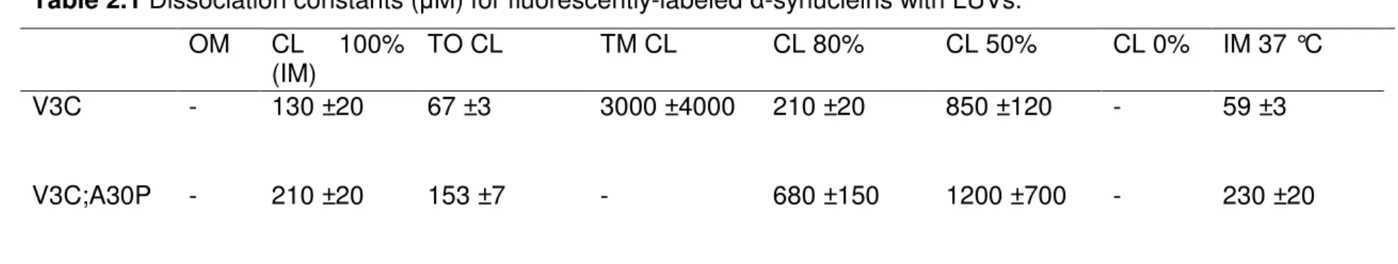
Figure 2.1 Schematic representation of α-synuclein and structural
formulas of the CLs ... 40
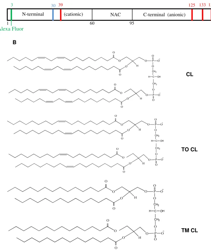
Figure 2.2 Confocal images of mitochondria incubated with α-synuclein ... 41
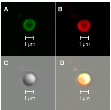
Figure 2.3 Size and polydispersity index of LUVs ... 42
Figure 2.4 Interaction of α-synucleins with OM and IM LUVs ... 43
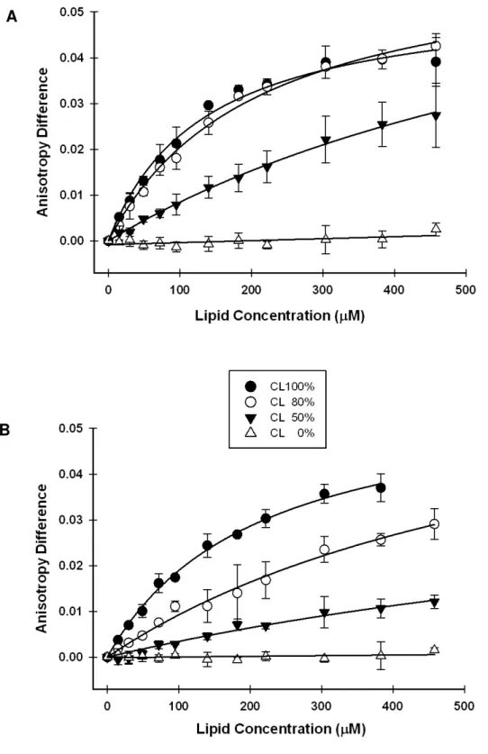
Figure 2.5 Interaction of α-synucleins with LUVs having different CL ratio ... 44
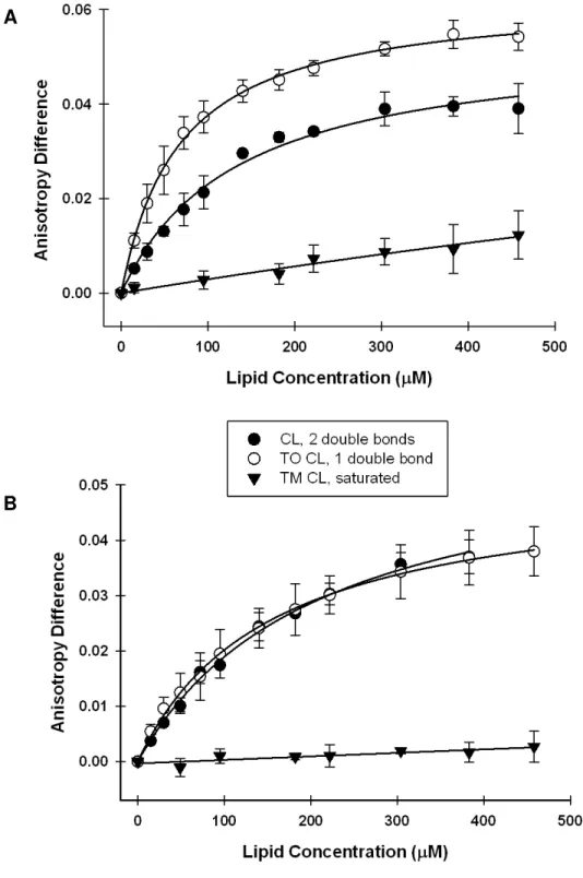
Figure 2.6 Interaction of α-synucleins with LUVs containing CLs with different side chains ... 45
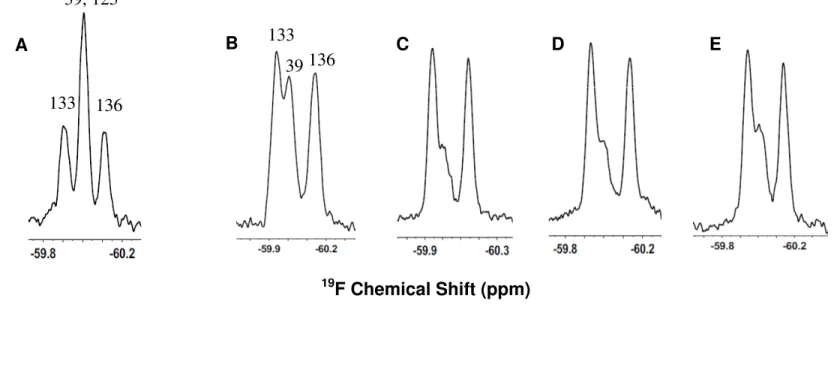
Figure 2.7 19F NMR spectra of α-synuclein interaction with LUVs containing CLs with different side chains ... 46
Figure 2.8 Interaction of α-synucleins with LUVs at 37 °C ... 47
Figure 2.9 DPH anisotropy upon the interaction of α-synucleins with LUVs... 48
Chapter 3
Figure 3.1 Schematic representation of CTP covalently attached to the N-terminus of α-synuclein ... 68Figure 3.2 Chromatograms and SDS-PAGE of wild-type and CTP α-synuclein ... 69
Figure 3.3 MALDI/MS of wild-type and CTP α-synuclein ... 70
Figure 3.4 Purified CTP α-synuclein ... 71
xiii
Figure 3.6 NMR tubes containing cells suspended in Ficoll ... 73
Figure 3.7 19F NMR spectra of wild-type and Y125F α-synuclein ... 74
Figure 3.8 α-Synuclein in Ficoll - controls ... 75
Figure 3.9 Interaction of 19F-labeled Y39F α-synuclein with CHO-K1 cells ... 76
Figure 3.10 Interaction of 19F-labeled CTP α-synuclein with CHO-K1 cells ... 77
Figure 3.11 Attempted assignments of the Y-11 resonance in CTP α-synuclein ... 78
Figure 3.12 CTP mediated delivery of α-synuclein into cells ... 79
Chapter 4
Figure 4.1 Schematic representation of the PEP-1/α-synuclein complex ... 103Figure 4.2 Schematic representation of α-synuclein translocation into higher eukaryotic cells using the PEP-1 peptide as a carrier ... 104
Figure 4.3 Translocation of CTP α-synuclein into cells ... 105
Figure 4.4 Cell lysates after translocation of PEP-1/α-synuclein complex ... 106
Figure 4.5 Intracellular degradation of α-synuclein ... 107
Figure 4.6 In-cell NMR of wild-type α-synuclein ... 108
Figure 4.7 In-cell NMR of wild-type α-synuclein with and without NH4Cl ... 109
Figure 4.8 The sensitivity of two NMR probes tested using 3-fluoro-L-tyrosine ... 110
Figure 4.9 Translocation of CTP α-synuclein into cells ... 111
Figure 4.10 SDS-PAGE of cell lysates containing PULS-in/ α-synuclein with and without NH4Cl ... 112
xiv
Figure 4.12 Distribution of QQ modified α-synuclein upon interaction
xv
List of Abbreviations and Symbols
AF488 Alexa Fluor 488
amp ampicillin
C Celsius
CH Cholesterol
CHO-K1 Chinese hamster ovary
CI2 chymotrypsin inhibitor 2
CL Cardiolipin
cm centimeter
CTP cytoplasmic transduction peptide
Da Dalton
DMEM Dulbecco’s Modified Eagle Medium
DOPC 1,2-dioleoyl-sn-glycero-3-phosphocholine
DOPE 1,2-dioleoyl-sn-glycero-3-phosphoethanolamine
DPH 1,6-diphenyl-1,3,5-hexatriene
DTT dithiothreitol
E. coli Escherichia coli
EDTA ethylenediaminetetraacetic acid
xvi
3FY 3-fluoro-L-tyrosine
g standard gravity
GFP green fluorescent protein
h hour
HeLa Human epithelial carcinoma cells
HSQC heteronuclear single quantum correlation
IPTG isopropyl β-D-1-thiogalactopyranoside
Kd dissociation constant
kDa kilodalton
L liter
LB Luria broth
LUV large unilamellar vesicle
M molar
MALDI/MS matrix-assisted laser desorption/ionization
mass spectrometry
MHz megahertz
min minute
mg milligram
mL milliliter
mM millimolar
MWCO molecular weight cut off
NAC non amyloid component
nm nanometer
xvii
OD optical density
PAGE polyacrylamide gel electrophoresis
PBS phosphate buffer saline
PCR polymerase chain reaction
PDI polydispersity index
PMSF phenylmethylsulphonyl fluoride
rpm revolutions per minute
RT room temperature
SDS sodium dodecyl sulfate
SDS-PAGE sodium dodecyl sulfate polyacrylamide gel electrophoresis
T temperature
TAT trans-acting activator of transcription
TM CL 1',3'-Bis[1,2-dimyristoyl-sn-glycero-3-phospho]-sn -glycerol
TO CL 1',3'-bis[1,2-dioleoyl-sn-glycero-3-phospho]-sn -glycerol
Tris trishydroxymethylaminomethane
v/v volume/volume
w/v weight/volume
X. laevis Xenopus laevis
α-syn α-synuclein
µg microgram
CHAPTER 1
Introduction
Reproduced in part with permission from Biochemistry. Copyright 2009 American
Chemical Society
1.1 Parkinson’s Disease
Parkinson’s disease is the second most common progressive neurological
disorder [1, 2] affecting more than 500,000 people in United States with more
than 50,000 new cases reported annually [3]. Its symptoms include rest tremor,
bradykinesia, rigidity and loss of postural reflexes [4]. The disease is more
prevalent in subjects older than 50, with the symptoms progressing more quickly
in some individuals [3, 5].
Parkinson’s disease is caused by the loss of dopaminergic neurons within
the substantia nigra pars compacta [6-8]. The disease is also characterized by
the presence of Lewy bodies in the cytoplasm of neurons in the substantia nigra.
Lewy bodies are pathological inclusions of 5-25 µm in diameter [9] having α-synuclein as a primary component [9-12]. Parkinson’s disease is diagnosed only
based on clinical symptoms; no single laboratory test exists. The factors that are
2
animal models) are: environmental [13-18], genetic [19, 20], and age-related [5,
21].
1.2 α-Synuclein
α-Synuclein is an intrinsically-disordered protein found in the presynaptic nerve terminals [22-28]. The 140-residue protein comprises a positively-charged
N-terminus, a hydrophobic middle region, and a negatively charged C-terminus
[29-31]. In its monomeric form, α-synuclein is natively unfolded but can assume a
β-sheet character or α-helical structure depending on the solution conditions [32-34]. Also, α-synuclein forms protofibrils [35, 36] and fibrils [37, 38] that are toxic for the neurons.
The N-terminal region of α-synuclein forms an α-helix upon interaction with vesicles of different lipid composition [39], phospholipid headgroups [40],
sizes [41], and surfaces [42, 43]. It is known that the protein has a higher affinity
for small unilamellar vesicles than for large unilamellar vesicles (LUVs), and that
the protein binds more strongly to vesicles containing anionic phospholipids [39,
40, 44, 45].
Two α-synuclein variants, A30P and A53T, associated with Parkinson’s disease have been identified [46]. In vitro studies confirm that fibrils form at an
increased rate for these mutants compared to wild-type α-synuclein, revealing the importance of these α-synuclein mutations in protein aggregation [6]. α-Synuclein is suspected to have multiple functions including roles in
3
A recent study shows that α-synuclein is implicated in SNARE-complex assembly at presynaptic vesicles via its C-terminal region while its N-terminal
region is anchored to the vesicle [47]. The protein is involved in synaptic activity
when the nerve terminals repeatedly release neurotransmitters necessary for
assembly and disassembly of the complex. However, another recent study,
shows that α-synuclein isolated under non-denaturing conditions from brain tissue and human cells is a partly-folded tetramer that is more resistant to
aggregation as compared to monomeric α-synuclein [52]. These recent findings
are important to gain insight into α-synuclein intracellular functions and represent
steps forward in elucidating the biochemistry of Parkinson’s disease. They also
indicate that a complete understanding of α-synuclein structure and functions and
how these properties relate to the disease need to be studied in the future.
1.3 Delivery Systems
Several peptides have been designed to cross the plasma membrane of
different cell types. Among these, the PEP family of amphipathic carriers have
been used to transport proteins and peptides [53, 54], antibodies [54], and
nucleic acids [55-57] into higher eukaryotic cells. The main component of this
class is the peptide, PEP-1 (Ac-KETWWETWWTEWSQPKKKRKV-Cya). This
peptide comprises a hydrophobic tryptophan-rich domain that interacts with the
cargo, a hydrophilic lysine-rich domain designed to improve intracellular delivery
and increase the solubility of the peptide, and a linker between the two domains
(SQP) [54]. The peptide has a cysteamine at its C-terminus, which improves the
4
cargo through non-covalent interactions. This complex is efficiently translocated
in a large number of cell lines [54]. The mechanism of PEP-1 translocation with
its cargo into higher eukaryotic cells is not well understood [59, 60]. Morris et al.
proved, using scanning electron microscopy and dynamic light scattering, that
PEP-1 forms nanoparticles with the cargo [61]. The particle’s size and
morphology is unaffected by the size of cargo, but the molar ratio of PEP-1 to
cargo has an important role in defining these parameters. After translocation, the
PEP-1/cargo complex rapidly dissociates in the cytoplasm, the peptide localizing
mostly in the nucleus while the cargo’s intracellular localization remains
unaffected (if the cargo’s function is in the cytosol than it remains there) [54].
PEP-1/cargo nanoparticles enter cells independently of the endosomal pathway
and deliver the cargo efficiently in a fully biological active form to a wide range of
cell lines [53, 54]. An important criterion for designing a delivery system is the
toxicity of the carrier. PEP-1 was tested on different cell lines, and no toxicity was
observed for concentrations of peptide of 100 µM [54].
Another common method of protein translocation involves making genes
that fuse the peptide to the cargo protein and expressing the construct in
Escherichia coli. One popular peptide is the trans-acting activator of transcription
(TAT) from the human immunodeficiency virus (HIV-1). This peptide has been
used to deliver proteins [62], oligonucleotides [63, 64], liposomes [65], and
nanoparticles [66]. TAT targets the cargo to the nucleus [67]. A more efficient
peptide developed by Kim et al. [68], is designed to translocate important
5
tested on live cells. The sequence YGR2AR6 proved to be the most efficient in
translocating β-galactosidase into several cell lines. After transduction, the peptide is cleaved in the cells by cytoplasmic enzymes and the cargo is released
[68]. PEP-1 and the YGR2AR6 peptide were selected to translocate α-synuclein into higher eukaryotic cells. These peptides were chosen because of their ability
to translocate large amounts of cargo into the cells and because of their
capability to release the cargo into the cytosol.
1.4 In-cell NMR
1.4.1 In-cell NMR in E. coli
1.4.1.1Protein-Protein Interactions and Signal Transduction
In-cell NMR spectroscopy characterizes one protein at a time while a more
interesting and valuable aspect would be to characterize the whole complex by
enriching all the proteins in the complex with one of the NMR active nuclei.
Unfortunately, the resulted spectra would be too complicated to analyze.
Shekhtman et al. [69] developed a method to study a protein in a complex by
NMR. STINT-NMR (structural interactions using NMR) works by enriching only
one protein in a complex and expressing it using the orthogonal induction
system. Shekhtman et al. [69] overexpressed ubiquitin in 15N-enriched media by
using the pBAD promoter. Next, the cells were collected by centrifugation and
resuspended in unenriched media. The PT7/lac system was used to express the
other protein in the complex. The interactions between the two proteins were
noted by changes in the width or chemical shift of the resonances of the enriched
6
Protein phosphorylation can be studied using NMR. Shekhtman‘s
laboratory used STINT-NMR to understand signal transduction by simultaneously
expressing three proteins: the target protein (ubiquitin), a large (>100 kDa)
heterodimeric affector complex, and a specific kinase [70]. The ubiquitin was
expressed under three conditions: alone, with coexpression of the affector
complex, and with coexpression of the complex and kinase. They determined the
interactions between ubiquitin and the complex and phosphorylation of ubiquitin
induced by expression of the kinase. Changes induced by phosphorylation can
be sensed by ubiquitin even though the complex is large.
1.4.1.2 Intrinsically Disordered Proteins
Intrinsically disordered proteins lack stable tertiary structure in dilute
solution. Many members of this recently defined class play important roles in cell
signaling, regulation, and control. Disordered proteins are associated with
disease states, including amyloidoses and neurodegenerative disorders [71].
Although the properties of globular proteins do not change significantly over a
wide range of solution conditions, the properties of intrinsically disordered
proteins can vary [72]. This sensitivity to solution conditions should make them
attractive targets for studies of physiologically relevant conditions. Despite this
sensitivity, and the known relationships between protein disorder and disease,
little is known about the intracellular structure of this protein class. Disordered
proteins are easier to detect by in-cell NMR than are globular proteins of the
same size. This increased sensitivity arises from differences in global and local
7
relaxation rates for globular proteins are most sensitive to global motion, which is
described by a single rotational correlation time. Disordered proteins, on the
other hand, are flexible. Their motions involve an ensemble of interconverting
conformers where different residues have different effective correlation times.
That is, the flexible nature of disordered proteins mitigates the deleterious effect
of viscosity on their spectra.
The first in-cell NMR study of an intrinsically disordered protein was
reported by Dedmon et al. [74]. The protein, FlgM, regulates flagellar synthesis
upon binding a transcription factor. The intracellular environment in E. coli
causes the C-terminal half of FlgM to gain structure while the N-terminal half
remains unstructured. Similar characteristics were noted in vitro in solutions
containing high levels of glucose, bovine serum albumin (BSA), and ovalbumin.
These data show that it is important to study disordered proteins under
physiologically relevant conditions. α-Synuclein, a 140-residue cytosolic eukaryotic protein, is the primary component of the intracellular protein
aggregates called Lewy bodies [73]. These aggregates are present in substantia
nigra neurons of patients with Parkinson’s disease. Studying α-synuclein under crowded conditions may provide information related to its role in the disease.
McNulty et al. [75] used in-cell NMR to investigate the structure of α-synuclein in
E. coli. These authors first noticed a difference between 1H-15N HSQC spectra in
dilute solution acquired at 10 and 35 °C [76]. Spectra collected at the higher
temperature presented fewer cross-peaks (~35) compared to spectra collected at
8
residues showed that cross-peaks in the spectrum acquired at 35 °C are from the
C-terminal third of the protein. This temperature dependent behavior was
associated with an increase in hydrodynamic radius and a gain in the level of
secondary structure at 35 °C. These changes were reversible when the
temperature was decreased to 10 °C. The authors concluded that the N-terminal
two-thirds of the protein exchanges between a more structured extended state
and more disordered but more compact state. Heating the protein increases the
rate of exchange between these states, causing the crosspeaks to disappear.
The in-cell spectrum of α-synuclein at 35 °C looks like the spectrum recorded at 10 °C in dilute solution. In vitro experiments in 300 g/L BSA yield
spectra similar to those acquired from E. coli at 35 °C. These observations give
rise to the idea that crowding in these cells or in vitro keeps α-synuclein in a disordered but more compact state. These conclusions, however, have been
questioned. Croke et al. [77] suggest the difference between the in-cell and dilute
solution results reflects a mismatch in pH between the samples, which results in
higher amide proton exchange rates and a concomitant loss of resonances at 35
°C.
1.4.1.3 The effect of intracellular crowding on proteins
Li et al. [78] studied globular and disordered proteins in E. coli. Globular
15N-enriched proteins cannot be detected in bacteria by using NMR. While 1H-15N
HSQC spectra are not resolved in intact cells, the 19F spectra can provide good
information about intracellular dynamics of globular and disordered proteins.
9
spectra while larger size proteins (up to ~100 kDa) provide good spectra only
when labeled with trifluoromethyl-L-phenylalanine.
Schlesinger et al. [79] studied the effect of E. coli cytosol on a variant of
Protein L. The globular protein, Protein L (~7 kDa), is folded in cells and in dilute
solution but the variant having seven lysine residues replaced by glutamic acids
is mostly unfolded in dilute solution at room temperature. The Protein L variant is
unfolded in cells suggesting that nonspecific interactions between cytoplasmic
components are more important than the excluded-volume effect.
1.4.1.4 Determination of protein structure in E. coli
The structure of the heavy-metal binding protein TTHA1718 from Thermus
thermophilus HB8 was determined de novo in E. coli [80]. This study represents
one of the most important breakthroughs of the in-cell NMR in prokaryotic cells.
The 3D NMR spectra were collected and the calculated structure reveals that the
protein structure in E. coli is similar with the structure of the protein in vitro.
1.4.2 In-cell NMR in Higher Eukaryotic Cells
In-cell studies in E. coli will continue to provide fundamental information
about crowding effects, but the medical relevance of higher eukaryotic cells
makes them attractive targets for in-cell NMR. In addition, in-cell NMR in higher
eukaryotic cells may be easier because the cytosol of eukaryotic cells seems to
have a lower apparent viscosity [81].
The first in-cell NMR study of isotopically enriched proteins in nucleated
higher eukaryotic cells was conducted in Xenopus laevis oocytes [82]. Their large
10
the cytosol. The B1 domain of streptococcal protein G (GB1) was expressed and
15N-enriched in E. coli, purified, and injected into 200 oocytes. The authors
studied a range of intracellular GB1 concentrations between 50 and 500 µM. The
positions of the GB1 resonances in the HSQC spectra remained the same in
dilute solution, in oocytes, and in solutions containing 250-300 mg/mL BSA,
showing that neither crowding nor the environment in the cell changes the
structure of GB1. Different behavior was observed, however, in terms of
cross-peak intensity. Resonances of amides involved in intramolecular hydrogen bonds
showed diminished intensity in cells and in BSA compared to dilute solution. This
observation is consistent with the idea that more dynamic parts of the protein are
less affected by the increased viscosity in cells [73]. Sakai et al. [83] studied
protein behavior after microinjection of 15N-enriched ubiquitin and calmodulin into
Xenopus oocytes. The in-cell spectrum of wild-type ubiquitin presented fewer
resonances compared to its in vitro spectrum. Mutations in the hydrophobic patch
on the β-sheet of ubiquitin confirmed that the loss of the resonances is associated with interactions between ubiquitin and other proteins. Calmodulin
spectra in oocytes presented two patterns correlated with the presence or
absence of extracellular calcium ions. When calcium ions were coinjected with
the calcium-free protein, the in-cell spectrum was characteristic of calcium-bound
calmodulin, while the spectrum was characteristic of the calcium-free form if
calcium was not coinjected. These observations show that the physiological
intracellular calcium concentration (~0.1 µM) does not affect calcium-free
11
conditions that preserve them will facilitate longer acquisition times, thereby
decreasing the amount of protein required for detection by NMR. Bodart et al.
[84] showed that oocytes can be preserved by embedding them in a 20% Ficoll
solution. The embedded cells remained intact for 20 h, allowing the detection of 5
µM intracellular protein. Nevertheless, care must be taken to monitor protein
leakage. Sakai et al. [85], working with enriched calmodulin and ubiquitin, have
developed an oocyte coinjection method involving GFP that is useful for
detecting leaks.
1.4.2.1 Phosphorylation in Higher Eukaryotic Cells
Selenko et al. [86] used NMR to detect phosphorylation of the viral SV40 T
antigen in intact X. laevis oocytes by endogenous casein kinase 2 (CK2). CK2
phosphorylates two serine residues in the regulatory region of the T antigen,
altering the nuclear-import properties of the full-length protein. These
investigators provide the first NMR observation of an in vivo protein substrate
phosphorylation event inside living cells by an endogenous protein kinase.
Time-resolved NMR spectra in oocytes show sequential phosphorylation of the
substrate. The investigators conclude that CK2 phosphorylation occurs in a
two-step reaction with intermediate release of the substrate and preference of the
kinase for the unphosphorylated substrate. The results obtained in oocytes agree
with NMR spectra acquired in dilute solution and in egg extracts, implying that
the kinetics of phosphorylation are not affected by macromolecular crowding.
Phosphorylation of the intrinsically disordered protein tau in X. laevis oocytes has
12
Bodart et al. [84] detected novel signals in the in vivo spectrum that they
assigned to phosphorylated residues of tau. Comparison of the in-cell spectrum
to the in vitro spectrum containing two known oocyte kinases revealed shifts in
resonance positions. These shifts suggest that unidentified kinases in oocytes
may alter the positions of resonances observed in vitro. This study provides the
first analysis of an intrinsically disordered protein in live eukaryotic cells by in-cell
NMR.
1.4.2.2 In-cell NMR in human cells
In-cell NMR spectra of 15N-enriched ubiquitin and the B1 domain of
streptococcal protein G (GB1) were conducted in HeLa cells [87]. The proteins
were either fussed to the cell-penetrating peptides or bound trough disulphide
links. Upon translocation, the proteins are released in the cytosol by specific
intracellular enzymes that cleave the covalent bond or by the reduced
intracellular pH for the disulphide links. The intracellular structure of all the
proteins tested is similar with their in vitro NMR structure indicating that
intracellular crowding does not have an effect on these proteins. However,
important information for drug screening was obtained for FKBP12 when the cells
were treated with extracellular immunosuppressants.
1.4.3 Caveats of in-cell NMR
A series of caveats are associated with NMR spectroscopy in live cells
and Ito and Selenko reviewed them recently [88]. These include: the insensitivity
of NMR spectroscopy [89] which requires large amounts of 15N-enriched or 19
13
the protein to other macromolecules or membranes in cells, protein leakage from
the cells, degradation and instability of translocated proteins in cells, and sample
inhomogeneity.
Studying α-synuclein in live cells and its interaction with the membranes using NMR provides atomic level information about this protein inside the cells
and helps elucidate the function of α-synuclein in Lewy bodies formation in Parkinson’s disease. This information may be relevant for prevention and
14
1.5 References
1. M. Martinez, A. Brice, J.R. Vaughan, A. Zimprich, M.M. Breteler, G. Meco, A. Filla, M.J. Farrer, C. Betard, J. Hardy, G. De Michele, V. Bonifati, B. Oostra, T. Gasser, N.W. Wood, A. Durr, Genome-wide scan linkage analysis for Parkinson's disease: the European genetic study of Parkinson's disease, Journal of medical genetics 41 (2004) 900-907.
2. C. Haass, P.J. Kahle, Parkinson's pathology in a fly, Nature 404 (2000) 341, 343.
3. http://www.ninds.nih.gov/disorders/parkinsons_disease/ parkinsons_disease_backgrounder.htm, Parkinson's Disease Backgrounder, as of 2009.
4. J. Jankovic, M. Stacy, Medical management of levodopa-associated motor complications in patients with Parkinson's disease, CNS drugs 21 (2007) 677-692.
5. J.H. Bower, D.M. Maraganore, S.K. McDonnell, W.A. Rocca, Incidence and distribution of parkinsonism in Olmsted County, Minnesota, 1976-1990, Neurology 52 (1999) 1214-1220.
6. R.A. Fredenburg, C. Rospigliosi, R.K. Meray, J.C. Kessler, H.A. Lashuel, D. Eliezer, P.T. Lansbury, Jr., The impact of the E46K mutation on the properties of alpha-synuclein in its monomeric and oligomeric states, Biochemistry 46 (2007) 7107-7118.
7. R.G. Perez, T.G. Hastings, Could a loss of alpha-synuclein function put dopaminergic neurons at risk?, Journal of neurochemistry 89 (2004) 1318-1324.
8. M.F. Chesselet, Dopamine and Parkinson's disease: is the killer in the house?, Molecular psychiatry 8 (2003) 369-370.
9. M.G. Spillantini, R.A. Crowther, R. Jakes, M. Hasegawa, M. Goedert, alpha-Synuclein in filamentous inclusions of Lewy bodies from Parkinson's disease and dementia with lewy bodies, Proceedings of the national academy of sciences of the United States of America 95 (1998) 6469-6473.
15
11. P. Desplats, H.J. Lee, E.J. Bae, C. Patrick, E. Rockenstein, L. Crews, B. Spencer, E. Masliah, S.J. Lee, Inclusion formation and neuronal cell death through neuron-to-neuron transmission of alpha-synuclein, Proceedings of the national academy of sciences of the United States of America 106 (2009) 13010-13015.
12. M.S. Parihar, A. Parihar, M. Fujita, M. Hashimoto, P. Ghafourifar, Mitochondrial association of alpha-synuclein causes oxidative stress, Cellular and molecular life sciences 65 (2008) 1272-1284.
13. J.W. Langston, P. Ballard, J.W. Tetrud, I. Irwin, Chronic Parkinsonism in humans due to a product of meperidine-analog synthesis, Science (New York, N.Y 219 (1983) 979-980.
14. P.R. Castello, D.A. Drechsel, M. Patel, Mitochondria are a major source of paraquat-induced reactive oxygen species production in the brain, The Journal of biological chemistry 282 (2007) 14186-14193.
15. J.S. Bus, J.E. Gibson, Paraquat: model for oxidant-initiated toxicity, Environmental health perspectives 55 (1984) 37-46.
16. T.B. Sherer, R. Betarbet, C.M. Testa, B.B. Seo, J.R. Richardson, J.H. Kim, G.W. Miller, T. Yagi, A. Matsuno-Yagi, J.T. Greenamyre, Mechanism of toxicity in rotenone models of Parkinson's disease, Journal neuroscience 23 (2003) 10756-10764.
17. G. Meco, V. Bonifati, N. Vanacore, E. Fabrizio, Parkinsonism after chronic exposure to the fungicide maneb (manganese ethylene-bis-dithiocarbamate), Scandinavian journal of work, environment & health 20 (1994) 301-305.
18. J. Zhang, V.A. Fitsanakis, G. Gu, D. Jing, M. Ao, V. Amarnath, T.J. Montine, Manganese ethylene-bis-dithiocarbamate and selective dopaminergic neurodegeneration in rat: a link through mitochondrial dysfunction, Journal of neurochemistry 84 (2003) 336-346.
19. S. Lesage, A. Brice, Parkinson's disease: from monogenic forms to genetic susceptibility factors, Human molecular genetics 18 (2009) R48-59.
16
21. A.E. Lang, A.M. Lozano, Parkinson's disease. First of two parts, The New England journal of medicine 339 (1998) 1044-1053.
22. S. Chandra, G. Gallardo, R. Fernandez-Chacon, O.M. Schluter, T.C. Sudhof, Alpha-synuclein cooperates with CSPalpha in preventing neurodegeneration, Cell 123 (2005) 383-396.
23. G. Di Rosa, D. Puzzo, A. Sant'Angelo, F. Trinchese, O. Arancio, Alpha-synuclein: between synaptic function and dysfunction, Histology and histopathology 18 (2003) 1257-1266.
24. S. Chandra, F. Fornai, H.B. Kwon, U. Yazdani, D. Atasoy, X. Liu, R.E. Hammer, G. Battaglia, D.C. German, P.E. Castillo, T.C. Sudhof, Double-knockout mice for alpha- and beta-synucleins: effect on synaptic functions, Proceedings of the national academy of sciences of the United States of America 101 (2004) 14966-14971.
25. P.H. Jensen, J.Y. Li, A. Dahlstrom, C.G. Dotti, Axonal transport of synucleins is mediated by all rate components, The European journal of neuroscience 11 (1999) 3369-3376.
26. L. Maroteaux, J.T. Campanelli, R.H. Scheller, Synuclein: a neuron-specific protein localized to the nucleus and presynaptic nerve terminal, Journal of Neuroscience 8 (1988) 2804-2815.
27. D.L. Fortin, M.D. Troyer, K. Nakamura, S. Kubo, M.D. Anthony, R.H. Edwards, Lipid rafts mediate the synaptic localization of alpha-synuclein, Journal of Neuroscience 24 (2004) 6715-6723.
28. V.N. Uversky, Neuropathology, biochemistry, and biophysics of alpha-synuclein aggregation, Journal of neurochemistry 103 (2007) 17-37.
29. V.N. Uversky, J. Li, A.L. Fink, Evidence for a partially folded intermediate in alpha-synuclein fibril formation, The Journal of biological chemistry 276 (2001) 10737-10744.
30. D.P. Hong, W. Xiong, J.Y. Chang, C. Jiang, The role of the C-terminus of human alpha-synuclein: intra-disulfide bonds between the C-terminus and other regions stabilize non-fibrillar monomeric isomers, FEBS letters 585 561-566.
17
32. J.C. Kessler, J.C. Rochet, P.T. Lansbury, Jr., The N-terminal repeat domain of alpha-synuclein inhibits beta-sheet and amyloid fibril formation, Biochemistry 42 (2003) 672-678.
33. L.C. Serpell, J. Berriman, R. Jakes, M. Goedert, R.A. Crowther, Fiber diffraction of synthetic alpha-synuclein filaments shows amyloid-like cross-beta conformation, Proceedings of the national academy of sciences of the United States of America 97 (2000) 4897-4902.
34. T. Antony, W. Hoyer, D. Cherny, G. Heim, T.M. Jovin, V. Subramaniam, Cellular polyamines promote the aggregation of alpha-synuclein, The journal of biological chemistry 278 (2003) 3235-3240.
35. M.J. Volles, P.T. Lansbury, Jr., Vesicle permeabilization by protofibrillar alpha-synuclein is sensitive to Parkinson's disease-linked mutations and occurs by a pore-like mechanism, Biochemistry 41 (2002) 4595-4602.
36. T.T. Ding, S.J. Lee, J.C. Rochet, P.T. Lansbury, Jr., Annular alpha-synuclein protofibrils are produced when spherical protofibrils are incubated in solution or bound to brain-derived membranes, Biochemistry 41 (2002) 10209-10217.
37. K.C. Luk, C. Song, P. O'Brien, A. Stieber, J.R. Branch, K.R. Brunden, J.Q. Trojanowski, V.M. Lee, Exogenous alpha-synuclein fibrils seed the formation of Lewy body-like intracellular inclusions in cultured cells, Proceedings of the national academy of sciences of the United States of America 106 (2009) 20051-20056.
38. M. Vilar, H.T. Chou, T. Luhrs, S.K. Maji, D. Riek-Loher, R. Verel, G. Manning, H. Stahlberg, R. Riek, The fold of alpha-synuclein fibrils, Proceedings of the national academy of sciences of the United States of America 105 (2008) 8637-8642.
39. G.F. Wang, C. Li, G.J. Pielak, 19F NMR studies of alpha-synuclein-membrane interactions, Protein science 19 (2010) 1686-1691.
40. E. Rhoades, T.F. Ramlall, W.W. Webb, D. Eliezer, Quantification of alpha-synuclein binding to lipid vesicles using fluorescence correlation spectroscopy, Biophysical journal 90 (2006) 4692-4700.
41. L. Kjaer, L. Giehm, T. Heimburg, D. Otzen, The influence of vesicle size and composition on alpha-synuclein structure and stability, Biophysical journal 96 (2009) 2857-2870.
18
fluorescence, Proceedings of the national academy of sciences U S A 106 (2009) 5645-5650.
43. C.C. Jao, B.G. Hegde, J. Chen, I.S. Haworth, R. Langen, Structure of membrane-bound alpha-synuclein from site-directed spin labeling and computational refinement, Proceedings of the national academy of sciences U S A 105 (2008) 19666-19671.
44. W.S. Davidson, A. Jonas, D.F. Clayton, J.M. George, Stabilization of alpha-synuclein secondary structure upon binding to synthetic membranes, The journal of biological chemistry 273 (1998) 9443-9449.
45. M. Zhu, J. Li, A.L. Fink, The association of alpha-synuclein with membranes affects bilayer structure, stability, and fibril formation, The journal of biological chemistry 278 (2003) 40186-40197.
46. J. Li, V.N. Uversky, A.L. Fink, Effect of familial Parkinson's disease point mutations A30P and A53T on the structural properties, aggregation, and fibrillation of human alpha-synuclein, Biochemistry 40 (2001) 11604-11613.
47. J. Burre, M. Sharma, T. Tsetsenis, V. Buchman, M.R. Etherton, T.C. Sudhof, Alpha-synuclein promotes SNARE-complex assembly in vivo and
in vitro, Science 329 1663-1667.
48. M. Vila, D. Ramonet, C. Perier, Mitochondrial alterations in Parkinson's disease: new clues, Journal of neurochemistry 107 (2008) 317-328.
49. L. Devi, H.K. Anandatheerthavarada, Mitochondrial trafficking of APP and alpha synuclein: Relevance to mitochondrial dysfunction in Alzheimer's and Parkinson's diseases, Biochimica et biophysica acta 1802 (2010) 11-19.
50. N.B. Cole, D. Dieuliis, P. Leo, D.C. Mitchell, R.L. Nussbaum, Mitochondrial translocation of alpha-synuclein is promoted by intracellular acidification, Experimental cell research 314 (2008) 2076-2089.
51. I. Mikolaenko, O. Pletnikova, C.H. Kawas, R. O'Brien, S.M. Resnick, B. Crain, J.C. Troncoso, Alpha-synuclein lesions in normal aging, Parkinson disease, and Alzheimer disease: evidence from the Baltimore Longitudinal Study of Aging (BLSA), Journal of neuropathology and experimental neurology 64 (2005) 156-162.
19
53. S. Deshayes, M. Morris, F. Heitz, G. Divita, Delivery of proteins and nucleic acids using a non-covalent peptide-based strategy, Advenced drug delivery reviews 60 (2008) 537-547.
54. M.C. Morris, J. Depollier, J. Mery, F. Heitz, G. Divita, A peptide carrier for the delivery of biologically active proteins into mammalian cells, Nature biotechnology 19 (2001) 1173-1176.
55. L. Crombez, A. Charnet, M.C. Morris, G. Aldrian-Herrada, F. Heitz, G. Divita, A non-covalent peptide-based strategy for siRNA delivery, Biochemical society transaction 35 (2007) 44-46.
56. S. Deshayes, F. Simeoni, M.C. Morris, G. Divita, F. Heitz, Peptide-mediated delivery of nucleic acids into mammalian cells, Methods in molecular biology 386 (2007) 299-308.
57. M.C. Morris, E. Gros, G. Aldrian-Herrada, M. Choob, J. Archdeacon, F. Heitz, G. Divita, A non-covalent peptide-based carrier for in vivo delivery of DNA mimics, Nucleic acids research 35 (2007) e49.
58. K. Weller, S. Lauber, M. Lerch, A. Renaud, H.P. Merkle, O. Zerbe, Biophysical and biological studies of end-group-modified derivatives of Pep-1, Biochemistry 44 (2005) 15799-15811.
59. S. Deshayes, A. Heitz, M.C. Morris, P. Charnet, G. Divita, F. Heitz, Insight into the mechanism of internalization of the cell-penetrating carrier peptide Pep-1 through conformational analysis, Biochemistry 43 (2004) 1449-1457.
60. S.T. Henriques, J. Costa, M.A. Castanho, Translocation of beta-galactosidase mediated by the cell-penetrating peptide pep-1 into lipid vesicles and human HeLa cells is driven by membrane electrostatic potential, Biochemistry 44 (2005) 10189-10198.
61. M.A. Munoz-Morris, F. Heitz, G. Divita, M.C. Morris, The peptide carrier Pep-1 forms biologically efficient nanoparticle complexes, Biochemical and biophysical research communication 355 (2007) 877-882.
62. E. Kubo, N. Fatma, Y. Akagi, D.R. Beier, S.P. Singh, D.P. Singh, TAT-mediated PRDX6 protein transduction protects against eye lens epithelial cell death and delays lens opacity, American journal of physiology 294 (2008) C842-855.
20
transduction domain of HIV-1 Tat protein promotes efficient delivery of DNA into mammalian cells, The Journal of biological chemistry 276 (2001) 26204-26210.
64. A. Ziegler, J. Seelig, High affinity of the cell-penetrating peptide HIV-1 Tat-PTD for DNA, Biochemistry 46 (2007) 8138-8145.
65. V.P. Torchilin, R. Rammohan, V. Weissig, T.S. Levchenko, TAT peptide on the surface of liposomes affords their efficient intracellular delivery even at low temperature and in the presence of metabolic inhibitors, Proceedings of the national academy of sciences of the United States of America 98 (2001) 8786-8791.
66. C.C. Berry, J.M. de la Fuente, M. Mullin, S.W. Chu, A.S. Curtis, Nuclear localization of HIV-1 tat functionalized gold nanoparticles, IEEE transactions on nanobioscience 6 (2007) 262-269.
67. E. Vives, P. Brodin, B. Lebleu, A truncated HIV-1 Tat protein basic domain rapidly translocates through the plasma membrane and accumulates in the cell nucleus, The Journal of biological chemistry 272 (1997) 16010-16017.
68. D. Kim, C. Jeon, J.H. Kim, M.S. Kim, C.H. Yoon, I.S. Choi, S.H. Kim, Y.S. Bae, Cytoplasmic transduction peptide (CTP): new approach for the delivery of biomolecules into cytoplasm in vitro and in vivo, Experimental cell research 312 (2006) 1277-1288.
69. D.S. Burz, K. Dutta, D. Cowburn, A. Shekhtman, Mapping structural interactions using in-cell NMR spectroscopy (STINT-NMR), Nature methods 3 (2006) 91-93.
70. D.S. Burz, A. Shekhtman, In-cell biochemistry using NMR spectroscopy, PloS one 3 (2008) e2571.
71. V.N. Uversky, A.L. Fink, Conformational constraints for amyloid fibrillation: the importance of being unfolded, Biochimica et biophysica acta 1698 (2004) 131-153.
72. G.W. Daughdrill, Pielak, G. J., Uversky, V. N., Cortese, M. S.,, A.K. and Dunker, Natively disordered proteins. In Protein folding handbook (Buchner, J., and Kiefhaber, T., Eds.), (2005) pp 275- 357, Wiley-VCH, Weinheim, Germany.
21
globular protein: implications for in-cell NMR spectroscopy, Journal of the American Chemical Society 130 (2008) 6310-6311.
74. M.M. Dedmon, C.N. Patel, G.B. Young, G.J. Pielak, FlgM gains structure in living cells, Proceedings of the National Academy of Sciences of the United States of America 99 (2002) 12681-12684.
75. B.C. McNulty, G.B. Young, G.J. Pielak, Macromolecular crowding in the Escherichia coli periplasm maintains alpha-synuclein disorder, Journal of molecular biology 355 (2006) 893-897.
76. B.C. McNulty, A. Tripathy, G.B. Young, L.M. Charlton, J. Orans, G.J. Pielak, Temperature-induced reversible conformational change in the first 100 residues of alpha-synuclein, Protein science 15 (2006) 602-608.
77. R.L. Croke, C.O. Sallum, E. Watson, E.D. Watt, A.T. Alexandrescu, Hydrogen exchange of monomeric alpha-synuclein shows unfolded structure persists at physiological temperature and is independent of molecular crowding in Escherichia coli, Protein science 17 (2008) 1434-1445.
78. C. Li, G.F. Wang, Y. Wang, R. Creager-Allen, E.A. Lutz, H. Scronce, K.M. Slade, R.A. Ruf, R.A. Mehl, G.J. Pielak, Protein 19F NMR in Escherichia coli, Journal of the American chemical society 132 (2009) 321-327.
79. A.P. Schlesinger, Y. Wang, X. Tadeo, O. Millet, G.J. Pielak, Macromolecular crowding fails to fold a globular protein in cells, Journal of the American chemical society 133 8082-8085.
80. D. Sakakibara, A. Sasaki, T. Ikeya, J. Hamatsu, T. Hanashima, M. Mishima, M. Yoshimasu, N. Hayashi, T. Mikawa, M. Walchli, B.O. Smith, M. Shirakawa, P. Guntert, Y. Ito, Protein structure determination in living cells by in-cell NMR spectroscopy, Nature 458 (2009) 102-105.
81. K. Luby-Phelps, Cytoarchitecture and physical properties of cytoplasm: volume, viscosity, diffusion, intracellular surface area, International review of cytology 192 (2000) 189-221.
82. P. Selenko, Z. Serber, B. Gadea, J. Ruderman, G. Wagner, Quantitative NMR analysis of the protein G B1 domain in Xenopus laevis egg extracts and intact oocytes, Proceedings of the national academy of sciences of the United States of America 103 (2006) 11904-11909.
22
84. J.F. Bodart, J.M. Wieruszeski, L. Amniai, A. Leroy, I. Landrieu, A. Rousseau-Lescuyer, J.P. Vilain, G. Lippens, NMR observation of Tau in
Xenopus oocytes, Journal of magnetic resonance 192 (2008) 252-257.
85. T. Sakai, H. Tochio, K. Inomata, Y. Sasaki, T. Tenno, T. Tanaka, T. Kokubo, H. Hiroaki, M. Shirakawa, Fluoroscopic assessment of protein leakage during Xenopus oocytes in-cell NMR experiment by co-injected EGFP, Analytical biochemistry 371 (2007) 247-249.
86. P. Selenko, D.P. Frueh, S.J. Elsaesser, W. Haas, S.P. Gygi, G. Wagner, In situ observation of protein phosphorylation by high-resolution NMR spectroscopy, Nature structural & molecular biology 15 (2008) 321-329.
87. K. Inomata, A. Ohno, H. Tochio, S. Isogai, T. Tenno, I. Nakase, T. Takeuchi, S. Futaki, Y. Ito, H. Hiroaki, M. Shirakawa, High-resolution multi-dimensional NMR spectroscopy of proteins in human cells, Nature 458 (2009) 106-109.
88. Y. Ito, P. Selenko, Cellular structural biology, Current opinion in structural biology 20 (2010) 640-648.
CHAPTER 2
Interaction of
α
-Synuclein and its A30P Variant with Vesicles of
Composition Similar to Mitochondrial Membranes
Summary
α-Synuclein, an intrinsically-disordered protein associated with Parkinson’s disease, interacts with mitochondria but the details of this interaction
are unknown. Cardiolipin is the main anionic lipid in mitochondria where almost
all of its acyl chains possess two double bonds. We probed the interaction of α-synuclein and its A30P variant with lipid vesicles by using fluorescence
anisotropy and 19F nuclear magnetic resonance. Both proteins interact strongly
with large unilamellar vesicles of composition similar to that of the inner
mitochondrial membrane, which contains cardiolipin, but have no affinity for
vesicles mimicking the outer mitochondrial membrane, which lacks cardiolipin.
The 19F data show that the interaction involves α-synuclein’s N-terminal region. These data indicate that the middle portion of the protein, which contains the
KAKEGVVAAAE repeats, is involved in binding, probably via electrostatic
interactions between the lysines and cardiolipin. However, the saturation of the
24
dramatically decreases affinity. Increasing the temperature increases the binding
of wild-type, but not the A30P variant. These data suggest that membrane
packing density is important in determining α-synuclein affinity. This idea was confirmed by examining the anisotropy of 1,6-diphenyl-1,3,5-hexatriene in the
presence of vesicles with or without the proteins. The results advance our
understanding of α-synuclein’s interaction with mitochondrial membranes.
2.1 Introduction
Parkinson’s disease and other neurodegenerative disorders are
characterized by cytoplasmic neuronal inclusions known as Lewy bodies [1-4].
α-Synuclein, a 140-amino acid intrinsically disordered protein, is the main
component of Lewy bodies [5-8]. The protein (Figure 2.1 A) comprises a
positively-charged N-terminal region (residues 1-60) with an imperfect consensus
repeat KTKEGV involved in lipid binding, a hydrophobic middle segment known
as the NAC region (Non Amyloid Component, residues 61-95), and a
negatively-charged C-terminal region (residues 96-140) [9, 10].
The protein helps maintain SNARE-complex assembly at presynaptic
vesicles through its C-terminus, while the N-terminus is anchored to the vesicle
[11]. Its function is important during increased synaptic activity when the nerve
terminals repeatedly release neurotransmitters, which requires the assembly and
disassembly of the SNARE complex. The protein is also involved in aging, but
the reason for its age-related loss of function is unknown [11].
25
localization is associated with mitochondrial dysfunction [13-15] including
impaired complex I function [12, 16], oxidative stress [17], and mitochondrial lipid
abnormalities [16]. More specifically, a decrease in cardiolipin (CL) and a
change in its acyl side chains were noted in mitochondria from the brains of mice
lacking α-synuclein [16]. The side chain changes involve a reduction in polyunsaturated fatty acids [16, 18] and an increase in saturated fatty acids [16].
Since CL is a mitochondria-specific phospholipid, the implied importance of α-synuclein at the mitochondrial level is evident. Although the exact link between
the loss of α-synuclein and the decrease in CL is unclear, the findings suggest that α-synuclein helps control intracellular transport of lipids [16].
The interaction of α-synuclein with vesicles of different lipid composition [19], phospholipid headgroups [20], sizes [21], and surfaces [22, 23] has been
studied. Despite these efforts little is known about the interaction of α-synuclein with the mitochondrial membrane, the mechanism of α-synuclein transport through the outer mitochondrial membrane, and the localization of the protein in
the inner mitochondrial membrane [17].
We used fluorescence anisotropy and high resolution nuclear magnetic
resonance spectroscopy (NMR) to investigate the interaction of wild-type α-synuclein and one of its familial Parkinson’s disease variants [24], A30P, with
large unillamelar vesicles (LUVs) having lipid compositions similar to the inner
and outer mitochondrial membranes. We evaluated the importance of CL as well
26
investigated the effect of temperature on affinity, and using 19F-labeled α-synuclein, we determined which region of the protein interacts with LUVs
containing CL. The results advance our understanding of α-synuclein’s interaction with mitochondrial membranes.
2.2 Materials and Methods
2.2.1 Expression, Purification, and Labeling of Human α-Synuclein
and its A30P Variant
Wild-type α-synuclein, A30P α-synuclein, V3C α-synuclein and V3C;A30P
α-synuclein were expressed from a T7-7 plasmid in Escherichia coli BL21-Gold(DE3) competent cells (Stratagene Cloning Systems, La Jolla, CA). The
proteins were purified as described [25, 26].
The V3C mutation at position 3 was made by using a Stratagene
site-directed mutagenesis kit. The cysteine was used to attach Alexa Fluor 488
(Invitrogen, Carlsbad, CA). For labeling, 12 mg of V3C α-synuclein were dissolved in sterile, degassed water to a final concentration of 2 mg/mL.
Tris(2-carboxyethyl)phosphine and NaHCO3 were added in a 10-fold molar excess over
the protein. The mixture was incubated at room temperature with shaking for 30
min. Next, Alexa Fluor 488 C5-maleimide was added in a 10-fold molar excess
over protein and the mixture incubated at room temperature with shaking for 2 h.
The labeled protein was purified with gel filtration chromatography by using a
Superdex 75 column eluted with 20% acetonitrile in phosphate buffered saline
(NaCl 137 mM, KCl 2.7 mM, Na2HPO4 10 mM, KH2PO4 1.8 mM). The labeled
27
The extinction coefficient of the dye at 494 nm (71,000 M-1cm-1) was used
to quantify the labeled protein. The Lowry method (Pierce, Rockford, IL) was
used to quantify the total protein. The degree of labeling was 84% for V3C α-synuclein and 99% for V3C;A30P α-synuclein. Aliquots containing 1 mg of labeled protein were lyophilized and stored at –80 °C.
For NMR experiments, wild-type and Y125F α-synuclein labeled with 3-fluoro-L-tyrosine were prepared as described [27].
2.2.2 Cell Culture and Mitochondria Isolation
Human epithelial carcinoma cells (HeLa) from ATCC were cultured in
Dulbelcco’s modified Eagle’s medium (Invitrogen) supplemented with 10% fetal
bovine serum, penicillin and streptomycin (100 µg/mL). The cultures were incubated at 37 °C in 5% CO2. Mitochondria were isolated from HeLa cells (2 x
107 cells) by using a Mitochondria Isolation Kit for Cultured Cells (Pierce,
Rockford, IL). Their activity was tested by assessing the cytochrome c oxidase
activity with the Mitochondria Activity Assay Kit (BioChain Institute, Inc.,
Haywatd, CA).
2.2.3 Mitochondrial Import of α-Synuclein
Freshly isolated mitochondria were incubated with fluorescently-labeled
α-synuclein in the presence of an energy mixture and transport buffer as described
[28]. Trypsin was used to remove α-synuclein attached to the surface of mitochondria. Mito Tracker Red CMXRos (Invitrogen) was used to stain the
active mitochondria after protein import and trypsinization. Fused silica and glass
α-28
synuclein were allowed to attach to the poly-L-lysine coated slide for 30 min and
then rinsed with minimal media. α-Synuclein import was tested using confocal microscopy on an inverted laser scanning microscope (Zeiss 510 Meta,
Thornwood, NY) equipped with a 63 x, 1.4 NA, Plan-Apochromat, oil immersion
objective.
2.2.4 Vesicle Preparation
Components were purchased from Avanti Polar Lipids, Inc. (Birmingham,
AL) in chloroform, except cholesterol (CH, from ovine wool), which was dissolved
in chloroform to a concentration of 1 mg/mL and stored at –20 °C. The
components were used without further purification.
Aliquots of the components, in chloroform, were mixed in glass vials and
the solvent removed overnight in a vacuum concentrator. The dried mixtures
were suspended in 1 mL of 50 mM sodium phosphate buffer, pH 7.4, to a final
lipid concentration of 2 mM. The following phospholipids were used for preparing
LUVs: CL (bovine heart), 1,2-dioleoyl-sn-glycero-3-phosphocholine (DOPC),
1,2-dioleoyl-sn-glycero-3-phosphoethanolamine (DOPE), 1',3'-bis[1,2-dimyristoyl-sn
-glycero-3-phospho]-sn-glycerol (TM CL), and 1',3'-bis[1,2-dioleoyl-sn
-glycero-3-phospho]-sn-glycerol (TO CL). Figure 2.1 B shows the structures of CLs.
Vesicles of composition DOPC:DOPE:CL:CH 2.0:1.3:1.0:0.6 molar ratio
and DOPC:DOPE:CH 4.0:2.0:0.9, corresponding to inner and outer membrane of
mitochondria [30], respectively, were prepared. LUVs with different ratios of CL
(1.0, 0.8, 0.5, and 0) or different CL acid side chains (TM and TO) were also
29
LUVs were prepared by multiple extrusion [20, 31, 32] through a 0.1 µm polycarbonate membrane (Whatman Inc., Sanford, ME). For NMR experiments,
the vesicles were prepared with the same protocol, but at a concentration of 4
mM. The dried mixture corresponding to the inner mitochondrial membrane was
resuspended in 50 mM sodium phosphate buffer, pH 7.4, containing 10% D2O.
The final concentration of 19F labeled protein in the NMR tube was 100 µM and
the protein:lipid molar ratio was ~1:100. Protein in buffer alone was used as a
reference.
2.2.5 Fluorescence Anisotropy of Labeled Proteins
The experiments were conducted on a FluoroLog®-322 spectrofluorometer
(HORIBA Jobin Yvon Inc., Edison, NJ) with an excitation wavelength of 495 nm
and an emission wavelength of 519 nm. Four hundred µL of protein (100 nM) in
50 mM sodium phosphate buffer, pH 7.4, were titrated with LUVs at 25 or 37 °C.
Each point is the average of five measurements, and each condition was tested
in triplicate. Control measurements using only LUVs or unlabeled wild-type α-synuclein in the same buffer were performed to assess background fluorescence,
which was negligible. The anisotropy was calculated as described [33]. The
dissociation constants (Kd) were calculated by fitting the data to a one site
binding model (SigmaPlot 11.0).
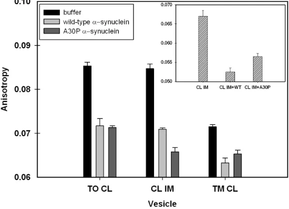
2.2.6 DPH Fluorescence Anisotropy
1,6-Diphenyl-1,3,5-hexatriene (DPH) was incubated with LUVs containing
TO, TM, or CL IM for one h at room temperature either in the presence or
30
anisotropy was recorded at 25 °C by using an excitation wavelength of 360 nm
and an emission wavelength of 440 nm. Fluorescence anisotropy was also
measured at 37 °C for CL IM LUVs in the presence or absence of wild-type or
A30P α-synuclein. The result for each condition represents the average of five measurements.
2.2.7 NMR
19F spectra were acquired at 25 °C on a Varian Inova 600 MHz
spectrometer equipped with a 5 mm triple resonance probe. The spectra
comprised 2048 transients with a 30 kHz sweep width. The 19F chemical shifts
were referenced to trifluoroethanol at 0 ppm. The experiments were conducted in
triplicate.
2.3 Results
2.3.1 In vitro Mitochondrial Import of α-Synuclein
Fluorescently-labeled α-synuclein localized in isolated mitochondria (Figure 2.2A). Mito Tracker Red CMXRos is an indicator of active mitochondria
because its accumulation is related to membrane potential. The red fluorescence
shows that mitochondria are active after protein import and trypsinization (Figure
2.2B). Co-localization of fluorescently-labeled protein and Mito Tracker Red
indicates that the protein is localized in close proximity of the membranes, not in
the matrix (Figure 2.2D). These findings agree with those of Devi et al. [15]. To
31
membrane, we used lipid vesicles with a composition similar to these
membranes.
2.3.2 Interaction with LUVs Having LipidCompositions Similar to the Inner and Outer Mitochondrial Membrane
Mitochondria contain 25.3% phospholipid by mass [30]. In terms of
phospholipid composition, the mitochondria comprise 40.8% phosphatidyl
choline, 37.4% phosphatidyl ethanolamine, 19.1% CL, and ~3% phosphatidyl
inositol. The major phospholipids of the outer membrane are phosphatidyl
choline (55.2%), and phosphatidyl ethanolamine (25.3%). The major
phospholipids of the inner membrane are phosphatidyl choline (44.5%),
phosphatidyl ethanolamine (27.7%), and CL (21.5%). CL is essentially absent
from the outer membrane [30].
Fluorescence anisotropy was used to quantify the interaction of
fluorescently-labeled V3C α-synuclein and V3C;A30P α-synuclein with LUVs. Alexa Fluor 488 dye was chosen because of its high photostability [34]. Labeling
at position 3 was selected because this region of the protein is highly dynamic,
decreasing the possibility that the modification will disrupt the native
conformation [35]. The average diameter of the LUVs, as determined by dynamic
light scattering, is ~140 nm (Figure 2.3A-D), consistent with values for vesicles
extruded through membranes with 100 nm pores [20, 31]. The vesicles were
stable for at least three days if kept at 4 °C in 50 mM sodium phosphate buffer,
32
absence of CL (Figure 2.3A and B) or CH (Figure 2.3C and D) does not affect the
size of the vesicles.
First, we studied the interaction of fluorescently-labeled proteins with
LUVs having a molar DOPC:DOPE:CH ratio of 4.0:2.0:0.90, which corresponds
to the composition of the outer membrane. No change in the anisotropy was
noted when titrating 100 nM protein with LUVs, suggesting no affinity of
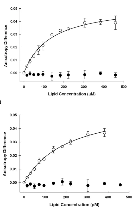
fluorescently labeled V3C or V3C;A30P α-synuclein for this type of vesicle (Figure 2.4 A and B).
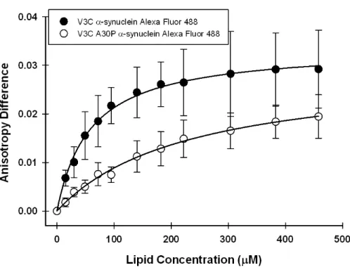
LUVs with a molar ratio corresponding to the inner membrane
(DOPC:DOPE:CL:CH of 2.0:1.3:1.0:0.60) gave strikingly different results; both
the wild-type protein and the A30P variant bind with sub mM dissociation
constants. As shown in Table 1, V3C α-synuclein binds slightly more strongly (Kd
130 µM) than the V3C;A30P variant (Kd 210 µM). Since only the inner membrane
contains CL, these observations implicate CL in the binding of α-synuclein and are consistent with studies showing that α-synuclein interacts with vesicles containing anionic phospholipids [10, 20, 36, 37].
2.3.3 Importance of CL
α-Synuclein is mostly associated with the inner membrane of mitochondria in brains from patients with Parkinson’s disease [12, 15]. Brains of
mice lacking α-synuclein have reduced levels of only CL and phosphatidylglycerol (a CL precursor) [16]. Since CL is mitochondria-specific, we