Marcos XIMENES(a) Mariane CARDOSO(a) Fernando ASTORGA(b) Roland ARNOLD(c) Luiz André PIMENTA(d) Ricardo de Sousa VIERA(a)
(a) Universidade Federal de Santa Catarina – UFSC, Dental School, Department of Pediatric Dentistry, Florianópolis, SC, Brazil.
(b) University of Colorado – UC, Dental School of Dental Medicine, Department of Restorative Dentistry, Denver, CO, USA.
(c) University of North Carolina – UNC, School of Dentistry, Deparment of Dental Research, Chapel Hill, NC, USA.
(d) University of North Carolina – UNC, School of Dentistry, Deparment of Dental Ecology, Chapel Hill, NC, USA.
Antimicrobial activity of ozone and
NaF-chlorhexidine on early
childhood caries
Abstract: An early childhood carie (ECC) is an extremely destructive form of tooth decay. The aim of this study was to investigate the action of ozone (O3), and the association of sodium fluoride (NaF) with chlorhexidine (CHX) on bacteria related to ECC. Overnight culture of the bacteria was performed. On exponential phase the suspension was adjusted (101-108 CFU/mL). A drop (10μL) of each concentration of bacteria was applied on sheep blood agar plates and treated with O3 (2, 20, 200, and 2,000 ppm); after 18 hours, recovery analysis
of CFU verified the reduction of bacterial activity. For NaF-CHX,
sterile 96-well plates were prepared and divided into groups:
G1 (150 µL TSB); G2 (20 µL of bacteria + 25 µL CHX + 25 µL NaF); and G3 (150 µL TSB + 20 µL of bacteria + 50 µL water). The plates were verified
by analysis of the optical density (0, 12, 14, 16, and 18 hours). The data from O3 test were submitted to ANOVA and Tukey’s test (p < 0.05). For the
data from NaF-CHX, the ANOVA 2-way and Bonferroni’s test (p < 0.05) were used. The number of CFU/mL showed death > 3log10 (99.9%) for
all bacteria (ozone ≥ 20ppm), while the combination of NaF-CHX was more effective (p < 0.001) compared to each substance tested alone and
the control group. The antimicrobial agents tested were able to inhibit all bacteria tested; O3 seemed to be a good alternative for controlling
progression of carious lesions, while the association of NaF-CHX showed
to be a good antimicrobial with easy and inexpensive application.
Keywords: Chlorhexidine; Dental Caries; Sodium Fluoride; Tooth
Deciduous; Ozone.
Introduction
The American Academy of Pediatric Dentistry (AAPD) defines Early
Childhood Caries (ECC) as the presence of one or more decayed deciduous
teeth, missing (due to caries) or restored before 71 months of age. Studies
indicate large quantities of S. mutans and Lactobacillus in dental caries of deciduous teeth1. Restorative therapy in primary teeth exposed to ECC is essential, but not always possible. It is relatively common for children to present resistant behavior, especially at an early age.
Partial caries removal, based on the philosophy of minimal intervention,
may be the treatment of choice in some cases. Unfortunately, in some
children, the caries lesions progress to a point that treatment under general
Declaration of Interests: The authors
certify that they have no commercial or associative interest that represents a conflict of interest in connection with the manuscript.
Corresponding Author: Marcos Ximenes
E-mail: [email protected]
DOI: 10.1590/1807-3107BOR-2017.vol31.0002
Submitted: Dec 3, 2015
anesthesia is required if there is no collaboration of the child.2 Other limiting factors are the size and
shape of the ECC’s lesion, which often do not allow
the adaptation of a suitable restorative material.2
Therefore, there is a need to find a way to immediately
treat those lesions and consequently inhibit or halt the caries lesion in these patients until the appropriate behavior and cooperation are achieved. Materials available for this purpose include fluoride and chlorhexidine, and the use of ozone gas (O3) has also been recently reported.
The use of fluoride in its various forms is a major
contributor to the decrease in the prevalence of caries worldwide, and also reduces the severity
and progression of lesions. Fluoride has three main
mechanisms of action: amendment of bacterial metabolism after diffusion into the interior of the
bacteria in the form of hydrofluoric acid (HF), inhibition
of demineralization, and aid in remineralization.3 Chlorhexidine is the gold standard anti plaque agent. Its ability to bind to soft and hard tissues in the oral cavity enables it to act for a long period after application. It can be bacteriostatic or bactericidal depending on the dose. It acts against a wide array of bacteria including Gram positive and Gram negative.4
Ozone gas (O3) has been tested in restorative dentistry and endodontics.5 Topical administration can be performed in gaseous form through an open system or through a suction sealing, as a prerequisite to avoid inhalation and adverse effects. The action of O3 is related to its reacting capacity with lipid double bonds, thus leading to bacterial wall lysis
and bacterial cell content extravasation. By entering
the cell, O3 promotes oxidation of nucleic and amino acids; and cell lysis depends on the extent of these reactions.6 Several questions about the effect of ozone remain unclear, for example: the ideal ozone concentration; delivery forms; and the ideal time to
reach full antimicrobial efficacy. Studies have shown
controversial results on S. mutans and L. casei,7,8 and negative results on E. faecalis.5,9 Therefore, despite the fact that E.faecalis is not related to ECC, in order
to test the efficacy of O3, the technique should be tested in bacteria considered more vulnerable and more resistant.
Due to the high prevalence of ECC in the world
population and the difficulty of behavior of many
patients facing the immediate restorative treatment, there is a need to find a treatment option that is effective in controlling the progression of caries lesions in deciduous teeth. Thus, we intend to investigate an antimicrobial agent that can be used clinically in pediatric patients, and is easily applied and accessible to the dentist. This study aims to test the hypothesis that ozone, and the association between chlorhexidine
and NaF, will have positive results in antimicrobial
action in bacteria related to early decay.
Methodology
Bacterial growth condition
The antimicrobial activity of agents was tested against
standard strains of microorganisms. Specimens of
Streptococcus mutans (ATCC 10449 serotype c), Lactobacillus acidophilus (ATCC 4356), and Enterococci faecalis (V583) were used. The stock cultures were stored in skim milk at −80°C. Inoculum from those stock cultures were cultivated in Wilkins-Chalgren Anaerobe broth (Oxoid Ltd., Basingstoke, Hampshire, UK) in an atmosphere of 5% CO2/10% H2/85% N2 (Coy anaerobic chamber,
Coy Laboratory Products Inc., Ann Arbor, MI, USA)
after being screened by Gram-staining to confirm purity. The bacteria were also cultivated on sheep blood agar plates for the analysis of colony morphology
and confirmation of purity. Loopful inoculations of S. mutans, L. acidophilus, and E. faecalis were transferred to
10 mL of appropriate broth and incubated at 37°C under
anaerobic conditions. Each microbial strain suspension was adjusted to the turbidity level, corresponding to
tube #1 of the McFarland scale, for an approximate
concentration of 3 x 10⁸ cells per mL.
Effect of ozone on S. mutans, L. acidophilus, and E. faecalis
We used two ozone generators with different
gas production capacity: OL80A (2 and 20 ppm) and OL80W (200 e 2,000 ppm), both fabricated by Yanco Industries LTD, USA. These devices produce
An overnight culture (early exponential phase) of all three bacteria was adjusted in suspension of 108 CFU/mL (#1 MacFarland Standard), followed by log dilutions ranging in concentrations of 108-100 CFU/ml.
Before the treatment with ozone, we applied 10μL
of each concentration of bacteria as droplets on the
surface of sheep blood agar (Trypticase™ Soy Agar with 5% Sheep Blood) plates. Six plates were prepared
as described in Table 1. Then, each plate was placed separately into the chambers according to the ozone concentration to be tested. The exposure time in this experiment was 4 minutes, based on results obtained in a previous study.10
The plates were incubated in an anaerobic chamber
(10% H2 - 5% CO2 - 85% N2) at 37°C for 18 hours. After this period, the number of colonies tested in different concentrations was calculated using positive control (no treatment) as the standard plate. The analyses were performed in triplicate.
Effect of sodium fluoride (NaF) and
chlorhexidine (CHX) on S. mutans, L.
acidophilus, and E. faecalis
The substances tested were 0.12% CHX (Peridex®,
3M, WA, USA) and 5% NaF (JT Baker, Center Valley, PA, USA). In previous tests, we observed that
concentrations of the substances mentioned above were able to inhibit bacterial growth completely under the conditions tested. Therefore, we decided to dilute the concentrations to the point that there was no inhibition of bacterial growth. Thus, we chose to test
the substances at the following concentrations: NaF (5/ 2.5/ 1.25/ 0.625/ 0.31/ 0.15/ 0.07/ 0.03) and CHX (0.0004/ 0.0002/ 0.0001/ 0.00005/ 0.000025/ 0.000012/
0.00006). All concentrations of these substances were
tested separately and together using the mixing
method known as Checkboard.11
Sterile 96-well plates were prepared so that each well contained a final volume of 220 µL. The columns were distributed in numbers 1-8 (NaF) and the lines
in letters “A” through “G” (CHX). The compositions of the wells were:
a. Experimental wells: 150 µL TSB, 20 µL of each
bacterium 106UFC/mL, 25 µL CHX, and 25µL NaF.
b. Bacteria control: 150 µL TSB, 20 µL of each
bacterium 106UFC/mL, 50 µL sterile distilled water.
c. Medium control (TSB): 150 µL TSB + 70 µL
sterile distilled water.
After the preparation of microplates, the optical density (λ610 nM) of the samples was verified
(T=0 hours), using the Vmax kinetic microplate reader/ SoftMaz Pro 3.1 (Sunnyvale, California, United States). Analyses of optical density were performed
on the following times: 12 hours, 14 hours, 16 hours,
and 18 hours. We kept the plates in the chamber at 37°C throughout the experiment period. A single
operator performed this study in triplicate.
Statistical analysis
To analyze the action of ozone, the mean and standard deviation of the data were calculated
and then the Analysis of Variance (ANOVA) was
applied. There was statistical difference between
the means, so Tukey’s test was used to evaluate
statistical differences for each of the criteria and their interactions.
For the experiments with CHX and NaF, the
data was submitted to analysis of variance of
two factors (two-way ANOVA). Then, Bonferroni
correction for multiple comparisons was performed. In both tests, statistical analyzes were blinded to the type of bacteria. Analyses were performed by
the Statistical Package for Social Sciences (SPSS, version 20.0, USA).
Results
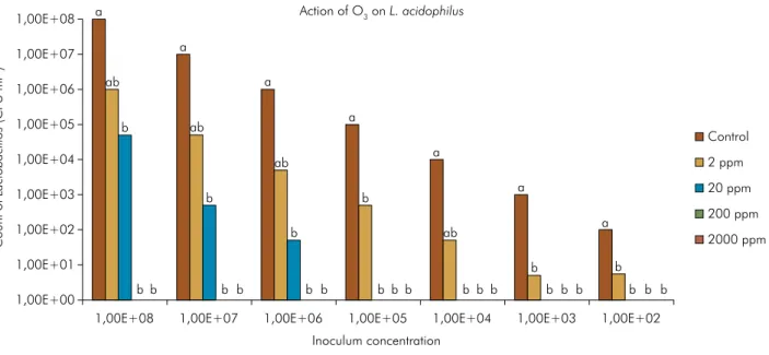
Antimicrobial activity of ozone
Figures 1, 2, and 3 show the inhibition of bacterial
growth on a logarithmic scale log10 after treatment at different concentrations of O3. Data shows a similar Table 1. Description of each plate.
Plate Inoculum O3 concentration
1- Negative control No No treatment
2- Positive control Yes No treatment
3- Experimental Yes 2 ppm
4- Experimental Yes 20 ppm
5- Experimental Yes 200 ppm
pattern of inhibition for all three bacteria tested. L. acidophilus (Figure 2) and E. faecalis (Figure 3),
when subjected to application of 2ppm of ozone, presented a slight resistance of these microorganisms to the gas, At concentrations ≥20 ppm, the inhibition was greater than 3log10 (99.9% kill) (p < 0.01) for all three bacteria.
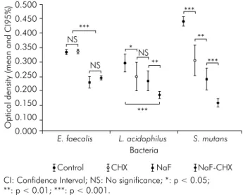
Antimicrobial activity of NaF-CHX
Table 2 shows the effect of each antibiotic in each bacteria compared to their control group (optical density “baseline” for each bacterial species). All three antibiotics tested on S. mutans acted better when compared to other microorganisms. Comparing E.faecalis and L.acidophilus, there was a weak tendency
1,00E+00 1,00E+01 1,00E+02 1,00E+03 1,00E+04 1,00E+05 1,00E+06 1,00E+07 1,00E+08
1,00E+08 1,00E+07 1,00E+06 1,00E+05 1,00E+04 1,00E+03 1,00E+02
Count of
S.
mutan
s
(CFU ml
-1)
Inoculum concentration Action of O3 on S. mutans
Control 2 ppm
20 ppm
200 ppm 2000 ppm
b
a b
b b
a
a a
ab a
ab
b a
ab
b
b
b b b b b b b b b b b b b b a
ab
b b b
Figure 1. Inhibition of S. mutans growth in logarithmic scale (log10) after O3 treatment. Logarithm values followed by the same
lower case letters (for comparasions between O3 concentrations) do not differ by Tukey’s test (significance level of 5%).
Count of
La
ctobacillo
s
(CFU ml
-1)
Action of O3 on L. acidophilus
1,00E+00 1,00E+01 1,00E+02 1,00E+03 1,00E+04 1,00E+05 1,00E+06 1,00E+07 1,00E+08
1,00E+08 1,00E+07 1,00E+06 1,00E+05 1,00E+04 1,00E+03 1,00E+02 Inoculum concentration
Control
2 ppm
20 ppm 200 ppm
2000 ppm
b a
b
b b
a
a a
a
ab ab
a
ab
b a
ab
b
b b b b b b b b b b b b b b b b b b
Figure 2. Inhibition of L. acidophilus growth in logarithmic scale (log10) after O3 treatment. Logarithm values followed by the same
to see a more favorable effect of L.acidophilus when
undergoing treatment with CHX and NaF separately; nonetheless, there was no difference for the NaF-CHX.
Figure 4 shows the analysis of the effect of each antibiotic and combination (NaF-CHX), by type
of bacteria. The two substances were effective in controlling bacterial growth for the three bacteria tested when compared to the control group (p < 0.001). On
E.faecalis there was no difference between CHX and
Control, NaF and NaF-CHX were better than control or CHX, and there was no difference between NaF-CHX or NaF. In the test for L.acidophilus, all antibiotics were
better than control: NaF-CHX was better than any of
the substance tested alone, and there was no difference
between CHX or NaF. For S. mutans, NaF-CHX was
more effective than any other substance tested alone
and the Control group, NaF was better than CHX,
and CHX was better than Control group.
Discussion
This study was developed with the purpose of identifying an antimicrobial agent able to inhibit or halt ECC caries until the appropriate behavior and
Table 2. The effect of antimicrobials in the optical density of bacterial growth
Antimicrobials Bacteria Mean difference of optical
density compared to control p-value
CHX
E. faecalis 0.002 0.089a
L. acidophilus - 0.049 0.028b
S. mutans - 0.138 < 0.001c
NaF
E. faecalis - 0.110 0.088a
L. acidophilus - 0.065 < 0.001b
S. mutans - 0.206 < 0.001c
CHX + NaF
E. faecalis - 0.093 0.326a
L. acidophilus - 0.115 < 0.001b
S. mutans - 0.292 < 0.001c
CHX: Chlorhexidine; NaF: Sodium fluoride. The p-values refer to the test of interaction between the effect of the antibiotic and the bacteria compared the following groups: a: E. faecalis and L. acidophilus; b: L. acidophilus and S.mutans; c: E. faecalis and S.mutans.
Count of
E.
faecalis
(CFU ml
-1)
Action of O3 on E. faecalis
1,00E+00 1,00E+01 1,00E+02 1,00E+03 1,00E+04 1,00E+05 1,00E+06 1,00E+07 1,00E+08
1,00E+08 1,00E+07 1,00E+06 1,00E+05 1,00E+04 1,00E+03 1,00E+02 Inoculum concentration
Control
2 ppm 20 ppm
200 ppm
2000 ppm a
a
c
b
b b b
b ab
a
bc
b b
b b
b a
b
a a
a a
Figure 3. Inhibition of E. feacalis growth in logarithmic scale (log10) after O3 treatment. Logarithm values followed by the same
lower case letters (for comparasions between O3 concentrations) do not differ by Tukey’s test (significance level of 5%).
CI: Confidence Interval; NS: No significance; *: p < 0.05; **: p < 0.01; ***: p < 0.001.
Figure 4. Effect of CHX, NaF and NaF-CHX on S. mutans,
L. acidophilus and E. feacalis.
Optical density (mean and CI95%)
Bacteria
Control CHX NaF NaF-CHX *
**
** ***
***
*** NS
NS
NS
*** 0.450
0.350
0.250
0.150 0.100 0.000 0.500
0.400
0.300
0.200
cooperation of the patient are achieved, allowing for routine dental care. The results showed that the hypothesis was accepted, since all the substances tested showed antimicrobial activity. Ozone is considered a strong oxidizer of the cell walls and cytoplasmic membranes of bacteria and is considered a potent bactericidal, antiviral, and antifungal agent.12 It is important to point out that this statement is based on the use of ozone in blood, not gassing ozone to a microfilm.12,13 This is a common finding in the literature, extrapolating the use and effect of systemic ozone dissolving it in the bloodstream, to the dental
field.12,14 These articles explain the indications and mechanism of action of ozone dissolved in blood but there is no reference to the effect in the oral cavity.
Therefore, the real ability of ozone to kill cariogenic
bacteria is yet to be determined.
From the three bacterial strains employed in this
study, two were selected to represent pathogenic bacteria commonly present in ECC (S. mutans and L. acidophilus), and E. faecalis was known to be more
resistant to antibiotics5,9. Our results showed that the application of O3 completely prevented the in vitro growth of all bacterial strains.
The use of agar-sheep-blood for bacterial culture in Petri dishes is a usual practice in microbiology, including the evaluation of bactericidal effects of different substances, such as ozone.15,16 Using similar methodology, an in vitro study revealed a reduction of viability of E. coli, S. aureus, and Listeria innocua under application of O3 (2ppm / 4 hours).16Another study showed that a mixture of 20 mg of O3/mL + O2
(1% O3/99%O2) in a single application for 5 minutes is able to effectively inhibit bacterial growth of E. faecalis, Staphylococcus aureus, and Escherichia coli.15 In all the studies cited above, the bacteria were cultured on agar in Petri dishes as well as in other culture media, and both studies considered agar as the best culture
media for measuring the efficacy of O3. This study showed success in applying O3 for 4 minutes in all three bacteria tested. It is pertinent to emphasize that
both generators (OL80A and OL80W) produce gas within chambers, and to achieve the desired final concentration takes about 20 minutes, adding 4 more
minutes in target concentration. Polydorou et al.17, showed bactericidal effects of O3 on S. mutans after
the application for 80 seconds, and suggested other studies on different bacteria. Estrela et al.5, reported satisfactory results on inhibiting growth of S. mutans, L. casei, and A. naeslundii using ozonated water, although the authors reported a limitation of delivering ozone gas from the equipment used. Polydorou et al.18, found complete inhibition of S.mutans growth after a
1-minute application even after a follow up of 8 weeks;
however, the method showed limited effect on L.casei. Hems et al.10, reported limitation using O
3 gas. However,
they showed a positive effect on planktonic E.feacallis with ozonated water after 4 minutes. As in the present study, most of the studies tested ozone generator for laboratory use only, we believe there is still a need to
find equipment for clinical use.
For the method that assesses bacterial death to be considered efficient, it is expected to decrease by at
least 3log10 (99.9%) in the number of bacteria detected19.
Using this parameter and standardizing the initial
concentration of the three bacteria, we observed a logarithmic ≥ 3log10 kill at all conditions tested, except for L. acidophilus and E. faecalis when subjected to application of 2 ppm of ozone. This suggests a slight resistance of these microorganisms to the gas, but this does not alter the results, since the equipment available and the methods that have been used and proven in the literature used a concentration above 2,000 ppm of ozone.7,10,17,18 Under the lab condition tested in this study, the results show complete inhibition of all the three bacteria using the concentration ≥ 20 ppm.
We suggest more studies in order to test the efficacy
of O3 under clinical situations and with lower doses of the gas. The present data show antimicrobial O3 effect on E. faecalis. This is in agreement with another study that found similar results testing ozone in the liquid (ozone bubbles), which demonstrated that the solution has a bactericidal effect on E. faecalis in planktonic
surface, and suspended in liquid.10 Nagayoshi et al.,20 also showed the sensitivity of E. faecalis to ozone, which the authors examined ex vivo, the effect of ozonated water on E. faecalis and S. mutans, and also verified cytotoxicity in mouse fibroblasts. They concluded that
action of antimicrobial agents.5,10 The main difference between our study and others is the ozone generator used and the method of application.
Both gaseous and aqueous ozone have been
reported to exert antimicrobial effects.10,21 The aqueous form of ozone is a potential antiseptic agent and shows less cytotoxicity than gaseous ozone.22 Overwhelming evidence shows that the bronchial-pulmonary system is very sensitive to ozone and that this gas should never be inhaled.
The respiratory tract lining fluid is constituted of a very thin, watery film containing a minimal amount of antioxidants that makes mucosal cells extremely
vulnerable to oxidation.23 Known side effects are epiphora and upper respiratory irritation, rhinitis, cough, headache, occasional nausea, and vomiting.24 Cytotoxicity is less relevant when applying ozone gas onto carious tooth hard substance via a sealing suction system as a prerequisite to avoid inhalation.22
Ozone generating equipment converts oxygen to ozone. It is delivered through a system composed of
a hand piece fitted with a silicone cup. This ensures
close contact between the silicone cup and the carious area of the tooth so that the ozone does not escape. In previous tests with commercialized ozone generators, it was not possible to achieve the ideal sealing enabling the creation of an impermeable layer or the so called vacuum effect. Therefore, we decided to use a device that produced O3 in
chambers. So, from the results found in this study,
we suggest that there is a need for improvement of equipment for clinical use, particularly with silicone
tips that fit onto deciduous teeth.
The data from this investigation, together with other in vitro studies cited here, indicate that the use of ozone gas may be an alternative for infections caused by S. mutans, L. acidophilus, and E. faecalis. However,
there is a need for other tests in bacterial biofilms and
clinical studies with proper controls and adequate sample size for the validation of this technique.
The antimicrobial activity of NaF-CHX on planktonic species of oral pathogens was evaluated
by minimum inhibitory concentration (MIC) assays.
Although NaF and CHX exhibited antimicrobial
activity against all bacteria tested, MIC results revealed
that the association of NaF + CHX is more effective
than agents tested alone. Although both substances presented antimicrobial activity alone in all tested microorganisms, it is not feasible to compare the
effect of NaF versus CHX.
Chlorhexidine (CHX) is one of the most widely used oral antimicrobial agents and is available in different formulations.25 The substance is known to have good
substantivity and at high concentrations (0.12% or more)
is bactericidal, causing a lethal damage to the bacterial membrane, being active on both gram-negative and gram-positive bacteria.4 The CHX as antiplaque and anti-gingivitis agent remains as the gold standard, but its use as an anti-caries agent particularly in established lesions has been considered controversial, based on
inconclusive clinical findings.26 In the presence of SLS (sodium lauryl sulfate), which is the most commonly
used surfactant in dentifrices, CHX has its efficiency decreased due to cationic (CHX) and anionic (SLS)
reactions.27 As the purpose of the study is to propose a substance that is effective in halting the ECC, the
tested solution (NaF-CHX) was formulated starting
from compounds in its purest form available on the
market, without adding any other substance. Under
the test condition, our results are similar to those found in the literature, proving its effectiveness in
reducing the number of S. mutans28,29on L. acidophilus30 and on E. Faecalis.31
In this study, fluoride was used in the form of sodium fluoride (NaF), and under the conditions tested, NaF had a satisfactory antimicrobial effect
on all bacteria tested. There are reports of direct antimicrobial activity against cariogenic bacteria.32
In the presence of low extracellular pH, the F- is
transported as hydrofluoric acid (HF) into the bacterial
cell, where it then dissociates into H+ and F-.33 The
excess acidification of the cytoplasm may also inhibit
the glucose transport mechanism inside the cell. Although these mechanisms have been demonstrated in cell culture, there is no proof of an antimicrobial
effect of fluoride clinically. It is well documented that fluoride has the ability to inhibit or even reverse
the initiation and progression of dental caries.34,35,36 Therefore, in clinical situations where there is a need for an urgent and effective treatment for halting the
ECC, the benefits of using NaF and CHX seem to be
The data obtained in this study suggest that there is
an additive effect when there is a combination of NaF and
CHX. This is clearly demonstrated by the fact that under the conditions of the study, both substances showed antimicrobial effects alone, but showed better results
when used in combination. This finding corroborates with the findings reported by other authors,37 testing in vitro the combination of Naf-CHX on S. mutans and S. sobrinus, and found significant increases in the antimicrobial effectiveness of the solutions. The benefit
of the association is attributed to the low molecular
weight of the fluoride ion, which allows it to reach niches
inaccessible to CHX, as in the case of incipient caries.38
Furthermore, extremely high concentrations of CHX did
not seem necessary for a lasting antimicrobial effect on bacterial colonization on root surfaces.39 Therefore, with the exception of a limited number of pathogens such as S. mutans, L. acidophilus, and E. faecalis, most indigenous
oral microorganisms are benign or beneficial. Therefore,
the use of high concentrations of any antimicrobial substance can be detrimental to the host.40
The bactericidal effect of NaF-CHX in S. mutans and L. acidophilus demonstrated in this study reinforces the advantage of combining these two substances for controlling the progression of dental caries. In general,
the idea of associating NaF-CHX aims to gain control of
bacterial proliferation (CHX) associated with reversing
the established caries (NaF). Therefore, this study
demonstrated that the combination of antimicrobials with different antimicrobial mechanisms allows for a more effective treatment strategy against pathogens related to severe childhood caries.
Conclusion
The antimicrobial agents tested were able to inhibit S. mutans, L. acidophilus, and E. faecalis. The technique of using ozone gas seems to be a good alternative for controlling the progression of carious lesions in children. However, there remains a need to develop a generator suitable for clinical use. The association
of NaF-CHX was shown to be a good antimicrobial
agent with an easy application method and faster
clinical applicability. Further investigation should be performed to confirm these results and to develop
protocols for the use of such products to prevent the progression of severe childhood caries.
Acknowledgments
This research was supported by CAPES (Brazilian Federal Agency for the Support and Evaluation of
Graduate Education) # 1342-12-6. The funders had no role in study design, data collection and analysis, decision to publish, or preparation of the manuscript.
1. American Academy of Peedodontics. American Academy of Pediatrics. Policy on early childhood caries (ECC): classifications, consequences, and preventive strategies. Ref Man. 2014;37(6):50-2.
2. Orhan AI, Oz FT, Ozcelik B, Orhan K. A clinical and
microbiological comparative study of deep carious lesion treatment in deciduous and young permanent molars. Clin Oral Investig. 2008;12(4):369-78.
doi:10.1007/s00784-008-0208-6
3. Featherstone JDB. Delivery challenges for fluoride, chlorhexidine and xylitol. BMC Oral Health. 2006;6 Suppl 1:S8. doi:10.1186/1472-6831-6-S1-S8
4. Marsh PD. Controlling the oral biofilm with antimicrobials. J. Dent. 2010;38 Suppl 1:S11-5. doi:10.1016/S0300-5712(10)70005-1
5. Estrela C, Estrela CR, Decurcio DA, Hollanda AC, Silva JA. Antimicrobial efficacy of ozonated water, gaseous
ozone, sodium hypochlorite and chlorhexidine in
infected human root canals. Int Endod J. 2007;40(2):85-93. doi:10.1111/j.1365-2591.2006.01185.x
6. Atiyeh BS, Dibo SA, Hayek SN. Wound cleansing, topical antiseptics and wound healing. Int Wound J. 2009;6(6):420-30. doi:10.1111/j.1742-481X.2009.00639.x
7. Johansson E, Claesson R, Dijken JWV. Antibacterial effect of ozone on cariogenic bacterial species. J Dent. 2009;37(6):449-53. doi:10.1016/j.jdent.2009.02.004
8. Sadatullah S, Mohamed NH, Razak FA. The antimicrobial
effect of 0.1 ppm ozonated water on 24-hour plaque
microorganisms in situ. Braz Oral Res. 2012;26(2):126-31. doi:10.1590/S1806-83242012000200007
9. Wali IE, Elhilaly G, Eid M, Omar WA, Elrafie S. The
antimicrobial efficacy of ozonated water, chlorhexidine and sodium hypochlorite against single species biofilms of Enterococcus faecalis and Candida albicans. Egypt J Med
Microbiol. 2008;17(3):419-28.
10. Hems RS, Gulabivala K, Ng Y-L, Ready D, Spratt DA. An in vitro evaluation of the ability of ozone to kill a
strain of Enterococcus faecalis. Int Endod J. 2005;38(1):22-9. doi:10.1111/j.1365-2591.2004.00891.x
11. Arnold RR, Wei HH, Simmons E, Tallury P, Barrow DA, Kalachandra S. Antimicrobial activity and local release
characteristics of chlorhexidine diacetate loaded within the dental copolymer matrix, ethylene vinyl acetate.
J Biomed Mater. Res B Appl Biomater. 2008;86(2):506-13. doi:10.1002/jbm.b.31049
12. Gomes BP, Ferraz CC, Vianna ME, Berber VB, Teixeira FB, Souza-Filho FJ. In vitro antimicrobial activity
of several concentrations of sodium hypochlorite and chlorhexidine gluconate in the elimination of
Enterococcus faecalis. Int Endod J. 2001;34(6):424-8. doi:10.1046/j.1365-2591.2001.00410.x
13. Bocci VA. Scientific and medical aspects of ozone therapy: State of the art. Arch Med Res. 2006;37(4):425-35. doi:10.1016/j.arcmed.2005.08.006
14. Lynch E, Swift EJ Jr. Evidence-based caries reversal using ozone. J Esthet Restor Dent. 2008;20(4):218-22. doi:10.1111/j.1708-8240.2008.00183.x
15. Fontes B, Heimbecker AMC, Brito GS, Costa SF, Heijden IM, Levin AS et al. Effect of low-dose gaseous ozone on pathogenic bacteria. BMC Infect Dis. 2012;12:358. doi:10.1186/1471-2334-12-358
16. Moore G, Griffith C, Peters A. Bactericidal properties
of ozone and its potential application as a terminal
disinfectant. J Food Prot. 2000;63(8):1100-6.
17. Polydorou O, Pelz K, Hahn P. Antibacterial effect
of an ozone device and its comparison with two
dentin-bonding systems. Eur J Oral Sci. 2006;114(4):349-53. doi:10.1111/j.1600-0722.2006.00363.x
18. Polydorou O, Halili A, Wittmer A, Pelz K, Hahn P. The
antibacterial effect of gas ozone after 2 months of in vitro evaluation. Clin Oral Investig. 2012;16(2):545-50.
doi:10.1007/s00784-011-0524-0
19. Thanomsub B, Anupunpisit V, Chanphetch S,
Watcharachaipong T, Poonkhum R, Srisukonth C. Effects
of ozone treatment on cell growth and ultrastructural
changes in bacteria. J Gen Appl Microbiol. 2002;48(4):193-9. doi:10.2323/jgam.48.193
20. Nagayoshi M, Kitamura C, Fukuizumi T, Nishihara
T, Terashita M. Antimicrobial effect of ozonated
water on bacteria invading dentinal tubules. J Endod 2004;30(11):778-81. doi:10.1097/00004770-200411000-00007
21. Baysan A, Lynch E. Effect of ozone on the oral microbiota and clinical severity of primary root caries. Am J Dent.
2004;17(1):56-60.
22. Huth KC, Jakob FM, Saugel B, Cappello C, Paschos E, Hollweck R et al. Effect of ozone on oral cells compared with established antimicrobials. Eur J Oral Sci. 2006;114(5):435-40. doi:10.1111/j.1600-0722.2006.00390.x
23. Bocci VA. Tropospheric ozone toxicity vs. usefulness
of ozone therapy. Arch Med Res. 2007;38(2):265-7.
doi:10.1016/j.arcmed.2006.09.011
24. Pattanaik B, Pattanaik S, Naitam D, Jetwa D, Manglekar S,
Dani A. Ozone therapy in dentistry: a literature review.
J Interdiscip Dent. 2011;1(2):87-92.
25. De Siena F, Del Fabbro M, Corbella S, Taschieri S, Weinstein R. Evaluation of chlorhexidine 0.05% with the adjunct of fluoride 0.05% in the inhibition of plaque
formation: a double blind, crossover, plaque regrowth
study. Int J Dent Hyg. 2012. doi:
26. Huang GJ, Roloff-Chiang B, Mills BE, Shalchi S, Spiekerman C, Korpak AM et al. Effectiveness of MI
Paste Plus and PreviDent fluoride varnish for treatment of white spot lesions: a randomized controlled trial.
Am J Orthod Dentofacial Orthop. 2013;143(1):31-41. doi:10.1016/j.ajodo.2012.09.007
27. Venu V, Prabhakar AR, Basappa N. Comparative
evaluation of antibacterial property and substantivity of chlorhexidine containing dentifrices with sodium lauryl sulfate and Tween as surfactants: an in vivo
study. Indian J Dent Res. 24(4):521-2.
doi:10.4103/0970-9290.118367
28. Emilson CG, Gisselsson H, Birkhed D. Potential efficacy
of chlorhexidine against mutans streptococci and
human dental caries. Eur J Oral Sci. 1999;107(3):170-5. doi:10.1046/j.0909-8836.1999.eos1070303.x
29. Lobo PLD, Carvalho CBM, Fonseca SG, Castro RS, Monteiro AJ, Fonteles MC et al. Sodium fluoride
and chlorhexidine effect in the inhibition of mutans streptococci in children with dental caries: a randomized, double-blind clinical trial. Oral Microbiol Immunol.
2008;23(6):486-91. doi:10.1111/j.1399-302X.2008.00458.x
30. Madléna M, Vitalyos G, Márton S, Nagy G. Effect of
chlorhexidine varnish on bacterial levels in plaque
and saliva during orthodontic treatment. J Clin Dent.
2000;11(2):42-6.
31. Wang CS, Arnold RR, Trope M, Teixeira FB. Clinical efficiency of 2% chlorhexidine gel in reducing intracanal bacteria. J Endod. 2007;33(11):1283-9. doi:10.1016/j.joen.2007.07.010
32. Baysan A, Lynch E, Ellwood R, Davies R, Petersson L, Borsboom P. Reversal of primary root caries using
dentifrices containing 5,000 and 1,100 ppm fluoride. Caries Res. 2001;35(1):41-6.
33. Li YH, Bowden GH. The effect of environmental pH
and fluoride from the substratum on the development
of biofilms of selected oral bacteria. J Dent Res.
1994;73(10):1615-26.
34. Marinho VCC. Evidence-based effectiveness of topical fluorides. Adv Dent Res. 2008;20(1):3-7 doi:10.1177/154407370802000102
36. Buzalaf MAR, Pessan JP, Honório HM, ten Cate JM.
Mechanisms of action of fluoride for caries control.
Monogr Oral Sci. 2011;22:97-114. doi:10.1159/000325151
37. Pinar Erdem A, Sepet E, Kulekci G, Trosola SC,
Guven Y. Effects of two fluoride varnishes and one
fluoride/chlorhexidine varnish on Streptococcus mutans and Streptococcus sobrinus biofilm formation in vitro. Int J Med Sci. 2012;9(2):129-36. doi:10.7150/ijms.3637
38. McDermid AS, Marsh PD, Keevil CW, Ellwood DC.
Additive inhibitory effects of combinations of fluoride and
chlorhexidine on acid production by Streptococcus mutans and Streptococcus sanguis. Caries Res. 1985;19(1):64-71. doi:10.1159/000260830
39. Ekenbäck SB, Linder LE, Lönnies H. Effect of four dental
varnishes on the colonization of cariogenic bacteria on exposed sound root surfaces. Caries Res. 2000;34(1):70-4.
doi:10.1159/000016572
40. He X, Lux R, Kuramitsu HK, Anderson MH, Shi W. Achieving
probiotic effects via modulating oral microbial ecology. Adv