Methods for Comprehensive RNA Structure and Dynamics Analysis using SHAPE Technologies
Kady-Ann Camele Steen-Burrell
A dissertation submitted to the faculty of the Univeristy of North Carolina at Chapel Hill in partial fulfillment of the requirements for the degree of Doctor of Philosophy in the Department of Chemistry.
Chapel Hill 2011
Approved by Michael Jarstfer
Jeffrey Johnson Linda Spremulli
© 2011
Kady-Ann Camele Steen-Burrell
ABSTRACT
KADY-ANN CAMELE STEEN-BURRELL: Methods for Comprehensive RNA Structure and Dynamics Analysis using SHAPE Technologies
(Under the direction of Kevin M. Weeks)
The many important cellular functions of RNA molecules depend on formation of complex RNA secondary and tertiary structures. The formation of these structures is
facilitated by the intrinsic motion of RNA nucleotides and can be influenced by various ligand or protein interactions. RNA SHAPE technology has made the determination of many
RNA secondary structures facile. However, the applicability of traditional SHAPE technology to short RNAs (less than 100 nucleotides) in their native state is limited by primer extension detection. Additionally, it is often difficult to discern the structural context of
constrained nucleotides in a traditional SHAPE experiment. In this work, I first develop an alternate SHAPE 2ʹ-O-adduct detection method, termed RNase-detected SHAPE, which
takes advantage of the RNA-specific activity of an exoribonuclease, RNase R. In the presence of a SHAPE 2ʹ-O-adduct or adducts at the nucleotide base-pairing face, RNase R
stops three or four nucleotides 3ʹ of the modification site, respectively. RNase-detected
SHAPE allowed for the structural characterization of a small, biologically relevant riboswitch in its ligand-free state and identification of a bulge register shift that facilitates
interactions, termed differential SHAPE reactivity analysis. This method uses two SHAPE electrophiles, N-methylisatoic anhydride (NMIA) and 1-methyl-6-nitroisatoic anhydride
(1M6) that detect slowly dynamic and one-sided stacking nucleotides, respectively. Together, both types of nucleotide behaviors provide a RNA tertiary structure “fingerprint” since both
tend to be over-represented in tertiary structure interactions and motifs. Third, I develop a chemical method for the removal of SHAPE 2ʹ-O-adducts by ester bond cleavage while
limiting RNA phosphodiester backbone degradation which allows for the downstream
manipulation of previously modified RNA. The extent of SHAPE 2ʹ-O-adduct removal can be modulated by varying the concentration of hydroxylamine and reaction times. Finally, I
evaluate the ability of SHAPE chemistry to detect ligand-induced conformational changes by comparing SHAPE reactivities and NMR measurements. Dynamics as measured by SHAPE reactivities and the NMR order parameter, S2, correlate well for small molecule binding to
First, I would like to thank my advisor, Kevin Weeks, for allowing me to work on this project and for giving me the opportunity to take the project in the direction that I wanted to go.
There are many people who have loved and supported me over the years. My family, many of whom I don’t get to see very often, thanks for supporting me through your calls, visits, and
prayers. My friends, both near and far, thank you all for your friendship, understanding and
support. In particular, thanks to Vanessa and Jen for all the support over the years. Graduate school was a little easier because of you. Thanks to my labmates who were always there to bounce ideas and talk science with. I will truly miss you all.
Finally, to my husband, Kevin, for all you have done over the past years in support of my dreams and aspirations. I love you and I could not have done it without you.
LIST OF TABLES ... x
LIST OF FIGURES ... xi
LIST OF ABBREVIATIONS AND SYMBOLS ... xiii
1. Towards Comprehensive Single Nucleotide Analysis of RNA Structure and Dynamics ... 1
1.1 Introduction ... 1
1.1.1 RNA structure and its importance to function ... 1
1.1.2 RNA ligand binding and dynamics ... 3
1.1.3 RNA structure determination methods ... 3
1.1.4 Research overview ... 7
1.1.5 Perspective ... 8
1.2 References ... 10
2. Selective 2ʹ-Hydroxyl Acylation analyzed by Protection from Exoribo- nuclease (RNase-detected SHAPE) ... 13
2.1 Introduction ... 13
2.2 Results ... 16
2.2.1 RNase R detects sites of 2ʹ-O-adduct formation in the TPP riboswitch RNA. ... 16
2.2.2 RNase R detects covalent 2ʹ-O- and kethoxal adducts in a two-site model in the enzyme catalytic site. ... 20
2.2.3 RNase-detected SHAPE yields quantitative structural information for the folding of the TPP riboswitch... 21
2.3 Discussion ... 28
2.4 Experimental ... 28
2.4.1 Screening of candidate RNases. ... 28
2.4.2 Heat inactivation of RNase R. ... 29
2.4.3 Synthesis of Escherichia coli thiamine pyrophosphate (TPP) riboswitch RNAs. ... 30
2.4.4 Structure-selective RNA modification. ... 31
2.4.5 RNase R digestion. ... 32
2.4.6 Kethoxal modification. ... 32
2.4.7 Cleavage of phosphorothioate-containing RNA. ... 32
2.4.8 Primer extension. ... 33
2.4.9 Data analysis. ... 33
2.4.10 RNase R Structure Modeling. ... 34
2.5 References ... 35
3. Fingerprinting RNA Tertiary Structure by Differential SHAPE Reactivity Analysis ... 38
3.1 Introduction ... 38
3.2 Results ... 42
3.2.1 Differential SHAPE analysis of the TPP riboswitch. ... 42
3.2.2 Analysis of the mechanism of differential 1M6 reactivity. ... 48
3.2.3 Analysis of diverse RNAs by differential SHAPE reactivities. ... 54
3.2.4 Differential reactivities as a function of the extent of RNA structure. ... 56
3.4.1 Synthesis of RNA constructs. ... 63
3.4.2 RNA folding and modification. ... 64
3.4.3 Primer extension. ... 64
3.4.4 Data analysis. ... 65
3.4.5 Differential SHAPE analysis. ... 66
3.4.6 Quantum mechanics calculations. ... 66
3.5 References ... 68
4. Hydroxylamine Cleavage of the SHAPE 2ʹ-O-adduct Ester Linkage ... 72
4.1 Introduction ... 72
4.2 Results ... 75
4.2.1 Hydroxylamine solution cleaves the ester bond of SHAPE 2ʹ-O-adducts. ... 75
4.2.2 Hydroxylamine cleavage of 2ʹ-O-adducts in TPP riboswitch. ... 76
4.3 Discussion ... 79
4.4 Experimental ... 82
4.4.1 Synthesis of [32P]-labeled AddC. ... 82
4.4.2 Detection of 2ʹ-O-adduct cleavage in AddC. ... 82
4.4.3 Synthesis of thiamine pyrophosphate RNA. ... 83
4.4.4 RNA 2ʹ-O-adduct cleavage in TPP riboswitch. ... 83
4.4.5 Primer extension. ... 84
4.5 References ... 85
5. Effects of Ligand Binding on RNA Nucleotide Dynamics as detected by SHAPE Chemistry and NMR ... 87
5.1 Introduction ... 87
5.2.1 Correlation between SHAPE and S2 values for the ligand-
free HIV-1 TAR RNA. ... 91
5.2.2 SHAPE detects argininamide binding to HIV-1 TAR RNA and correlates to S2... 94
5.2.3 SHAPE chemistry detects linear peptide binding to the TAR RNA construct. ... 99
5.3 Discussion ... 103
5.4 Experimental ... 105
5.4.1 Synthesis of TAR RNA construct. ... 105
5.4.2 Structure-selective SHAPE modification. ... 106
5.4.3 Primer extension ... 106
5.4.4 SHAPE data analysis. ... 107
5.4.5 NMR relaxation experiments and calculation of the generalized order parameter, S2. ... 107
LIST OF TABLES
Table 3.1 Summary of all the RNAs studied for differential SHAPE
LIST OF FIGURES
Figure 1.1 RNA structure levels ... 2
Figure 1.2 RNA SHAPE technology ... 4
Figure 2.1 Schematic for SHAPE chemistry. ... 15
Figure 2.2 Quantitative detection of 2ʹ-O-adducts by RNase R degradation.. ... 18
Figure 2.3 Heat inactivation of RNase R at 95 °Cand resulting half-lfe of inactivation. ... 19
Figure 2.4 Model of Mycoplasma genitalium RNase R and the interactions that mediate covalent adduct detection in RNA.. ... 22
Figure 2.5 Representative RNase-detected SHAPE experiment.. ... 24
Figure 2.6 Absolute SHAPE reactivities determined by RNase R degradation superimposed on the secondary structure models for the TPP riboswitch in the absence and presence of TPP ligand.. ... 25
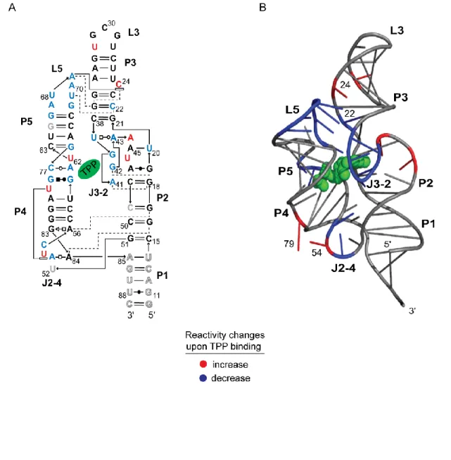
Figure 2.7 Base-pairing and tertiary structure of the ligand-bound TPP riboswitch showing structural features whose constituent nucleotides increase or decrease in SHAPE reactivity when TPP binds. ... 27
Figure 3.1 The mechanism of RNA SHAPE chemistry with the concurrent hydrolysis reaction. ... 40
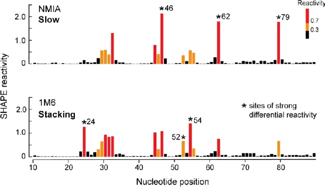
Figure 3.2 Absolute SHAPE reactivities for the ligand-bound state of the TPP riboswitch resulting from reaction with NMIA and 1M6.. ... 43
Figure 3.3 Absolute SHAPE reactivities for the ligand-bound TPP riboswitch resulting from reaction with NMIA and 1M6 are superimposed on the planar representation of the three-dimensional structure. ... 44
Figure 3.4 Differential reactivities for the ligand-bound state of the TPP riboswitch.. ... 46
Figure 3.5 Nucleotide conformations and structural context for all the differential reactivities in the ligand-bound state of the TPP riboswitch.. ... 47
Figure 3.8 Reagent differential reactivities superimposed on the three- dimensional structures and differential reactivity plots of the adenine and lysine ribsowitches, and specificity domain of RNase P. ... 55 Figure 3.9 Differential SHAPE reactivities as a function of the extent of
RNA structure.. ... 57 Figure 3.10 NMIA and 1M6 reagent enhancements are superimposed on
the planar representation of the three-dimensional structure of the ligand-bound; secondary structure model of the ligand- free and secondary structure model of the ion-free, ligand-free states of the TPP riboswitch. ... 59 Figure 4.1 Mechanism of RNA SHAPE reaction showing formation of the
ester linkage between the 2ʹ-hydroxyl of flexible nucleotides and the SHAPE reagent.. ... 74 Figure 4.2 Gel showing modification of a model nucleotide, AddC, with
1M7 and removal of 2ʹ-O-adduct by hydroxylamine.. ... 77 Figure 4.3 Capillary electropherograms showing fluorescent signal
corresponding to detection of 2ʹ-O-adduct as a function of hydroxylamine (NH2OH) concentration and reaction time.. ... 78 Figure 4.4 Proposed mechanism consistent with hydroxylamine reactivity
and SHAPE 2ʹ-O-adduct ester bond cleavage. ... 81 Figure 5.1 Schematic for the relationship between SHAPE and NMR
flexibility measurements. ... 89 Figure 5.2 Ligand-free TAR RNA from the HIV-1 RNA . ... 92 Figure 5.3 SHAPE reactivities of the free and argininamide-bound TAR
RNA. ... 95 Figure 5.4 SHAPE and S2 values for the argininamide-bound TAR RNA. ... 96 Figure 5.5 Structures of the free and argininamide-bound TAR RNA. . ... 98 Figure 5.6 SHAPE reactivities for the free and linear peptide-bound TAR
RNA. ... 101 Figure 5.7 SHAPE and S2 values compared for the linear peptide-bound
LIST OF ABBREVIATIONS AND SYMBOLS
1M6 1-methyl-6-nitroisatoic anhydride 1M6Br 1-methyl-6-bromoisatoic anhydride 1M6M 1-methyl-6-methylisatoic anhydride 1M7 1-methyl-7-nitroisatoic anhydride 2ʹ-OH 2ʹ-hydroxyl
2-AP 2-aminopurine
A adenine
AddC adenosine-2ʹ,3ʹ-dideoxycytidine triphosphate ATP adenosine triphosphate
C cytosine
cDNA complementary deoxyribonucleic acid
Ci curie
cm centimeter
CSD cold shock domain °C degree Celsius
dNTP deoxyribonucleotide triphosphate DMSO dimethylsulfoxide
DNA deoxyribonucleic acid DTT dithiothreitol
EDTA ethylenediaminetetraacetic acid
G guanosine
h hour
H2O water
HIV human immunodeficiency virus KCl potassium chloride
L liter
M molar
Mg2+ magnesium ion MgCl2 magnesium chloride
min minute
mg milligram
mL milliliter
mm millimeter
mM millimolar
μg microgram
μL microliter
μM micromolar
N nitrogen
NaCl sodium chloride NH2OH hydroxylamine
nM nanomolar
NMIA N-methylisatoic anhydride NMR nuclear magnetic resonance
NO2 nitro
NOE nuclear Overhauser effect
nt nucleotide
NTP ribonucleotide triphosphate
O oxygen
P phosphate
PAGE polyacrylamide gel electrophoresis PCR polymerase chain reaction
pmol picomole
PNK polynucleotide kinase
QM quantum mechanics
RNA ribonucleic acid RNase ribonuclease
RMSD root mean square deviation RNB ribonucleic acid binding
s second
SAFA semi-automated footprinting analysis SCF self-consistent field
SHAPE Selective 2ʹ-Hydroxyl Acylation analyzed by Primer Extension
σm sigma meta
T1 longitudinal relaxation rate T2 transverse relaxation rate TAR trans-activation region
Tat trans-activator of transcription TBE 90 mM Tris-borate, 2 mM EDTA TE 10 mM Tris-HCl (pH 8), 1 mM EDTA TPP thiamine pyrophosphate
Tris tris(hydroxymethyl)aminomethane tRNA transfer ribonucleic acid
U uridine
UTR untranslated region
v volume
w weight
CHAPTER 1
Towards Comprehensive, Single Nucleotide Analysis of RNA Structure
and Dynamics
1.1 Introduction
1.1.1 RNA structure and its importance to function
RNA serves dual roles as both an integral carrier of genetic information at the
primary nucleotide sequence level, and as an important biomolecular machine through formation of higher-order structures that are central to almost every biological process in the cell [1-3]. Many of these critical cellular functions including protein synthesis, catalysis,
gene expression and regulation, depend on the ability of RNA molecules to fold back on itself to form complex and specific dimensional structures [3-5]. RNA three-dimensional structures vary both in their size and complexity from small tRNAs to large
ribosomal RNAs [6-8]. Formation of these three-dimensional structures requires base-pairing interactions to form secondary structures (Figure 1.1A) and long-range contacts including
1.1.2 RNA ligand binding and dynamics
RNA structure, folding and function are also influenced by local nucleotide dynamics. Many cellular functions, such as gene regulation by riboswitches, require RNA molecule to undergo conformational changes in response to changes in ionic conditions or
external cofactors such as small molecules and proteins (compare A and B, Figure 1.1) [3, 11]. Additionally, RNAs in many ribonucleoprotein complexes rely on protein binding to
form the functional, active RNA conformation [12, 13]. These ligand-induced conformational changes are facilitated by the inherent motion of RNA nucleotides. RNA dynamics occur over a range of timescales from very fast bond vibrations to intermediate
helical and domain motions to very slow structural rearrangements [16, 17]. The ability to biochemically evaluate ligand-induced conformational changes of individual RNA
nucleotides regardless of RNA size and complexity is an unmet experimental challenge.
1.1.3 RNA structure determination methods
RNA secondary structure determination is a key first step towards comprehensive understanding of RNA structure-function relationships. Conventional biochemical methods
of secondary structure determination rely on small molecules or ribonucleases that primarily modify or cleave the RNA in a base-specific manner including kethoxal, which modifies G residues, and RNase T1, which cleaves after G residues [18, 19]. Additionally, some
ribonucleases cleave in a base non-specific manner such RNase V1, which cleaves base-paired nucleotides [18]. While these biochemical methods have provided valuable
information about RNA structures, no single method is capable of completely mapping the structure of any given RNA of interest.
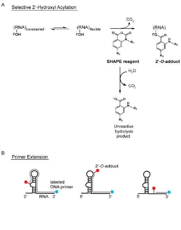
Acylation analyzed by Primer Extension (SHAPE) takes advantage of the intrinsic reactivity of the 2ʹ-hydroxyl on the ribose sugar. This feature allows structural information to be
obtained for all nucleotides in a single experiment. The ability of the 2ʹ-hydroxyl to deprotonate and react with electrophiles is strongly influenced by local nucleotide flexibility
[20]. Conformationally flexible nucleotides preferentially react with SHAPE electrophiles such as 1-methyl-7-nitroisatoic anhydride (1M7) resulting in the formation of ester adducts (2′-O-adducts) (Figure 1.2A). Base-paired or otherwise constrained nucleotides are less
reactive towards SHAPE electrophiles [21, 22]. The resulting 2ʹ-O-adducts are then identified as stops in a primer extension reaction using 5ʹ-end labeled primers (radiolabeled
or fluorescently labeled) that are annealed to the 3ʹ-end of the RNA (Figure 1.2B). Reverse transcriptase produces cDNAs that terminate one nucleotide before the modification site resulting in a cDNA library whose fragment lengths correspond to the sites of modification
and whose amounts correspond to degree of flexibility. The cDNA fragments are then separated by denaturing polyacrylamide gel electrophoresis (for radiolabeled fragments) or
capillary electrophoresis (for fluorescently-labeled fragments) [23, 24]. Dideoxy-terminated sequencing reactions are performed concurrently in order to map the modification site to the RNA sequence.
SHAPE allows for quantitative, robust, single nucleotide resolution structural information to be obtained for most RNAs in a relatively short time using picomolar amounts
of RNA. As local nucleotide flexibility correlates with SHAPE reactivity [25], SHAPE reactivities can be applied to the prediction of known and novel RNA structures by interpreting the reactivities as pseudo free energies that can be used to improve computer-
RNA tertiary structure determination has proven to be a more challenging undertaking. Biophysical methods such as NMR and x-ray crystallography have been
primarily employed in the tertiary structure determination of RNA. X-ray crystallography provides high quality atomic resolution of RNA structure and allows for the observation of
specific nucleotide conformations, tertiary structure motifs and contacts [8]. However, x-ray crystallography provides a snapshot of a static RNA state so information about RNA dynamics are difficult to detect. Additionally, many biologically relevant RNA states are too
dynamic to be crystallized. The most widely used method for studying RNA local nucleotide dynamics is NMR spectroscopy [17, 28]. However, NMR spectroscopy is limited by the size
and complexity of the RNA being studied.
Along with x-ray crystallography and NMR, biochemical methods have been employed to map the tertiary interactions in RNA. Current biochemical methods of RNA
tertiary structure analysis are primarily based on cleavage of the RNA backbone by hydroxyl radicals. Hydroxyl radicals are usually generated in situ by reagents that are either free in
solution or tethered to the RNA [19]. In-solution, free hydroxyl radical experiments provide information about solvent accessible nucleotides while tethered hydroxyl radical experiments provide information about nucleotide distance in three-dimensional space. However, some of
these experiments tend to be difficult to implement due to the requirement of a modified RNA sequence for tethering the reagent [29] and the results from hydroxyl radical
1.1.4 Research overview
Consequently, the overall goal of this project was to develop methods that address the problem of characterization of RNA structure and dynamics for any given RNA all within the framework of SHAPE technology.
SHAPE technology has revolutionized the RNA structure field by providing an accessible, relatively straightforward method for analyzing RNA structure. With
improvements in reagent reaction time [22], electrophoresis separation [24] and data analysis [30], SHAPE technology has allowed for the probing of a number of RNAs [26, 31-33]. Traditional SHAPE technology relies on primer extension as the method of detecting
modified nucleotides. Primer extension was ideal for probing long RNA structures, but the requirement of a primer binding site adjacent to the RNA sequence of interest prevented the
application of traditional SHAPE to many important native short RNAs including riboswitches and nucleotides at the ends of all RNAs.
In Chapter 2, I describe an alternate method of 2ʹ-O-adduct detection using the
exoribonuclease Mycoplasma genitalium RNase R. This method, termed RNase-detected SHAPE, is simple to use and allows for the structural analysis of short, authentic RNAs and
the ends of longer RNAs. RNase R is also capable of detecting covalent adducts on the base-pairing face of nucleotides. I then applied this new method to develop a secondary structure model for the ligand-free state of a biologically important RNA, the thiamine
pyrophosphate-sensing (TPP) riboswitch.
In Chapter 3, I develop a new method, termed Differential SHAPE Reactivity
N-preferentially with nucleotides undergoing slow dynamics while 1M6 reacts N-preferentially with nucleotides that possess an unoccupied nucleobase face for stacking interactions with 1M6. The combined preferential reactivities allows for the “fingerprinting” of RNA tertiary
structure by identifying nucleotides involved in tertiary contacts in a simple, robust manner.
In Chapter 4, I develop a method to chemically cleave the ester linkage of the stable 2ʹ-O-adduct while limiting phosphodiester backbone cleavage. This method results in
complete removal of the 2ʹ-O-adduct and allows for downstream biochemical manipulations
of the previously modified RNA provided the structural information was previously encoded by either primer extension or RNase R detection.
Finally, in Chapter 5, I characterize the correlation between SHAPE and RNA dynamics for ligand-induced conformational changes. I compare SHAPE reactivities and NMR measurements for ligands bound to the trans-activating region (TAR) of HIV RNA.
The absolute NMR measurements correlate strongly to SHAPE reactivities for binding of the small molecule argininamide, but the absolute measurements did not correlate with binding
of a larger molecule, an 11 nucleotide linear peptide mimic of the Tat protein. However, both SHAPE chemistry and NMR reflected the same changes in local nucleotide flexibility upon linear peptide binding and suggests that SHAPE can detect changes in conformational
dynamics upon ligand binding.
1.1.5 Perspective
The principles of molecular biology, biochemistry, organic chemistry and physical chemistry were integrated throughout this project to address the problem of accurate RNA structure determination regardless of RNA size or complexity. I demonstrate that SHAPE
an exoribonuclease; nucleotides involved in tertiary structure contacts can be detected using the differences in SHAPE reactivities between NMIA and 1M6; the ester linkage of SHAPE 2ʹ-O-adducts can be specifically cleaved with limited RNA degradation using
hydroxylamine; and changes in local nucleotide dynamics as a result of ligand binding can be
detected using SHAPE chemistry.
It is my hope that each of the methods that I have developed will be applied to understanding structure-function relationships of RNAs in general, and specifically to
predicting the secondary and tertiary structures of novel RNAs. I expect that these methods will contribute to the further development of SHAPE as a facile, high-throughput technology
1.2 References
1. Gesteland, R.F., Cech, T.R., and Atkins, J.F. The RNA world. 3rd ed. 2006, Cold Spring Harbor, New York: Cold Spring Harbor Laboratory Press.
2. Strobel, S.A. and Cochrane, J.C. RNA catalysis: ribozymes, ribosomes, and riboswitches. Curr Opin Chem Biol, 2007. 11(6): p. 636-43.
3. Breaker, R.R. Riboswitches and the RNA world. Cold Spring Harb Perspect Biol, 2010. 4. Korostelev, A. and Noller, H.F. The ribosome in focus: new structures bring new
insights. Trends Biochem Sci, 2007. 32(9): p. 434-41.
5. Sharp, P.A., The centrality of RNA. Cell, 2009. 136(4): p. 577-80.
6. Rich, A. and RajBhandary, U.L. Transfer RNA: molecular structure, sequence, and properties. Annu Rev Biochem, 1976. 45: p. 805-60.
7. Reiter, N.J., Chan, C.W., and Mondragon, A. Emerging structural themes in large RNA molecules. Curr Opin Struct Biol, 2011. 21(3): p. 319-26.
8. Holbrook, S.R., Structural principles from large RNAs. Annu Rev Biophys, 2008. 37: p. 445-64.
9. Tinoco, I., Jr. and Bustamante, C. How RNA folds. J Mol Biol, 1999. 293(2): p. 271-81. 10. Butcher, S.E. and Pyle, A.M. The molecular interactions that stabilize RNA tertiary
structure: RNA motifs, patterns, and networks. Acc Chem Res, 2011.
11. Haller, A., Souliere, M.F., and Micura, R. The dynamic nature of RNA as key to understanding riboswitch mechanisms. Acc Chem Res, 2011.
12. Shajani, Z., Sykes, M.T., and Williamson, J.R. Assembly of bacterial ribosomes. Annu Rev Biochem, 2011. 80: p. 501-26.
13. Duncan, C.D. and Weeks, K.M. Nonhierarchical ribonucleoprotein assembly suggests a strain-propagation model for protein-facilitated RNA folding. Biochemistry, 2010. 49(26): p. 5418-25.
14. Steen, K.A., Malhotra, A. and Weeks, K.M. Selective 2'-hydroxyl acylation analyzed by protection from exoribonuclease. J Am Chem Soc, 2010. 132(29): p. 9940-3.
15. Serganov, A., Polonskaia, A., Phan, A.T., Breaker, R.R., and Patel, D.J. Structural basis for gene regulation by a thiamine pyrophosphate-sensing riboswitch. Nature, 2006. 441(7097): p. 1167-71.
17. Rinnenthal, J., Buck, J., Ferner, J., Wacker, A., Furtig, B., and Schwalbe, H. Mapping the landscape of RNA dynamics with NMR spectroscopy. Acc Chem Res, 2011.
18. Ehresmann, C., Baudin, F., Mougel, M., Romby, P., Ebel, J.P., and Ehresmann, B. Probing the structure of RNAs in solution. Nucleic Acids Res, 1987. 15(22): p. 9109-28.
19. Weeks, K.M., Advances in RNA structure analysis by chemical probing. Curr Opin Struct Biol, 2010. 20(3): p. 295-304.
20. Merino, E.J., Wilkinson, K.A. Coughlan, J.L. and Weeks, K.M. RNA structure analysis at single nucleotide resolution by selective 2'-hydroxyl acylation and primer extension (SHAPE). J Am Chem Soc, 2005. 127(12): p. 4223-31.
21. Wilkinson, K.A., Merino, E.J. and Weeks, K.M. RNA SHAPE chemistry reveals nonhierarchical interactions dominate equilibrium structural transitions in tRNA(Asp) transcripts. J Am Chem Soc, 2005. 127(13): p. 4659-67.
22. Weeks, K.M. and Mauger, D.M. Exploring RNA structural codes with SHAPE chemistry. Acc Chem Res, 2011.
23. Wilkinson, K.A., Merino, E.J., and Weeks, K.M. Selective 2'-hydroxyl acylation analyzed by primer extension (SHAPE): quantitative RNA structure analysis at single nucleotide resolution. Nat Protoc, 2006. 1(3): p. 1610-6.
24. Wilkinson, K.A., Gorelick, R.J., Vasa, S.M., Guex, N., Rein, A., Mathews, D.H., Giddings, M.C., and Weeks, K.M. High-throughput SHAPE analysis reveals structures in HIV-1 genomic RNA strongly conserved across distinct biological states. PLoS Biol, 2008. 6(4): p. e96.
25. Gherghe, C.M., Shajani, Z., Wilkinson, K.A., Varani, G., and Weeks, K.M. Strong correlation between SHAPE chemistry and the generalized NMR order parameter (S2) in RNA. J Am Chem Soc, 2008. 130(37): p. 12244-5.
26. Deigan, K.E., Li, T.W., Mathews, D.H., and Weeks, K.M. Accurate SHAPE-directed RNA structure determination. Proc Natl Acad Sci U S A, 2009. 106(1): p. 97-102.
27. Low, J.T. and Weeks, K.M. SHAPE-directed RNA secondary structure prediction. Methods, 2010. 52(2): p. 150-8.
28. Latham, M.P., Brown, D.J., McCallum, S.A., and Pardi, A. NMR methods for studying the structure and dynamics of RNA. Chembiochem, 2005. 6(9): p. 1492-505.
29. Gherghe, C.M., Leonard, C.W., Ding, F., Dokholyan, N.V., and Weeks, K.M. Native-like RNA tertiary structures using a sequence-encoded cleavage agent and refinement by discrete molecular dynamics. J Am Chem Soc, 2009. 131(7): p. 2541-6.
31. Mortimer, S.A. and Weeks, K.M. Time-resolved RNA SHAPE chemistry. J Am Chem Soc, 2008. 130(48): p. 16178-80.
32. Duncan, C.D. and Weeks, K.M. SHAPE analysis of long-range interactions reveals extensive and thermodynamically preferred misfolding in a fragile group I intron RNA. Biochemistry, 2008. 47(33): p. 8504-13.
CHAPTER 2
Selective 2ʹ-Hydroxyl Acylation analyzed by Protection from
Exoribonuclease (RNase-detected SHAPE)
2.1 Introduction
The three-dimensional structures of RNAs play direct roles in gene expression and regulation. The sizes of important regulatory elements vary enormously from large catalytic and structural RNAs [1] to the small, compact structures of microRNA precursors, tRNAs,
and riboswitches [2-4]. The function of each of these motifs is dependent on the specific local structures and dynamics that characterize each nucleotide.
The local structural environment at most nucleotides in large RNAs can be probed using selective 2ʹ-hydroxyl acylation analyzed by primer extension (SHAPE) [5, 6]. SHAPE yields quantitative, nucleotide-resolution, structural information for RNAs ranging in size
from small tRNAs to entire RNA genomes [6-8]. SHAPE chemistry takes advantage of the discovery that the reactivity of the 2ʹ-hydroxyl position is highly sensitive to the precise
constrained nucleotides sample fewer conformations [9, 10]. Dynamic or conformationally flexible nucleotides react preferentially with electrophilic SHAPE reagents to form
2ʹ-O-adducts (Figure 2.1).
In principle, local nucleotide dynamics and flexibility can be assessed rapidly in a
simple chemical interrogation step since all four RNA nucleotides react similarly with the acylating reagent [11] and the extent of reaction is quantitatively proportional to local nucleotide dynamics [10, 12]. The sites of 2ʹ-O-adduct formation are then detected by primer
extension [13].
The use of primer extension to detect 2ʹ-O-adducts represents a weakness of SHAPE
technology, especially as applied to important short RNAs, and is also a shortcoming for many other probing approaches for analyzing RNA modification chemistries and structure. Using primer extension, RNA adducts are detected by annealing a labeled DNA primer to the
3ʹ end of a modified RNA and then a reverse transcriptase enzyme is used to extend these primers to the sites of modification. Chemical information is thus read out indirectly, as the
lengths and frequency of a given cDNA product, instead of by direct analysis of the RNA fragment. No structural data is obtained for the 40-60 nucleotides at the 3ʹ end of the RNA due to overlap with the primer binding site and to incomplete enzyme processivity in the
early stages of extension. Structural data for 10-20 nucleotides at the 5ʹ end of the RNA are also obscured due to overlap with full-length extension products. These two features make it
impossible to analyze the structures of biologically important short RNAs in their native forms.
The limitations of primer extension-based structure probing are also evident in longer
this problem involves appending non-native flanking sequences, a “structure cassette”, on both ends of the RNA to move the region of interest to the readable center of the RNA [13].
Finally, although powerful and highly quantitative, the primer extension process requires multiple biochemical manipulations, RNA-specific primer design, and optimization of
annealing and extension conditions.
I developed a direct method for detecting covalent adducts in RNA, including the 2ʹ-O-adducts created by SHAPE, based on adduct-selective protection from exoribonuclease
degradation. The RNase R family of exoribonucleases processively and nonspecifically hydrolyzes RNA in the 3ʹ→5ʹ direction to release 5ʹ-nucleotide monophosphates [15-17]. I
screened RNase R enzymes from three organisms and determined that the RNase R enzyme from Mycoplasma genitalium degrades structured RNAs, but cannot proceed past 2ʹ-O-methyl modifications [18], and is readily inactivated by heat treatment. I then evaluated whether RNase R degradation could be used to detect sites of 2ʹ-O-adduct formation in the
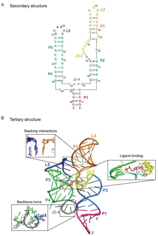
aptamer domain of the Escherichia coli thiM thiamine pyrophosphate (TPP) riboswitch
RNA, a small RNA (80 nts) that contains many features common to structured RNAs, such as canonical and non-canonical base-pairing, local stacking, and long-range docking
interactions [19].
2.2 Results
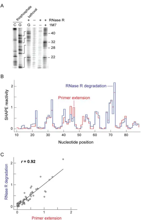
2.2.1 RNase R detects sites of 2ʹ-O-adduct formation in the TPP riboswitch RNA I modified a 5ʹ-end labeled TPP riboswitch using 1-methyl-7-nitroisatoic anhydride
0.25 mM MgCl2; 30 min, 50 °C) and the resulting end-labeled RNA fragments were resolved by gel electrophoresis (Figure 2.2A). Following the degradation step, the RNase R enzyme
can be completely heat inactivated by incubation at elevated temperature (95 °C, 3 min). The half-life of M. genitalium RNase R is 18.4 s at 95 °C (Figure 2.3). To facilitate comparison
with conventional primer extension analysis of the sites of modification, these initial experiments were performed using a TPP riboswitch RNA containing non-native 5ʹ and 3ʹ “structure cassette” sequences of 14 and 43 nucleotides, respectively [13].
The RNase R enzyme efficiently degraded the unmodified riboswitch RNA. In contrast, when the RNA was treated with 1M7 and then incubated with RNase R, a strong
pattern of banding was observed [compare (–) and (+) 1M7 lanes, Figure 2.2A]. The lengths of these 1M7-modified fragments were determined by comparison with sequencing ladders generated by iodine-mediated cleavage of phosphorothioate-substituted RNA [21] and by
RNase R degradation of kethoxal-modified RNA (sequencing lanes, Figure 2.2A). Unexpectedly, bands corresponding to guanosine residues in the two sequencing reactions
were offset by 5 nucleotide positions on the gel. Phosphorothioate cleavage results in a 2ʹ,3ʹ-cyclic phosphate fragment [21] that is one nucleotide shorter than the guanosine-terminated fragment, when visualized using 5ʹ-labeled RNA. Taking into account this offset for
phosphorothioate cleavage, the net offset for the phosphorothioate- and kethoxal-mediated sequencing reactions is 4 nucleotides.
To determine if RNase R degradation, in fact, recapitulated RNA 1M7 reactivity and to understand the large offset in the sequencing reactions, I performed identical SHAPE reactions on the TPP riboswitch but analyzed the results by conventional primer extension.
modified intensities for reactions analyzed by both RNase R degradation and primer extension, (blue and red, Figure 2.2B). Absolute reactivities detected by RNase R degradation were almost identical to those obtained by primer extension (Pearson’s linear r =
0.92; Figure 2.2C). However, in order to superimpose the reactivity profiles, RNase R
detected bands in the 1M7 reaction and in kethoxal-mediated sequencing required 3- and 4-nucleotide offsets, respectively, to yield agreement with the primer extension-detected reactions.
2.2.2 RNase R detects covalent 2ʹ-O- and kethoxal adducts in a two-site model in the enzyme catalytic site
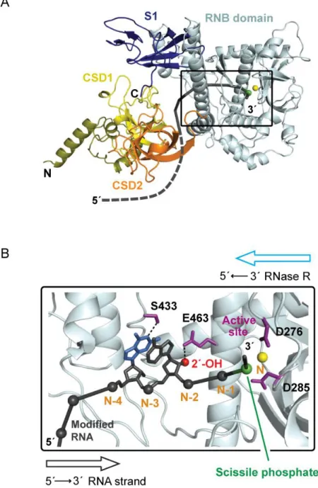
To understand the chemical basis of these offsets, I created a homology model [22, 23] for the M. genitalium RNase R enzyme based on the known structures of two close
homologs, E. coli RNase II (2ix0, 2ix1) [24] and S. cerevisiae Rrp44 (2vnu) [25]. The M. genitalium RNase R enzyme consists of four major domains: two N-terminal cold shock
domains, CSD1 and CSD2; a central, highly conserved, RNA binding (RNB) domain; and a
C-terminal S1 domain (Figure 2.4A). The RNB domain contains the RNA substrate-binding channel and the active site for hydrolytic degradation of RNA (Figure 2.4B). The RNA
strand is threaded into the RNB domain through an opening between the CSD1 and RNB domains (black strand, Figure 2.4A); makes numerous contacts with protein residues in the RNB domain (Figure 2.4B) and ultimately occupies the active site where phosphate cleavage
occurs (see green sphere, Figure 2.4B).
Critically, the enzyme also makes several contacts with RNA nucleotides 5ʹ of the
hydrogen bond with the base-pairing face of nucleotide N-4 (blue nucleotide, Figure 2.4B). Thus, the RNase structure is consistent with a model in which a 1M7-mediated 2ʹ-O-adduct
or a kethoxal-mediated cyclic adduct at guanosine [27] cause RNase R degradation to stop either 3 or 4 nucleotides, respectively, 3ʹ of the site of modification (Figure 2.4B). In sum,
RNase R-mediated degradation of end-labeled RNA yields quantitative detection (Figures 2.2B and C) of covalent adducts at both the ribose 2ʹ-OH position in the backbone and at the base-pairing face of guanosine. Sites of adduct formation can be readily assigned by noting
that kethoxal-mediated sequencing bands are exactly 1 nucleotide shorter than the corresponding 2ʹ-O-adduct (Figure 2.4B). RNase R degradation thus provides a novel, direct
and efficient one-step approach for detecting covalent adducts in RNA.
2.2.3 RNase-detected SHAPE yields quantitative structural information for the folding of the TPP riboswitch
Crystallographic and biochemical analyses have yielded a wealth of information
about the secondary and tertiary structures of the ligand-bound state of the TPP riboswitch [19, 28-32]. However, a single nucleotide resolution structure of the ligand-free state and the
changes in RNA dynamics that occur upon ligand binding are unknown. I, therefore, used RNase-detected SHAPE to analyze the structure of the native TPP riboswitch aptamer domain in the absence and presence of the TPP ligand. Significant differences in SHAPE
reactivities for the free and ligand-bound states were observed (Figure 2.5). Kethoxal-mediated sequencing at guanosine nucleotides were used to assign bands observed in the (+)
oligonucleotide fragments that reflect the short RNA “handle” by which RNase R binds
RNA, and correspond to the end products of 3ʹ→5ʹ exoribonuclease digestion. Using RNase
R-detected SHAPE, SHAPE reactivities for both the ligand-free and the ligand-bound riboswitch RNA states were resolved and quantified (Figure 2.5B).
Absolute SHAPE reactivities for the ligand-free state were used to create an experimentally supported secondary structure model using RNAStructure [14] (Figure 2.6A). The ligand-free secondary structure is characterized by three significant changes relative to
the ligand-bound state. First, all loops in the ligand-free RNA were highly flexible. Second, with the exception of the G19-A47 and A56-G83 base pairs, non-canonical base pairs did not
form stably in the ligand-free structure. Third, the P3 helix showed a significant register shift in the ligand-free relative to the ligand-bound state. In the TPP-bound state, nucleotides in P3 have SHAPE reactivities that are exactly consistent with their crystallographically visualized
structure: C24 is reactive while all base-paired positions are unreactive (see C24 in Figure 2.6B). In contrast, in the ligand-free state, C24 is unreactive while C22 is reactive, indicating
the latter nucleotide is the bulged nucleotide (Figure 2.6A). In sum, the SHAPE data emphasize that, in the ligand-free state, the TPP riboswitch consists of five well-formed and stable helices that are linked by highly dynamic single-stranded regions, suggestive of a
structure with little or no stable tertiary interactions.
Upon addition of TPP, large changes in local nucleotide dynamics occur throughout
the riboswitch RNA, especially in P3, J3-2, J2-4, L5, and nucleotides 60-62 (compare panels, Figure 2.5B). Several regions that became more constrained in the ligand-bound state reflect direct interactions between the ligand (specifically with the TPP aminopyrimidine and
(Figures 2.6B and 2.7A). A few nucleotides near the TPP binding site became more flexible in the ligand-bound state. For example, the SHAPE reactivity of nucleotide 62 increased,
consistent with becoming extrahelical to form the pyrophosphate binding pocket for TPP [19, 28] (Figures 2.5B and 2.7B).
Critically, regions that are spatially distant from the ligand binding site also showed large changes in SHAPE reactivity. L5 was highly dynamic in the ligand-free state but became completely constrained in the ligand-bound state, consistent with L5 docking into the
minor groove of P3 (Figure 2.5B). The register shift in P3 results in the bulged nucleotide migrating from C22 to C24 and allows C24 to stack with A69 (Figure 2.6B). The L5-P3
interaction forms early during TPP binding [29] and the C24-A69 stacking interaction makes a significant contribution to the stability of the RNA-ligand complex [31]. The large change in SHAPE reactivity observed in L5 nucleotides thus implies that the C22 to C24 bulge
migration functions to direct riboswitch folding and to stabilize the structure of the ligand-bound state.
The J2-4 region also undergoes large conformational changes to form interactions that stabilize the three-way junction (Figures 2.5 and 2.6). A53 becomes less reactive in the ligand-bound state, consistent with formation of the A53-G84 non-canonical base pair that
stabilizes the three-way junction [19]. The crystallographic data indicate that U54 and U79 form a stacking interaction in the ligand-bound state [19]; SHAPE data indicated that these
2.3 Discussion
Selective 2ʹ-hydroxyl acylation analyzed by protection from exoribonuclease
(RNase-detected SHAPE) combines quantitative and robust structure-selective RNA acylation with a simple one-tube exoribonuclease degradation step for detecting sites of covalent modification
in RNA. Degradation by M. genitalium RNase R was inhibited by 2ʹ-O-adducts and adducts at the base-pairing face of guanosine due to specific, but distinct, interactions in the substrate binding channel of the enzyme (Figure 2.4). RNase-detected SHAPE is thus likely to be
broadly useful for identifying and quantifying many additional classes of chemical adducts in RNA. Using this technology, I generated single-nucleotide resolution secondary structure
models for both the ligand-free and ligand-bound states of the 80 nt native sequence TPP riboswitch aptamer domain (Figures 2.6A and B); determined the nucleotides undergoing the largest conformational changes upon ligand binding, and characterized a single nucleotide
bulge shift from C22 in the ligand-free state to C24 in the ligand-bound state that is partly responsible for stabilizing the ligand-bound RNA structure.
The usefulness of RNase-detected SHAPE is general and it is anticipated that this approach will make possible structural analysis of miRNAs and their precursors, riboswitches, and small non-coding RNAs in their native forms. RNase-detected SHAPE will also facilitate complete analysis of functionally important structures at the 5ʹ and 3ʹ ends of
large RNAs, including the genomes of RNA viruses.
2.4 Experimental
2.4.1 Screening of candidate RNases
non-specifically degrades RNA (including highly structured RNA), is quantitatively inhibited by 2ʹ-O-adducts, and can be fully and permanently inactivated. All three enzymes efficiently
degraded structured RNA. The E. coli and M. genitalium enzymes are additionally readily inactivated by a simple heating step (95 °C, 3 min) whereas the A. aeolicus enzyme is very
difficult to heat inactivate. 2ʹ-O-adducts caused significantly stronger stops with the M. genitalium enzyme than with the E. coli version. For these reasons, I focused on using the M.
genitalium enzyme for detection of covalent adducts in RNA. RNase R from M. genitalium
was purified as described [18] and used for all subsequent experiments. Enzyme purification should be performed using equipment separate from that used for RNA-based research.
2.4.2 Heat inactivation of RNase R
RNase R enzymes require Mg2+ for activity and can be immediately inactivated by addition of excess EDTA. The enzyme is also permanently inactivated by heat denaturation. To assess the thermal inactivation of M. genitalium RNase R, individual 1 μL aliquots of the enzyme (1.5 μg/μL stock) were incubated at 95 °C [in 20 mM Tris-HCl (pH 8.0), 100 mM
KCl]. Each aliquot was removed at specific time points ranging from 15 s to 10 min and
placed on ice. To the heat-treated RNase, 8 μL of 5ʹ-[32P]-labeled RNA (~ 1 pmol) in the same buffer containing 0.25 mM MgCl2 were added. Reactions were subjected to the
standard exoribonuclease digestion step [20 mM Tris-HCl (pH 8.0), 100 mM KCl, 0.25 mM MgCl2 and 0.45 μg/μL M. genitalium RNase R; incubation at 50 °C for 30 min] and resolved on a 10% denaturing polyacrylamide gel. RNase R activity was quantified as the fraction of
other RNA experiments in the laboratory.
2.4.3 Synthesis of E. coli thiamine pyrophosphate (TPP) riboswitch RNAs
DNA templates for the aptamer domain of the E. coli thiamine pyrophosphate (TPP) riboswitch [19] both with and without 5ʹ and 3ʹ structure cassette flanking sequences [13] were generated by PCR [1 mL; 20 mM Tris-HCl (pH 8.4), 50 mM KCl, 2.5 mM MgCl2, 0.2
mM each dNTP, 250 nM each forward and reverse primer (IDT), 40 nM template (IDT), and 0.025 units/μL Taq polymerase; denaturation at 95 °C, 45 s; annealing at 55 °C, 30 s;
elongation at 72 °C, 1 min; 35 cycles]. The PCR product was recovered by ethanol precipitation and resuspended in 200 μL TE [10 mM Tris-HCl (pH 8.0), 1 mM EDTA]. RNA
constructs were synthesized by in vitro transcription [1 mL; 40 mM Tris-HCl (pH 8.0), 10
mM MgCl2, 10 mM dithiothreitol, 2 mM spermidine, 0.01% (v/v) Triton X-100, 4% (w/v) poly(ethylene) glycol 8000, 2 mM each NTP, 40 μL PCR-generated template, 0.1 mg/mL T7
RNA polymerase; 37 °C; 4 h]. Phosphorothioate-containing RNAs for sequencing were synthesized with the same protocol in 100 μL volumes using 10 μL PCR-generated template
and contained 0.2 mM guanosine or cytidine α-thiotriphosphate (Glen Research). RNAs were
purified by denaturing polyacrylamide gel electrophoresis (8% polyacrylamide, 7 M urea, 29:1 acrylamide:bisacrylamide, 0.4 mm × 28.5 cm × 23 cm; 32 W, 1.5 h), excised from the
gel, recovered by overnight passive elution at 4 °C, and precipitation with ethanol. Purified RNAs were resuspended in TE and stored at ˗20 °C.
Purified RNAs were 5ʹ-[32
P]-radiolabeled by: (1) dephosphorylation [300 μL; 50 mM
precipitation, and resuspension in TE (storage at ˗20 °C); and (3) treatment with T4 polynucleotide kinase [20 μL; 80 pmol dephosphorylated RNA, 70 mM Tris-HCl (pH 7.6), 10 mM MgCl2, 5 mM DTT, 2 μL T4 polynucleotide kinase (NEB, 10,000 units/mL), 80 μCi [γ-32
P]-ATP; 37 °C; 30 min]. Phosphorothioate RNA radiolabeling reactions contained 20-30 pmol RNA, 1 μL T4 polynucleotide kinase (10,000 units/mL), and 20 μCi [γ-32
P]-ATP. Radiolabeled RNAs were purified by denaturing (8%) gel electrophoresis, excised from the gel, and recovered by overnight passive elution at 4 °C. The purified 5ʹ-[32P]-labeled RNAs
were precipitated with ethanol, resuspended in 10 mM HEPES-NaOH (pH 8.0), and stored at ˗20 °C.
2.4.4 Structure-selective RNA modification
Unlabeled RNA with flanking sequences (5 pmol) or 5ʹ-[32P]-labeled RNA with and without flanking sequences (~1 pmol) in 5 μL 1/2× TE (for modified RNAs analyzed by
primer extension) or sterile water (for modified RNAs analyzed by RNase R degradation) was heated at 95 °C for 2 min, cooled on ice, treated with 3 μL 3.3× folding buffer [333 mM
HEPES-NaOH (pH 8.0), 333 mM NaCl, 33.3 mM MgCl2], and incubated at 37 °C for 10 min. The ligand (1 μL; 50 μM TPP) or sterile water was added and incubated at 37 °C for 20
min. After incubation, 9 μL of the folded RNA (+/– TPP) was added to 1 μL 80 mM 1M7 (in
DMSO) [20] and incubated at 37 °C for 2 min. No-reagent control reactions were performed with 1 μL neat DMSO. The RNA was recovered by ethanol precipitation. For the 5ʹ-[32
2.4.5 RNase R digestion 5ʹ-[32
P]-labeled RNA from the 1M7 or kethoxal modification reaction (8 μL) was supplemented with 1 μL 10× reaction buffer [200 mM Tris-HCl (pH 8.0), 1 M KCl, 2.5 mM MgCl2] and 1 μL M. genitalium RNase R [18] (4.5 μg/μL) and incubated at 50 °C for 30 min.
[Note: M. genitalium RNase R activity is very sensitive to Mg2+ concentration.] The RNase R enzyme was inactivated by the addition of 1 μL 100 mM EDTA followed by incubation at 95
°C for 3 min. The RNA fragments were recovered by precipitation with 2.5 vol ethanol plus 1 vol isopropanol, washed once with 70% ethanol, and resuspended in 2 μL sterile water and 7 μL stop dye [96% formamide, 1 mM EDTA (pH 8.0), bromophenol blue and xylene
cyanol]. RNA fragments were separated by denaturing polyacrylamide gel electrophoresis
(10% polyacrylamide, 29:1 acrylamide:bisacrylamide, 7 M urea, 1× TBE, 0.75 mm × 31 cm × 38.5 cm; 70 W) and visualized by phosphorimaging. Fragments were run for both 1.5 and 3.5-4 h to resolve most nucleotide positions.
2.4.6 Kethoxal modification 5ʹ-[32
P]-labeled RNA (1 μL) in 15 μL sterile water was heated at 95 °C for 2 min, cooled on ice, mixed with 2 μL 1 M HEPES-NaOH (pH 8.0), and incubated at 70 °C for 3 min. The RNA was then treated with 2 μL 20 mM kethoxal (in sterile water, from USB) and
incubated at 70 °C for 5 min [34]. The reaction was quenched with 20 μL 10 mM boric acid
followed by ethanol precipitation, washed twice with 70% ethanol, and resuspended in 8 μL sterile water.
2.4.7 Cleavage of phosphorothioate-containing RNA 5ʹ-[32
dye, and used directly for sequencing.
2.4.8 Primer extension
DNA primers were 5ʹ-end labeled with VIC or NED fluorophores (from Applied Biosystems). Unlabeled RNA from the 1M7 modification reaction (10 μL) was added to a
fluorescently labeled DNA primer (5ʹ-VIC-labeled GAA CCG GAC CGA AGC CCG; 3 μL, 0.3 μM) and allowed to anneal at 65 °C for 6 min and then cooled on ice. Reverse transcription buffer [6 μL; 167 mM Tris-HCl (pH 8.3), 250 mM KCl, 10 mM MgCl2, 1.67
mM each dNTP] and Superscript III (1 μL, 200 units) were added and incubated at 45 °C for
2 min, 52 °C for 20 min then 65 °C for 5 min. The reactions were quenched with 4 μL 50 mM EDTA. The cDNAs were recovered by ethanol precipitation, washed twice with 70% ethanol, dried in a SpeedVac for 5 min, and resuspended in 10 μL deionized formamide.
Dideoxy sequencing ladders were produced using unlabeled, unmodified RNA, annealing a
5ʹ-NED-labeled fluorescently labeled DNA primer (same sequence as above) (3 μL, 0.3 μM), and by adding 1 μL 2ʹ,3ʹ-dideoxycytosine triphosphate (10 mM) before addition of Superscript III. cDNA fragments were separated by capillary electrophoresis using an
Applied Biosystems 3130 DNA sequencing instrument.
2.4.9 Data analysis
Band intensities of fragments separated by gel electrophoresis were quantified using
SAFA (Semi-Automated Footprinting Analysis) [35] and raw capillary electrophoresis traces were analyzed using SHAPEFinder [36]. SHAPE reactivity profiles for both the RNase R degradation- and primer extension-detected experiments were obtained by subtracting the
nucleotides, and then dividing all intensities by this averaged value.
2.4.10 RNase R structure modeling
A homology model for the M. genitalium RNase R was generated using I-TASSER [22, 23, 37] with 2ix0 [24] and 2vnu [25] as the template structures. The center-most model (by RMSD) is shown in Figure 2.4. The RNA strand was generated from the RNAs in the
2ix1 [24] and 2vnu structures; the adenosine residue visualized in the 2ix1 structure at position N-4 was changed to a guanosine residue. RNase R likely uses a two Mg2+
mechanism for hydrolytic cleavage. Only one Mg2+ ion has been visualized crystallographically and is shown, along with its coordinating aspartic acid residues, in Figure 2.4. Images were composed using Pymol (Delano Scientific). The secondary structure
2.5 References
1. Holbrook, S.R., Structural principles from large RNAs. Annu Rev Biophys, 2008. 37: p. 445-64.
2. He, L. and Hannon, G.J. MicroRNAs: small RNAs with a big role in gene regulation. Nat Rev Genet, 2004. 5(7): p. 522-31.
3. Rich, A. and RajBhandary, U.L. Transfer RNA: molecular structure, sequence, and properties. Annu Rev Biochem, 1976. 45: p. 805-60.
4. Montange, R.K. and Batey, R.T. Riboswitches: emerging themes in RNA structure and function. Annu Rev Biophys, 2008. 37: p. 117-33.
5. Duncan, C.D. and Weeks, K.M. SHAPE analysis of long-range interactions reveals extensive and thermodynamically preferred misfolding in a fragile group I intron RNA. Biochemistry, 2008. 47(33): p. 8504-13.
6. Watts, J.M., Dang, K.K., Gorelick, R.J. Leonard, C.W., Bess, J.W., Swanstrom, R., Burch, C.L., and Weeks, K.M. Architecture and secondary structure of an entire HIV-1 RNA genome. Nature, 2009. 460(7256): p. 711-6.
7. Wang, B., Wilkinson, K.A., and Weeks, K.M. Complex ligand-induced conformational changes in tRNA(Asp) revealed by single-nucleotide resolution SHAPE chemistry. Biochemistry, 2008. 47(11): p. 3454-61.
8. Wilkinson, K.A., Gorelick, R.J., Vasa, S.M., Guex, N., Rein, A., Mathews, D.H., Giddings, M.C., and Weeks, K.M. High-throughput SHAPE analysis reveals structures in HIV-1 genomic RNA strongly conserved across distinct biological states. PLoS Biol, 2008. 6(4): p. e96.
9. Wilkinson, K.A., Merino, E.J., and Weeks, K.M. RNA SHAPE chemistry reveals nonhierarchical interactions dominate equilibrium structural transitions in tRNA(Asp) transcripts. J Am Chem Soc, 2005. 127(13): p. 4659-67.
10. Merino, E.J., Wilkinson, K.A., Coughlan, J.L., and Weeks, K.M. RNA structure analysis at single nucleotide resolution by selective 2'-hydroxyl acylation and primer extension (SHAPE). J Am Chem Soc, 2005. 127(12): p. 4223-31.
11. Wilkinson, K.A., Vasa, S.M. Deigan, K.E., Mortimer, S.A., Giddings, M.C., and Weeks, K.M. Influence of nucleotide identity on ribose 2'-hydroxyl reactivity in RNA. RNA, 2009. 15(7): p. 1314-21.
13. Wilkinson, K.A., Merino, E.J., and Weeks, K.M. Selective 2'-hydroxyl acylation analyzed by primer extension (SHAPE): quantitative RNA structure analysis at single nucleotide resolution. Nat Protoc, 2006. 1(3): p. 1610-6.
14. Deigan, K.E., Li, T.W., Mathews, D.H., and Weeks, K.M. Accurate SHAPE-directed RNA structure determination. Proc Natl Acad Sci U S A, 2009. 106(1): p. 97-102.
15. Vincent, H.A. and Deutscher, M.P. Substrate recognition and catalysis by the exoribonuclease RNase R. J Biol Chem, 2006. 281(40): p. 29769-75.
16. Vincent, H.A. and Deutscher, M.P. Insights into how RNase R degrades structured RNA: analysis of the nuclease domain. J Mol Biol, 2009. 387(3): p. 570-83.
17. Matos, R.G., Barbas, A., and Arraiano, C.M. RNase R mutants elucidate the catalysis of structured RNA: RNA-binding domains select the RNAs targeted for degradation. Biochem J, 2009. 423(2): p. 291-301.
18. Lalonde, M.S., Zuo, Y., Zhang, J., Gong, X., Wu, S., Malhotra, A., and Li, Z. Exoribonuclease R in Mycoplasma genitalium can carry out both RNA processing and degradative functions and is sensitive to RNA ribose methylation. RNA, 2007. 13(11): p. 1957-68.
19. Serganov, A., Polonskaia, A., Phan, A.T., Breaker, R.R., and Patel, D.J. Structural basis for gene regulation by a thiamine pyrophosphate-sensing riboswitch. Nature, 2006. 441(7097): p. 1167-71.
20. Mortimer, S.A. and Weeks, K.M. A fast-acting reagent for accurate analysis of RNA secondary and tertiary structure by SHAPE chemistry. J Am Chem Soc, 2007. 129(14): p. 4144-5.
21. Heidenreich, O., Pieken, W., and Eckstein, F. Chemically modified RNA: approaches and applications. FASEB J, 1993. 7(1): p. 90-6.
22. Wu, S., Skolnick, J., and Zhang, Y. Ab initio modeling of small proteins by iterative TASSER simulations. BMC Biol, 2007. 5: p. 17.
23. Zhang, Y., I-TASSER server for protein 3D structure prediction. BMC Bioinformatics, 2008. 9: p. 40.
24. Frazao, C., McVey, C.E., Amblar, M., Barbas, A., Vonrhein, C., Arraiano, C.M., and Carrondo, M.A. Unravelling the dynamics of RNA degradation by ribonuclease II and its RNA-bound complex. Nature, 2006. 443(7107): p. 110-4.
25. Lorentzen, E., Basquin, J., Tomecki, R., Dziembowski, A., and Conti, E. Structure of the active subunit of the yeast exosome core, Rrp44: diverse modes of substrate recruitment in the RNase II nuclease family. Mol Cell, 2008. 29(6): p. 717-28.
mutation turns RNase II into a "SUPER-ENZYME". J Biol Chem, 2009. 284(31): p. 20486-98.
27. Shapiro, R. and Hachmann, J. The reaction of guanine derivatives with 1,2-dicarbonyl compounds. Biochemistry, 1966. 5(9): p. 2799-807.
28. Edwards, T.E. and Ferre-D'Amare, A.R. Crystal structures of the thi-box riboswitch bound to thiamine pyrophosphate analogs reveal adaptive RNA-small molecule recognition. Structure, 2006. 14(9): p. 1459-68.
29. Lang, K., Rieder, R., and Micura, R. Ligand-induced folding of the thiM TPP riboswitch investigated by a structure-based fluorescence spectroscopic approach. Nucleic Acids Res, 2007. 35(16): p. 5370-8.
30. Rentmeister, A., Mayer, G., Kuhn, N., and Famulok, M. Conformational changes in the expression domain of the Escherichia coli thiM riboswitch. Nucleic Acids Res, 2007. 35(11): p. 3713-22.
31. Kulshina, N., Edwards, T.E., and Ferre-D'Amare, A.R. Thermodynamic analysis of ligand binding and ligand binding-induced tertiary structure formation by the thiamine pyrophosphate riboswitch. RNA, 2010. 16(1): p. 186-96.
32. Ali, M., Lipfert, J. Seifert, S., Herschlag, D., and Doniach, S. The ligand-free state of the TPP riboswitch: a partially folded RNA structure. J Mol Biol, 2010. 396(1): p. 153-65.
33. Cheng, Z.F. and Deutscher, M.P. Purification and characterization of the Escherichia coli exoribonuclease RNase R. Comparison with RNase II. J Biol Chem, 2002. 277(24): p. 21624-9.
34. Mortimer, S.A., Johnson, J.S., and Weeks, K.M. Quantitative analysis of RNA solvent accessibility by N-silylation of guanosine. Biochemistry, 2009. 48(10): p. 2109-14.
35. Das, R., Laederach, A., Pearlman, S.M., Herschlag, D., and Altman, R.B. SAFA: semi-automated footprinting analysis software for high-throughput quantification of nucleic acid footprinting experiments. RNA, 2005. 11(3): p. 344-54.
36. Vasa, S.M., Guex, N., Wilkinson, K.A., Weeks, K.M., and Giddings, M.C. ShapeFinder: a software system for high-throughput quantitative analysis of nucleic acid reactivity information resolved by capillary electrophoresis. RNA, 2008. 14(10): p. 1979-90.
37. Zhang, Y., Template-based modeling and free modeling by I-TASSER in CASP7. Proteins, 2007. 69 Suppl 8: p. 108-17.
CHAPTER 3
Fingerprinting RNA Tertiary Structure by Differential SHAPE
Reactivity Analysis
3.1 Introduction
RNA molecules are involved in essentially every aspect of cellular information transfer and regulation [1, 2]. The information encoded in RNA, through which it carries out
these functions, is encoded in both the primary sequence and the higher-order secondary and tertiary structure of the RNA [3]. RNA three-dimensional structure is comprised of defined secondary structure elements typically held together by a few key tertiary interactions [4, 5].
These tertiary interactions include long-range stacking interactions, loop-loop and loop-helix contacts, pseudoknots, and well-defined turns in the RNA backbone [5-8]. The regions of an
RNA that contain significant tertiary structures play numerous important functional roles, as elegantly revealed in the dramatic increase in available high resolution structures [7, 8].
Nucleotides that participate in either base-pairing or higher-order tertiary structure interactions can often be detected using chemical probing strategies. In general, nucleotides
that base-pair or form key tertiary structure motifs tend to be unreactive to solution phase reagents while single-stranded and relatively unstructured elements are reactive [9]. Selective 2ʹ-Hydroxyl Acylation analyzed by Primer Extension (SHAPE) has become an especially
informative approach for probing the structure of RNA [9-11]. SHAPE chemistry exploits the discovery that the reactivity of the ribose 2ʹ-hydroxyl is highly sensitive to local
nucleotide flexibility (Figure 3.1A). Flexible nucleotides sample many conformations, a few of which preferentially react with hydroxyl-selective, electrophilic reagents to form a
2ʹ-O-adduct (Figure 3.1A). The strong relationship between SHAPE reactivity and molecular motion [12] makes it possible to use this chemistry to achieve accurate secondary structure predictions, to monitor RNA dynamics and folding, and to explore RNA-protein interactions
[10, 13-15].
SHAPE thus provides a robust measure of molecular motion and makes it
straightforward to detect local conformational changes due to base-pairing, formation of tertiary interactions, or induced by protein or small molecule binding. However, in the absence of additional information, it is often not obvious whether a given constraining
interaction reflects base-pairing or higher-order tertiary interactions. Consequently, there is a pressing need for a biochemical method that precisely and reliably identifies specific
nucleotides involved in tertiary interactions.
To this end, I have developed an approach for rapidly “fingerprinting” RNA tertiary structure derived from additional, orthogonal, chemical reactivity information based on the
highly overrepresented in RNA motifs that form tertiary structures. Such features include unusual backbone and stacking geometries [5, 7], prevalence of syn nucleotides [16], and
nucleotides that undergo local conformational changes on slow timescales [14]. Thus, identification of chemical probes that react selectively with nucleotides in these diagnostic
categories would make possible de novo discovery of tertiary structures in arbitrary RNAs. Here I explore two such distinctive features using RNAs of known structures.
First, nucleotides that undergo local conformational changes on slow timescales can
be selectively detected using SHAPE reagents that also react slowly. These nucleotides are usually in the relatively rare C2ʹ-endo conformation [13] and, in some cases, govern the
folding of entire RNA domains [14]. Second, nucleotide base moieties typically form strong, stacking interactions that are almost fully saturated in well folded RNAs [5]. Nucleotides whose bases stack on only one side (one-sided stackers) can be robustly detected using a
SHAPE reagent that binds at the unoccupied stacking face.
Using two reagents, N-methylisatoic anhydride (NMIA) and 1-methyl-6-nitroisatoic
anhydride (1M6), that differ from each other only in the identity of a single functional group (compare NMIA and 1M6, Figure 3.1B), nucleotides displaying slow, local dynamics and one-sided stacking interactions can be selectively detected. The reactivities of these two
reagents can be combined to make possible very concise analysis and discovery of tertiary structure elements by simply calculating the difference in their reactivities. The resulting
differential reactivities provide an information-rich “fingerprint” for RNA tertiary structure, as exemplified by the ability to recapitulate tertiary structure information for four RNAs of known structure. Differential SHAPE reactivity analysis was then applied to detect sites of